Feeds:
Posts
Comments

Posts Tagged ‘giraffe’

Today is the 210th anniversary of Charles R. Darwin’s birthday so I put together a quick post. I’d been meaning to blog about some of our latest scientific papers, so I chose those that had an explicit evolutionary theme, which I hope Chuck would like. Here they are, each with a purty picture and a short explainer blurb! Also please check out Anatomy To You’s post by Katrina van Grouw on Darwin’s fancy pigeons.

Stomach-Churning Rating: 1/10 science!

First, Brandon Kilbourne at the Naturkunde Museum in Berlin kindly invited me to assist in a paper from his German fellowship studying mustelid mammals (otters, weasels, wolverines, badgers, etc.; stinky smaller carnivorous mammals). Here we (very much driven by Brandon; I was along for the ride) didn’t just look at how forelimb bone shape changes with body size in this ecologically diverse group. We already knew bigger mustelids would have more robust bones, although it was cool to see how swimming-adapted and digging-adapted mustelids evolved similarly robust bones; whereas climbing ones had the skinniest bones.

The really exciting and novel (yes I am using that much-abused word!) aspect of the paper is that Brandon conjured some sorcery with the latest methods for analysing evolutionary trends, to test how forelimb bone shapes evolved. Was their pattern of evolution mostly a leisurely “random walk” or were there early bursts of shape innovation in the mustelid tree of life, or did shape evolve toward one or more optimal shapes (e.g. suited to ecology/habitat)? We found that the most likely pattern involved multiple rates of evolution and/or optima, rather than a single regime. And it was fascinating to see that the patterns of internal shape change deviated from external shape change such as bone lengths: so perhaps selection sometimes works independently at many levels of bone morphology?

Various evolutionary models applied to the phylogeny of mustelids.

Then there, coincidentally, was another paper originating in part from the same museum group in Berlin. This one I’d been involved in as a co-investigator (author) on a Volkswagen (yes! They like science) grant back about 8 years ago and since. There is an amazing ~290 million year old fossil near-amniote (more terrestrial tetrapod) called Orobates pabsti, preserved with good skeletal material but also sets of footprints that match bones very well, allowing a rare match of the two down to this species level. John Nyakatura’s team had 3D modelled this animal before, so we set out to use digital techniques to test how it did, or did not, move—similar to what I’d tried before with Tyrannosaurus, Ichthyostega and so forth. The main question was whether Orobates moved in a more “ancestral” salamander-like way, a more “derived” lizard-like way (i.e. amniote-ish), or something else.

The approach was like a science sledgehammer: we combined experimental studies of 4 living tetrapods (to approximate “rules” of various sprawling gaits), a digital marionette of Orobates (to assess how well its skeleton stayed articulated in various motions), and two robotics analysis (led by robotics guru Auke Ijspeert and his amazing team): a physical robot version “OroBOT” (as a real-world test of our methods), and a biomechanical simulation of OroBOT (to estimate hard-to-measure things in the other analyses, and matches of motions to footprints). And, best of all, we made it all transparent: you can go play with our interactive website, which I still find very fun to explore, and test what motion patterns do or do not work best for Orobates. We concluded that a more amniote-like set of motions was most plausible, which means such motions might have first evolved outside of amniotes.

OroBOT in tha house!

You may remember Crassigyrinus, the early tetrapod, from a prior post on Anatomy To You. My PhD student Eva Herbst finished her anatomical study of the best fossils we could fit into a microCT-scanner and found some neat new details about the “tadpole from hell”. Buried in the rocky matrix were previously unrecognized bones: vertebrae (pleurocentra; the smaller nubbins of what may be “rhachitomous” bipartite classic tetrapod/omorph structure), ribs (from broad thoracic ones to thin rear ones), pelvic (pubis; lower front), and numerous limb bones. One interesting trait we noticed was that the metatarsals (“sole bones” of the foot) were not symmetrical from left-to-right across each bone, as shown below. Such asymmetry was previously used to infer that some early tetrapods were terrestrial, yet Crassigyrinus was uncontroversially aquatic, so what’s up with that? Maybe this asymmetry is a “hangover” from more terrestrial ancestry, or maybe these bones get asymmetrical for non-terrestrial reasons.

The oddly asymmetrical metatarsals of Crassigyrinus.

Finally, Dr. Peter Bishop finished his PhD at Griffith University in Australia and came to join us as a DAWNDINOS postdoc. He blasted out three of his thesis chapters (starting here) with me and many others as coauthors, all three papers building on a major theme: how does the inner bone structure (spongy or cancellous bone) relate to hindlimb function in theropod dinosaurs (including birds) and how did that evolve? Might it tell us something about how leg posture or even gait evolved? There are big theories in “mechanobiology” variously named Wolff’s Law or the Trajectorial Theory that explain why, at certain levels, bony struts tend to align themselves to help resist certain stresses, and thus their alignment can be “read” to indicate stresses. Sometimes. It’s complicated!

Undaunted, Peter measured a bunch of theropod limb bones’ inner geometry and found consistent differences in how the “tracts” of bony struts, mainly around joints, were oriented. He then built a biomechanical model of a chicken to test if the loads that muscles placed on the joints incurred stresses that matched the tracts’ orientations. Hmm, they did! Then, with renewed confidence that we can use this in the fossil record to infer approximate limb postures, Peter scanned and modelled a less birdlike Daspletosaurus (smaller tyrannosaur) and more birdlike “Troodon” (now Stenonychosaurus; long story). Nicely fitting many other studies’ conclusions, Peter found that the tyrannosaur had a more straightened hindlimb whereas the troodontid had a more crouched hindlimb; intermediate between the tyrannosaur and chicken. Voila! More evidence for a gradual evolution of leg posture across Mesozoic-theropods-into-modern-birds. That’s nice.

Three theropods, three best-supported postures based on cancellous bone architecture.

If you are still thirsty for more papers even if they are less evolutionary, here’s the quick scoop on ones I’ve neglected until now:

(1) Former PhD student Chris Basu published his thesis work w/us on measuring giraffe walking dynamics with force plates, finding that they move mostly like other quadrupeds and their wobbly necks might cost them a little.

(2) Oh, and Chris’s second paper just came out as I was writing this! We measured faster giraffe gaits in the wilds of South Africa, as zoo giraffes couldn’t safely do them. And we found they don’t normally go airborne, just using a rotary gallop (not trot, pace or canter); unlike some other mammals. Stay tuned: next we get evolutionary with this project!

(2) How do you safely anaesthetize a Nile crocodile? There’s now a rigorous protocol (from our DAWNDINOS work).

(3) Kickstarting my broad interest in how animals do “extreme” non-locomotor motions, we simulated how greyhounds stand up, finding that even without stretchy tendons they should, barely, be able to do it, which is neat. Expect much more about this from us in due time.

(4) Let’s simulate some more biomechanics! Ashley Heers, an NSF research fellow w/me for a year, simulated how growing chukar birds use their wing muscles to flap their way up steeper inclines (“WAIR” for devotees), and the results were very encouraging for simulating this behaviour in more detail (e.g. tendons seem to matter a lot) and even in fossil species; and finally…

(5) Hey did you ever think about how bone shape differs between hopping marsupials (macropods) and galloping artiodactyl (even-toed) mammals? We did, in long-the-making work from an old BBSRC grant with Michael Doube et al., and one cool thing is that they mostly don’t change shape with body size that differently, even though one is more bipedal at faster speeds—so maybe it is lower-intensity, slower behaviours that (sometimes?) influence bone shape more?

So there you have the skinny on what we’ve been up to lately, messing around with evolution, biomechanics and morphology.

Read Full Post »

One of my favourite museums in the world, and certainly one of the best natural history museums in the UK, is Cambridge’s Museum of Zoology, AKA “University Museum of Zoology at Cambridge” (UMZC). It is now nearing a lengthy completion of renovations; the old museum exhibits and collections were excellent but needed some big changes along with the re-fabbed “David Attenborough Building” that houses them. As a longtime fan of the exhibits and user of the collection (and microCT scanner), I hurried to see the new museum once it officially opened.

And that makes a great excuse to present a photo-shoot from my visit. This focuses on the “mammal floor” below the entrance- the upper floor(s?) are still being completed and will have the birds, non-avian tetrapods, fish, etc. But the UMZC is strong in mammals and so it is natural for them to feature them in this chock-full-o-specimens display. Less talk, more images. Here we go!

All images can be clicked to mu-zoom in on them.

Stomach-Churning Rating: 3/10; bones and taxidermy and innocuous jars.

The building. The whale skeleton that hung outside for years is now cleaned up and housed right inside; you walk under it as you enter.

Entrance.

First view past the entryway: lots of cool specimens.

View from the walkway down into the ground/basement level from the entry. As specimens-per-unit-volume goes, the UMZC still scores highly and that is GOOD!

Explanation of frog dissection image below.

Gorgeous old frog dissection illustration; such care taken here.

Leeuwenhoek’s flea woodcut; I think from Arcana Naturae Detecta (1695). There is an impressive display of classic natural history books near the entryway.

Dürer/other rhino art image and info.

Darwin was famed for collecting beetles when he should have been studying theology at Cambridge as a youth, and here is some of his collection. Dang.

Darwin’s finches!

Darwin kicked off some of his meticulous work with volumes on barnacles; specimens included here; which helped fuel insights into evolution (e.g. they are “retrograde” crustaceans, not mollusks).

Darwin’s voyage: fish & other preserved specimens.

I think this is a solitaire weka (flightless island bird; see Comment below). I’ve never seen them displayed w/skeleton + taxidermy; it’s effective here.

Eryops cast. More early tetrapods will surely be featured on the upper floor; this one was on the timeline-of-life-on-Earth display.

I LOVE dioramas and this seabird nesting ground display is very evocative, especially now that I’ve visited quite a few such islands.

Mammal introduction; phylogenetic context.

Monotreme glory.

UMZC is well endowed with thylacines and this one is lovely.

“TAZ FEEL NAKED!”

Narwhal above!

Rhinocerotoidea past, present, and fading glory. 😦

Ceratotherium white rhino. The horn is not real; sadly museums (and even zoos) across the world have to worry about theft of such things, given that some people think these horns are magic.

Ceratotherium staring match. You lose.

Ceratotherium stance.

Foot of a Sumatran rhino juxtaposed with a horse’s for Perissodactyla didaction.

A tapir. As a kid, I used to wander around the house pretending to be a tapir but I did not know what noise they’d make so I’d say “tape tape tape!”.

Big Southern Elephant Seal.

Squat little fur seal.

Hippopotamus for the lot of us. (baby included)

Hippo facedown.

Skull of a dwarf Madagascar hippo.

Cave bear and sabretooth cat make an impressive Ice Age demo.

It’s a wombat.

Ain’t no don like a Diprotodon! (also note its modern miniature cousin the wombat, below)

Diprotodon facial.

Diprotodon shoulder: big clavicles bracing that joint region.

Diprotodon knee: even in big marsupials, the “parafibula”/lateral sesamoid of the knee is still generally present. And why it is there/what it does deserves much more study.

Diprotodon hip. I just find this animal’s anatomy fascinating head-to-tail.

Diprotodon front foot. Absolutely freakish.

Diprotodon hind foot. Even weirder.

Your view after having been trampled in a supine position by a Diprotodon. Not a good way to go.

Diprotodon got back.

Elephant seal’s butt continues my series of photos of big animals’ bottoms.

Asian elephant’s butt view.

African elephant butt.

Sectioned elephant skull to show pneumatic resonating chambers.

Paenungulates: hyraxes, Sirenia, elephants & kin (evolutionary demo).

AND MY HYRAX!
Sorry. Had to.

Megatherium side view.

Megatherium. Yeah!

Megatherium hindlegs fascinate me. Well-heeled.

Tamandua duo.

Silky anteater; wonderful.

Armadillos.

Anteaters round out a fab display on Xenarthra.

The UMZC has everything from aardvarks to zebus. Here, conceptualized with other Afrotheria.

Golden moles: the more I read about them, the more they fascinate me.

We can all use some more solenodons in our lives!

Example of the phylogenetic context used throughout exhibits.

If you’ve got a good Okapi taxidermy, you’d better use it.

It’s a giraffe. Did you guess right?

Gerenuk showing off its bipedal capacity.

Warthogs have an inner beauty.

Pangolin. Glad to see it back on exhibit.

Nice little brown bear.

Double-barrelled shot of hyenas.

Colugo!

Nice to see some Scandentia featured.

My brain says this is a springhare (Pedetes) so I am going with what my brain says and anyway I really like this display.

When I saw this I thought, “That’s a nice… rodent thingy.” And so “rodent thing” it shall be labelled here. Enjoy the rodent thingy. Some serious taxidermy-fu in action.

Moonrats– now there’s something you seldom see a full display of. Well done!

That’s part I of this sneak peek at the evolving exhibits- I will put up a part II once the upper floor exhibits open. I highly encourage a visit!

For Mike: gimlet

Read Full Post »

Nice GIF of the human biceps in action- By Niwadare - Own work, CC BY-SA 4.0, https://commons.wikimedia.org/w/index.php?curid=38718790

GIF of the human biceps (above) and its antagonist triceps (below) in action- By “Niwadare” – own work, CC BY-SA 4.0, https://commons.wikimedia.org/w/index.php?curid=38718790

Last year on Darwin Day I debuted “Better Know A Muscle” (BKAM), which was intended to be a series of posts focusing on one cool muscle at a time, and its anatomical, functional and evolutionary diversity and history. A year later, it’s another post on another muscle! Several dozen more muscles to go, so I’ve got my work cut out for me… But today: get ready to FLEX your myology knowledge! Our subject is Musculus biceps brachii; the “biceps” (“two-headed muscle of the arm”). Beloved of Arnie and anatomists alike, the biceps brachii is. Let’s get pumped up!

Stomach-Churning Rating: 7/10. Lots of meaty elbow flexion!

While the previous BKAM’s topic was a hindlimb muscle with a somewhat complex history (and some uncertainties), the biceps brachii is a forelimb muscle with a simpler, clearer history. Fish lack a biceps, just having simple fin ab/adductor muscles with little differentiation. Between fish and tetrapods (limb-bearing vertebrates), there was an explosion in the number of muscles; part of transforming fins into limbs; and the biceps is thenceforth evident in all known tetrapods in a readily identifiable anatomical form. In salamanders and their amphibian kin, there is a muscle usually called “humeroantebrachialis” that seems to be an undivided mass corresponding to the biceps brachii plus the brachialis (shorter humerus-to-elbow) muscle:

Most of the humerobrachialis muscle (purplish colour), in dorsal (top) view of the right forelimb of the fire salamander Salamandra salamandra (draft from unpublished work by my team).

Most of the humerobrachialis muscle (purplish colour), in dorsal (top) view of the right forelimb of the fire salamander Salamandra salamandra (draft from unpublished work by my team).

In all other tetrapods; the amniote group (reptiles, mammals, etc.); there is a separate biceps and brachialis, so these muscles split up from the ancestrally single “humeroantebrachialis” muscle sometime after the amphibian lineage diverged from the amniotes. And not much changed after then– the biceps is a relatively conservative muscle, in an evolutionary (not political!) sense. In amniote tetrapods that have a biceps, it develops as part of the ventral mass of the embryonic forelimb along with other muscles such as the shorter, humerus-originating brachialis, from which it diverges late in development (reinforcing that these two muscles are more recent evolutionary divergences, too).

Biceps brachialis or humerobrachialis, the “biceps group” tends to originate just in front of the shoulder (from the scapula/coracoid/pectoral girdle), running in front of (parallel to) the humerus. It usually forms of two closely linked heads (hence the “two heads” name), most obviously in mammals; one head is longer and comes from higher/deeper on the pectoral girdle, whereas the other is closer to the shoulder joint and thus is shorter. The two heads fuse as they cross the shoulder joint and we can then refer to them collectively as “the biceps”. It can be harder to see the longer vs. shorter heads of the biceps in non-mammals such as crocodiles, or they may be more or less fused/undifferentiated, but that’s just details of relatively minor evolutionary variation.

The biceps muscle then crosses in front of the elbow to insert mainly onto the radius (bone that connects your elbow to your wrist/thumb region) and somewhat to the ulna (“funny bone”) via various extra tendons, fascia and/or aponeuroses. The origin from the shoulder region tends to have a strong mark or bony process that identifies it, such as the coracoid process in most mammals (I know this well as I had my coracoid process surgically moved!). The insertion onto the radius tends to have a marked muscle scar (the radial tuberosity or a similar name), shared with the brachialis to some degree. A nice thing about the biceps is that, because it may leave clear tendinous marks on the skeleton, we sometimes can reconstruct how its attachments and path evolved (and any obvious specializations; even perhaps changes of functions if/when they happened).

Here are some biceps examples from the world of crocodiles:

Crocodile's right forelimb showing the huge pectoralis, and the biceps underlying it on the bottom right.

Crocodile’s right forelimb showing the huge pectoralis, and the biceps underlying it; on the bottom right (“BB”- click to embiceps it).

Crocodile left forelimb with biceps visible (

Crocodile left forelimb with biceps visible (“BB”) on the left.

Crocodile biceps muscle cut off, showing the proximal and distal tendons (and long parallel muscle fibres) for a typical amniote vertebrate.

Crocodile biceps muscle cut off, showing the proximal (to right) and distal (to left) tendons (and long parallel muscle fibres) for a typical amniote vertebrate.

What does the biceps muscle do? It flexes (draws forward) the shoulder joint/humerus, and does the same for the elbow/forearm while supinating it (i.e. rotating the radius around the ulna so that the palm faces upwards, in animals like us who can rotate those two bones around each other). In humans, which have had their biceps muscles studied by far the most extensively, we know for example that the biceps is most effective at flexing the elbow (e.g. lifting a dumbbell weight) when the elbow is moderately straight. These same general functions (shoulder and elbow flexion; with some supination) prevail across the biceps muscle of [almost; I am sure there are exceptions] all tetrapods, because the attachments and path of the biceps brachii are so conservative.

And this flexor function of the biceps brachii stands in contrast to our first BKAM muscle, the caudofemoralis (longus): that muscle acts mainly during weight support (stance phase) as an antigravity/extensor muscle, whereas the flexor action of biceps makes it more useful as a limb protractor or “swing phase” muscle used to collapse the limb and draw it forwards during weight support. However, mammals add some complexity to that non-supportive function of the biceps…

Hey mammals! Show us your biceps!

Jaguar forelimb with biceps peeking out from the other superficial muscles, and its cousin brachialis nicely visible.

Jaguar forelimb with biceps peeking out from the other superficial muscles, and its cousin brachialis nicely visible, running along the front of the forearm for a bit.

Elephant's left forelimb with the biceps labelled.

Elephant’s left forelimb with the biceps labelled.

Longitudinal slice thru the biceps of an elephant, showing the internal tendon.

Longitudinal slice thru the biceps of an elephant, showing the internal tendon that helps identify where the two bellies of the biceps fuse.

In certain mammals; the phylogenetic distribution of which is still not clear; the biceps brachii forms a key part of a passive “stay apparatus” that helps keep the forelimb upright against gravity while standing (even sleeping). The classic example is in horses but plenty of other quadrupedal mammals, especially ungulate herbivores, show evidence of similar traits:

Giraffe biceps cut away proximally to show the

Giraffe biceps cut away proximally to show the “stay apparatus” around the shoulder joint (upper right).

Zooming in on the

Zooming in on the “stay apparatus”; now in proximal view, with the biceps tendon on the left and the humeral head (showing some arthritic damage) on the right, with the groove for the biceps in between.

Hippo's humerus (upper left) and biceps muscle cut away proximally, displaying the same sort of

Hippo’s humerus (upper left) and biceps muscle cut away proximally, displaying the same sort of “stay apparatus” as in the giraffe. Again, note the stout proximal and distal tendons of the biceps. The proximal tendon fits into the groove of the humerus on the far left side of the image; becoming constrained into a narrow circular “tunnel” there. It’s neat to dissect that region because of its fascinating relationships between bone and soft tissues.

The biceps brachii, in those mammals with a stay apparatus, seems to me to have a larger tendon overall, especially around the shoulder, and that helps brace the shoulder joint from extending (retracting) too far backward, whilst also transmitting passive tension down the arm to the forearm, and bracing the elbow (as well as distal joints via other muscles and ligaments). It’s a neat adaptation whose evolution still needs to be further inspected.

Otherwise, I shouldn’t say this but the biceps is sort of boring, anatomically. Whether you’re a lizard, croc, bird or mammal, a biceps is a biceps is a biceps; more or less-ceps. But the biceps still has a clear evolutionary history and Darwin would gladly flex his biceps to raise a pint in toast to it.

So now we know a muscle better. That’s two muscles now. And that is good; be you predator or prey. Let’s shake on it!

Read Full Post »

My Summer in the SML

Excellent post by a summer research student on my team!

Luke Grinham

I spent this summer, the second of my undergraduate degree, in the Royal Veterinary College’s Structure and Motion Laboratory, as I undertook a BBSRC-funded Summer Research Experience Placement. The purpose of the REP is to give undergrad students a taste of what research would be like as a career. In my case, I was given the fantastic opportunity to study giraffe locomotion. Mentored by Christopher Basu, a PhD student in the SML, and Professor John Hutchinson, my ten-week project began at the start of July.

First things first, I had some ground work to do. All the data had been collected prior to my placement, though I will be joining Chris next week to collect fresh data for his future work. Giraffes were recorded using high speed video cameras walking parallel to the edge of their enclosure, over concealed force-plates measuring ground reaction forces. I was provided with 3 days’…

View original post 650 more words

Read Full Post »

Why should you care
If you have to trim my hooves?
I’ve got to move with good feet
Or be put down fast.
I know I should trot
But my old vet she cares a lot.
And I’m still living on stone
Even though these feet won’t last.

(mutated from The Who, “Cut My Hair“, Quadrophenia… from the heyday of concept albums and grandiose rock!)

Talkin' bout my osteitis?

Talkin’ bout my osteitis

Day Four of Freezermas. Four posts to go. I can see through time… Hence the silly title for today’s concept album track. Quadrupedophilia did not have a good ring to it, anyway.

Stomach-Churning Rating: 4/10. Reasonably tame; bones and hooves. Some pathologies of those, but not gory.

If Quadrophenia was the story of a man with four personalities (metaphor for the four band members), then quadrupedopheniaphilia is the story of how diverse forms of four-legged animals have lots of problems because of our exploitation of them, which leaves a crisis to resolve: Who are we? Are we caring enough to fix a bad situation we’ve created for our four-legged ungulate comrades?

Four legs good, two legs bad? Not really. I featured ostriches earlier this week and two legs are indeed pretty good. Four-legged cats are great, too. But four-footed big beasties with deformed hooves: those are bad all around. That leads to today’s topic…

But hey, happy 205th funkin’ birthday Charles freakin’ Robert Darwin!

Charles Darwin on his horse “Tommy” in 1868- from the Darwin Correspondence Project, https://www.darwinproject.ac.uk/darwins-photographic-portraits

Today’s post concerns a phenomenon that (Western) civilization has wrought with large hoofed mammals, and evolution is a big part of it (as well as biomechanics and anatomy) . Cynical perspective, with some truth to it: We’ve evolved larger and heavier animals to either do harder and harder work on tough surfaces like concrete floors and tarmac roads, or to stand around while we gawk at them or wait for them to get fat and tasty. Either way, the outcome should come as no surprise: their feet, the interface of that hard ground and their body, eventually start falling apart.

I’ve posted about this several times with respect to rhinos and elephants (here and here and here and here and here), but this post hits closer to home: what goes wrong with the humble hoof of our friend the horse, cow, sheep or other ungulate. It’s where the rubberkeratin hits the road. Ungulates have not evolved to live on dirty, wet concrete floors; to be obese and inactive; or to have hooves that don’t get worn down. So they suffer when they do encounter those modern conditions.

“No foot no horse,” they say, and it’s so true- once the feet start to go (due to hoof overgrowth or cracks, abscesses or other trouble), it’s hard to reverse the pathologies that ensue (arthritis, osteomyelitis, infections, fractures, etc.) and the animals start going lame, then other limbs (supporting greater loads than the affected limb) start to go, too, sometimes.

Jerry the obese, untrimmed-hoof-bearing horse.

Jerry the obese, untrimmed-hoof-bearing horse. “Turkish slippers” is an apt description. DM has more here.

We can do plenty about these problems, and the title track above explains one of them: trimming hooves. Hooves often get overgrown, and if animals are tame enough (requires training!) or are sedated (risky!), hoof care experts (farriers) can rasp/file/saw them down to a more acceptable conformation. If we don’t, and the animals don’t do the trimming themselves by digging or walking around or living on varied surfaces, then the feet can suffer. But there’s still not much evidence for most common species kept in captivity by humans that indicates what the best methods are for avoiding or fixing foot problems.

What we’ve been trying to do at the RVC is use our expertise in evolution, anatomy and biomechanics to find new ways to prevent, detect, monitor or reverse these foot problems. We had BBSRC grant funding from 2009-2012 to do this, and the work continues, as it behooves us to do… Past posts have described some of this research, which spun off into other benefits like re-discovering/illuminating the false sixth toes of elephants. We’re working with several zoos in the UK to apply some of the lessons we’re learning to their animals and management practices.

Above: Thunderous hoof impacts with nasty vibrations, and large forces concentrated on small areas, seem to contribute to foot problems in hoofed mammals. From our recent work published in PLOS ONE.

Foot health check on a white rhino at a UK zoo. Photo by Ann & Steve Toon, http://www.toonphoto.com/

Foot health check on a white rhino at a UK zoo; one of the animals we’ve worked with. Photo by Ann & Steve Toon, http://www.toonphoto.com/

If it works, it’s the most satisfying outcome my research will have ever had, and it will prevent my freezers from filling up with foot-influenced mortality victims.

Again, I’ll tell this tale mainly in photos. First, by showing some cool variations evolved in the feet of hoofed mammals (artiodactyls and perissodactyls; mostly even/odd-toed ungulates of the cow/sheep and horse lineages, respectively). Second, by showing some pretty amazing and shocking images of how “normal” hooves go all wonky.

Two ways to evolve a splayed hoof for crossing soft ground: 2 toes that are flexible and linked to big pads (camel), and 2 main toes that allow some extra support from 2 side toes when needed (elk). At Univ. Mus. Zoology- Cambridge.

Two ways to evolve a splayed hoof for crossing soft ground: 2 toes that are flexible and linked to big pads (camel), and 2 main toes that allow some extra support from 2 side toes when needed (elk). At Univ. Mus. Zoology- Cambridge.

Diversity of camelid foot forms: big clunky, soft Old World camel feet and dainty, sharp highland New World camelids.

Diversity of camelid foot forms: big clunky, soft Old World camel feet and dainty, sharp highland New World camelids. [Image source uncertain]

Moschus, Siberian musk deer with remarkable splayed hooves/claws; aiding it in crossing snowy or swampy ground. At Univ. Mus. Zoology- Cambridge.

Moschus, Siberian musk deer with remarkable splayed hooves/claws; aiding it in crossing snowy or swampy ground. At Univ. Mus. Zoology- Cambridge.

Tragulus, or mouse-deer, with freaky long "splint bones" (evolutionarily reduced sole bones or metatarsals) and dainty hooved feet. At Univ. Mus. Zoology- Cambridge.

Tragulus, or mouse-deer, with freaky long “splint bones” (evolutionarily reduced sole bones or metatarsals) and dainty hooved feet. At Univ. Mus. Zoology- Cambridge.

Overgrown giraffe hooves. An all-too-common problem, and one we're tacking with gusto lately, thanks to PhD student Chris Basu's NERC-funded giraffe project!

Overgrown giraffe hooves. An all-too-common problem, and one we’re tacking with gusto lately, thanks to PhD student Chris Basu’s NERC-funded giraffe project!

Wayyyyyyyyy overgrown hooves of a ?sheep, from the RVC's pathology collection.

Wayyyyyyyyy overgrown hooves of a ?sheep, from the RVC’s pathology collection.

Craaaaaaazy overgrown ?cow hooves, from the RVC's pathology collection.

Craaaaaaazy overgrown ?sheep hooves, from the RVC’s pathology collection.

If we understand how foot form, function and pathology relate in diverse living hoofed mammals, we can start to piece together how extinct ones lived and evolved- like this giant rhinoceros! At IVPP museum in Beijing.

If we understand how foot form, function and pathology relate in diverse living hoofed mammals, we can start to piece together how extinct ones lived and evolved- like this giant rhinoceros! At IVPP museum in Beijing.

So, what do we do now? If we love our diverse hoofed quadrupeds, we need to exert that quadrupedopheniaphilia and take better care of them. Finding out how to do that is where science comes in. I’d call that a bargain. The best hooves ever had?

Read Full Post »

A photo blog post for ya here! I went to Dublin on a ~28 hour tour, for a PhD viva (now-Dr Xia Wang; bird feather/flight evolution thesis) earlier this month. And I made a beeline for the local natural history museum (National Museum of Ireland, Natural History building) when I had free time. So here are the results!

Stomach-Churning Rating: Tame; about a 1/10 for most, but I am going to break my rule about showing human bodies near the end. Just a warning. The bog bodies were too awesome not to share. So that might be 4/10-8/10 depending on your proclivities. They are dry and not juicy or bloody, and don’t look as human as you’d expect.

Simple Natural History museum entrance area.

Simple Natural History museum entrance area.

Adorable frolicking topiaries outside the NHM.

Adorable frolicking topiaries outside the NHM.

Inside, it was a classical Victorian-style, dark wood-panelled museum stuffed with stuffed specimens. It could use major refurbishment, but I do love old-fashioned exhibits. Get on with it and show us the animals; minimize interpretive signage and NO FUCKING INTERACTIVE COMPUTER PANELS! So by those criteria, I liked it. Some shots of the halls: hall2 hall1 hall3 hall4 hall5 hall6 And on to the specimens!

Giant European deer ("Irish elk"). I looked at these and thought, "why don't we see female deer without antlers ever? then noticed one standing next to these; photo was crappy though. :(

Giant European deer (“Irish elk”). I looked at these and thought, “why don’t we see female deer without antlers ever? then noticed one standing next to these (you can barely see it in back); too bad my photo is crappy.

Superb mounted skeleton of giraffe (stuffed skin was standing near it).

Superb mounted skeleton of giraffe (stuffed skin was standing near it).

A sheep or a goat-y thingy; I dunno but it shows off a nice example of the nuchal ligament (supports the head/neck).

A sheep-y or a goat-y beastie; I dunno but it shows off a nice example of the nuchal ligament (supports the head/neck).

Yarr, narwhals be internet gold!

Yarr, narwhals be internet gold!

Giant blown glass models of lice!

Giant blown glass models of lice!

Who doesn't like a good giant foramanifera image/models? Not me.

Who doesn’t like a good giant foramanifera image/model?

"That's one bigass skate," I murmured to myself.

“That’s one bigass skate,” I murmured to myself.

"That's one bigass halibut," I quipped.

“That’s one bigass halibut,” I quipped.

Tatty basking shark in entry hall.

Tatty basking shark in entry hall.

Irish wolfhound, with a glass sculpture of its spine hanging near it, for some reason.

Irish wolfhound, with a glass sculpture of its spine hanging near it, for some reason.

Stand back folks! The beaver has a club!

Stand back everyone! That beaver has a club!

Skull of a pilot whale/dolphin.

Skull of a pilot whale/dolphin.

Nice anteater skeleton and skin.

Nice anteater skeleton and skin.

Nice anteater skeleton and skin.

Nice wombat skeleton and skin.

Sad display of a stuffed rhino with the horn removed, and signage explaining the problem of thefts of those horns from museum specimens of rhinos worldwide.

Sad display of a stuffed rhino with the horn removed, and signage explaining the problem of thefts of those horns from museum specimens of rhinos worldwide.

But then the stuffed animals started to get to me. Or maybe it was the hangover. Anyway, I saw this…
creepy proboscis (1) creepy proboscis (2)

A proboscis monkey mother who seemed to be saying “Hey kid, you want this yummy fruit? Tough shit. I’m going to hold it over here, out of reach.” with a disturbing grimace. That got me thinking about facial expressions in stuffed museum specimens of mammals more, and I couldn’t help but anthropomorphize as I toured the rest of the collection, journeying deeper into surreality as I progressed. What follows could thus be employed as a study of the Tim-Burton-eseque grimaces of stuffed sloths. Click to emslothen.

sloths (1) sloths (5)sloths (4) sloths (3) sloths (2)

Tree anteater has a go at the awkward expression game.

Tree anteater has a go at the awkward expression game.


This completed my tour of the museum; there were 2 more floors of specimens but they were closed for, sigh, say it with me… health and safety reasons. Balconies from which toddlers or pensioners or drunken undergrads could accidentally catapult themselves to their messy demise upon the throngs of zoological specimens below. But the National Museum’s Archaeology collection was just around the block, so off I went, following whispered tales of bog bodies. There will be a nice, calm, pretty photo, then the bodies, so if peaty ~300 BCE cadavers are not your cup of boggy tea, you can depart this tour now and lose no respect.

Impressive entrance to the National Museum's Archaeology building.

Impressive entrance to the National Museum’s Archaeology building.

The bog bodies exhibit is called “Kingship and Sacrifice“. It is packed with cylindrical chambers that conceal, and present in a tomb-like enclosed setting, the partial bodies of people that were killed and then tossed in peat bogs as honoraria for the ascension of a new king. The peaty chemistry has preserved them for ~2300 years, but in a dessicated, contorted state. The preservation has imparted a mottled colouration and wrinkled texture not far off from a Twix chocolate bar’s. Researchers have studied the bejesus out of these bodies (including 3D medical imaging techniques) and found remarkable details including not just wounds and likely causes of death (axes, strangling, slit throats etc) but also clothing, diet, health and more.

Here they are; click to (wait for it)… emboggen:

BogBodies (1) BogBodies (2) BogBodies (3) BogBodies (4) BogBodies (5) BogBodies (6)

Did you find the Celtic armband on one of them?

Finally (actually this happened first; my post is going back in time), I visited UCD’s zoology building for the PhD viva and saw a few cool specimens there, as follows:

Giant deer in UCD zoology building foyer.

Giant deer in UCD zoology building foyer, with a lovely Pleistocene landscape painted on the wall behind it.

Sika deer in awkward posture in Univ Coll Dublin zoology building's foyer.

Sika deer in an awkward posture (what is it supposed to be doing?) in Univ Coll Dublin zoology building’s foyer.

The pose of this ?baboon? struck me as very peculiar, and menacing- reminiscent of a vampire bat's pose, to me.

The pose of this ?baboon?mandrill struck me as very peculiar and menacing- reminiscent of a vampire bat’s pose.

A whole lotta chicken skeletons in a UCD teaching lab.

A whole lotta chicken skeletons in a UCD teaching lab.

After the viva we went out for some nice Chinese food and passed some Dublin landmarks like this:

Trinity College entrance, I think.

Trinity College entrance, I think.Former Irish Parliament; now the Bank of Ireland.

And we wandered into a very posh Irish pub called the Bank (on College Green), which displayed this interesting specimen, as well as some other features shown below:

Replica of illuminated old Gaelic manuscript.

Replica of illuminated 9th Century gospel manuscript “The Book of Kells”, with gorgeous Celtic art.

Vaults near toilets in the Bank pub.

Vaults near toilets in the Bank pub. Almost as cool as having giant freezers down there.

Nice glass ceiling of the Bank pub.

Nice glass ceiling of the Bank pub.

And Irish pub means one big, delicious thing to me, which I will finish with here– much as I finished that night off:

Ahhh...

Ahhh… ice cold.

Read Full Post »

I have a lot to be thankful for as a scientist, including a great, steady set of blog readers interested in my freezer and its sundry tenants. And now and then I get a fun surprise, like Redditors stumbling across my posts and ramping up my blog views by a factor of 10-20 fold. So this weekend I did (and am still doing at this moment) an “Ask Me Anything” (AMA) on Reddit, by suggestion, and I just crossed 1000 Twitter followers. So I figure I should give some thanks.

And I will give those thanks in a way that I can only do on this blog. With kickass pictures of incredible animal anatomy! Much as I started this blog with giraffes, I will return to them now. And I will let the pictures, with brief captions, tell the tale. These photos are from a dissection our team did quite a few years ago, on an adult giraffe that died suddenly in a local zoo. I forget who snapped these photos– my thanks to them anyway, as I didn’t take them but it was someone from our team.

Stomach-Churning Rating: a 7/10 or even 8/10, depending on your fortitude. Blood, a freshly dead animal, guts, brains, and more.  So before we go further, while you brace yourself if need be, a pic to liven things up. Here I am with my cat (taken a few years ago, too), wishing you Happy Holidays — and much fortitude.

Away we go!

TOSHIBA Exif JPEG

Left side of the neck. Purplish-blue vessel is the jugular vein, shown next.

Left side of the neck. Purplish-blue vessel toward the bottom/eft is the jugular vein, shown next. Nuchal ligament, shown further below, is toward the top.

The jugular vein, opened to show the valves, which prevent blood from flowing back down the neck.

The jugular vein, opened to show the valves (little pockets), which prevent blood from flowing back down the neck.

Cross-section of trachea (windpipe). A narrow tube should give less dead space to move in/out with each breath, so it makes sense for such a huge, long-necked animal to have such a thin trachea.

Cross-section of trachea (windpipe). A narrow tube should give less dead space to move in/out with each breath, so it makes sense for such a huge, long-necked animal to have such a thin trachea.

The nuchal ligament, which runs along the spine and helps hold up that long neck.

The nuchal ligament, which runs along the spine and helps hold up that long neck.

The big heart, needed to pump blood up that long neck to the head.

The big heart, needed to pump blood up that long neck to the head. Compare with the elephant and rhino hearts posted here before.

Left shoulder and ribcage, muscles peeled back.

Left shoulder and ribcage, muscles of the triceps peeled back. Shoulder blade (scapula) visible. The neck extends up to the left corner.

Left side of chest, rumen showing through behind ribcage.

Left side of chest, rumen (fermenting tank) showing through behind ribcage. Forelimb has been entirely removed here.

The left cheek's teeth-- and check out the spines on the inside of the cheek! Keratinous growths to aid in chewing, food movement, digestion etc. These extend into the stomach, too! Amazed me first time I saw them, in an okapi (giraffe cousin).

The left cheek’s teeth (molars)– and check out the spines on the inside of the cheek! They are keratinous growths to aid in chewing, food movement, digestion, protection against thorns, etc. These extend into the stomach, too! These amazed me the first time I saw them, in an okapi (giraffe cousin).

The brain.

The brain, in bottom view. Olfactory nerves leading to the nostrils near the top (whitish), and optic chiasm for the eyes (“X” shape behind the olfactory nerves) are visible, then the medulla oblongata, smallish cerebellum and the spinal cord. For a human brain diagrammed and labelled in similar view, see here.

Like rhinos, elephants and many other large mammals, giraffes (especially in captivity) are vulnerable to foot/hoof pathologies, such as this very skewed/divergent pair of nails. This can lead to them walking very abnormally, getting infections or arthritis and other problems, so it is very serious.

Like rhinos, elephants and many other large mammals, giraffes (especially in captivity) are vulnerable to foot/hoof pathologies, such as this very skewed/divergent pair of nails on the right front foot. This can lead to them walking very abnormally, getting infections or arthritis and other problems, so it is very serious.

The tapetum; reflective coating of the eye that can aid in night vision and protect the eye a bit. Gorgeous!
The tapetum lucidum; reflective coating of the eye that can aid in night vision and protect the eye a bit. Gorgeous!

Hope to see you again here soon!

Read Full Post »

Last time I gave a quick overview of the museum and anatomy’s long history at the RVC. I’ll cover some of the cool skulls we have on display first, then in future posts the dissected specimens, then some of the neat birds and other exotic animals, and I’ll return to bones with a general overview of rockin’ skeletons at the end. Or something like that order. I am a fickle beast and might do otherwise. On with the show then!

So, skulls… I dread talking about skulls. As people on my research team know, I have at least one Cardinal Rule: I don’t work on skulls or heads. Except maybe skulls of Cardinals? Skull anatomy is too complex for my feeble old mind to grasp, and there are too many people working on them that are really great scientists, so I feel I have little to contribute to that area. But I do appreciate them, even if with a glazed look of apprehension and lack of comprehension. 🙂

It may not surprise you to learn that we have dog skulls in our vet anatomy museum, but you might be impressed by the selection! We have a case full of a diverse set of domestic breeds and non-domestic species (fox, wolf, etc.) of canids, shown above. There is bound to be a good research project in there somewhere- the plasticity of dog skeletons under humans’ artificial selection has done some amazing, and disturbing, stuff, especially to skulls and heads!

But I’m a cat lover and, lest the felids get jealous and enact their final vengeance upon us all, let me move quickly to our felid skull collection:

Yep, not just domestic cats but a nice assortment of lions, tigers and other hypercarnivorous predators! Many from local zoos, especially London Zoo over the past centuries/decades.

Not to be outdone, the ungulates are braying for predationattention, so let’s give them some love:

With some requisite horses thrown in but also quite a few exotic species! And right next to them is another case full of coneheads:

With one prominent member that definitely deserves a closeup, since giraffes are what got this blog started, and this is a lovely old ?male? specimen with honking big ossicones:

Don’t worry old bean, your artiodactyl chum the hippo is keeping you company, pip pip!

Look closely at the right side of the mandible (lower jaw) of that hippo and observe the lumpy bit in the middle of the jaw, which would be inside the cheek in life. That is apparently (I am no skull pathologist by any stretch) an impacted or otherwise severely wonky tooth, and it shows some signs of having been operated on, presumably to keep it from puncturing the cheek or getting infected. Ouch! (For comparison see this more normal, more toothsome skull) It is unilateral (just right side) so I presume this is a sound, if rough, diagnosis. Anyway, stunning skull nonetheless!

Stunning as that may be, archosaurs aren’t going to take this skull-off lying down! Here is one of our crocodylian species; an American Alligator (I think, but I know if I am wrong one of my readers will steer me to an alternative ID, presumably a common caiman, which I know we also have, along with a Tomistoma), with a turtle and python cowering behind it:

Indeed, some Alligator teeth,  with a horse jaw on display, make a lovely halo-like effect when viewed from a certain angle, in a set of shelves devoted to the diversity of tooth forms and functions:

But let’s get real, folks. Fish (OK systematists, non-tetrapod vertebrate) skulls could be said to blow away the diversity of tetrapod skulls. Or at least that’s what the wolffish is trying to tell us, from his vantage point in a display about jaws and ears (more evolutionary context, yay!):

And because I am not one to argue with a wolffish, I will let him have the last word. Hope you enjoyed another quick tour! I’ve only scratched the surface of our selection of skulls; there are plenty more good ones, so come visit sometime!

Read Full Post »

Title is so meta?

OK Londoners, and Olympics visitors, and anatomy (or just science/biology) buffs, and those not lucky enough to see other versions of the animal Body Worlds show. You have a mission. And that mission is to go see “Animal Inside Out”, a special (£9 for adults is well worth it!) exhbit at the Natural History Museum, open until September 16. This blog will self destruct, very messily, by turning itself inside out in 5 seconds… Boom.

Hippopotamus attempting to outdo elephant guts.

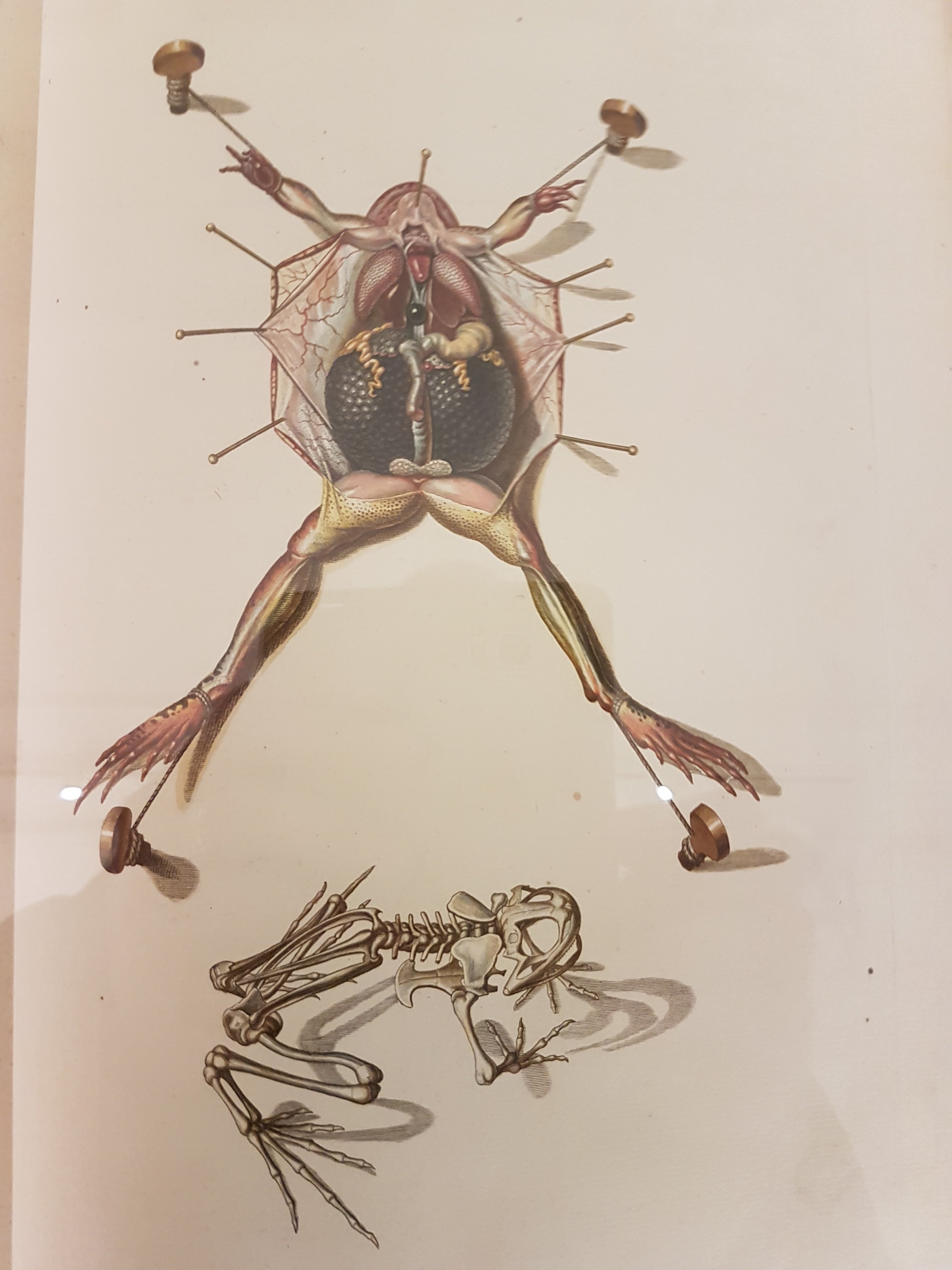
Anatomy to me is beautiful even when it’s “ugly” (messy, wet, mucosal, intestinal, asymmetrical, unlike human, whatever), and that’s a major theme of this blog. Hence I am embarrassed that I hadn’t yet gone to see this Body Worlds spinoff exhibit until now, but can begin to shake off that shame by means of an almost exclusively effusive gushing of blood love for said exhibit. Wow, wow, wow! I went in with no particular expectations, having seen some pictures and knowing some of what to expect, and having other things on my mind. I came out very pleased; the NHM exhibits folks and von Hagens’s crew have created an inspirational spectacle that could do wonders for anatomical sciences and natural history. More about that at the end.

(Warning: possibility of spoilers, but the exhibit is so visual that I don’t think my descriptions can spoil it)

The entrance

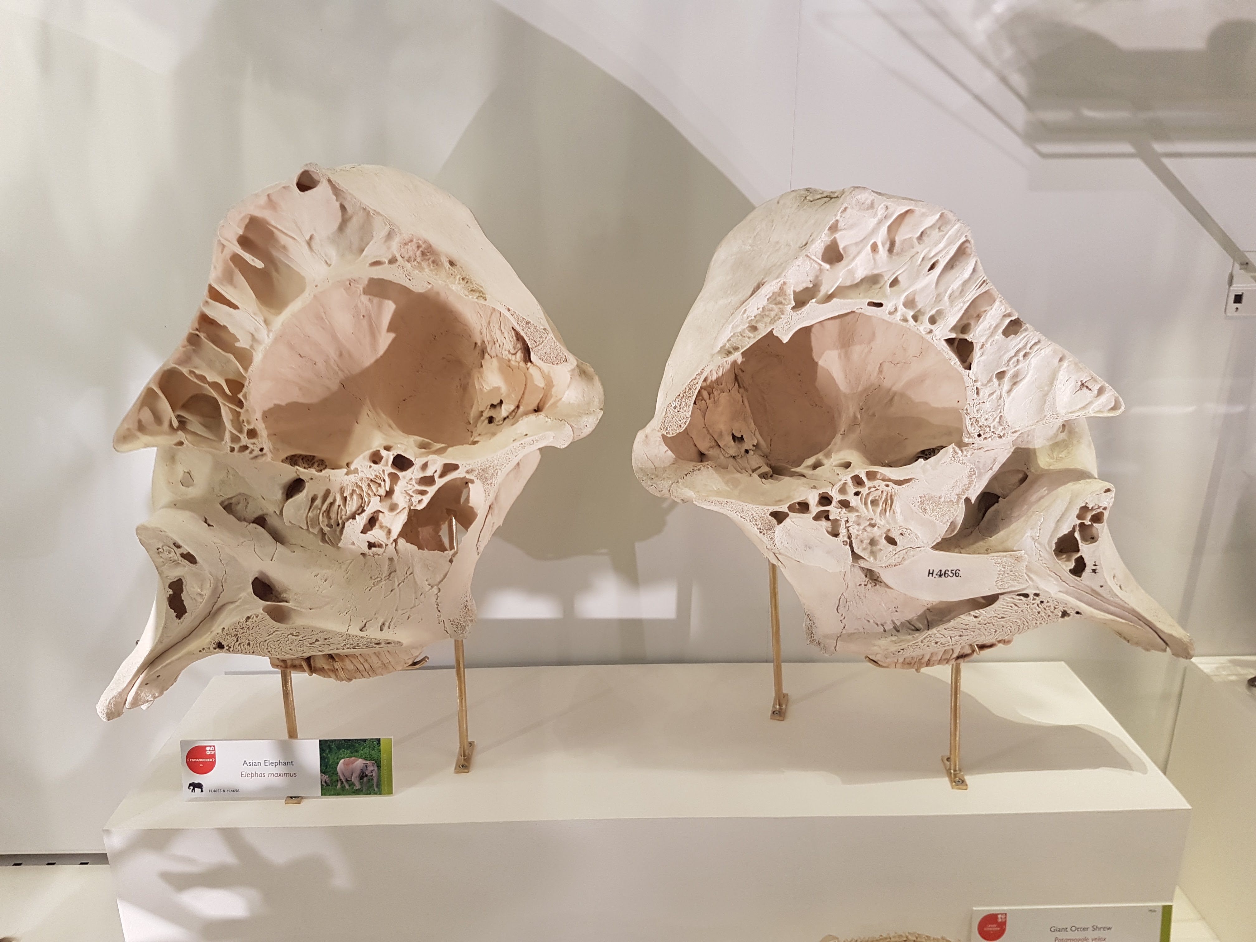
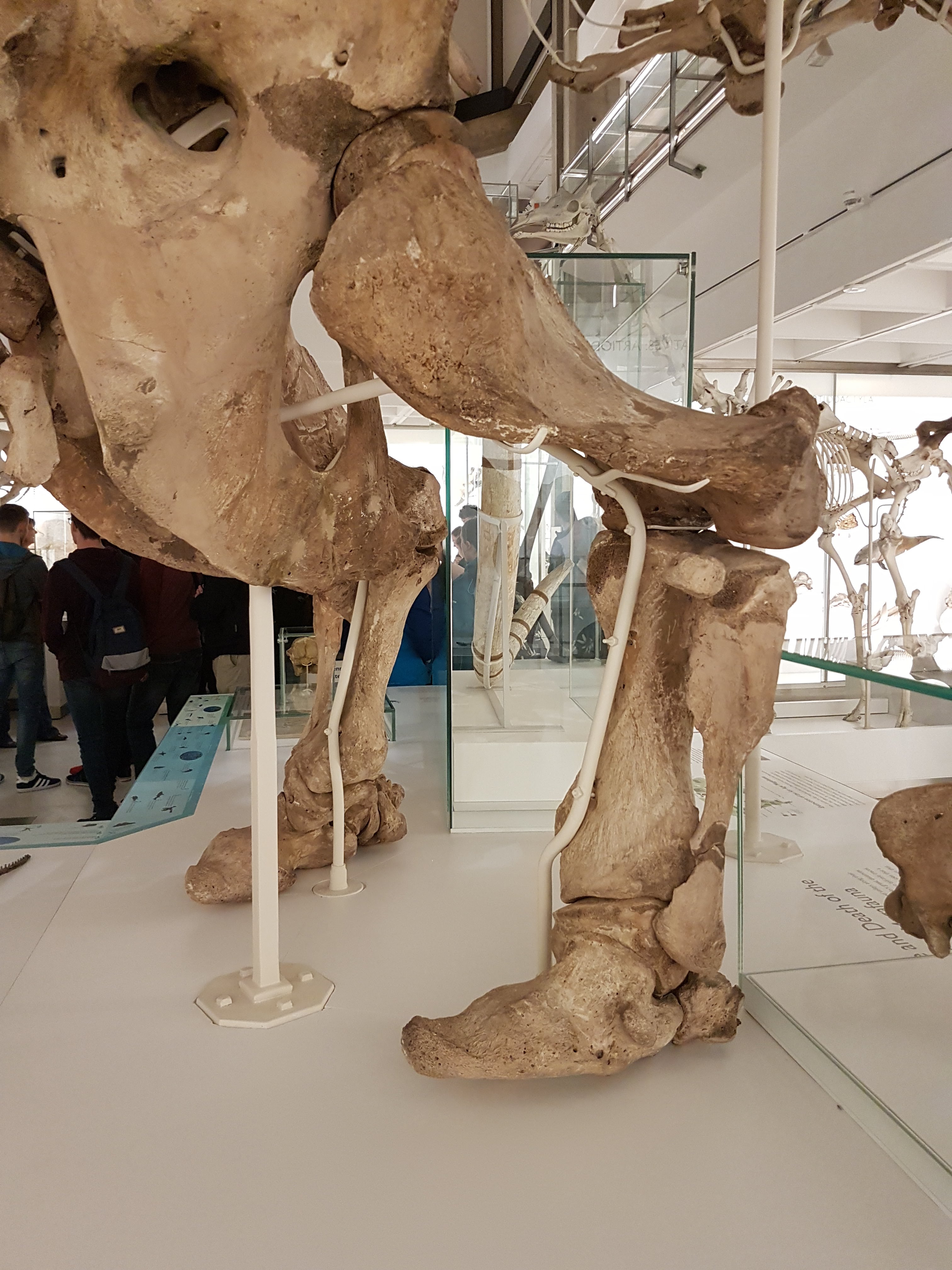
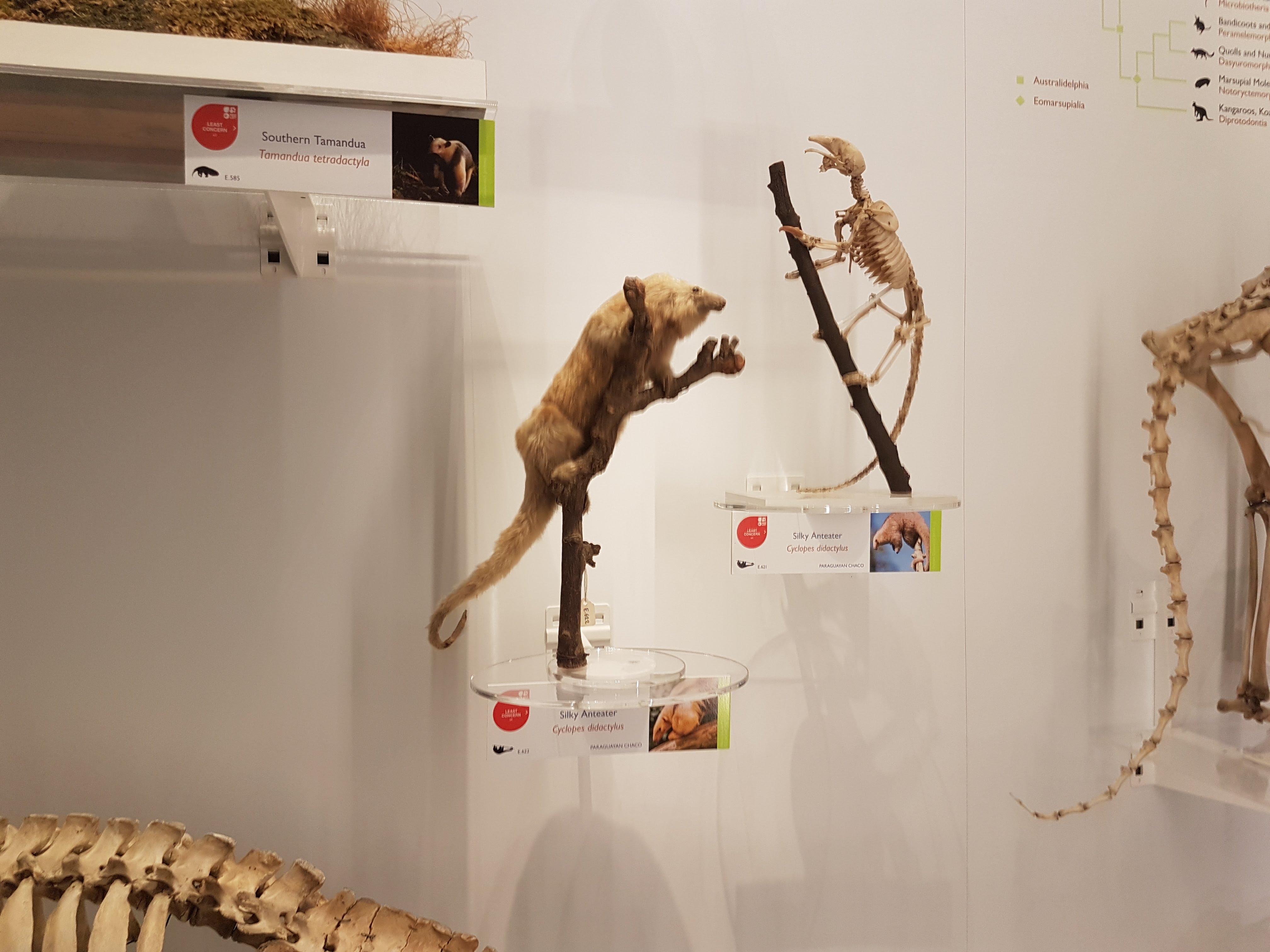
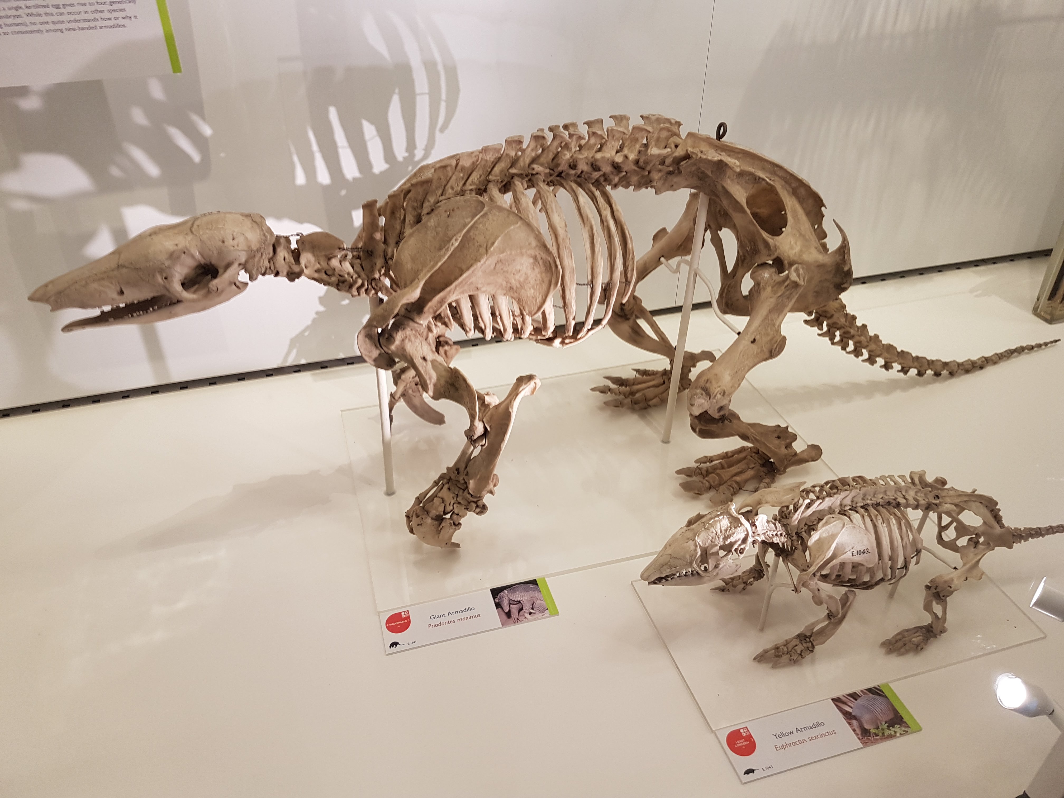
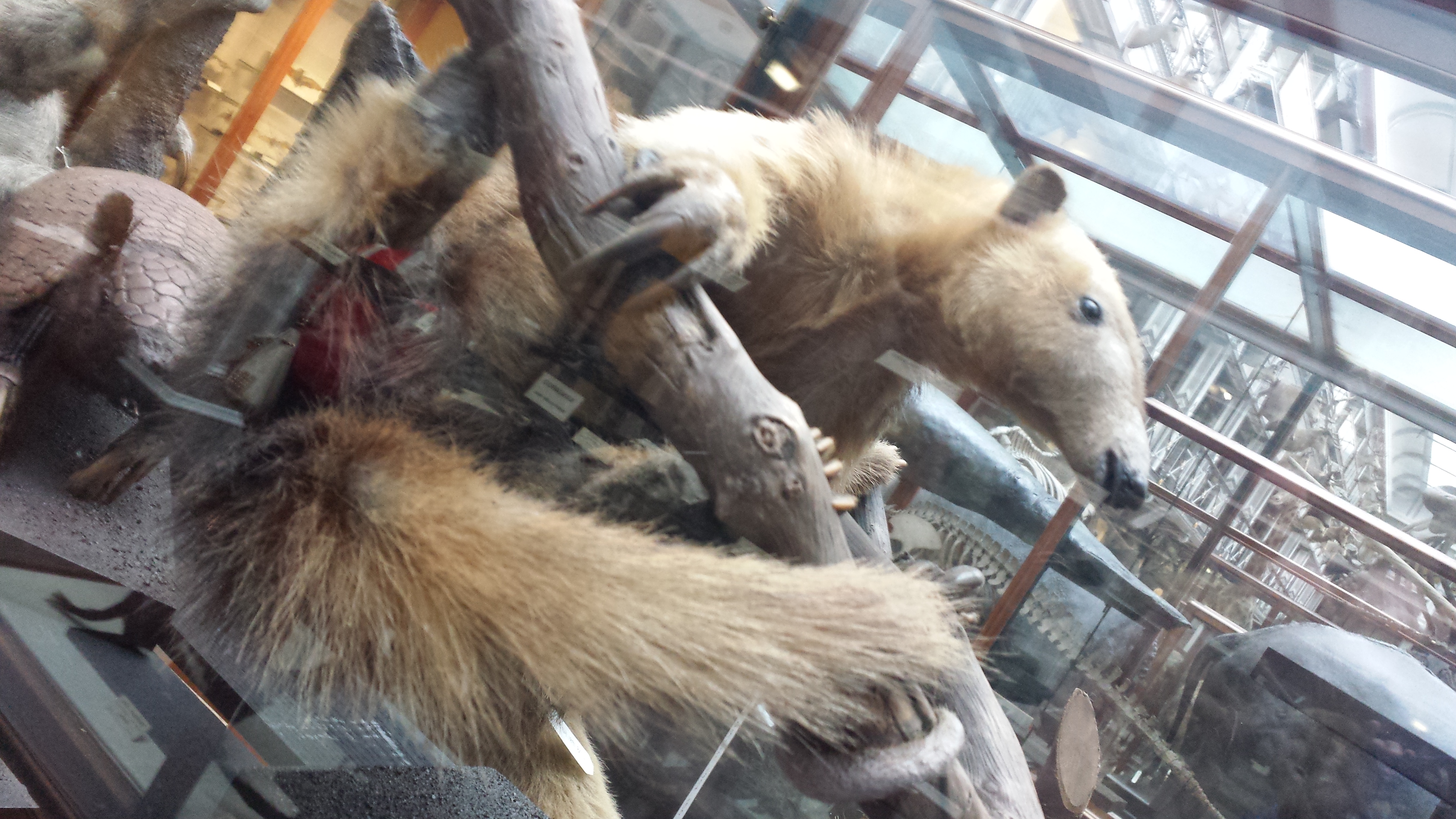
No photos are allowed as usual, so all I have to show you is the entrance and some anatomy pics I’ve interspersed from my team’s research to lighten up the text. I suppose I could have asked for special permission to take photos for review usage but this was a very impromptu visit, and with ~4 months of showing left I may well be back again.

Weighing a hippo; spot on at 1600 kg!

There is a brief panel on homology and why it is the major concept underlying comparative anatomy (and a key part of evolution, co-opted from the not-so-evolutionary ideas of Sir Richard Owen, whom the NHM rightly mentions here). Another panel rightly brings up the issue of ethics, which has plagued Body Worlds before. It comforts the visitors that animals were not slaughtered just for this display and that the NHM applied its strict collections criteria to them. Convincing enough for me, and absolutely necessary to bring up early on.

The entry hall then presents you with about five cephalopods (labelled “squid” and “octopus”—a gripe is that species names/details are not given for most specimens on show) prominently occupying the view. The cephalopods, like basically everything else, are plastinated (by a now US-patented set of procedures, I learned from the exhibit book detailed later). They are stunningly frozen in lifelike poses or with gaping cuts to show their interior anatomy, although there was very little explanation here about cephalopod biology and anatomy (about 1 smallish panel). No mention of Cthulhu. Damn. He’d approve of the Grand Guignol scenery.

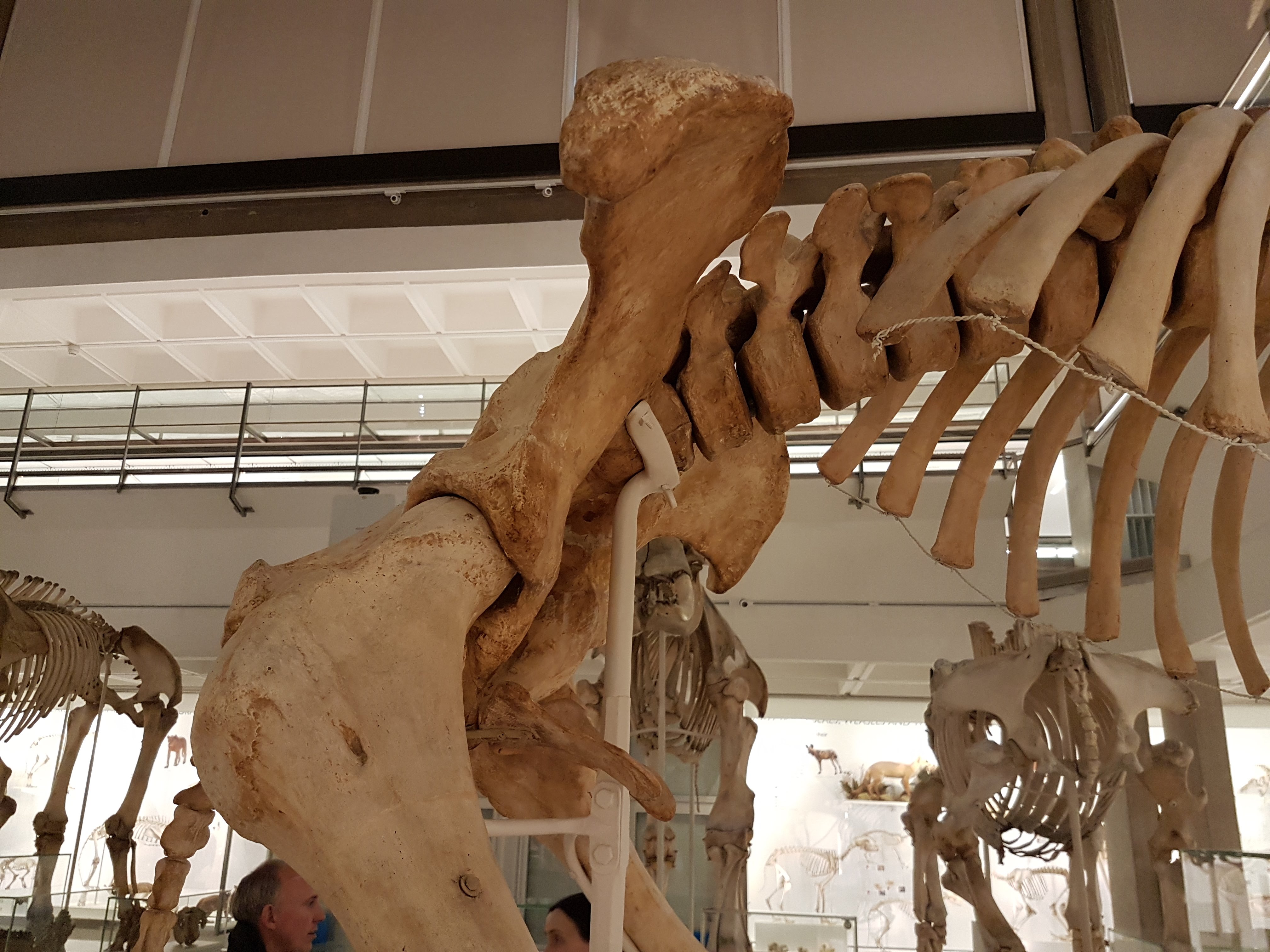
Toward the back of the first corridor of specimens and cases, there is a stunning scarlet haze outlining the body of a “shark” (species not given) with its huge liver lying below it. The haze, a technique used repeatedly throughout the exhibit, is some kind of corrosion cast of the circulatory system, I gather. A bunch of cross/longitudinal sections of cephalopods, crocodiles, fish, horse hooves and other animals decorate blank spaces on the walls, some with labels showing basic features and some just hung like paintings. Fair enough, but a missed opportunity for a bit more educational content here.

Gratuitious Melanosuchus (black caiman) shot.

A smallish whole shark confronts you as you turn the corner from the crimson chondrichthyan; again of unknown classification. One would think a museum exhibit would care about classification beyond “shark,” but oh well, I am banging the same drum here too much and missing the point, that the exhibit is really a visual, visceral expose rather than a deep prose-driven intellectual dissection. On one of the shark panels it is noted that sharks have red and white kinds of muscle used for slower and faster swimming, but not clarified that this is a very widespread vertebrate (chordate?) feature. This forms my second gripe, that a truly evolutionary approach, such as that taken by dozens of the museum’s research staff as their major paradigm of phylogenetic systematics, could have helped the public grasp the evolutionary, hierarchical nature of homology and depart with accurate information about what features characterize groups at which levels. I’m not asking for cladograms laid out on the floor as at the American Museum of Natural History, although maybe that could work, but the exhibit tended to fall back on an outmoded “this animal has this feature, and that animal has that feature, and these are cool adaptations” shopping list approach rather than a modern comparative approach. Granted, almost all museum exhibits fall into this trap, for various reasons and some of them justified. But with a spare word or phrase here or there, this could have been done better without drowning the visitors in that dreaded sea of bloodprose.

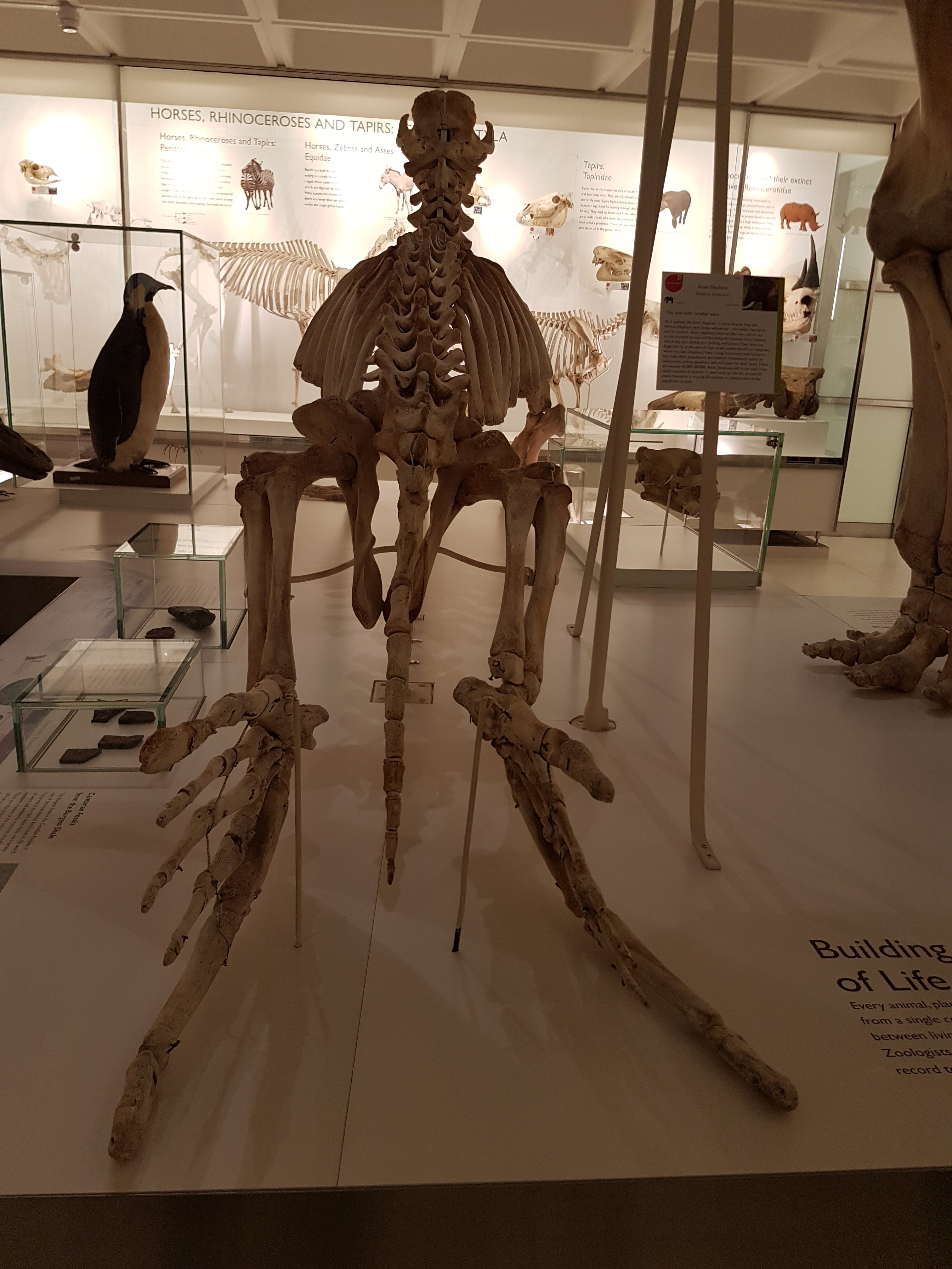
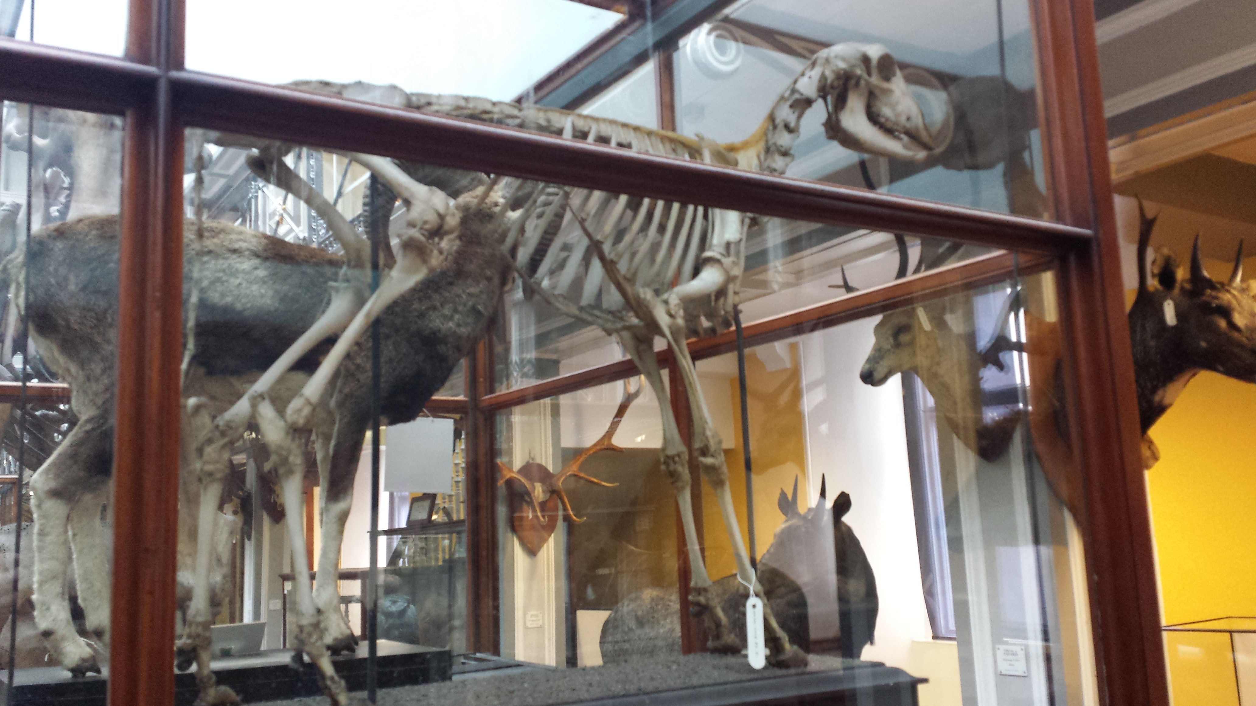
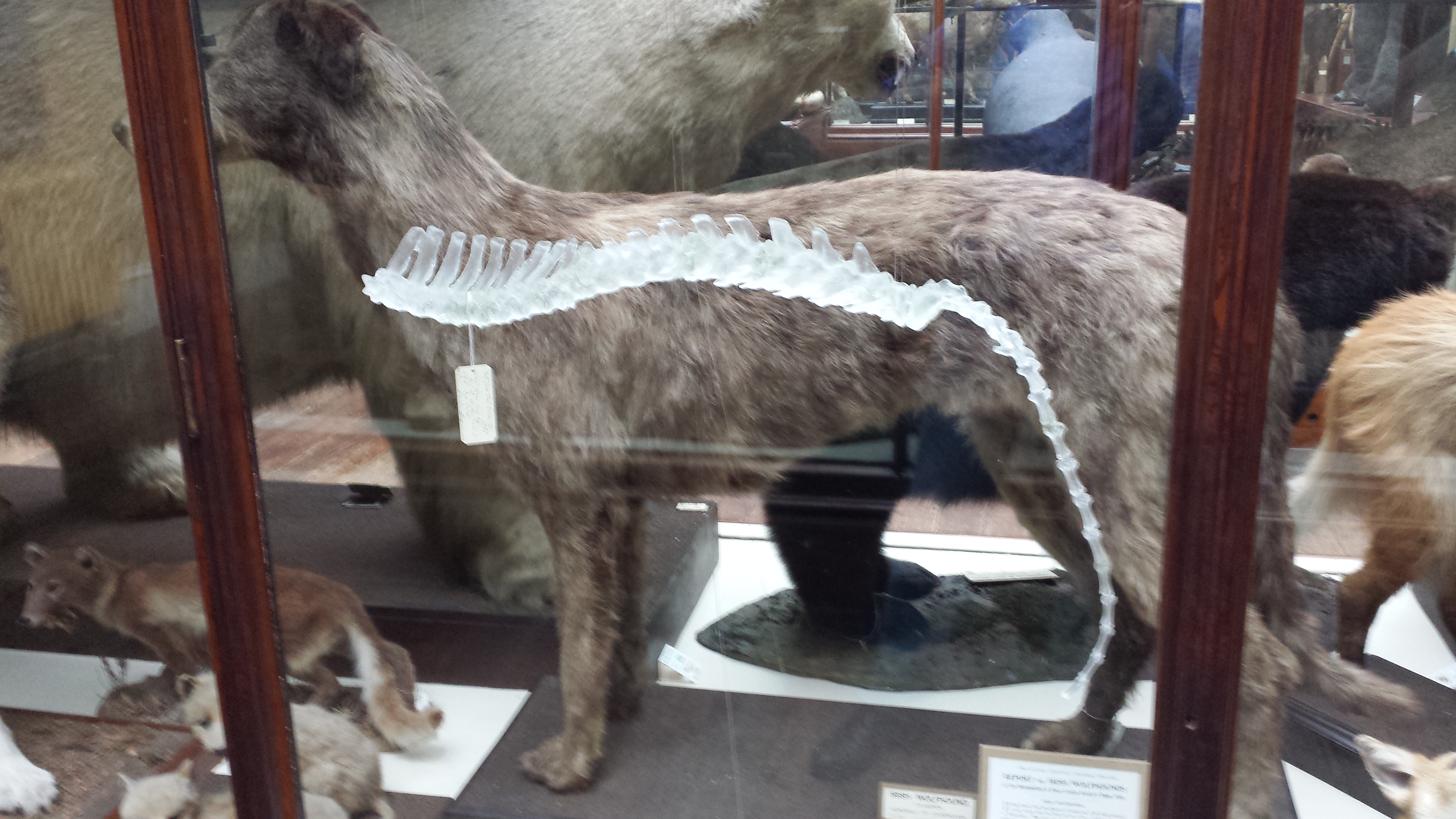
Passing the sharks, we come to one of several thematic sections about body systems, this first one on the skeleton (later, brain/nerves, circulation, muscles, etc.). A few small skeletal specimens of the type that are seen throughout the museum are presented, with a scallop reminding us that skeletons can come in many types among multicellular organisms. There is a horse skull and a stark white whole skeleton of a young-ish ostrich, which was very nicely mounted. However, I was caught off guard by the pelvis, which lacked the curved, ventral “boot” like connection of the pubic bones that ostriches have—presumably explained by its juvenile status although I wasn’t 100% sure it was even an ostrich pelvis. OK, I am having a serious pelvis-nerd moment here; forgive me as my PhD was on this stuff.

Ostrich in the midst of disassembling.

BUT, once again the small interpretive panel had a moment of Fail. The ostrich was explained to have two toes, in contrast to normal birds which have “five”.  HUH? Birds have three main toes and variably also a fourth, inner (first) toe called the hallux, used for perching and other activities including walking. None have a fifth toe; indeed their dinosaurian forebears lost that feature some 230ish million years ago. Just an embryonic vestige of the base of the fifth toe is visible in bird embryos today. Furthermore, the panel said that two toes in ostriches can grip the ground more strongly than more toes in other birds. I know of no evidence that shows this, and suspect that the contrary might be true. The standard explanation for toe reduction in ostriches is that it is a lightening feature characteristic of “cursorial” (long-legged, sometimes fleet/efficient) animals, to make swinging the long legs easier. These errors really should have been caught by involving experts in polishing the scientific content of the exhibit.

But I don’t want this post to grumble too much; wrong message. There was so much to celebrate in this exhibit, which was felt impressively spacious and full of cool specimens! Visitors pass some plastinated whole sheep and goats, with panels nicely explaining that goats and sheep look quite similar on the inside and are evolutionary relatives. Having “four stomachs” (technically, a four-chambered stomach; not four distinct organs that were duplicated) is attributed as a sheep trait, then being a ruminant is said to be a goat trait; this might get a little confusing for non—anatomists (both are ruminants and have similar stomachs).

I learned that goats have an extra tail muscle that allows them to swing up/down as well as side-to-side. Hey, I teach veterinary anatomy and I don’t know that!? I must tuck my tail between my legs in shame, but I am no goat so I do not think I can (do satyrs count?). But I wasn’t so sure that goats, as described, were the first animals to be domesticated—I thought that was dogs? Ahh, Wikipedia says dogs, then sheep, then pigs, then goats? I’m outside my expertise here, I admit, and resorting to Wikipedia out of ignorant desperation. Anyway, here, another instance of coulda-been-more-phylogenetically-specific presented itself: the forelimb of goats was said to be connected to the thorax by muscles and ligaments, not a joint, but this is a feature common to most Mammalia. Although audience attentions might be wandering at this point, waiting for the next big spectacle (goats and sheep are not a big crowd draw, even plastinated), some more care as to what was written would be good. Some reindeer and horses and other animals join in the fun later on. Good, but mostly ‘filler’ (wise to put these in the middle of the exhibit, after sharks/cephalopods and before climax) unless you’re a big fan of fairly familiar ungulates with fairly homogeneous postcrania. OK, my bias is showing…

Gratuitious image of emu curled up for CT scan.

Next along the path, a longitudinal section of a whole ostrich caught my attention. Wow again! I had no idea that one could make a section like this of such a large animal, all in one plastic sheet like a giant microscope slide! I stared at this for a while, wondering how both legs could be fit in a ~1cm thick panel, and gave up trying to understand the technology. Von Hagens, you got me there; I’m stumped. Were multiple sections glued together somehow to produce a pseudo-2D slice from many thin 3D sections? I could not tell, and felt humbled and deeply impressed by the technical skill shown in the exhibits so far…

And then the punches kept coming, one-two-three! The exhibit approaches its climax with a crescendo of great specimens in the final hall. First, another maroon marvel. A whole ostrich, standing with wings askew, showing off its entire circulatory system (plus a few wing plumes for aesthetics) from head to toes! Gorgeous, technically brilliant, and well worth at least a 5 minute walk around (you can stroll around many of the displays in 360 degrees- very good move!). A plastinated whole ostrich stands next to it, and for a muscular anatomy geek like me, it was nirvana. However, in a churlish moment I had to look away from a panel explaining that an ostrich is “too heavy to fly” (I admit some younger visitors may need reminding of this). But then I looked into the big open space of this main hall, and the climax was before me. I think I’d had my climax a few times since this, but wow this was enormous in so many ways. All the ways. Mind-blowingly, vastly, geektastically kewl.

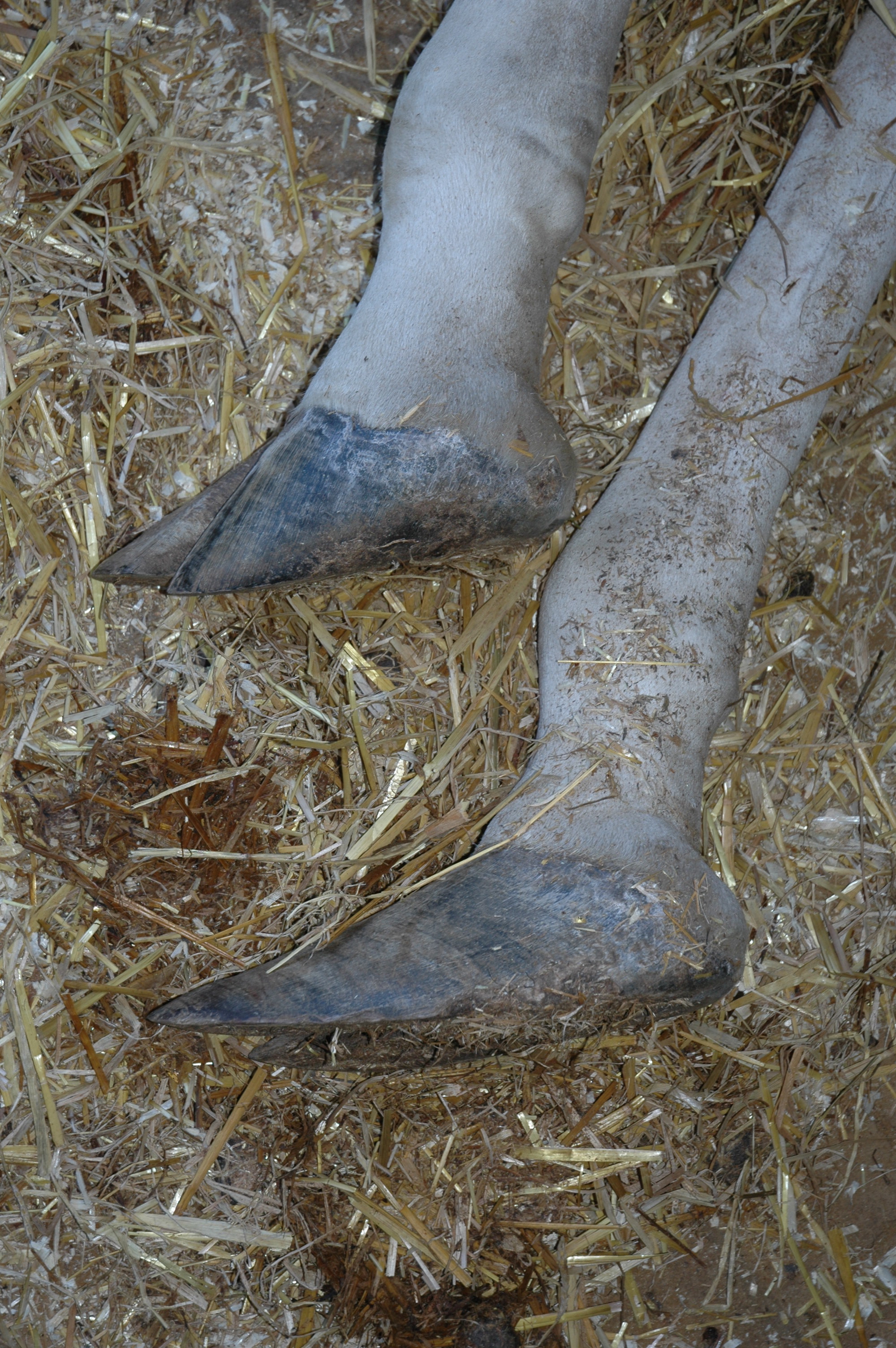
Gratuitious rhinoceros leg.

Across from the two posed ostriches and flanked by numerous smaller specimens, the elephant and giraffe stand frozen in vigil. There is also a lovingly detailed dissection of a huge male gorilla by the back wall and exit, with a panel reminding us that gorillas are (among) “our closest relatives.” The giraffe is precariously poised on one front toe-tip, in mid-gallop. What a great pose! There is the requisite explanation of how they solve the blood pressure problem in their neck (e.g. arterial valves), but also the statement, news to me, that they are the only animals able to ruminate while running. Who figured that out and how? I really want to know! Must be hard to check. (or was walking intended? Are my notes wrong?) Across from the full-fleshed plastinated giraffe (which I could see with my eyes closed after all our dissections from a month ago), there was another visually arresting and technically monumental giraffe on exhibit: one represented completely by small, reddish cross-sectional slices, from head to toes in a standing pose. That took me a while to absorb, it was so lovely, almost like a hanging mobile of morphological splendour.

There is a panel about genes and variation and inheritance. It is brief. (and it belongs there) Thank you. Let’s celebrate anatomy for anatomy’s sake for once!

“But John,” you might say, “What about the elephant? No love for the elephant? The star of the show?”

Zoinks! I want one! Stoic and triumphant (except against death and plastination), the Asian elephant is the centrepiece of the collection. (The book explains it was “Samba” from Neunkirchen Zoo, Germany, dead of some circulatory problem in 2005 and the first one plastinated, plus the inspiration for the animal show). I was speechless and paralyzed for a moment. I didn’t even know how to start looking at the partly-exploded-to-show-its-insides elephant. I actually avoided it for a while, looking closely at the other specimens, and building up anticipation, before stepping up and taking a long, intense look at this tall drink of water.

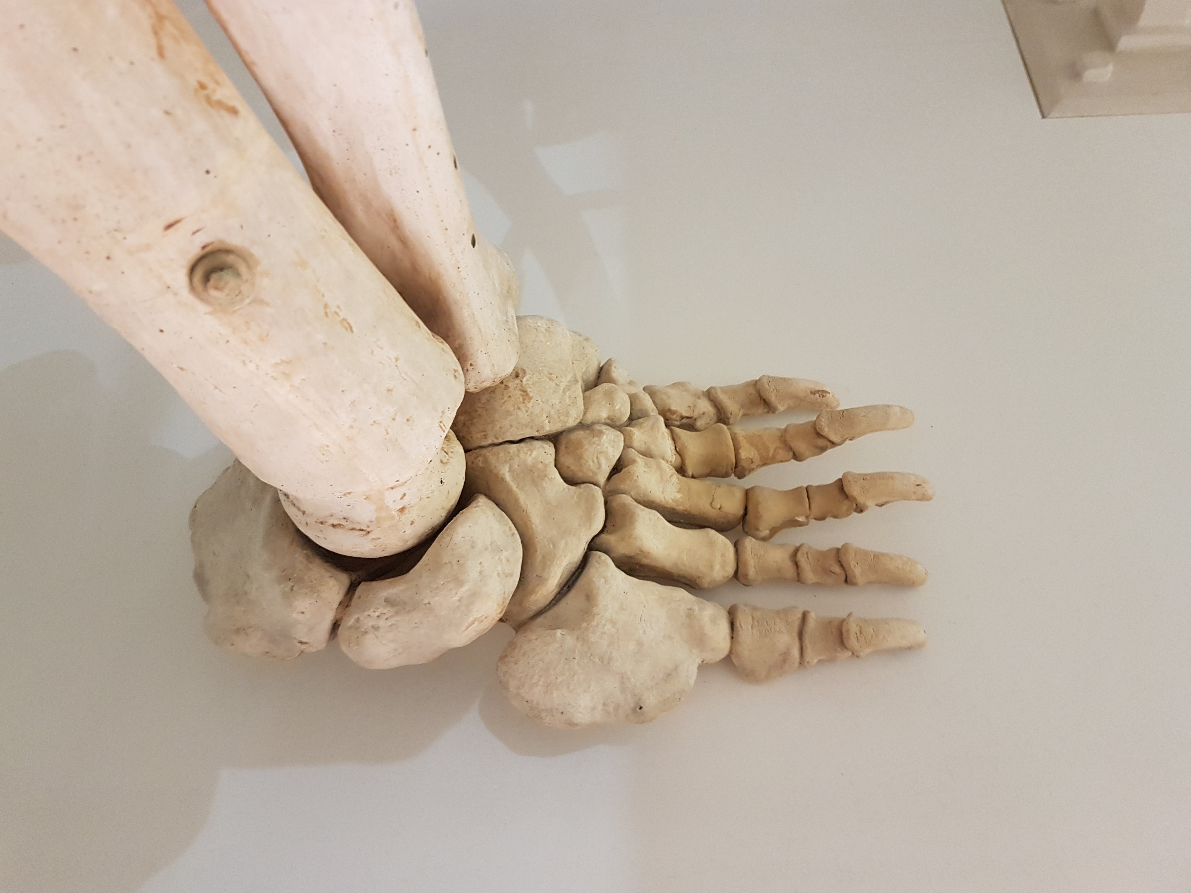
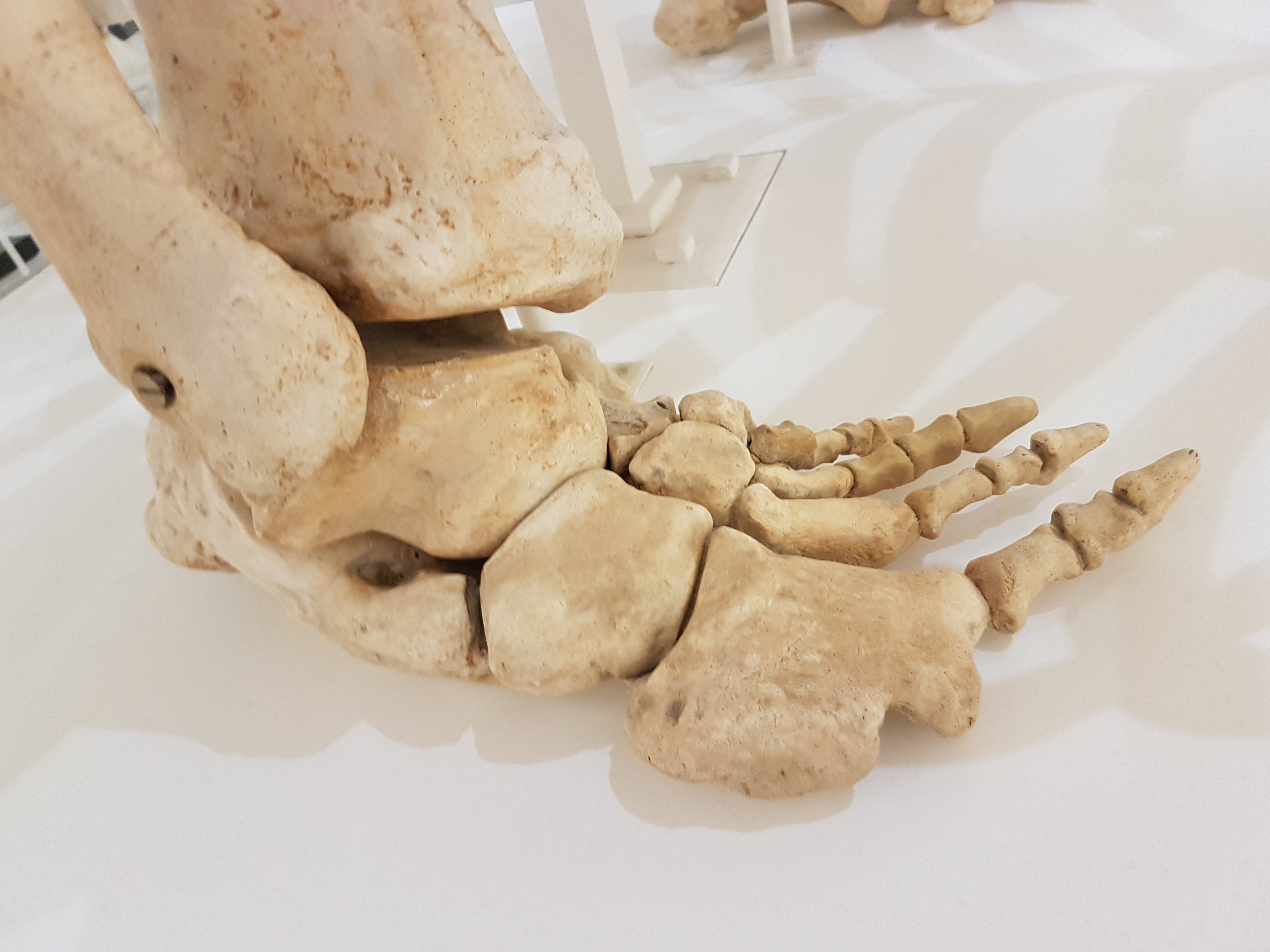
Go see the elephant. If you know basic anatomy, look at its leg muscles. Check out the huge triceps, still attached to the elbow; I like to say it is the size of a graduate student. Same for the analogous superficial gluteal and somewhat-fused biceps femoris muscles on the rear end, around the thigh/knee joint. Huge! I’ve never been able to view a standing dissected elephant, so this really impressed me more than a table full of giant muscle slabs like I normally deal with. And best of all, for me, the “false sixth toes”; the prepollex and prehallux; are visible in all four feet (but not noted anywhere, even in the book; too bad, these things were widely known by anatomists before my work on them). So much to marvel at here. It is an anatomical treasure. I wish I had a 3D image of it to use for anatomical studies- it was so easy to identify every single muscle group (except for a few missing around the shoulder/neck), even in the distal limbs. Hmm, photogrammetry might be possible (nugget of idea begins to crawl around John’s brain like a Zimmerian parasite)…

Behold, the triceps muscle of an elephant!

Behind that gorgeous elephant, don’t miss the wall mountings of two cross-sectional slices: through the head/neck of a moderate-sized elephant (How!?!?) and distal leg (no predigits but good features). And definitely don’t miss the stool (non-fecal, furniture form). I almost did. A wooden stool is shaped like a newborn elephant and a cross-section of the body is adhered on top of it. I assume you cannot sit there, and I am very glad that it was not, as I first imagined, an actual plastinated baby elephant turned into a stool. That would be bad taste.

The exhibit is in very good taste, without exception, and although I am gore-desensitized to say the least, it is not gory in my view. The plastination process preserves the reality and even some of the colour faithfully, but renders it just unreal enough (past uncanny valley territory?) that it should not be very disturbing to most viewers.

You can’t leave with your own photographs, but you can be schnookered into buying the exhibit book (£12.99) and a couple of packages of nice colour postcards (£4 for six; excellent quality images and cardstock IMO). The book and postcards show many of the exhibit specimens but not all, and include some others that are not on exhibit. I was saddened that the bear was left out—very cool image of that in the book. I’ve only skimmed the book a bit. I was annoyed by a few mistruths about elephants (25mph running speed, “have no ankle joints, which is one of the reasons why elephants cannot jump”, the bones “do not contain any marrow”—wrong, 15mph and there are ankles, they just are not very flexible (but not immobile either); also the bones do contain marrow (how could a large vertebrate survive entirely without it???) but just not as much of it per unit volume, due to lots of spongy bone). But I am still very happy with the 139 pages chock fulla pretty images, which is all I really wanted. Indeed, the book is a great pictorial anatomical reference- some of the species such as elephants and giraffe lack a really good anatomical resource in the modern, or any, literature! The exhibit shop also sells some good anatomy texts, mostly on humans but I recommend “Animal Anatomy for Artists” very strongly; I use that regularly in my own work.

So, £29.99 of schnookering later (haha, poor victimized me!), I emerged and reflected more on what I’d seen. I’m still a bit giddy about it all. I like the minimalism in most aspects- black backgrounds, minimal signage (but just enough to make it educational—when they got the facts right), focus kept on the specimens. Well done there. The spectacle of the specimens I’ve raved plenty about- it is not at all disappointing. It is AWESOME in every sense. I feel I easily got £9 of value from the ticket, and would (probably will!) pay it again. It is a profound experience to see the rich anatomical detail exposed, and be able to circumnavigate the specimens to absorb multiple perspectives. If you know some anatomy, you’ll be doubly rewarded at least, and if you bring your own phylogenetic perspective that can be trebled.

Baby white rhinoceros. Sad infant mortality.

What makes me happiest after my visit is realizing that we are in an anatomical renaissance for science and public interest therein. Exhibits like this and documentaries like “Inside Nature’s Giants” have tapped a public interest and curiosity in the wonders of basic anatomy. Anatomy is at the core of so many biological sciences and is so immediately accessible to people, because we all have anatomy. Anatomy is at the crossroads of art and science; it is visual, variable and complex, yet concrete, objective and easy to relate to. “Animal Inside Out” is a spectacular blend of art and science. They nail the artistic aspect, and the science is done reasonably well (despite my few gripes)—the exhibit’s science speaks for itself, in a way, although many visitors will need a nudge to grasp that.

I’d like to make a call for a permanent exhibit of the likes of “Animal Inside Out” in the UK. We deserve this! Museum exhibits could use something new, other than lame, quickly broken digital pushbuttons and bland skeletons devoid of soft tissue context (although the latter can be sufficient, e.g. at the Paris NMNH). That’s what makes “Animal Inside Out” (and Body Worlds) such a hit- as Hagens is quoted on the book dustcover, animal anatomy that goes beyond digitized abstractions and dusty bones is able “to sharpen our sense of the extraordinary by looking at the self-evident.” I could not say it better myself. This exhibit is extraordinary; that is self-evident after even a peek. It is a loving tribute to how fantastic the totality of animal structure is. Go! Enjoy. Absorb. Gape. Stare. Thrill. Revel. Think. Question. IT’S BEAUTIFUL.

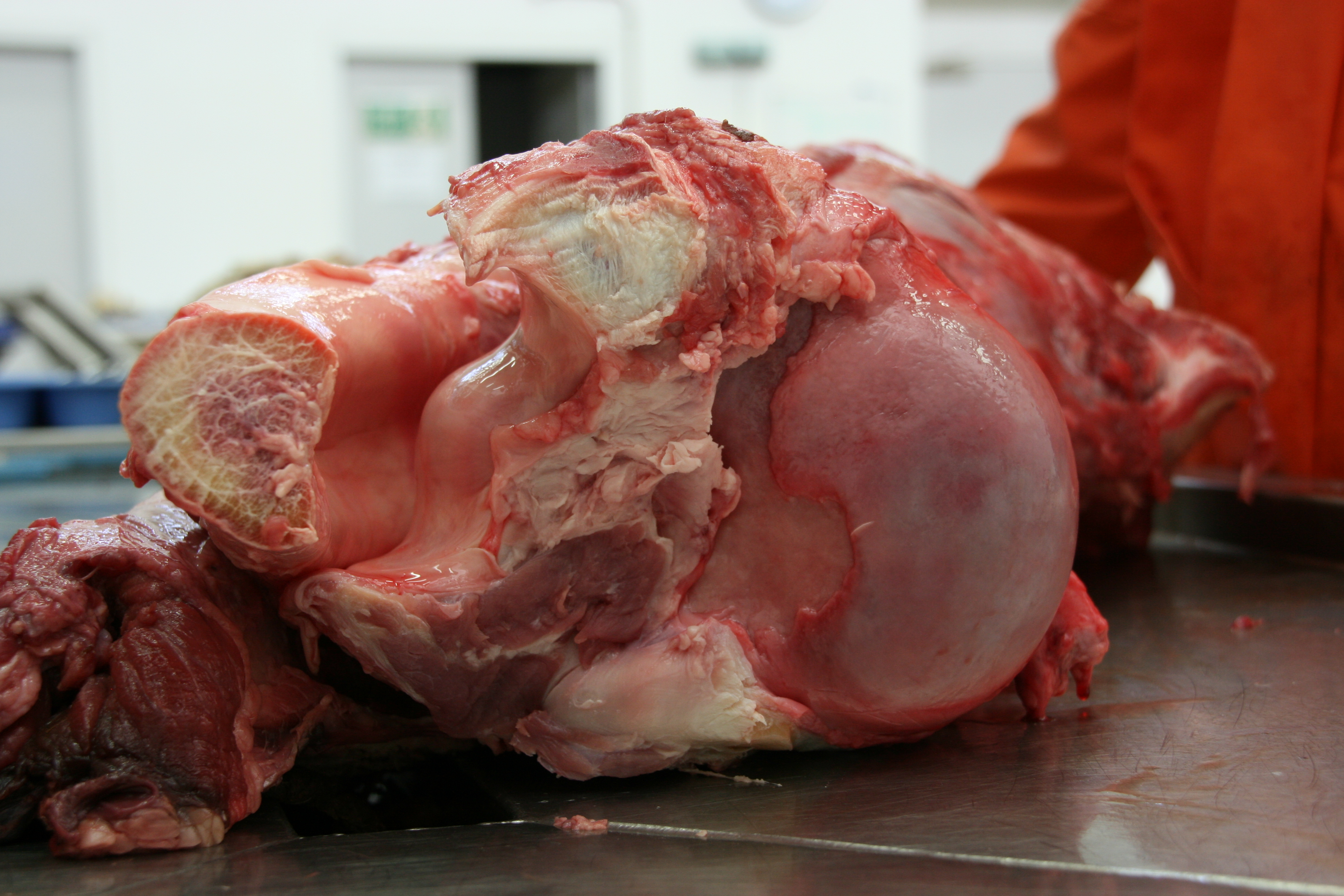
Impressive hippo mouth says “Farewell for now.”

Edit: @samjamespearson on Twitter has kindly posted some photos (for free NHM/AIO publicity) of the exhibits and here are the links, now that they’re out there– SPOILERS! And thanks, Sam! I don’t think these really spoil the intense visual experience of actually being there and walking around the specimens, not at all.

octopus, whelk, squid, needlefish, scarlet haze of shark, hare brain, cat nerves,  bactrian camel, another camel,  bull (I forgot to mention it; this one was pretty great!)

Read Full Post »

There’s no better way to kick things off after a holiday than with a celebration of the Inside Nature’s Giants series, which I had a small part in early on, including these shots I took during the time they spent filming at the RVC >3 years ago (!?!?); most of these animals spent multiple holidays inside The Freezers:

Elephant arriving…

Elephant revealed

Private moment with elephant

Stunning emergence of The Guts

So you are impressed by the guts too, ehh? It was pretty amazing to watch it happen. The tension was intense- the animal had been dead for a while and was rather bloated. So cutting it open was a task gingerly taken…

Bloated elephant

RVC dissector Richard Prior stuck a scalpel in the upper abdomen when the time was right… the piercing whistle and the sulphuric odour silenced the crowd watching… and then quickly out came the guts.

Everyone was pretty amazed by the scale.

The guts just went on and on…

Not a 1-person job by any means.

Spreading them out to see the whole GI tract.

I waited patiently and watched the show filming; what a great, professional crew. Then I got to take the legs away for our research.

But not just elephants, no sirree! The Windfall Films/ING team filmed giraffe, crocodile and big cats episodes (4 total) at the RVC too; a crazy period of a few weeks (including a major blizzard that hit us during the croc filming) in 2009. Some of the stars follow:

Frozen lion waiting for CT scan, shot 1

Frozen lion waiting for CT scan, leg shot

Frozen lion waiting for CT scan, shot 2; eerily contorted pose

Frozen tiger waiting for CT scan, shot 1

Frozen tiger waiting for CT scan, shot 2

Frozen tiger waiting for CT scan, shot 3

…and here is the tiger’s head after scanning

…and I’m rather fond of that tiger’s neck– check out the hyoids (roaring/tongue apparatus in throat; bottom of movie)!

…and here is the adult Nile crocodile’s head after scanning

…and another view of that big Nile croc, just because I like how this reconstruction turned out

…and here’s one of the small (~1m long, 10kg) juvenile Nile crocodiles from the show, with a pilot CT scan showing the skeleton nicely- and possibly a last meal or stomach stone on the left side of the abdomen (bright white blob; I need to check this now that we’ve dissected it)

Foetal giraffe; stillborn; from the show, in process of dissection in our lab to measure its limb anatomy. Trust me, it looked –and smelled– better on the inside than it did from the outside. Eew.

How most of the specimens from the first 4 episodes ended up after all dissection was done (part of my/RVC’s collection of skeletons). Sadly, I did not get great photos of the 3.7m Nile crocodile or the two giraffes before they were reduced to bits, but I do have the skeletons and CT scans.

Giving a tour (including The Freezers) to A Certain Esteemed Visitor.

(Another) Gratuitous shot with one of the sweet old Red Kangaroos at Alma Park Zoo near Brisbane, Australia. Experiments on hopping we did there will be briefly featured in the new Inside Nature’s Giants show on Channel 4, 16 April @2000- details at http://t.co/SkjsMeVC.

Read Full Post »

Older Posts »