I heard that the UMZC has some new exhibits open, so back I went! For the prior posts see here (mammals/basement) and here (everything else). Another photo tour! There’s a special (art) exhibit, too, so stick around to the end.
All images can be clicked to mu-zoom in on them.
Stomach-Churning Rating: 3/10 mainly skeletons, some preserved critters in jars.
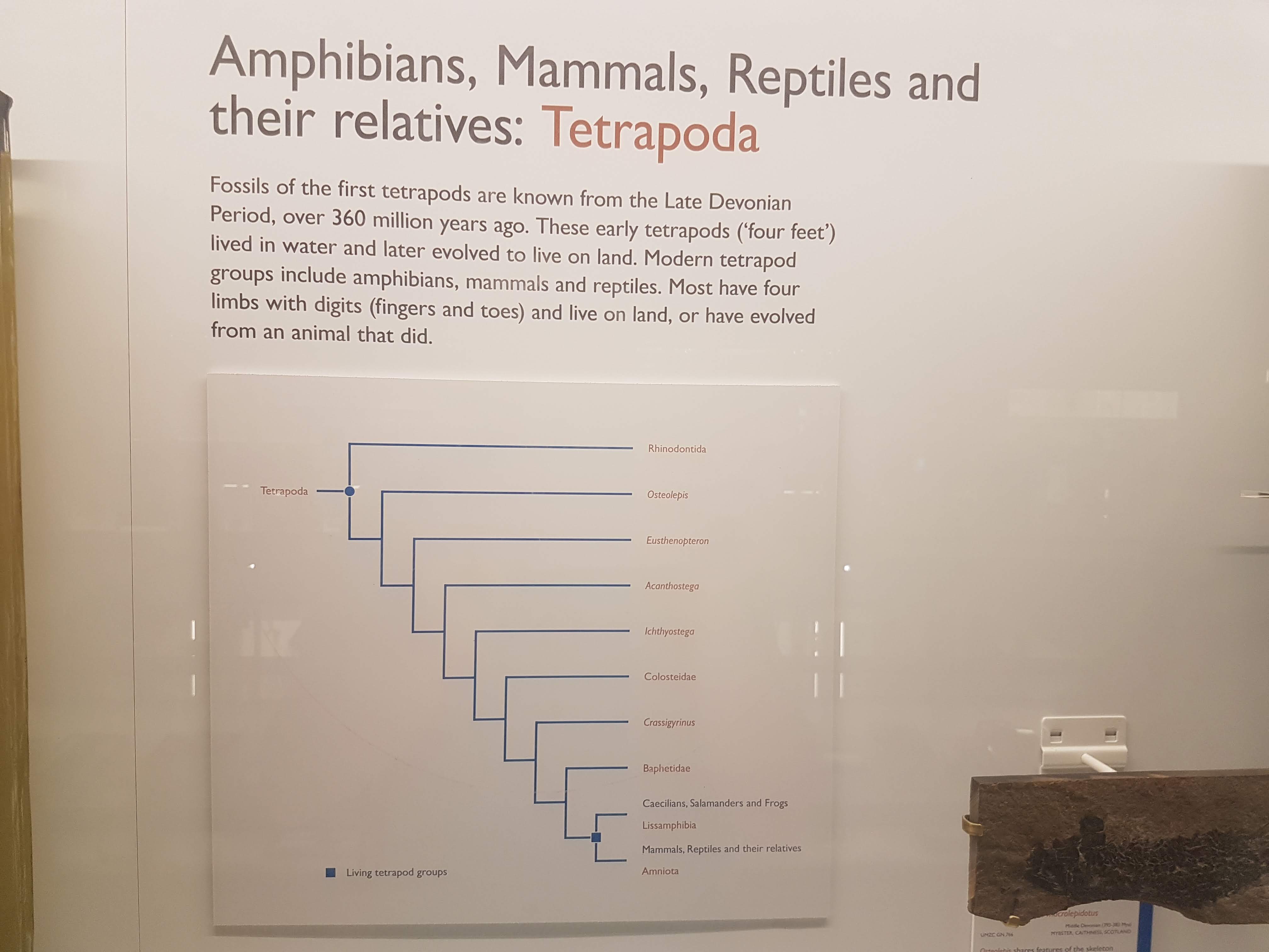
The first new section is an elaborated display on reptiles.
Clevosaurus, a Triassic relative of the living tuatara reptile, Sphenodon. Nice fossil hindlimbs!
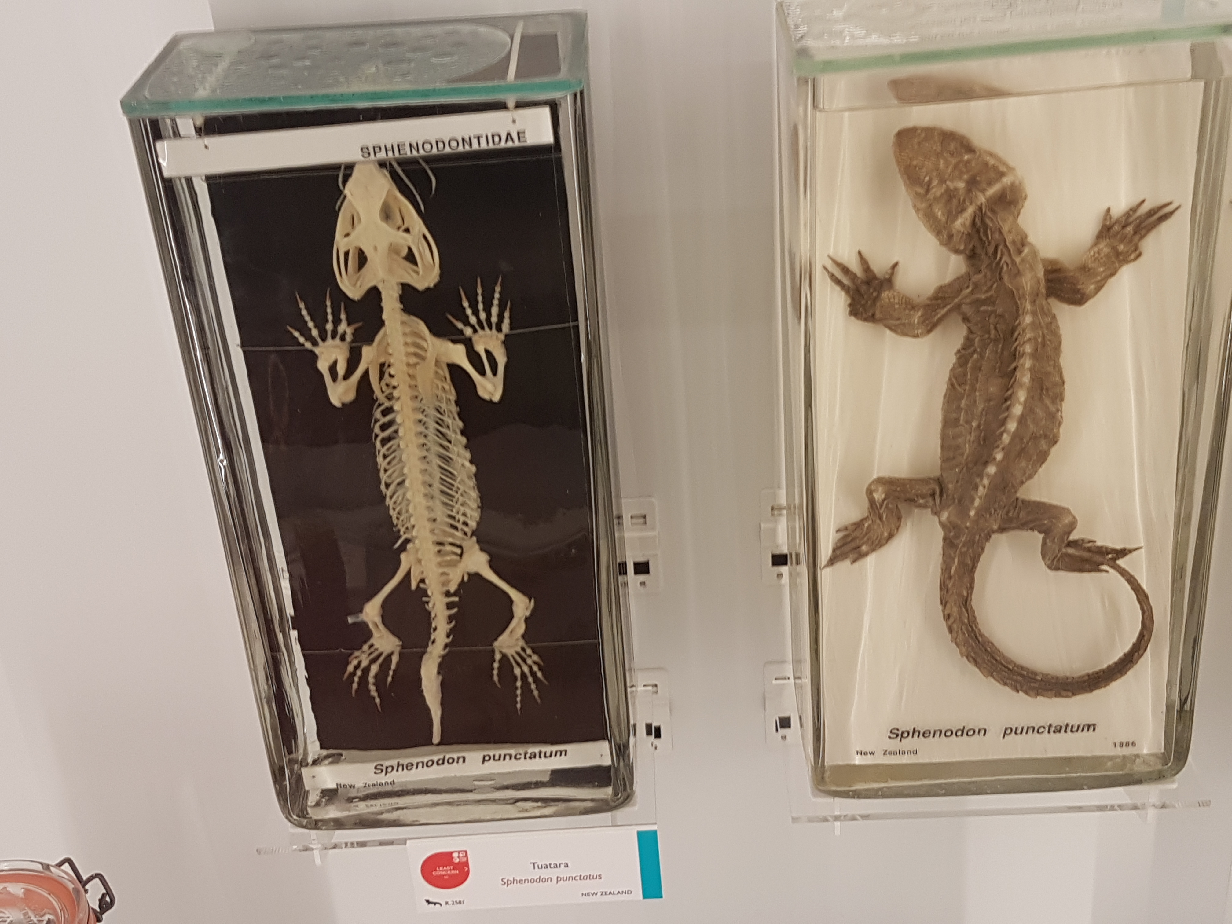
Tuataras (Sphenodon), skeletal and preserved.
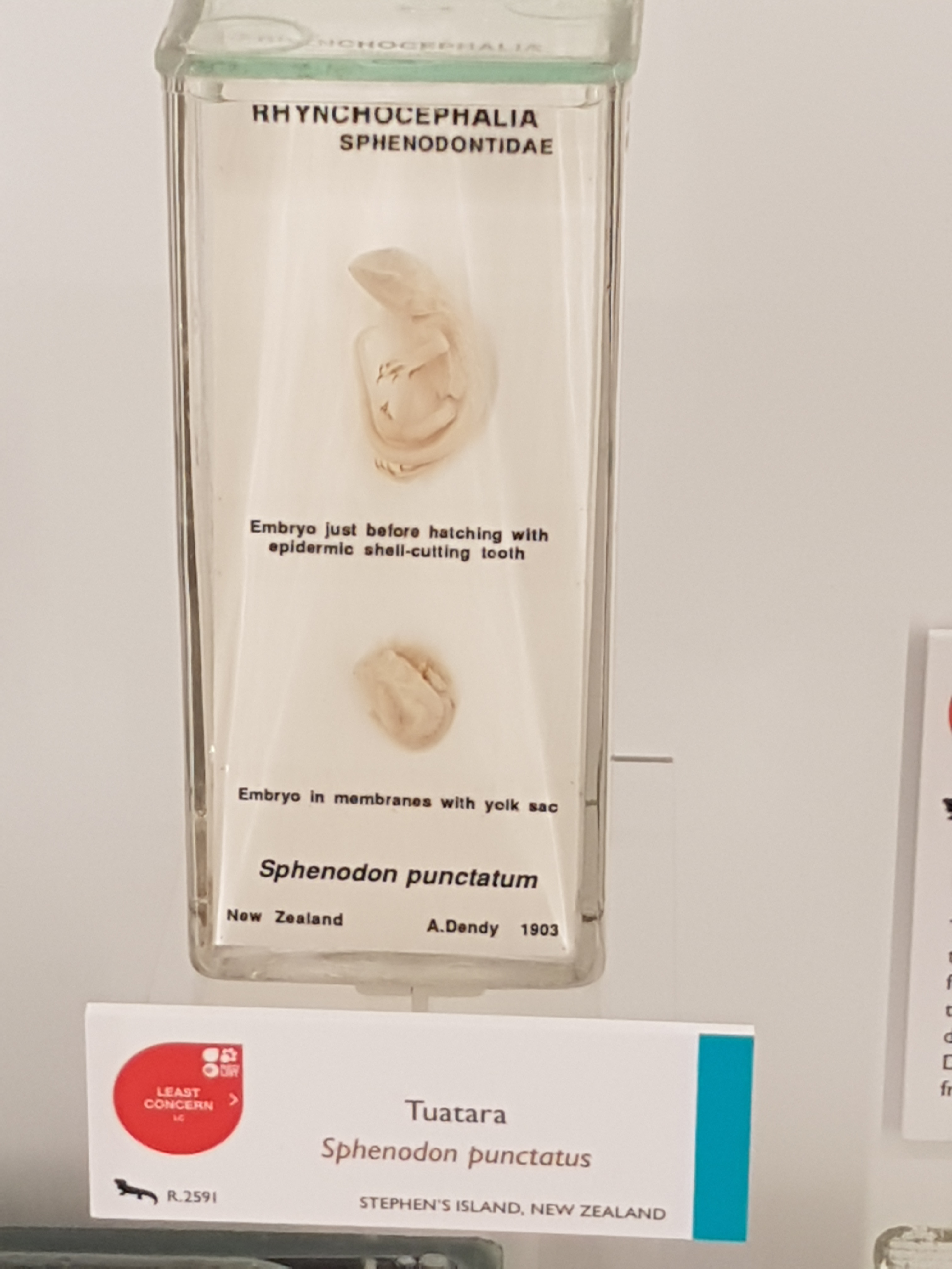
Tuatara embryos!
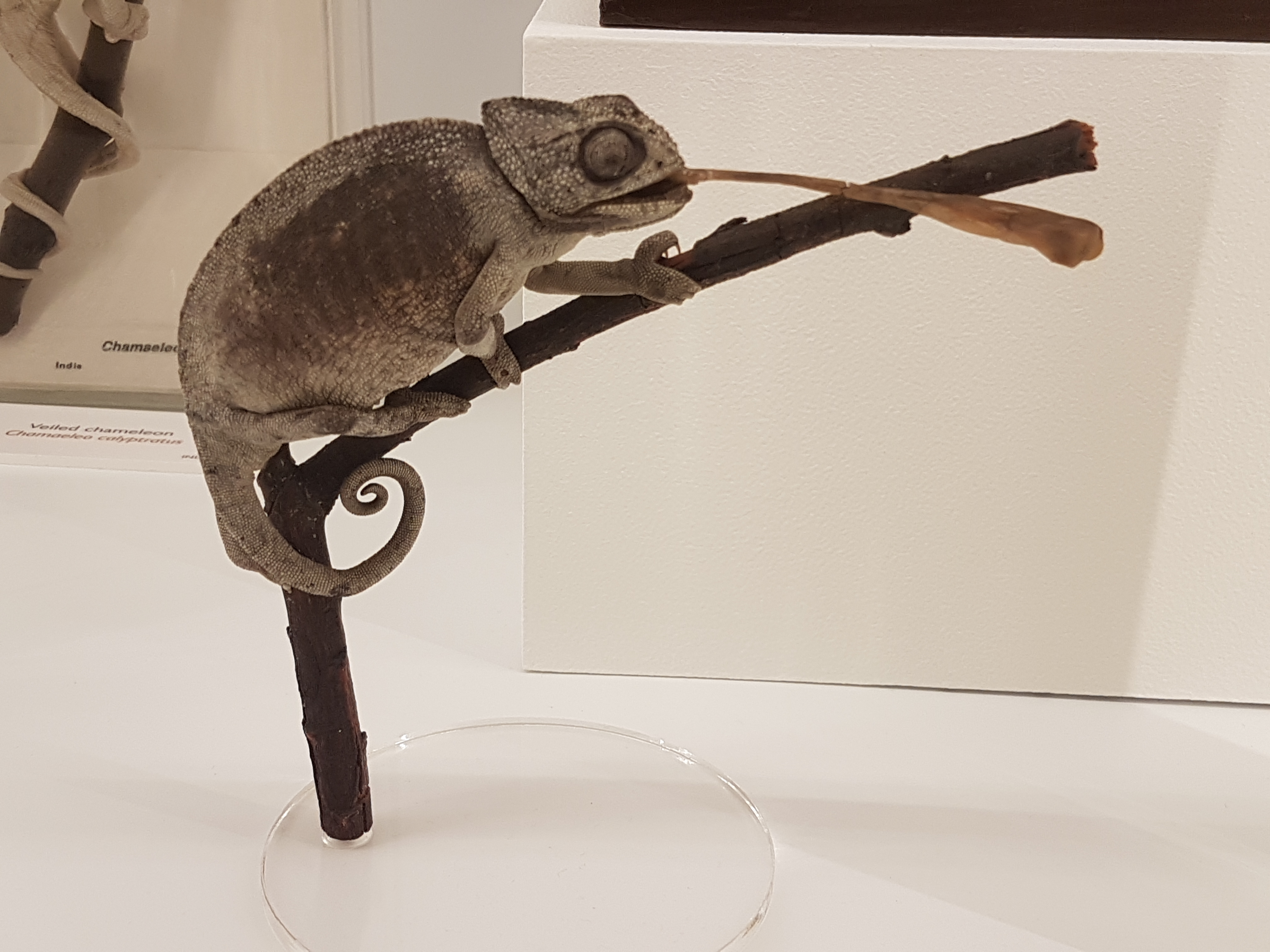
Nice chameleon mount w/tongue extended.
Thorny devil (Moloch), de-thorned and in the flesh.
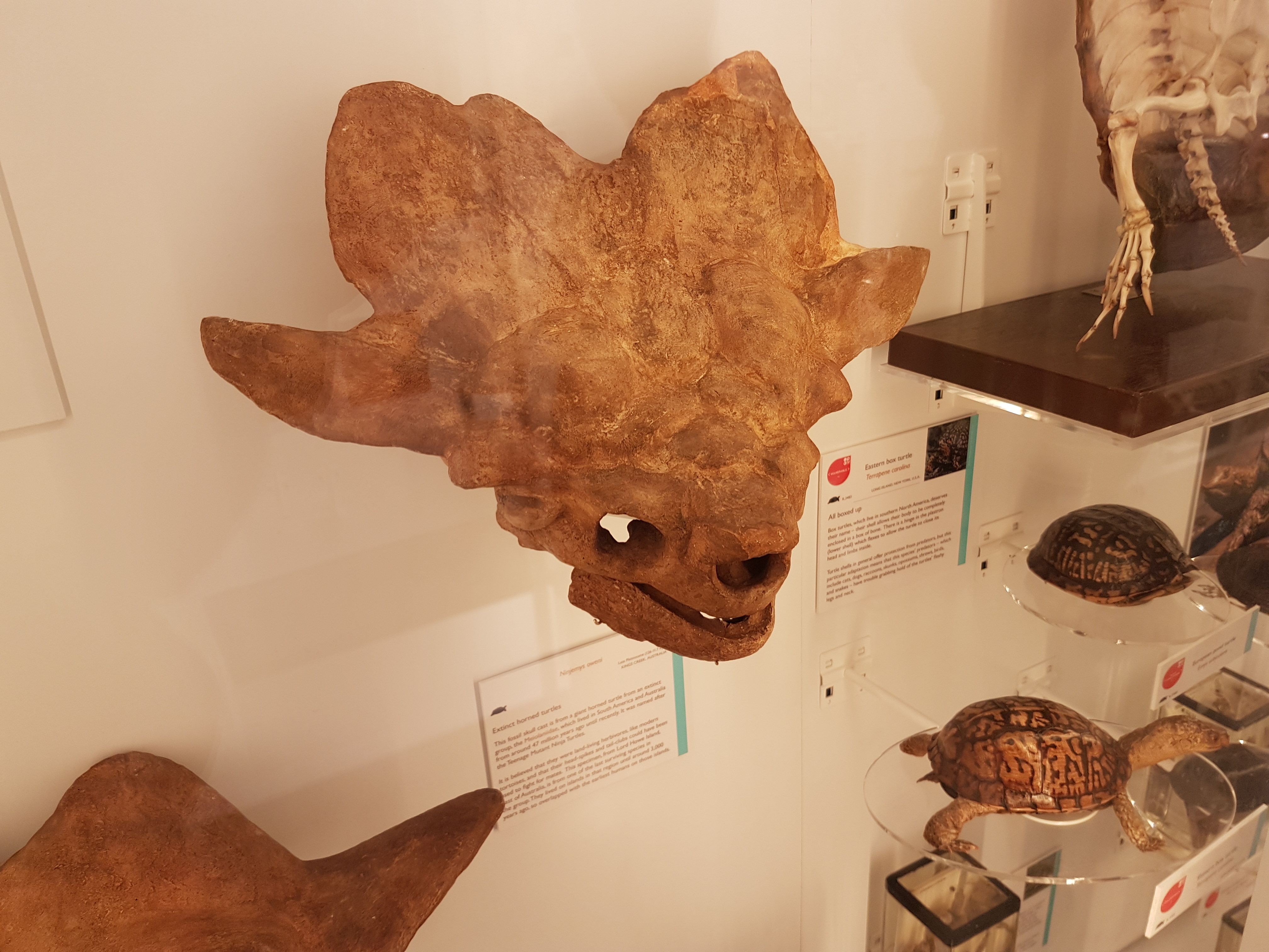
Skull (cast) of Ninjemys, the giant turtle.
Pipe snakes! Snakes with vestigial hindlegs.
Istiodactylus pterosaur snout-tip (real fossil) from the Isle of Wight, UK. Nice 3D fossil.
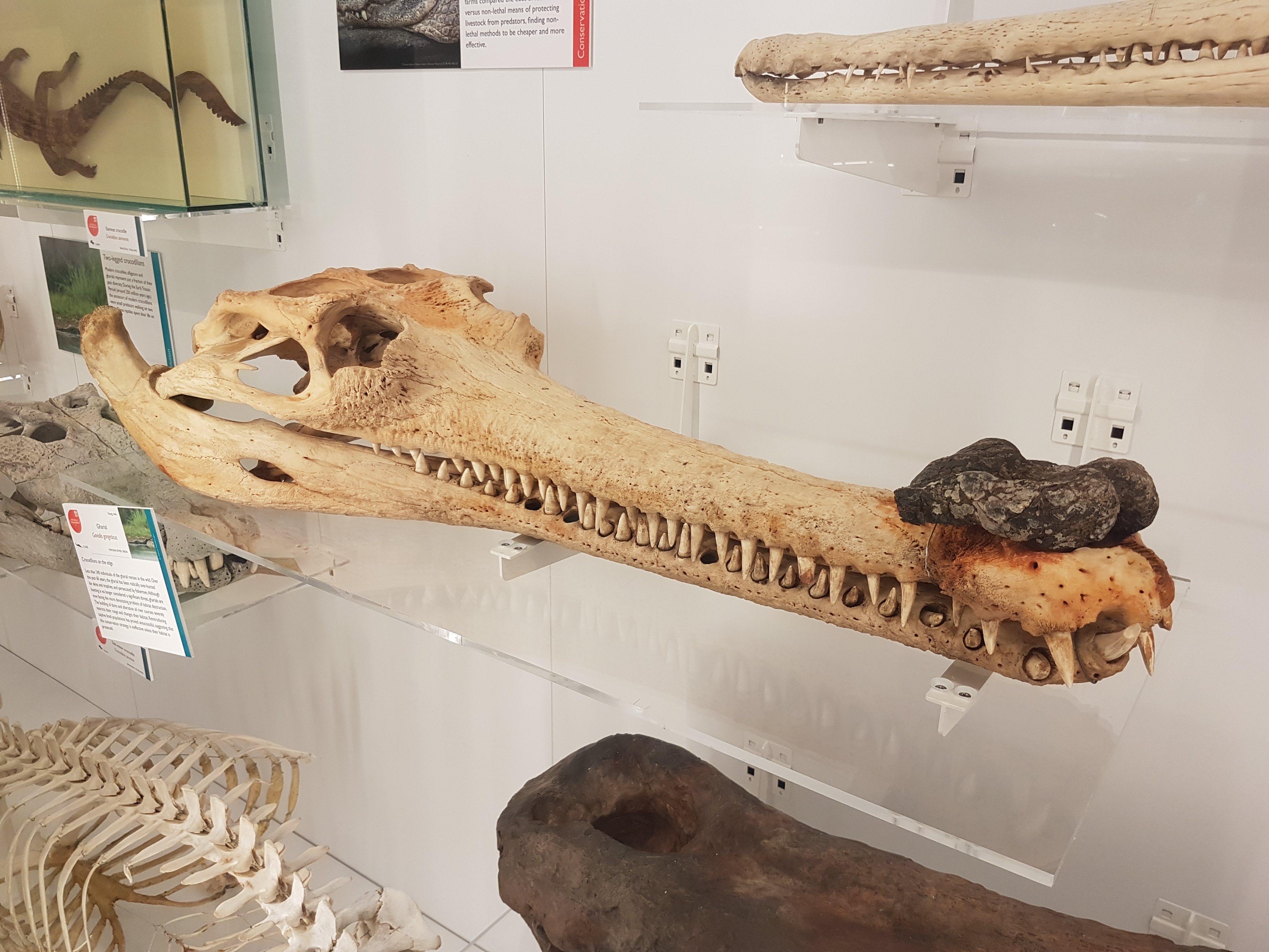
The gharial (Gavialis), male with protuberance on snout (mating-related).
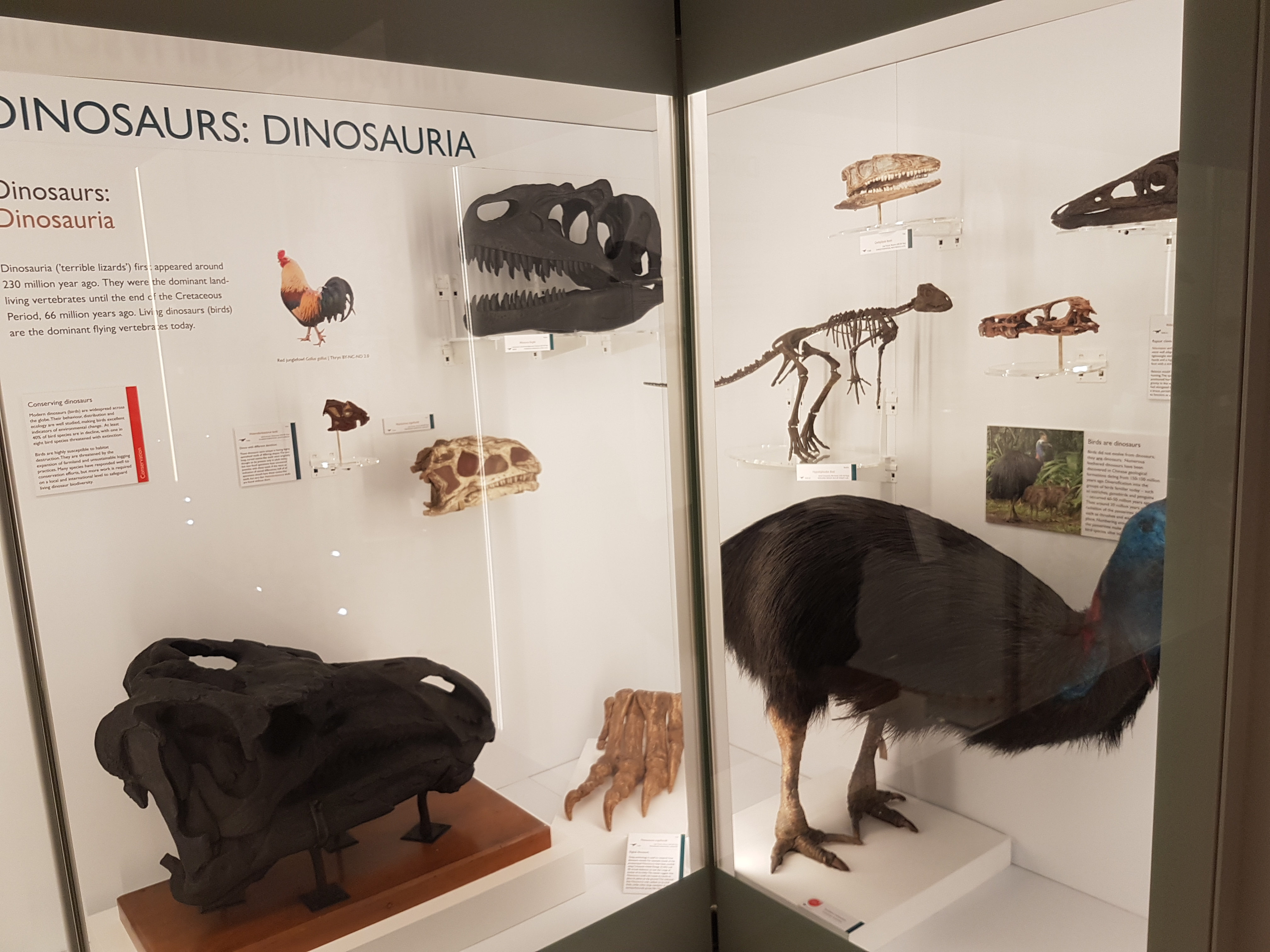
I dub thee Dinosaur Corner! For dinosaurs, the Sedgwick Museum across the street (also free; also classic and awesome) is the place to go but this corner does a good job fighting for the scientific conclusion that birds are dinosaurs.
And now a change of pace. On to the special exhibit!
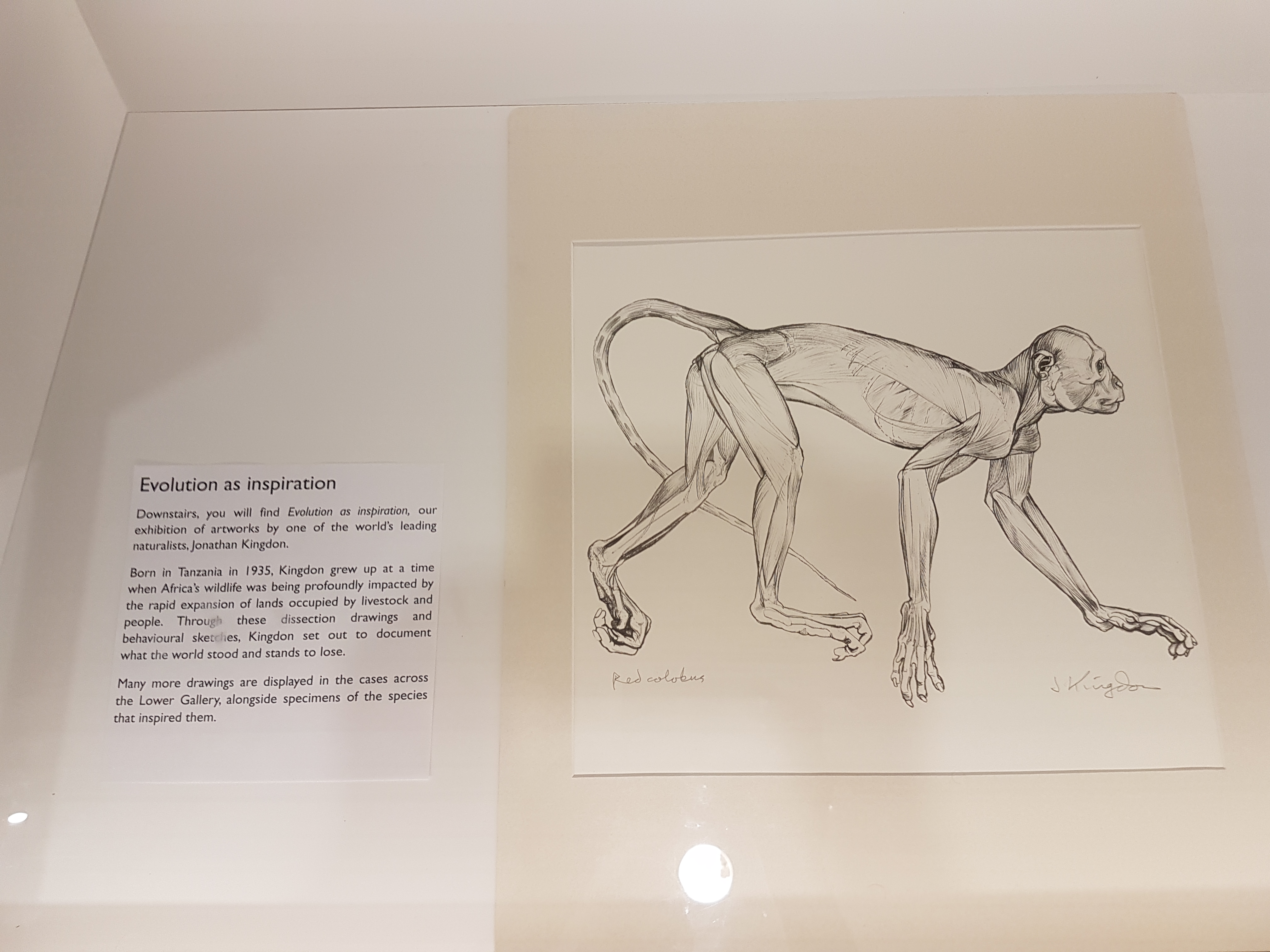
A nice surprise to see naturalist superstar Jonathan Kingdon‘s scientific illustrations and nature-inspired artwork displayed here. I’ve added photos of ones I liked the most.
As the caption explains, Kingdon used art to explain the value of nature; via realistic images of life, dissections, and creative abstractions drawn from them.
Hammerhead bats: even freakier when skinned.
Begone if ye find not joy in aardvarks!
White-toothed shrew looking extra-ghoulish with flensed face.
Skinned sengis in action.
More sengis (elephant shrews); with a note explaining that they are not rodents/insectivores but afrotheres, cousins of aardvarks, elephants and kin.
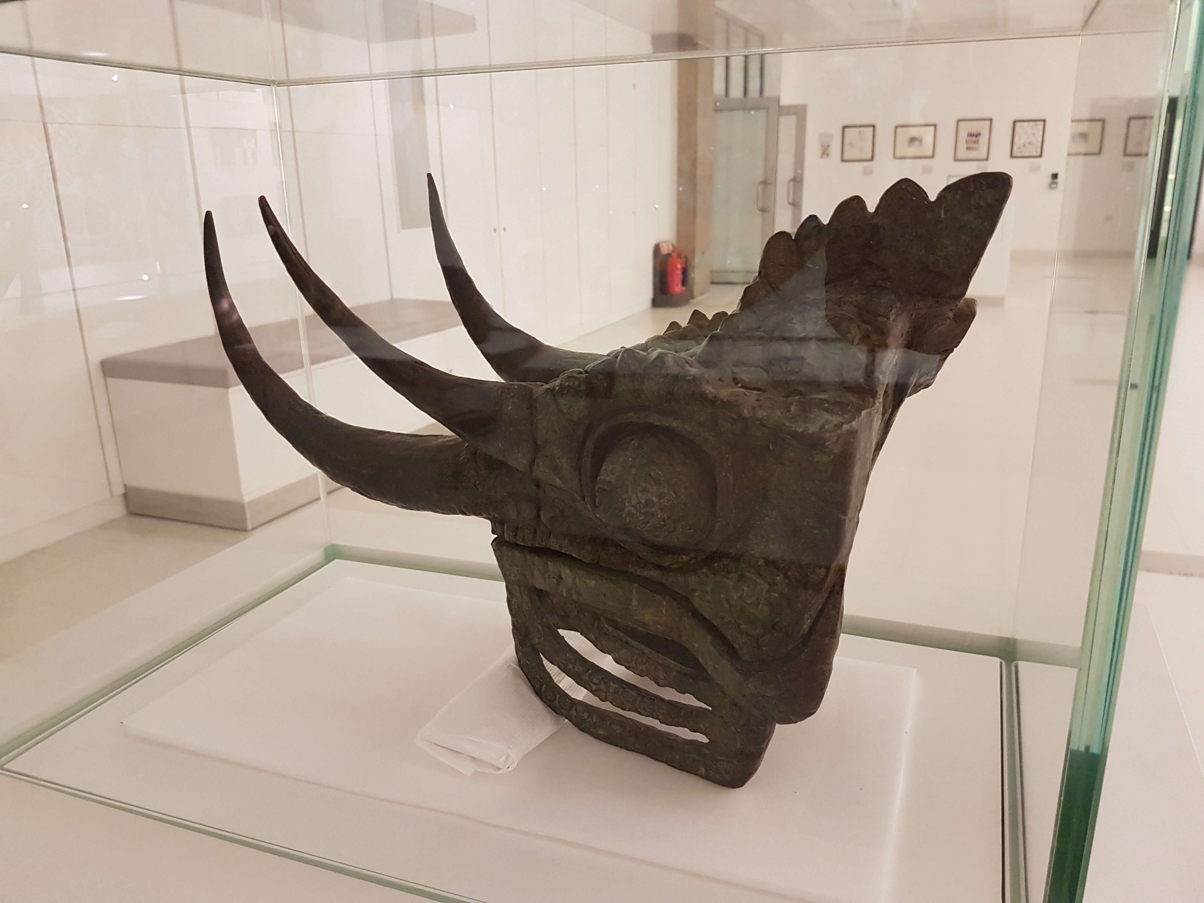
Bronze Jackson’s chameleon bust.
Asian barbet faces: this was fascinating. Kingdon used the paintings to explain how barbet faces vary across species as recognition devices to aid in territorial defense, especially of their nest-holes in trees, in which they face outwards to display their coloured faces. The middle image shows one lone species that has no such territorial competitors and has evolved back into brown colour, perhaps due to relaxed selective pressure for colour. Neat!
Oh my, this took my breath away! Mixed media depicting the varied forms of facial ornaments in vultures; soft tissues used in communcation. And here mounted on a butcher’s rack. Do vulture bits mimic their grisly food?
Today is the 210th anniversary of Charles R. Darwin’s birthday so I put together a quick post. I’d been meaning to blog about some of our latest scientific papers, so I chose those that had an explicit evolutionary theme, which I hope Chuck would like. Here they are, each with a purty picture and a short explainer blurb! Also please check out Anatomy To You’s post by Katrina van Grouw on Darwin’s fancy pigeons.
Stomach-Churning Rating: 1/10 science!
First, Brandon Kilbourne at the Naturkunde Museum in Berlin kindly invited me to assist in a paper from his German fellowship studying mustelid mammals (otters, weasels, wolverines, badgers, etc.; stinky smaller carnivorous mammals). Here we (very much driven by Brandon; I was along for the ride) didn’t just look at how forelimb bone shape changes with body size in this ecologically diverse group. We already knew bigger mustelids would have more robust bones, although it was cool to see how swimming-adapted and digging-adapted mustelids evolved similarly robust bones; whereas climbing ones had the skinniest bones.
The really exciting and novel (yes I am using that much-abused word!) aspect of the paper is that Brandon conjured some sorcery with the latest methods for analysing evolutionary trends, to test how forelimb bone shapes evolved. Was their pattern of evolution mostly a leisurely “random walk” or were there early bursts of shape innovation in the mustelid tree of life, or did shape evolve toward one or more optimal shapes (e.g. suited to ecology/habitat)? We found that the most likely pattern involved multiple rates of evolution and/or optima, rather than a single regime. And it was fascinating to see that the patterns of internal shape change deviated from external shape change such as bone lengths: so perhaps selection sometimes works independently at many levels of bone morphology?
Various evolutionary models applied to the phylogeny of mustelids.
Then there, coincidentally, was another paper originating in part from the same museum group in Berlin. This one I’d been involved in as a co-investigator (author) on a Volkswagen (yes! They like science) grant back about 8 years ago and since. There is an amazing ~290 million year old fossil near-amniote (more terrestrial tetrapod) called Orobates pabsti, preserved with good skeletal material but also sets of footprints that match bones very well, allowing a rare match of the two down to this species level. John Nyakatura’s team had 3D modelled this animal before, so we set out to use digital techniques to test how it did, or did not, move—similar to what I’d tried before with Tyrannosaurus, Ichthyostegaand so forth. The main question was whether Orobates moved in a more “ancestral” salamander-like way, a more “derived” lizard-like way (i.e. amniote-ish), or something else.
The approach was like a science sledgehammer: we combined experimental studies of 4 living tetrapods (to approximate “rules” of various sprawling gaits), a digital marionette of Orobates (to assess how well its skeleton stayed articulated in various motions), and two robotics analysis (led by robotics guru Auke Ijspeert and his amazing team): a physical robot version “OroBOT” (as a real-world test of our methods), and a biomechanical simulation of OroBOT (to estimate hard-to-measure things in the other analyses, and matches of motions to footprints). And, best of all, we made it all transparent: you can go play with our interactive website, which I still find very fun to explore, and test what motion patterns do or do not work best for Orobates. We concluded that a more amniote-like set of motions was most plausible, which means such motions might have first evolved outside of amniotes.
OroBOT in tha house!
You may remember Crassigyrinus, the early tetrapod, from a prior post on Anatomy To You. My PhD student Eva Herbst finished her anatomical study of the best fossils we could fit into a microCT-scanner and found some neat new details about the “tadpole from hell”. Buried in the rocky matrix were previously unrecognized bones: vertebrae (pleurocentra; the smaller nubbins of what may be “rhachitomous” bipartite classic tetrapod/omorph structure), ribs (from broad thoracic ones to thin rear ones), pelvic (pubis; lower front), and numerous limb bones. One interesting trait we noticed was that the metatarsals (“sole bones” of the foot) were not symmetrical from left-to-right across each bone, as shown below. Such asymmetry was previously used to infer that some early tetrapods were terrestrial, yet Crassigyrinus was uncontroversially aquatic, so what’s up with that? Maybe this asymmetry is a “hangover” from more terrestrial ancestry, or maybe these bones get asymmetrical for non-terrestrial reasons.
The oddly asymmetrical metatarsals of Crassigyrinus.
Finally, Dr. Peter Bishop finished his PhD at Griffith University in Australia and came to join us as a DAWNDINOS postdoc. He blasted out three of his thesis chapters (starting here) with me and many others as coauthors, all three papers building on a major theme: how does the inner bone structure (spongy or cancellous bone) relate to hindlimb function in theropod dinosaurs (including birds) and how did that evolve? Might it tell us something about how leg posture or even gait evolved? There are big theories in “mechanobiology” variously named Wolff’s Law or the Trajectorial Theory that explain why, at certain levels, bony struts tend to align themselves to help resist certain stresses, and thus their alignment can be “read” to indicate stresses. Sometimes. It’s complicated!
Undaunted, Peter measured a bunch of theropod limb bones’ inner geometry and found consistent differences in how the “tracts” of bony struts, mainly around joints, were oriented. He then built a biomechanical model of a chicken to test if the loads that muscles placed on the joints incurred stresses that matched the tracts’ orientations. Hmm, they did! Then, with renewed confidence that we can use this in the fossil record to infer approximate limb postures, Peter scanned and modelled a less birdlike Daspletosaurus (smaller tyrannosaur) and more birdlike “Troodon” (now Stenonychosaurus; long story). Nicely fitting many other studies’ conclusions, Peter found that the tyrannosaur had a more straightened hindlimb whereas the troodontid had a more crouched hindlimb; intermediate between the tyrannosaur and chicken. Voila! More evidence for a gradual evolution of leg posture across Mesozoic-theropods-into-modern-birds. That’s nice.
Three theropods, three best-supported postures based on cancellous bone architecture.
If you are still thirsty for more papers even if they are less evolutionary, here’s the quick scoop on ones I’ve neglected until now:
(1) Former PhD student Chris Basu published his thesis work w/us on measuring giraffe walking dynamics with force plates, finding that they move mostly like other quadrupeds and their wobbly necks might cost them a little.
(2) Oh, and Chris’s second paper just came out as I was writing this! We measured faster giraffe gaits in the wilds of South Africa, as zoo giraffes couldn’t safely do them. And we found they don’t normally go airborne, just using a rotary gallop (not trot, pace or canter); unlike some other mammals. Stay tuned: next we get evolutionary with this project!
(2) How do you safely anaesthetize a Nile crocodile? There’s now a rigorous protocol (from our DAWNDINOS work).
(3) Kickstarting my broad interest in how animals do “extreme” non-locomotor motions, we simulated how greyhounds stand up, finding that even without stretchy tendons they should, barely, be able to do it, which is neat. Expect much more about this from us in due time.
(4) Let’s simulate some more biomechanics! Ashley Heers, an NSF research fellow w/me for a year, simulated how growing chukar birds use their wing muscles to flap their way up steeper inclines (“WAIR” for devotees), and the results were very encouraging for simulating this behaviour in more detail (e.g. tendons seem to matter a lot) and even in fossil species; and finally…
(5) Hey did you ever think about how bone shape differs between hopping marsupials (macropods) and galloping artiodactyl (even-toed) mammals? We did, in long-the-making work from an old BBSRC grant with Michael Doube et al., and one cool thing is that they mostly don’t change shape with body size that differently, even though one is more bipedal at faster speeds—so maybe it is lower-intensity, slower behaviours that (sometimes?) influence bone shape more?
So there you have the skinny on what we’ve been up to lately, messing around with evolution, biomechanics and morphology.
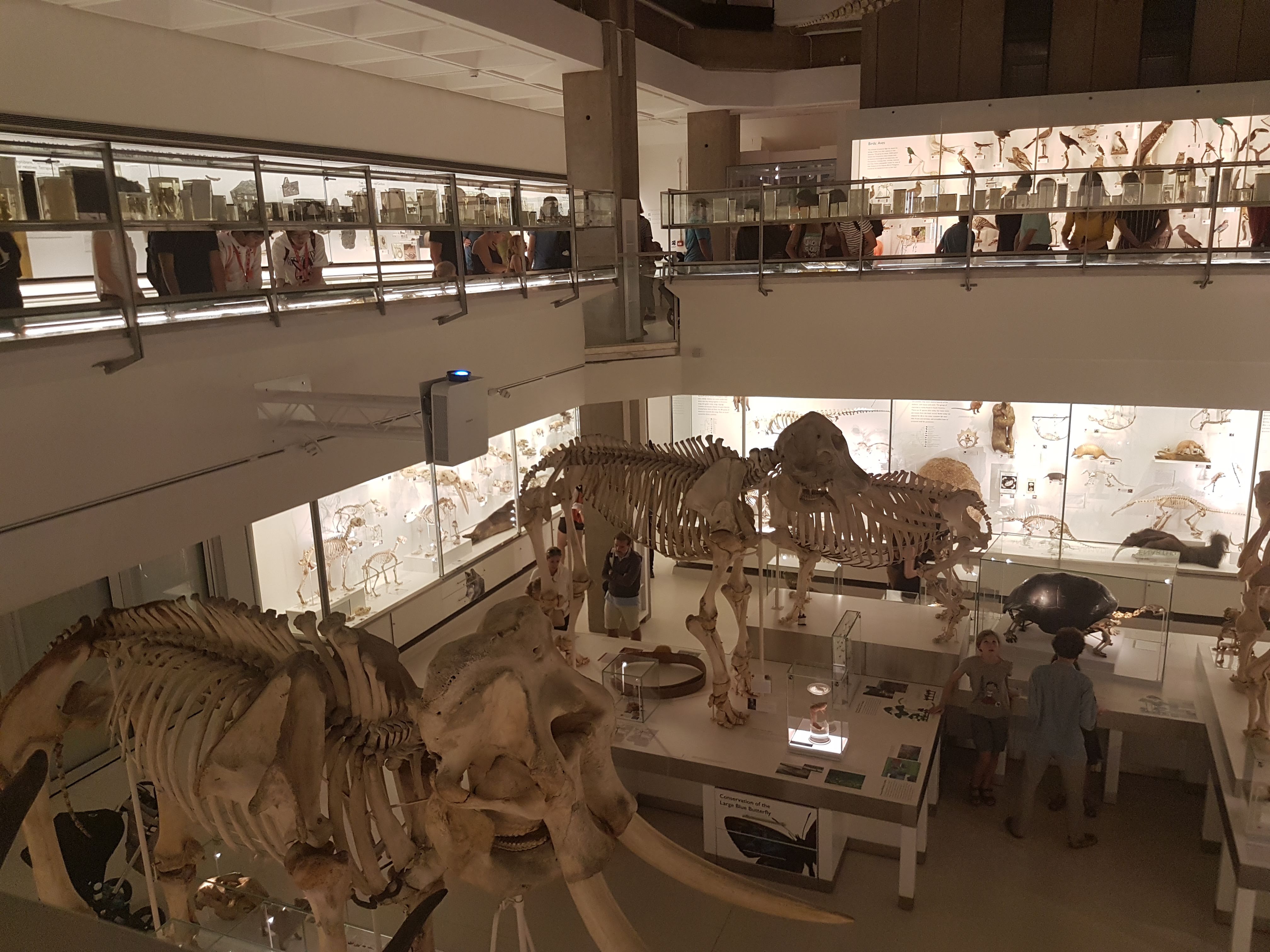
I had a spare hour in Cambridge this weekend so I dared the crowds in the revamped UMZC’s upper floor. In my prior visit and post I’d experienced and described the lower floor, which is almost exclusively mammals. This “new” floor has everything else that is zoological (animal/Metazoa) and again is organized in an evolutionary context. And here is my photo tour as promised!
Inviting, soft lighting perfuses the exhibits from the entryway onwards.
All images can be clicked to mu-zoom in on them.
Stomach-Churning Rating: 5/10 for spirit animals, by which I mean dissected/ghostly pale whole specimens of animals in preservative fluids.
The exhibits are on a square balcony overlooking the lower floor, so you can get some nice views. It does make the balcony crowded when the museum is busy, so take that in mind if visiting. Strollers on this upper floor could be really difficult. But the ceiling is very tall so it is not cramped in a 3D sense. The lower floor is more spacious.
Like phylogenies? You got em! Tucked away at the beginning of each major group; not occupying huge valuable space or glaringly obvious like AMNH in NYC but still noticeable and useful. To me, it strikes a good balance; gives the necessary evolutionary context for the displayed specimens/taxa.
Introductory panels explain how names are given to specimens, how specimens are preserved and more.
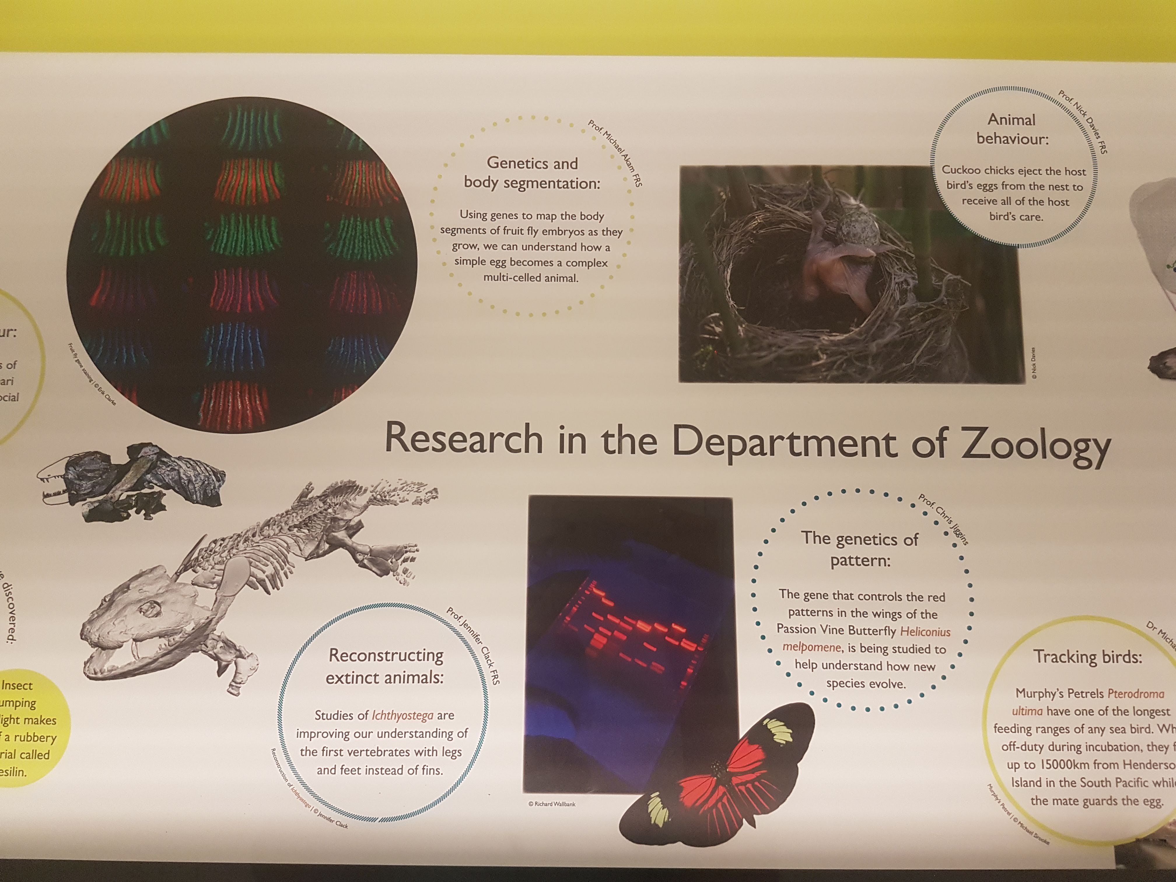
The exhibits give due focus to research that the UMZC is doing or has been famous for. Hey I recognize that 3D tetrapod image in the lower left! 🙂
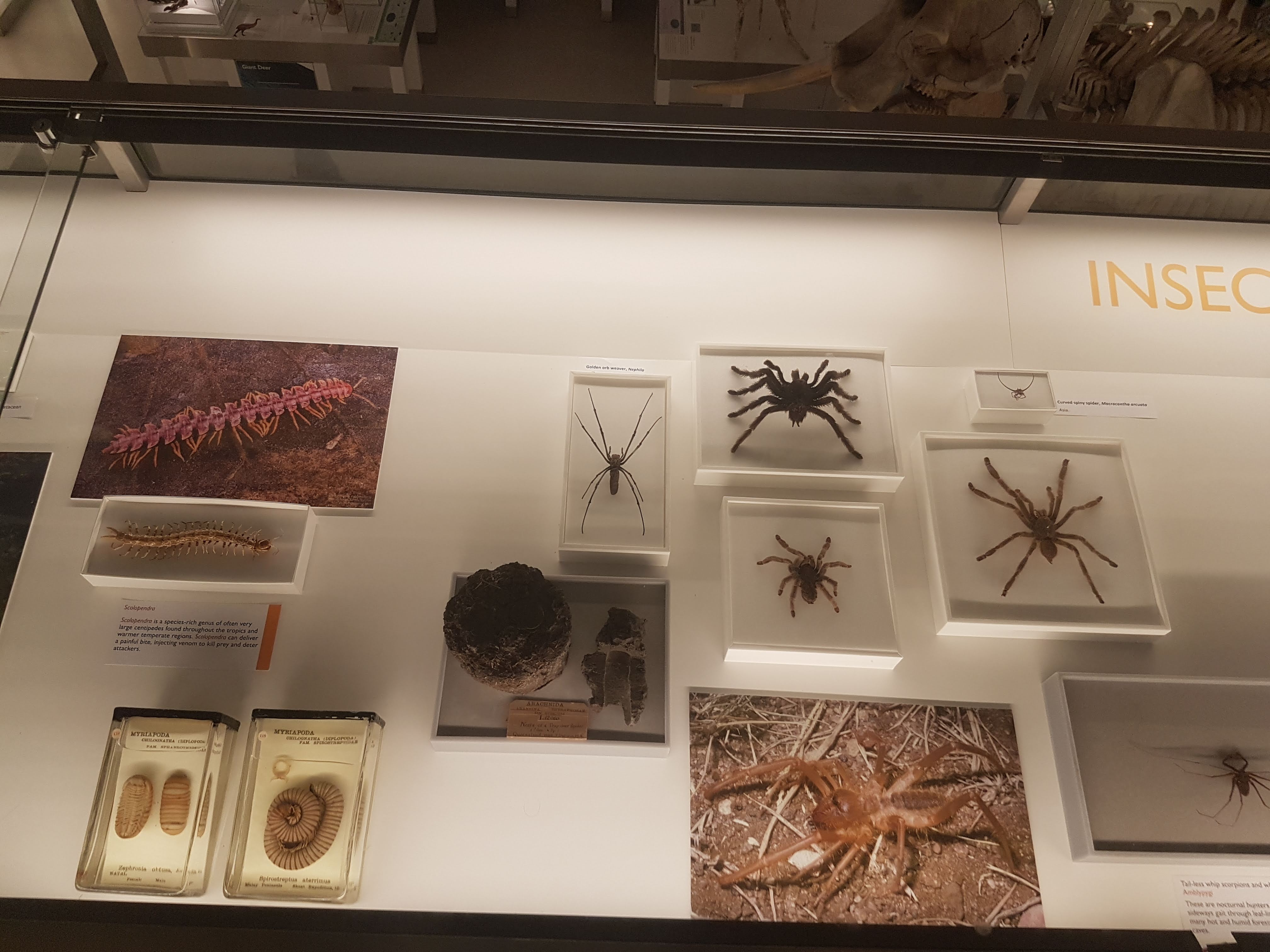
There is ample coverage of diversity throughout Metazoa but my camera tended to be drawn to the Vertebrata. Except in some instances like these.
Some larger chelicerates.
Some smaller, shadowy sea scorpion (eurypterid) fossils.
Watch here for more about ophiuroids (brittlestars) in not too long!
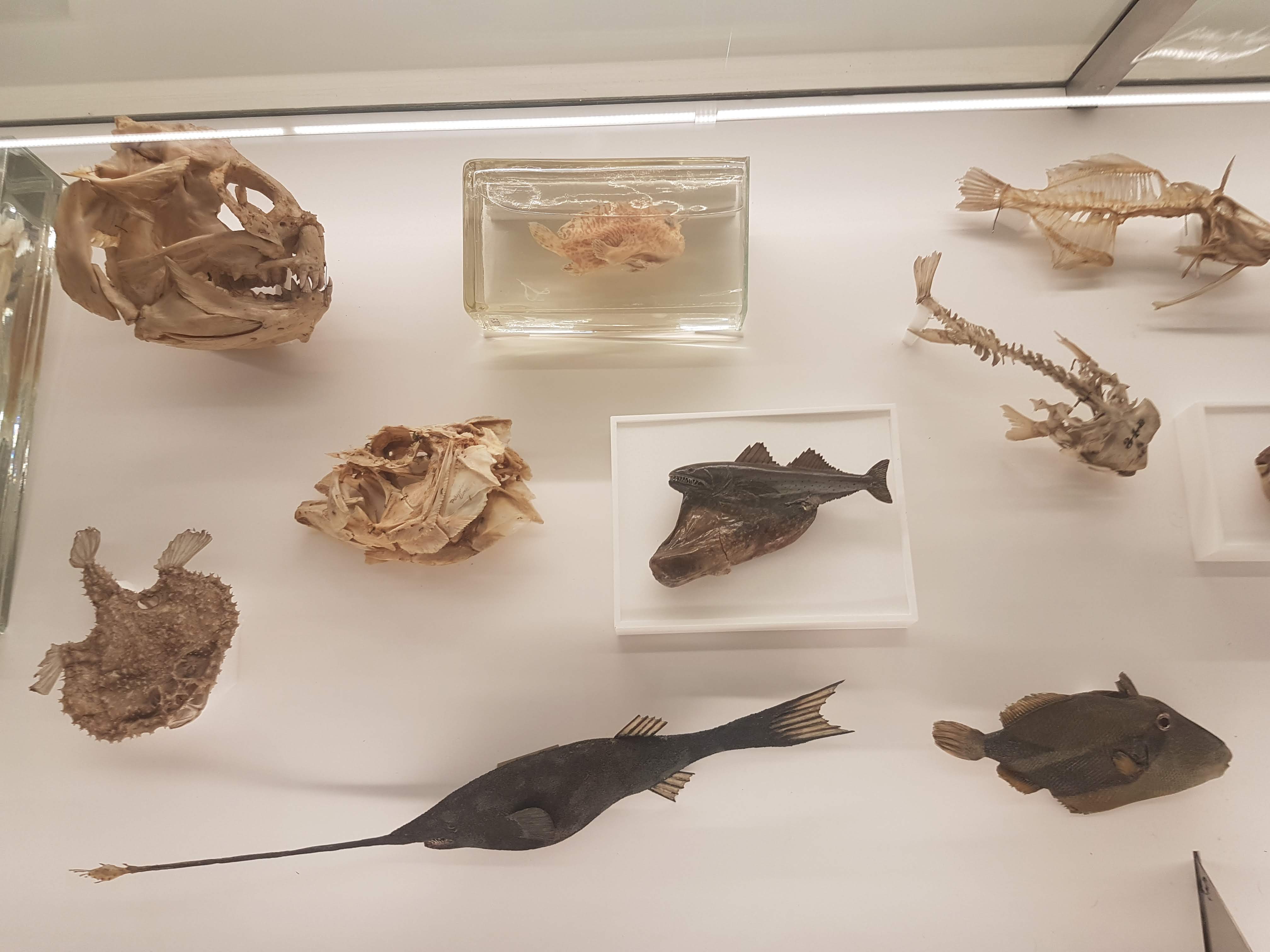
A BIG fish brain! Interesting! Before I go through specimens in evolutionary “sequence”, I will feature another thing i really liked: lots of dissected spirit-specimens that show off cool anatomy/evolution/adaptation (and technical skills in anatomical preparation). Mostly heads; mostly fish.
Salps and other tunicates! Our closest non-vertebrate relatives- and some insight into how our head and gut came to be.
Salp-reflection.
Lamprey head: not hard to spot the commonalities with the salps; but now into Vertebrata.
Hagfish head: as a fellow cyclostome/agnathan, much like a lamprey but never forget the slime glands!
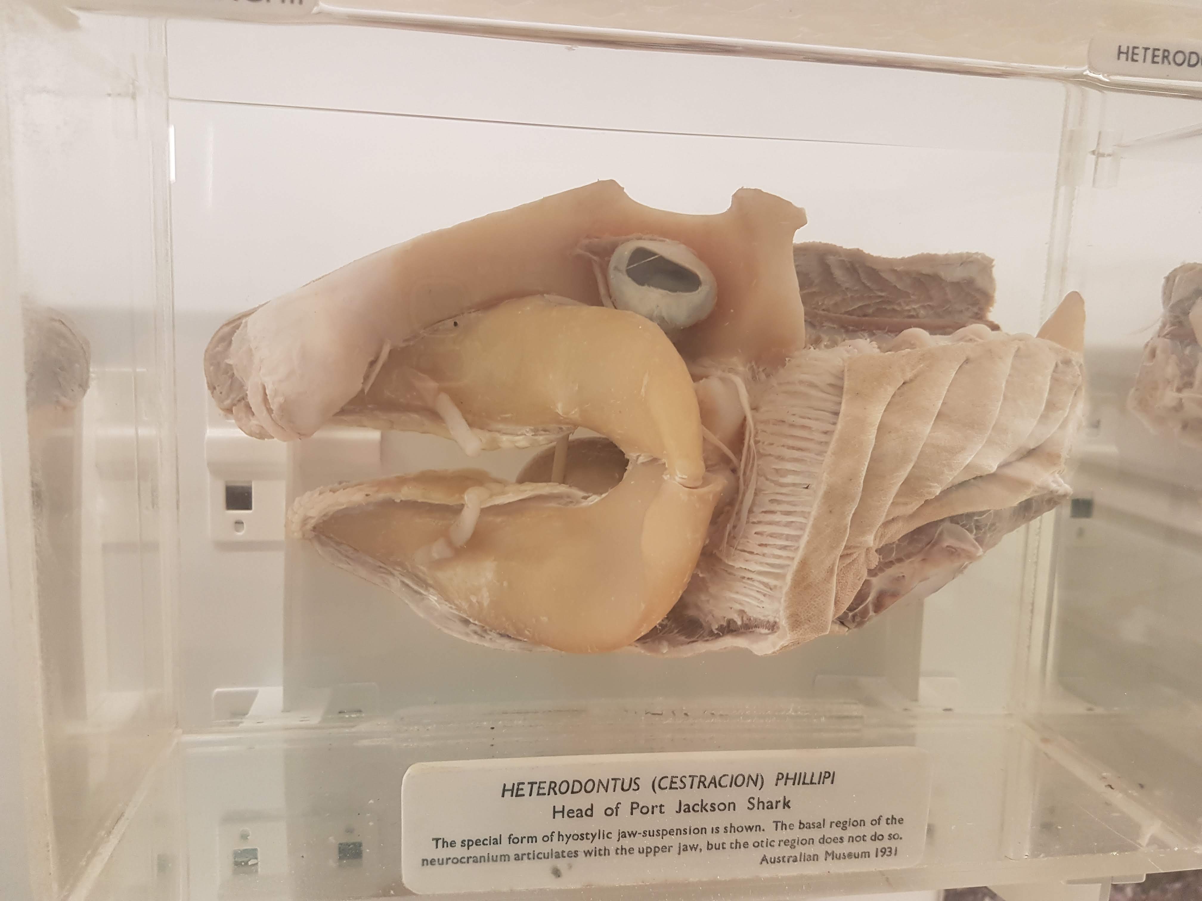
Shark head. Big fat jaws; all the better to bite prey with!
Lungfish (Protopterus) head showing the big crushing tooth plates (above).
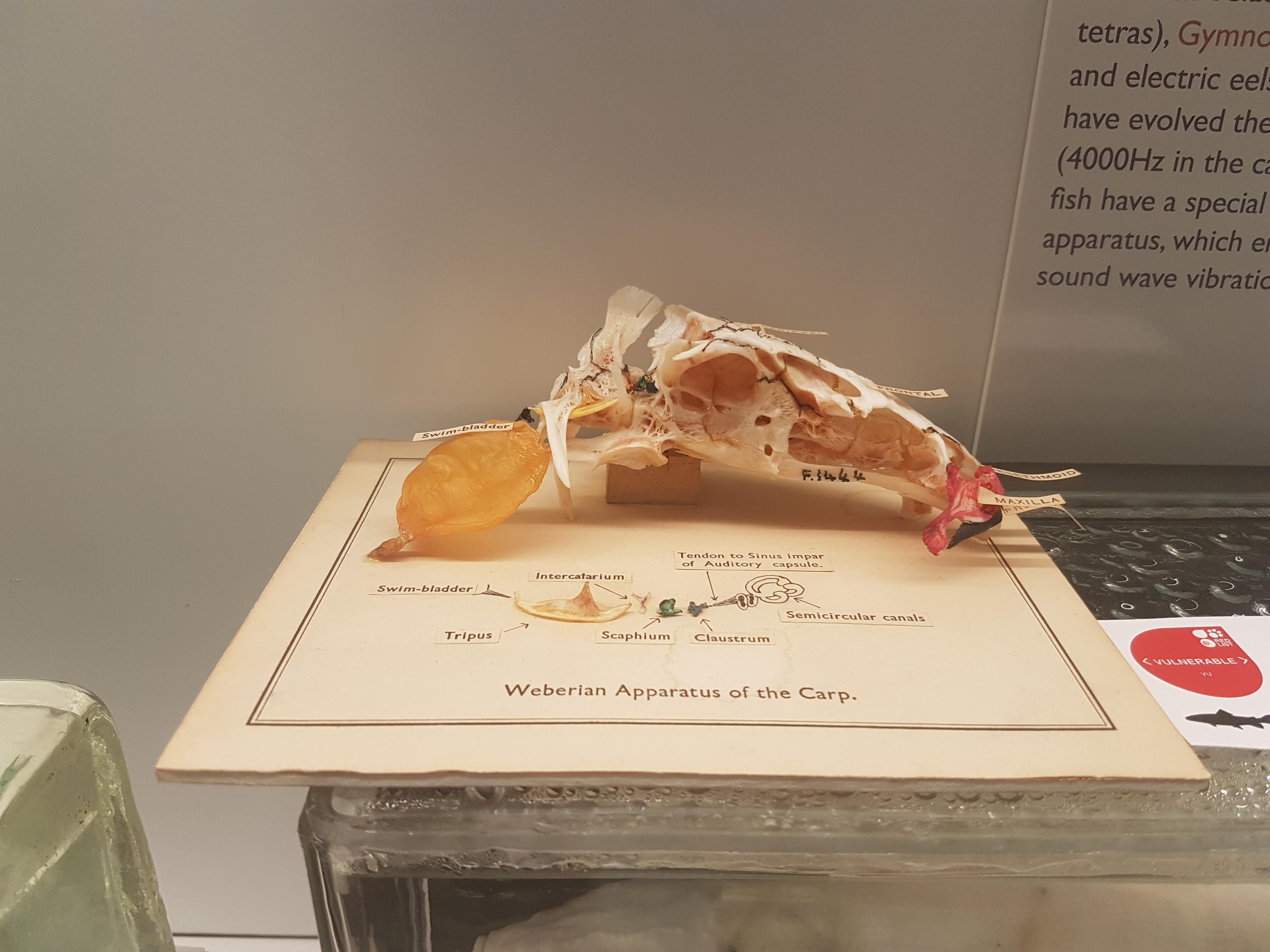
Sturgeon vertebrae: tweak some agnathan/shark bits and here you are.
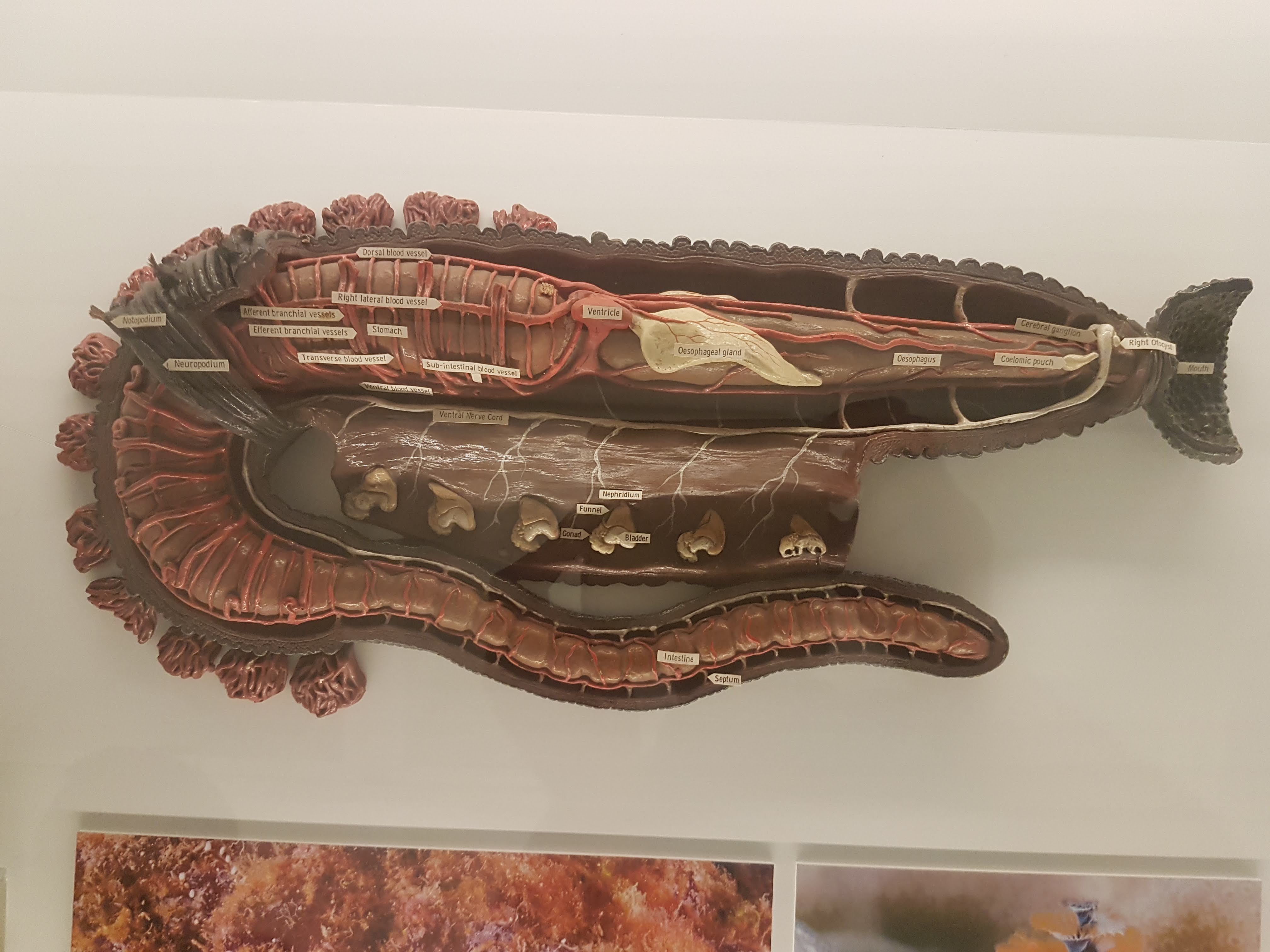
Worm (annelid) anatomy model, displaying some differences from/similarities to Vertebrata. (e.g. ventral vs. dorsal nerve cord; segmentation)
Dissected flipper from a small whale/other cetacean. Still five fingers, but other specializations make it work underwater.
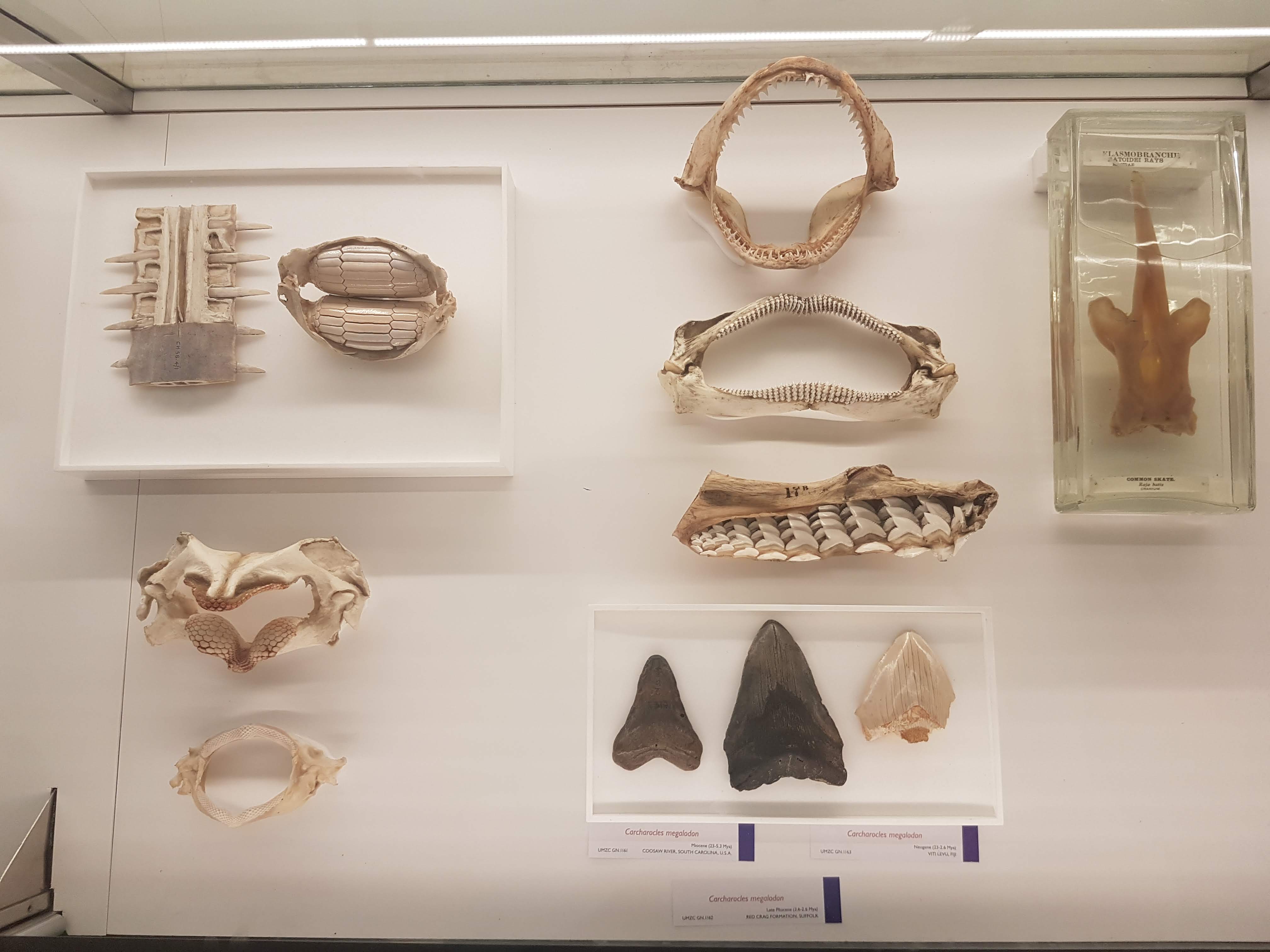
Wonderful diversity of tooth and jaw forms in sharks, rays and relatives. I like this display a lot.
More of the above, but disparate fossil forms!
On with the evolutionary context! Woven throughout the displays of modern animals are numerous fossils, like these lovely placoderms (lineage interposed between agnathans, sharks and other jawed fish).
Goblin shark head.
I seem to always forget what ray-finned fish this is (I want to say wolffish? Quick Googling suggests maybe I am right), but see it often and like its impressive bitey-ness.
Bichir and snakefish; early ray-finned fish radiations.
Armoured and similar fish today.
Armoured fish of the past; some convergent evolution within ray-fins.
Convergence- and homology- of amphibious nature in fish is another evolutionary pattern exemplified here.
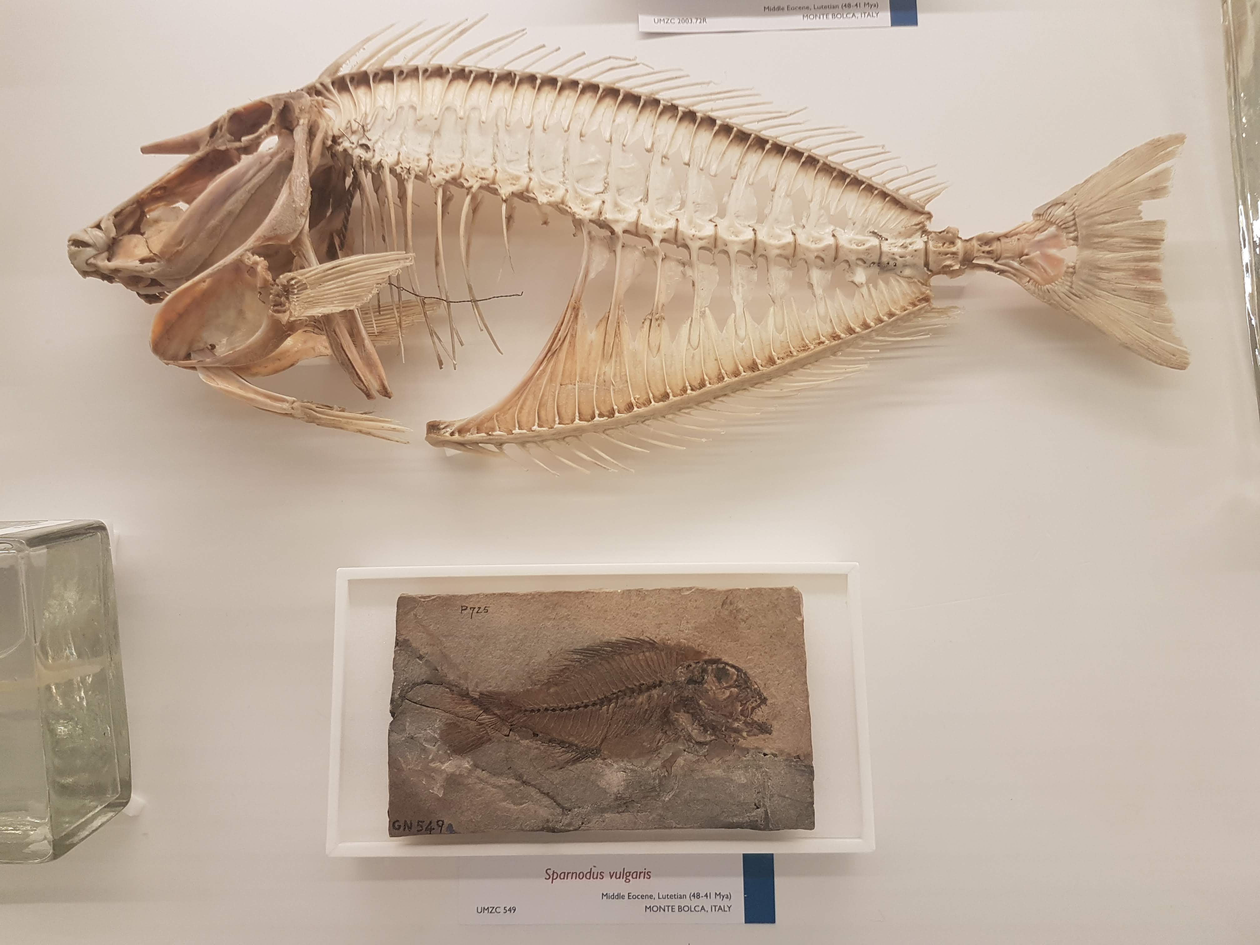
Gorgeous fossils of ray-finned fish lineages that arose after the Permian extinctions, then went extinct later in the Triassic.
Note the loooooong snout on this cornetfish but the actual jaws are just at the tip.
Flying fish– those ray-fins are versatile.
Diversity of unusual ray-finned fish, including deep-water and bottom-dwelling forms.
Can you find the low-slung jaws of a dory?
Recent and fossil perch lineage fish.
It’s hard to get far into talking about evolution without bringing up the adaptive radiation of east African cichlid fish, and UMZC researchers are keen on this topic too.
Lobe-fins! Everybody dance!
Rhizodonts & kin: reasons to get out of Devonian-Carboniferous waters.
A Cretaceous fossil coelacanth (skull); not extremely different from living ones’.
Let’s admire some fossil and modern lungfish skulls, shall we? Big platey things (here, mainly looking at the palate) with lots of fusions of tiny bones on the skull roof.
Eusthenopteron fossils aren’t that uncommon but they are still great to see; and very important, because…
OK let’s stop messing around. The UMZC has one of the best displays of fossil stem-tetrapods in the world! And it should.
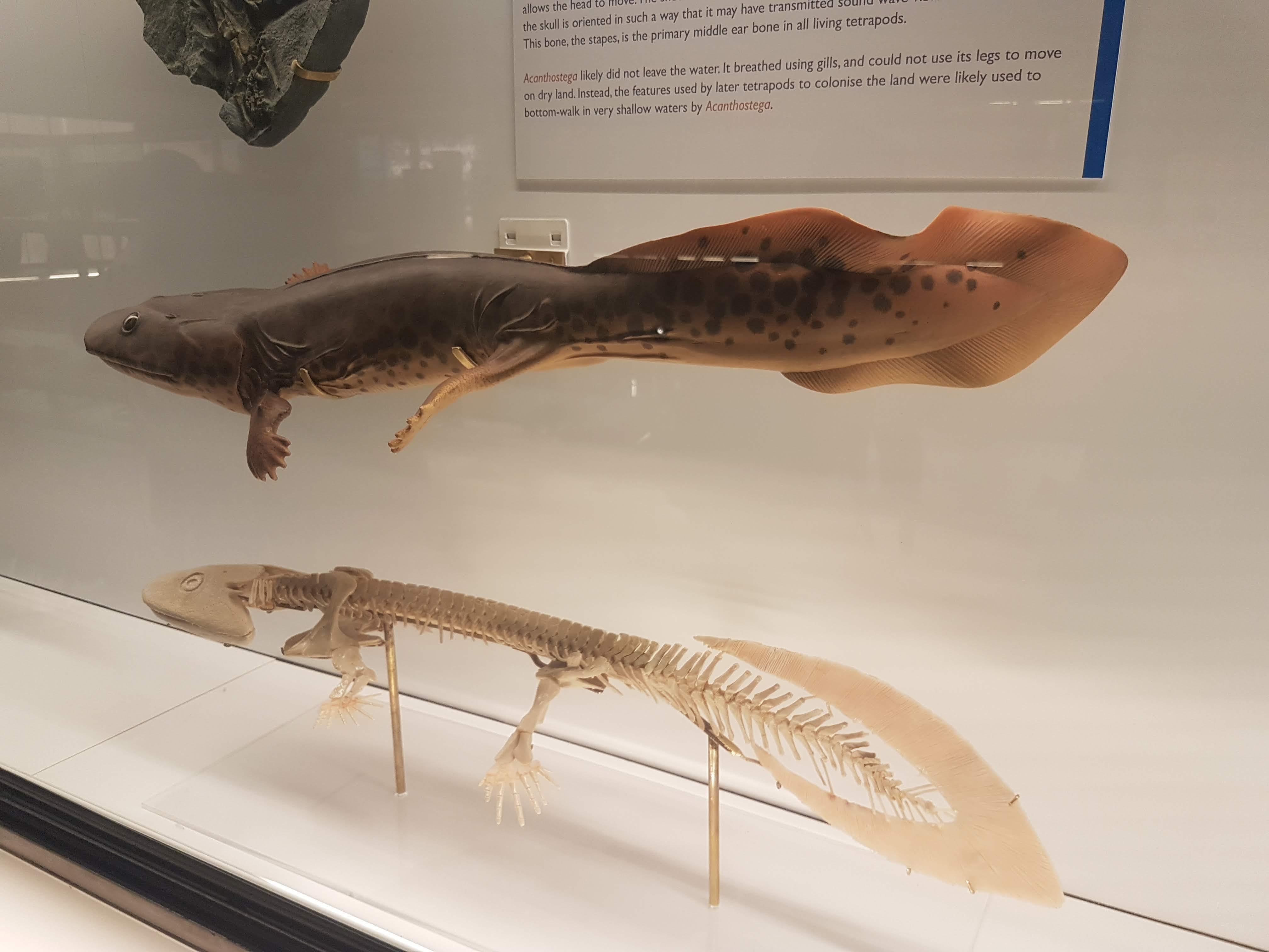
Another look at the pretty Acanthostega models.
Acanthostega vs. primate forelimb: so like us.
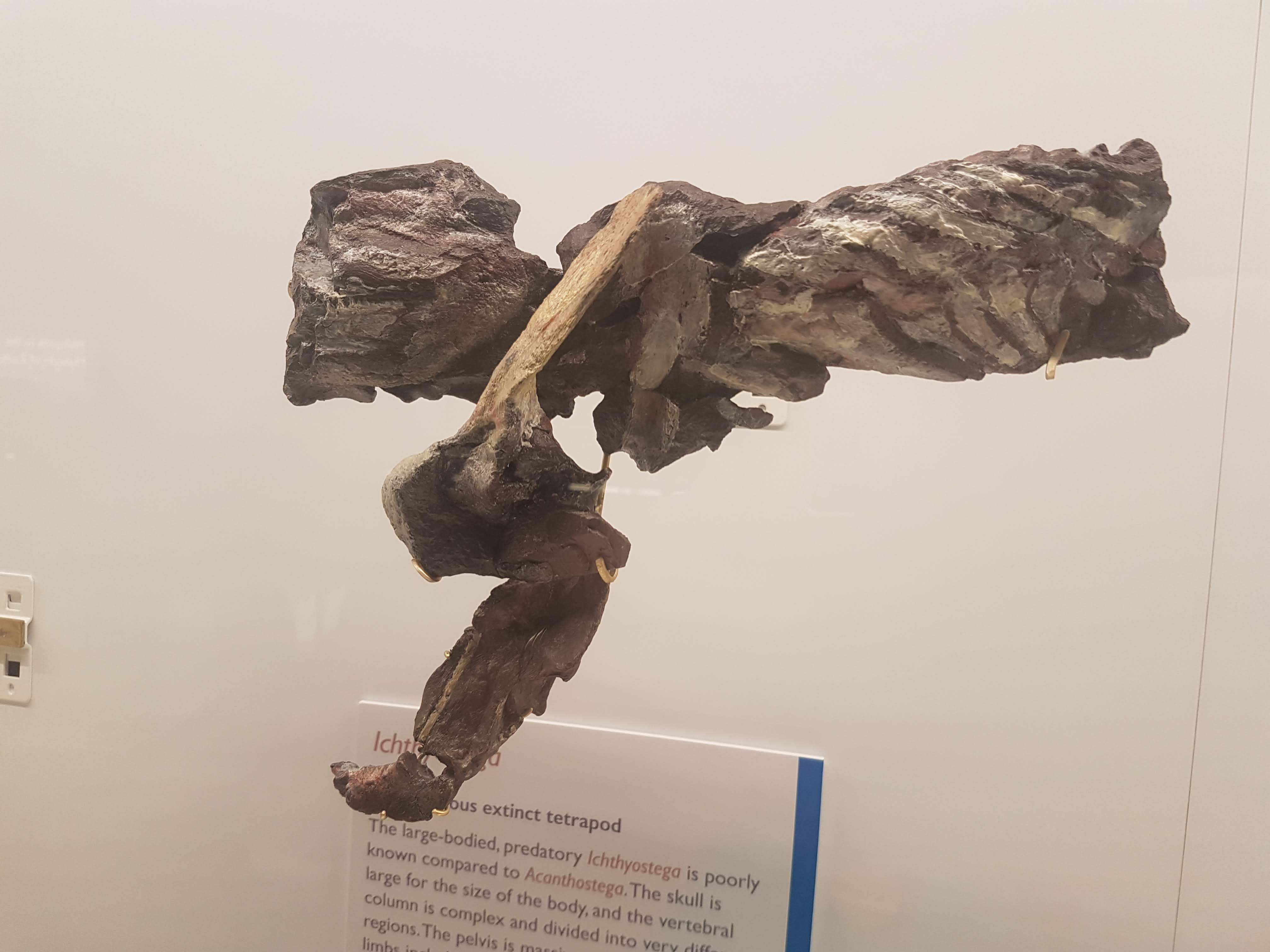
Ichthyostega parts keep Acanthostega company.
A closer look at the “Mr. Magic” Ichthyostega specimen, which takes some unpacking but is incredibly informative and was a mainstay of our 2012 model. Back of skull, left forelimb, and thorax (from left to right here).
Eucritta, another stem-tetrapod.
Closer look at Eucritta‘s skull.
Weird stem-tetrapod Crassigyrinus, which we’re still trying to figure out. It’s a fabulous specimen in terms of completeness, but messy “roadkill” with too many damn bones.
The large skull of Crassigyrinus, in right side view.
Early temnospondyl (true amphibian-line) skulls and neck.
Nectrideans or the boomerangs of the Palaeozoic.
Cool fossil frogs.
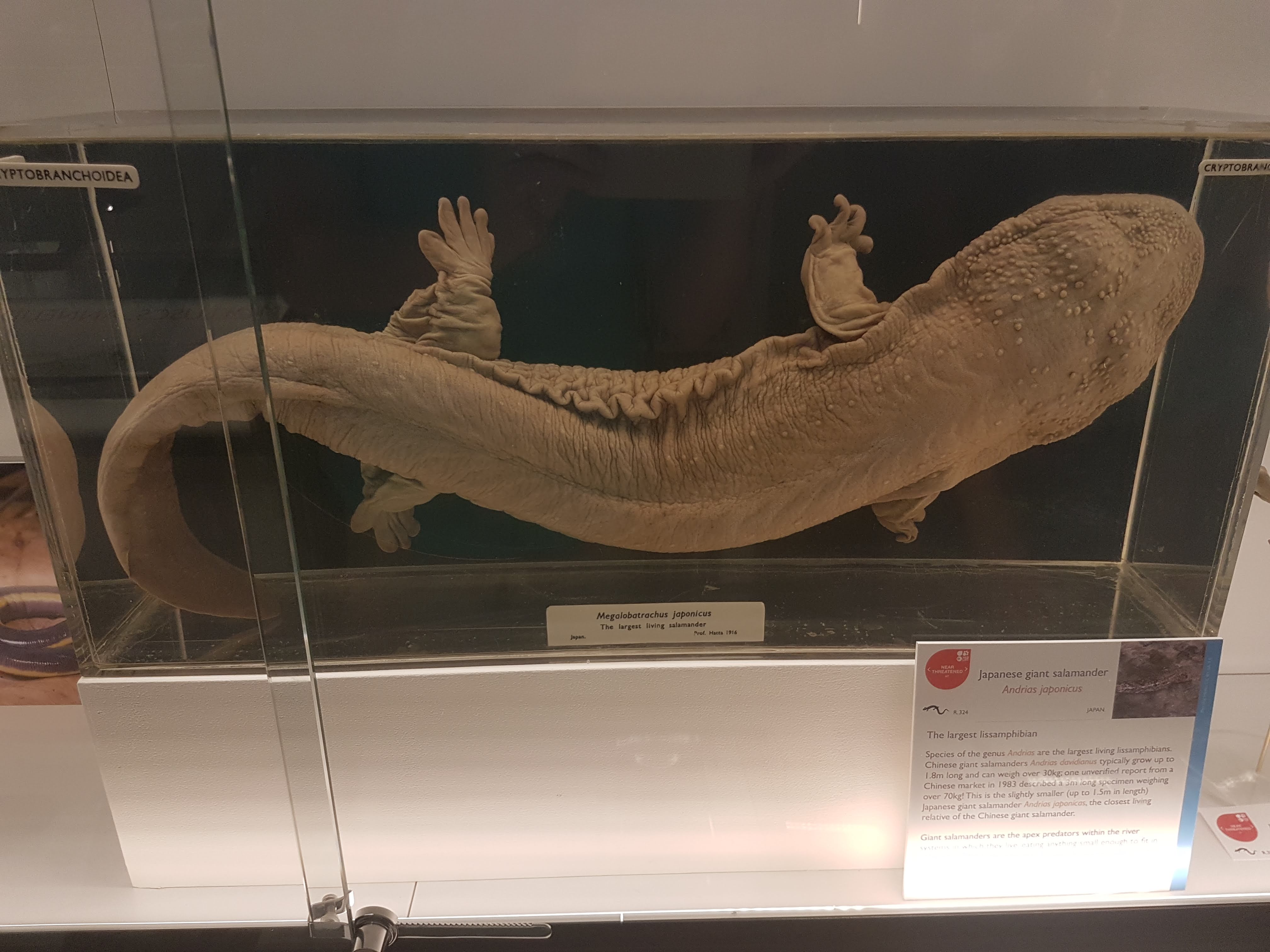
Giant Japanese salamander!
Fire salamanders: not as colourful as the real thing, but here revealing their reproductive cycle in beautiful detail.
Closeup of oviduct in above.
Sexual dimorphism in Leptodactylus frogs: the males have bulging upper arms to (I am assuming) help them hold onto females during amplexus (grasping in mating competitions).
Did I forget that Leptodactylus has big flanges on the humerus in males, to support those muscles? Seems so.
An early stem-amniote, Limnoscelis (close to mammals/reptiles divergence); cast.
Grand sea turtle skeleton.
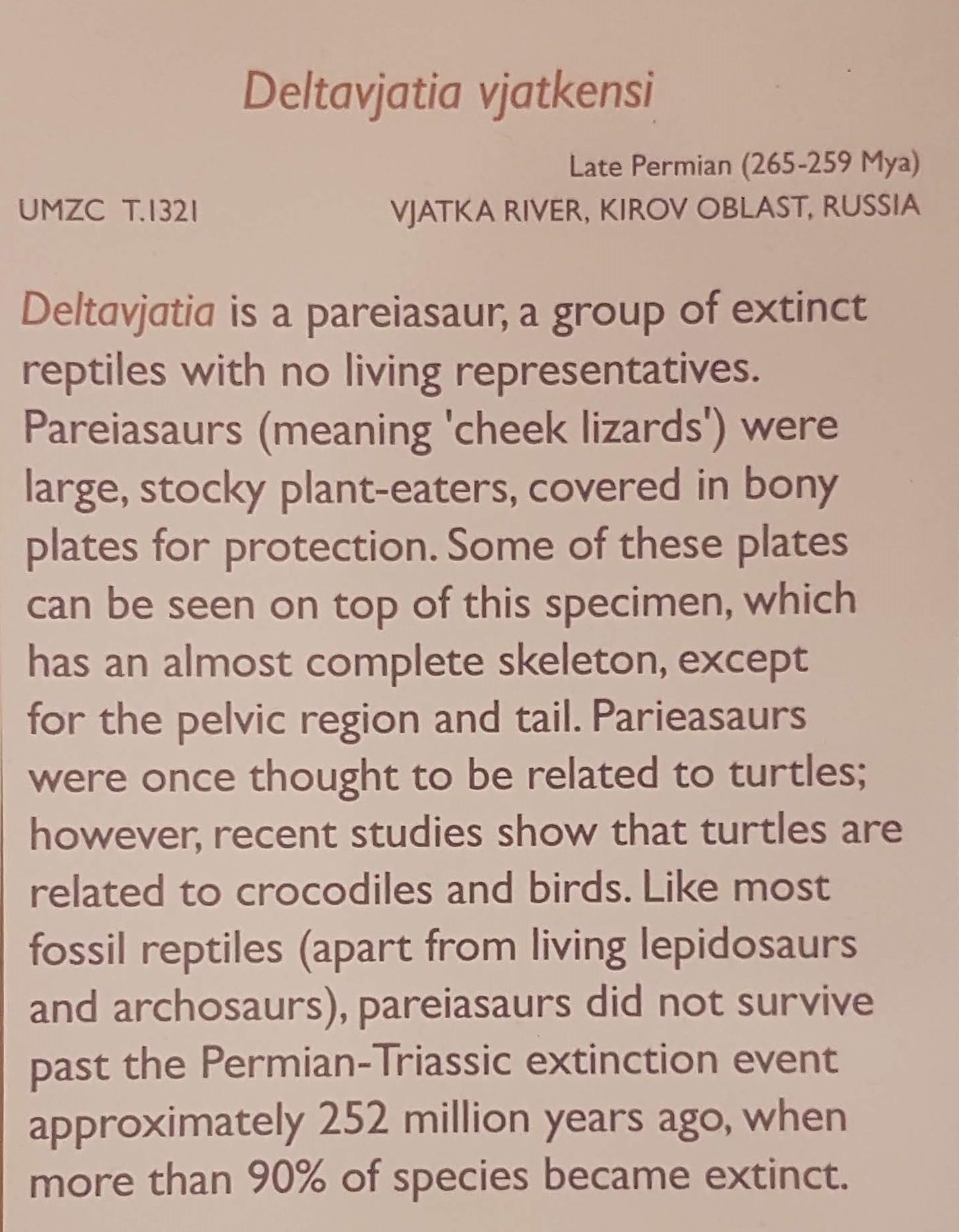
One of my faves on display: a real pareiasaurian reptile skeleton, and you can get a good 3D look around it.
Details on above pareiasaurian.
Mammals are downstairs, but we’re reminded that they fit into tetrapod/amniote evolution nonetheless.
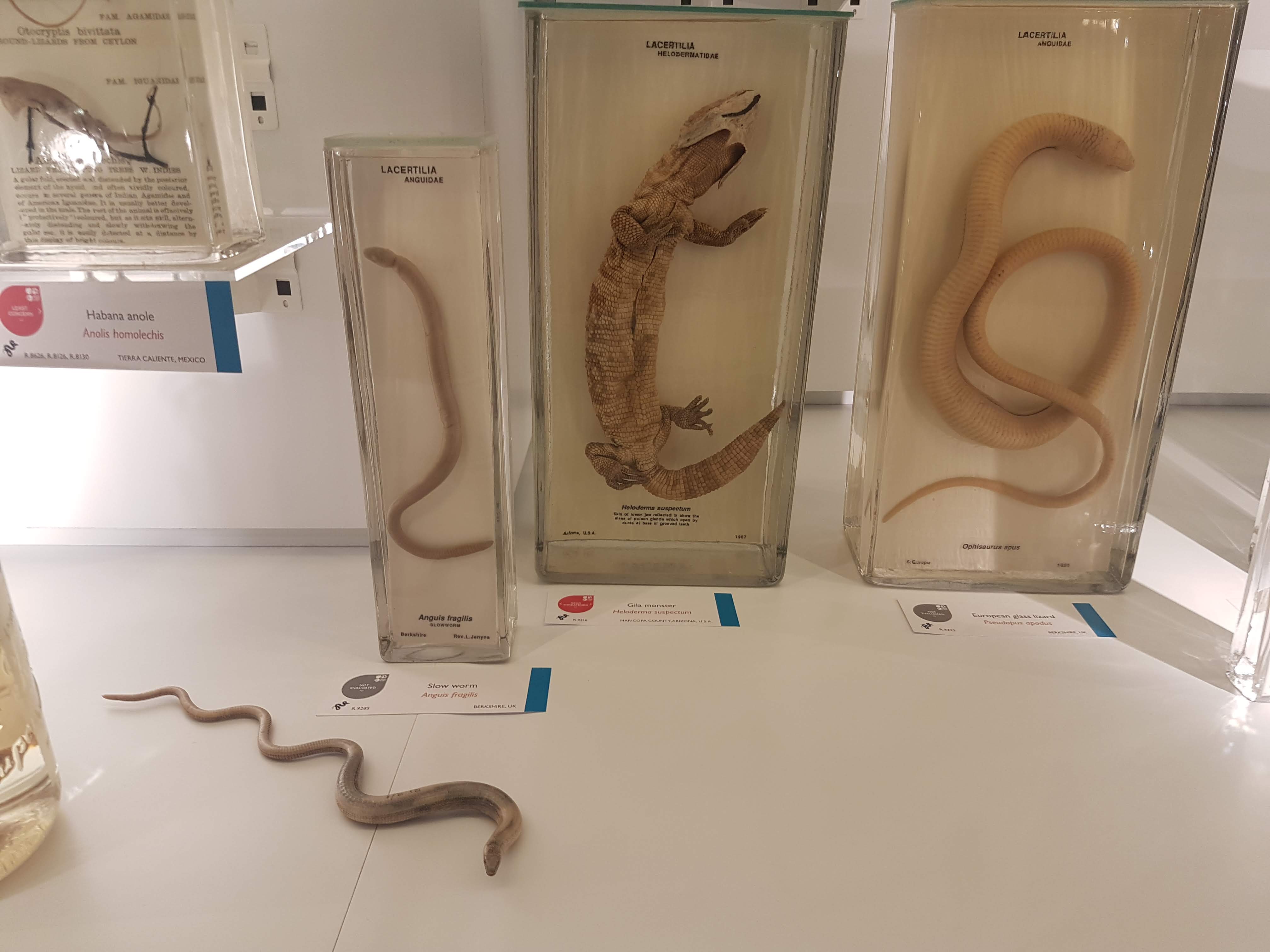
Let there be reptiles! And it was good.
Herps so good. (slow worm, Gila monster, glass lizard)
A curator is Dr Jason Head so you bet Titanoboa is featured!
Crocodylia: impressive specimens chosen here.
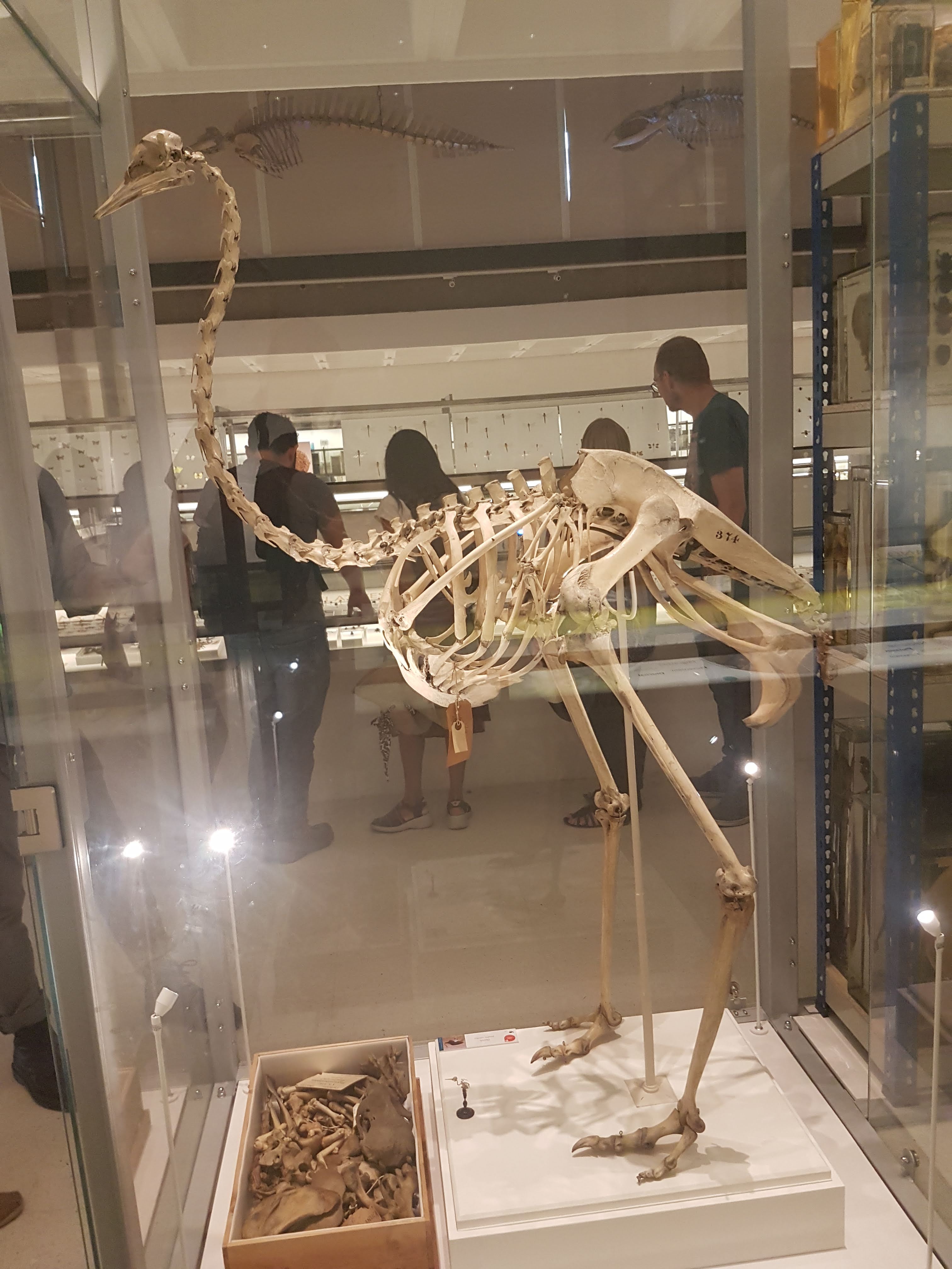
It ain’t a museum without a statuesque ratite skeleton. (There are ~no non-avian dinosaurs here– for those, go to the Sedgwick Museum across the street, which has no shortage!)
Avian diversity takes off.
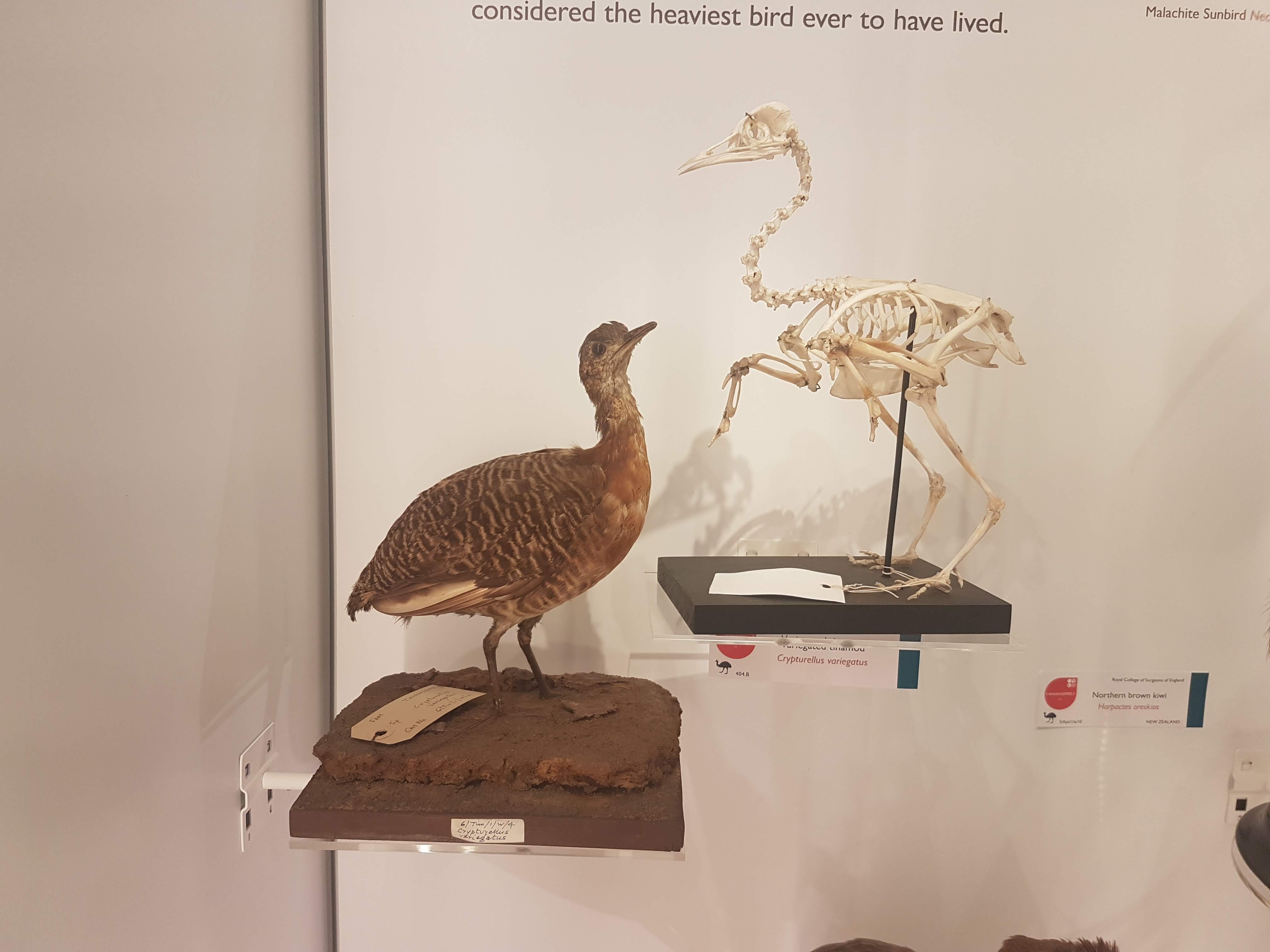
Glad to see a tinamou make an appearance. They get neglected too often in museums- uncommon and often seemingly unimpressive, but I’m a fan.
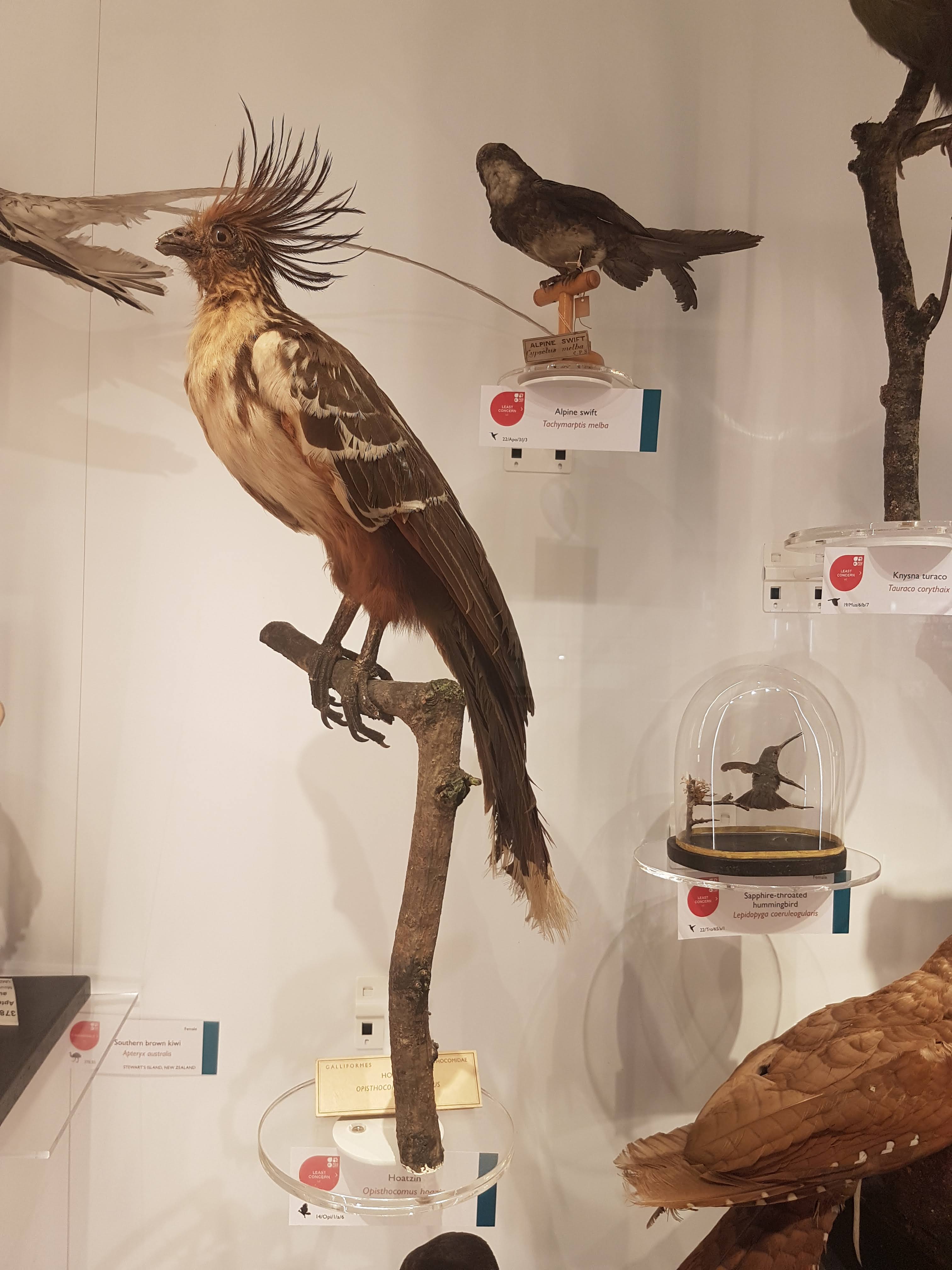
I still do not understand hoatzins; the “cuckoo” gone cuckoo.
Dodo parts (and Great Auk) near the entrance.
Wow. What an oilbird taxidermy display! :-O
There we have it. Phew! That’s a lot! And I left out a lot of inverts. This upper floor is stuffed with specimens; easier there because the specimens are smaller on average than on the lower floor. Little text-heavy signage is around. I give a thumbs-up to that– let people revel in the natural glory of what their eyes show them, and give them nuggets of info to leave them wanting more so they go find out.
Now it’s in your hands– go find out yourself how lovely this museum is! I’ve just given a taste.
As 2017 approaches its end, there have been a few papers I’ve been involved in that I thought I’d point out here while I have time. Our DAWNDINOS project has been taking up much of that time and you’ll see much more of that project’s work in 2018, but we just published our first paper from it! And since the other two recent papers involve a similar theme of muscles, appendages and computer models of biomechanics, they’ll feature here too.
Stomach-Churning Rating: 0/10; computer models and other abstractions.
Mussaurus patagonicus was an early sauropodomorph dinosaur from Argentina, and is now widely accepted to be a very close relative of the true (giant, quadrupedal) sauropods. Here is John Conway’s great reconstruction of it:
We have been working with Alejandro Otero and Diego Pol on Mussaurus for many years now, starting with Royal Society International Exchange funds and now supported by my ERC grant “DAWNDINOS”. It features in our grant because it is a decent example of a large sauropodomorph that was probably still bipedal and lived near the Triassic-Jurassic transition (~215mya).
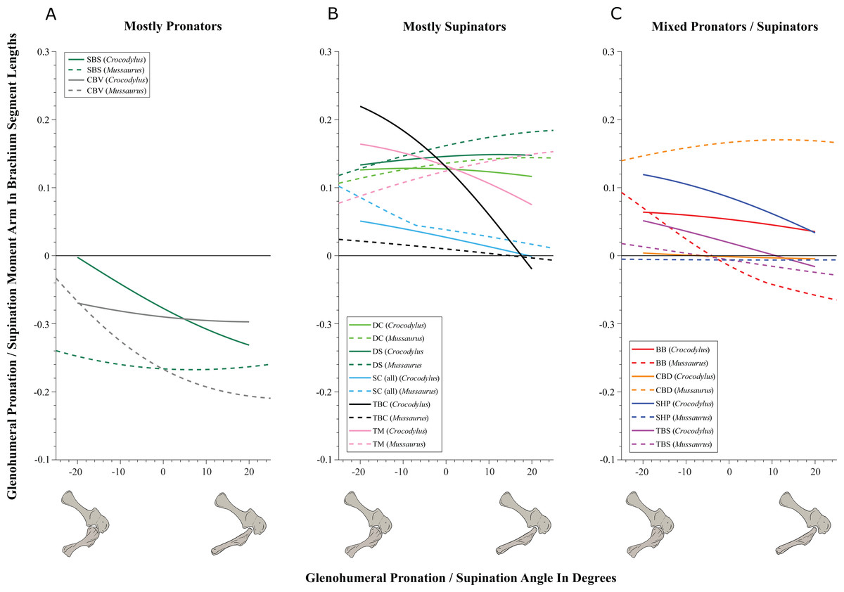
In our new study, we applied one of my team’s typical methods, 3D musculoskeletal modelling, to an adult Mussaurus’s forelimbs. This is a change of topic from the hindlimbs that I’ve myopically focused on before with Tyrannosaurus and Velociraptor [in an obscure paper that I should never have published in a book! pdf link], among other critters my team has tackled (mouse, elephant [still to be finished…], ostrich, horse, Ichthyostega… dozens more to come!). But we also modelled the forelimbs of Crocodylus johnstoni (Australian “freshie”) for a key comparison with a living animal whose anatomy we actually knew, rather than reconstructed.
Mussaurus above; Crocodylus below; forelimb models in various views; muscles are red lines.
The methods for this biomechanical modelling are now standard (I learned them from their creator Prof. Scott Delp during my 2001-2003 postdoc at Stanford): scan bones, connect them with joints, add muscle paths around them, and then use the models to estimate joint ranges of motion and muscle moment arms (leverage) around joints. I have some mixed feelings about developing this approach in our 2005 paper that is now widely used by the few teams that study appendicular function in extinct animals. As a recent review paper noted and I’ve always cautioned, it has a lot of assumptions and problems and one must exercise extreme caution in its design and interpretation. Our new Mussaurus paper continues those ruminations, but I think we made some progress, too.
On to the nuts and bolts of the science (it’s a 60 page paper so this summary will omit a lot!): first, we wanted to know how the forelimb joint ranges of motion in Mussaurus compared with those in Crocodylus and whether our model of Mussaurus might be able to be placed in a quadrupedal pose, with the palms at least somewhat flat (“pronated”) on the ground. Even considering missing joint cartilage, this didn’t seem very plausible in Mussaurus unless one allowed the whole forearm to rotate around its long axis from the elbow joint, which is very speculative—but not impossible in Crocodylus, either. Furthermore, the model didn’t seem to have forelimbs fully adapted yet for a more graviportal, columnar posture. Here’s what the model’s mobility was like:
So Mussaurus, like other early sauropodomorphs such as Plateosaurus, probably wasn’t quadrupedal, and thus quadrupedalism must have evolved very close to in the Sauropoda common ancestor.
Second, we compared the muscle moment arms (individual 3D “muscleactions” for short) in different poses for all of the main forelimb muscles that extend (in various ways and extents) from the pectoral girdle to the thumb, for both animals, to see how muscle actions might differ in Crocodylus (which would be closer to the ancestral state) and Mussaurus. Did muscles transform their actions in relation to bipedalism (or reversal to quadrupedalism) in the latter? Well, it’s complicated but there are a lot of similarities and differences in how the muscles might have functioned; probably reflecting evolutionary ancestry and specialization. What I found most surprising about our results was that the forelimbs didn’t have muscles well-positioned to pronate the forearm/hand, and thus musculoskeletal modelling of those muscles reinforced the conclusions from the joints that quadrupedal locomotion was unlikely. I think that result is fairly robust to the uncertainties, but we’ll see in future work.
You like moment arms? We got moment arms! 15 figures of them, like this! And tables and explanatory text and comparisons with human data and, well, lots!
If you’re really a myology geek, you might find our other conclusions about individual muscle actions to be interesting—e.g. the scapulohumeralis seems to have been a shoulder pronator in Crocodylus vs. supinator in Mussaurus, owing to differences in humeral shape (specialization present in Mussaurus; which maybe originated in early dinosaurs?). Contrastingly, the deltoid muscles acted in the same basic way in both species; presumed to reflect evolutionary conservation. And muuuuuuch more!
Do you want to know more? You can play with our models (it takes some work in OpenSim free software but it’s do-able) by downloading them (Crocodylus; Mussaurus; also available: Tyrannosaurus, Velociraptor!). And there will be MUCH more about Mussaurus coming soon. What is awesome about this dinosaur is that we have essentially complete skeletons from tiny hatchlings (the “mouse lizard” etymology) to ~1 year old juveniles to >1000kg adults. So we can do more than arm-wave about forelimbs!
But that’s not all. Last week we published our third paper on mouse hindlimb biomechanics, using musculoskeletal modelling as well. This one was a collaboration that arose from past PhD student James Charles’s thesis: his model has been in much demand from mouse researchers, and in this case we were invited by University of Virginia biomechanical engineers to join them in using this model to test how muscle fibres (the truly muscle-y, contractile parts of “muscle-tendon units”) change length in walking mice vs. humans. It was a pleasure to re-unite in coauthorship with Prof. Silvia Blemker, who was a coauthor on that 2005 T. rex hindlimb modelling paper which set me on my current dark path.
Mouse and human legs in right side view, going through walking cycles in simulations. Too small? Click to embiggen.
We found that, because mice move their hindlimb joints through smaller arcs than humans do during walking and because human muscles have large moment arms, the hindlimb muscles of humans change length more—mouse muscles change length only about 48% of the amount that typical leg muscles do in humans! This is cool not only from an evolutionary (mouse muscles are probably closer to the ancestral mammalian state) and scaling (smaller animals may use less muscle excursions, to a point, in comparable gaits?) perspective, but it also has clinical relevance.
Simulated stride for mouse and human; with muscles either almost inactive (Act=0.05) or fully active (Act=1). Red curve goes through much bigger excursions (along y-axis) than blue curve), so humans should use bigger % of their muscle fibre lengths in walking. Too small? Click to embiggen.
My coauthors study muscular dystrophy and similar diseases that can involve muscle stiffness and similar biomechanical or neural control problems. Mice are often used as “models” (both in the sense of analogues/study systems for animal trials in developing treatments, and in the sense of computational abstractions) for human diseases. But because mouse muscles don’t work the same as human muscles, especially in regards to length changes in walking, there are concerns that overreliance on mice as human models might cause erroneous conclusions about what treatments work best to reduce muscle stiffness (or response to muscle stretching that causes progressive damage), for example. Thus either mouse model studies need some rethinking sometimes, or other models such as canines might be more effective. Regardless, it was exciting to be involved in a study that seems to deliver the goods on translating basic science to clinical relevance.
Muscle-by-muscle data; most mouse muscles go through smaller excursions; a few go through greater; some are the same as humans’.
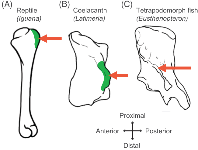
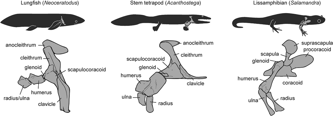
Finally, a third recent paper of ours was led by Julia Molnar and Stephanie Pierce (of prior RVC “Team Tetrapod” affiliation), with myself and Rui Diogo. This study tied together a bunch of disparate research strands of our different teams, including musculature and its homologies, the early tetrapod fossil record, muscle reconstruction in fossils, and biomechanics. And again the focus was on forelimbs, or front-appendages anyway; but turning back the clock to the very early history of fishes, especially lobe-finned forms, and trying to piece together how the few pectoral fin muscles of those fish evolved into the many forelimb muscles of true tetrapods from >400mya to much more recent times.
Humerus in ventral view, showing muscle attachments. Extent (green) is unknown in the fossil but the muscle position is clear (arrow).
We considered the homologies for those muscles in extant forms, hypothesized by Diogo, Molnar et al., in light of the fossil record that reveals where those muscles attach(ed), using that reciprocal illumination to reconstruct how forelimb musculature evolved. This parallels almost-as-ancient (well, year 2000) work that I’d done in my PhD on reconstructing hindlimb muscle evolution in early reptiles/archosaurs/dinosaurs/birds. Along the way, we could reconstruct estimates of pectoral muscles in various representative extinct tetrapod(omorph)s.
Disparity of skeletal pectoral appendages to work with from lobe-fins to tetrapods.
Again, it’s a lengthy, detailed study (31 pages) but designed as a review and meta-analysis that introduces readers to the data and ideas and then builds on them in new ways. I feel that this was a synthesis that was badly needed to tie together disparate observations and speculations on what the many, many obvious bumps, squiggles, crests and tuberosities on fossil tetrapods/cousins “mean” in terms of soft tissues. The figures here tell the basic story; Julia, as usual, rocked it with some lovely scientific illustration! Short message: the large number of pectoral limb muscles in living tetrapods probably didn’t evolve until limbs with digits evolved, but that number might go back to the common ancestor of all tetrapods, rather than more recently. BUT there are strong hints that earlier tetrapodomorph “fishapods” had some of those novel muscles already, so it was a more stepwise/gradual pattern of evolution than a simple punctuated event or two.
Colour maps of reconstructed right fin/limb muscles in tetrapodomorph sarcopterygian (~”fishapod”) and tetrapod most recent common ancestors. Some are less ambiguous than others.
That study opens the way to do proper biomechanical studies (like the Mussaurus study) of muscle actions, functions… even locomotor dynamics (like the mouse study)– and ooh, I’ve now tied all three studies together, tidily wrapped up with a scientific bow! There you have it. I’m looking forward to sharing more new science in 2018. We have some big, big plans!
We’d been wanting to do a family holiday in Ireland for years and so we finally did. I’d been to Dublin twice before for work visits and we wanted a more rural experience. On others’ recommendations, we started in the city of Cork. With some sleuthing and asking around, I realized that we weren’t far then from gorgeous Killarney National Park, and then it wasn’t far west from there to get to Valentia Island, where incidentally there is something amazing for palaeontology-lovers. There was no deterring me at that point from visiting what I’d only read about. I’ll mainly let the images tell the story.
Stomach-Churning Rating: 0/10; fossils and scenery. Kick back and enjoy.
Island map- it really is that simple to get around! The harbour town of Portmagee is damned adorable.
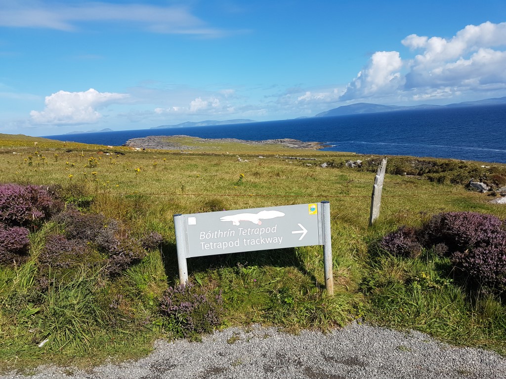
Driving in (no I am the passenger; not taking photo while at the wheel!)- excitement level = 8 and building… “Tetrapod carpark” sign ratcheted up the excitement and was amusing.
Headed to the trail; excitement level = 9…
Looking down onto the site (on the right); excitement level = 9.5; beauty level = 9.5 too!
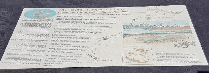
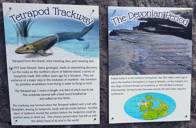
Now, the site of what is broadly accepted by experts as a ~Middle Devonian tetrapod(omorph)’s fossil trackway(s) was originally described by Stössel in 1995. To me, that feels like a recent discovery but it is 22 years ago. The only other well-preserved, widely-accepted, probably-terrestrial, Late Devonian tetrapod(omorph) trackways are from the Genoa River site in Australia; described by Warren et al. in 1972. Those trackways even reveal some details of the fingers and toes; these do not. Other tracks are either isolated footprints of minimal scientific value/clarity, subaerial (i.e. underwater), not clearly stem-tetrapod (or now argued to be arthropod or other origin), not Devonian, or controversial for reasons I won’t get into here. The famous Zachelmie tracks in Poland are strong contenders but remain controversial to more than a few researchers in terms of who made them and in what environmental/substrate context; but their Middle Devonian age seems robustly agreed. Clack and Lucas have reviewed the relevant evidence recently. So there are essentially at least two, and arguably three, places in the world that you can visit to view tracks like these and it was a joy to go visit one set. (Easter Ross, Scotland may be a fourth site but it is reasonably disputed in age and maker)
There is a “however,” however- Falkingham and Horner showed how lungfish can produce tracks (with fins and heads together) that look like these, to some viewers (but not to others) and in some substrates (mud; not sand as at the Valentia site)– so there is still uncertainty for some tracks although the lungfish-feeding claims have also been vehemently disputed too. Without finger and toe impressions, claims of discrete tetrapod tracks are risky, and it would be wrong to say that the Valentia Island footprints are uncontroversially or 100% certainly tetrapod in origin, although they are (late-Middle) Devonian and made by some sort of animal, and very likely a tetrapodomorph at least.
Stössel et al. also published a recent update on these Valentia Island tracks with more information. I wish I’d come across that before I visited (oops!). That study reports on a total of nine(!) trackways from the area, adding to the 1995’s first one (the “Dohilla locality, Do 1”– see diagrams below), and describes them as Middle Devonian (with a radiometric dating of 385 million years old). I’m not enough of a geologist to evaluate that; prior reports had focused on Late Devonian or so.
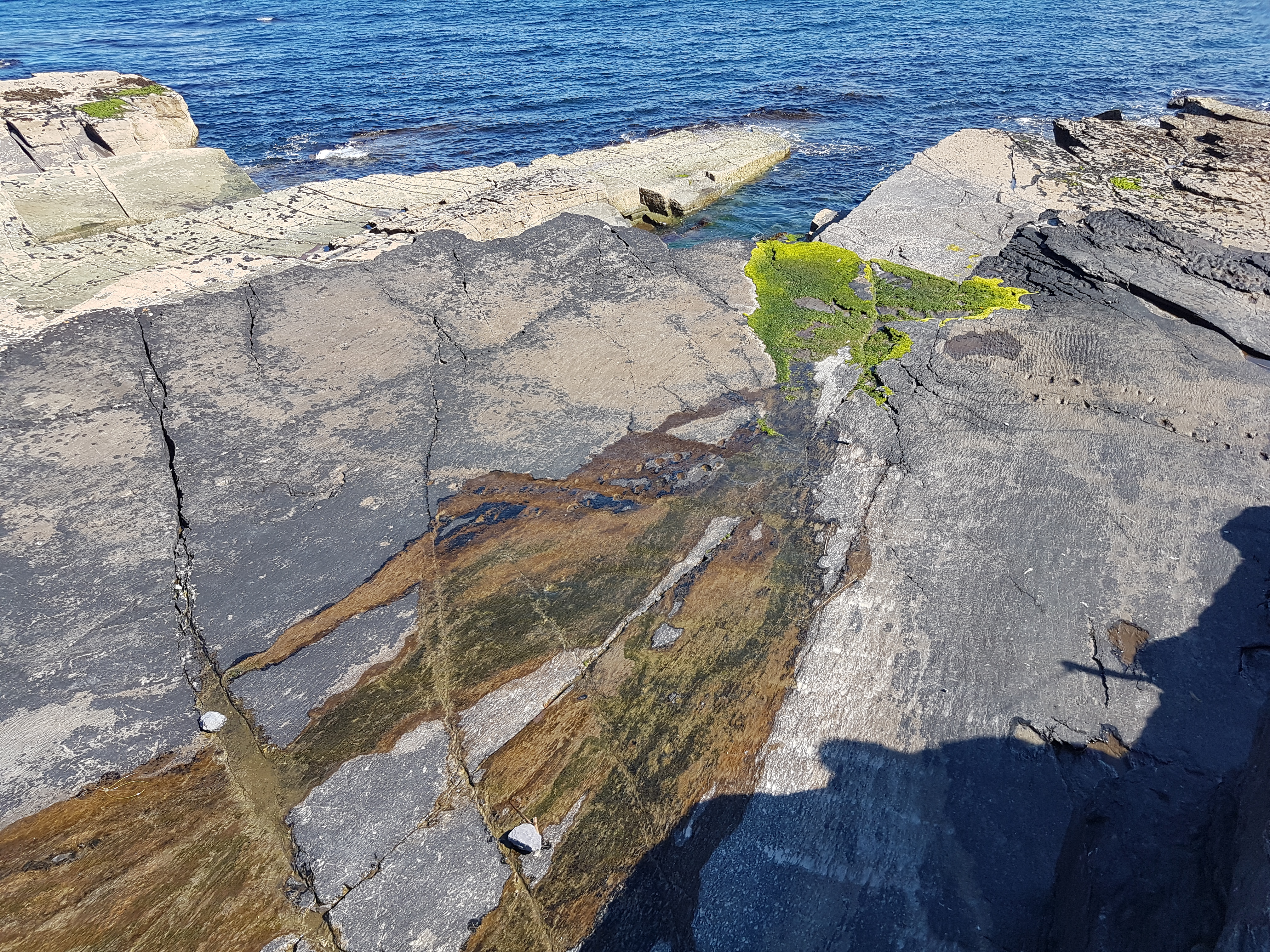
Rippled sandstone example; preservation characteristic of the trackway area/Valentia Slate Formation. It’s an alluvial deposit (freshwater floodplain), interpreted to lie inland from the coastal marine deposits. Raindrop impressions above the plane of the tracks raise the possibilities that the tracks were made on (moist) land.
The island has plenty of signs advertising the tracks as a tourist destination but happily(?) there are no knick-knack shops stocked with plush tetrapods, or other developments at or near the site. One simply winds down a very narrow road near a radio station and old lighthouse, and parks then walks to see the tracks. No fancy crap; just AWESOME sights to take in, and some good educational information.
Explanatory plaque at the viewing area. Pretty good!
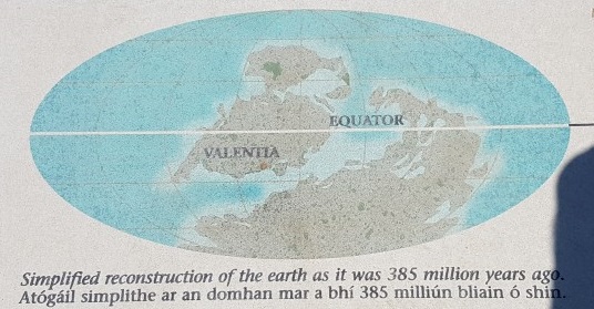
Nice image of where Valentia Island was; although the 385 My age may be exaggerated. It’s not clear how old the tracks are but “Mid-to-Late Devonian” might suffice depending on how you view the evidence. The tracks were the “oldest known” at the time of discovery and remain close to that, but challenged by the Zachelmie trackways (see references above).
Explanatory signs on the peak above the shore. Given the likely tetrapod(omorph) trackmakers like Acanthostega-style critters, the adult animal may have been able to breathe air with lungs and underwater with gills.
Enough exposition– let’s expose those tracks! (images can be clicked to enlarge)
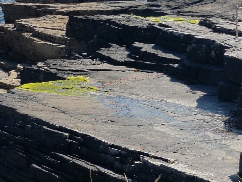
My first close-up look at the tracks. Whoa! Small tracks are presumably hand (manus) impressions; larger ones are foot (pes). The tracks go in an alternating fashion (like a salamander’s walk) and the animal was probably going from the bottom-right toward the top-left. Moss and moisture obscured some of the prints that day, sadly. The tracks are oval, with the long axes perpendicular to the direction of travel. There are some pesky geological deformations of the trackway, faults, and other distortions. 145 footprints in total are reported from this one trackway!
Trackway as it turns to the left and gets harder to follow. John-shadow for ~scale. Frustratingly for me, a little rivulet was coming down the hill across the left side of the trackway and hiding much of the detail of the end.
Alternative view of the majority of the tracks; turned ~90 degrees from above two views.
Zoomed-in view of the tracks from head-on (opposite the view in other photos); i.e. western position looking east (ish). I added red and blue dots to roughly outline the right side of the main trackway (red) and the second one (blue), which crosses it and may have been made after it.
Even these nice trackways, viewed by an expert, take some unpacking. Here is some:
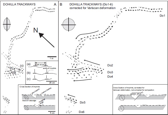
Diagram of known tracks at the site by Stössel et al. 2016.
Diagram at view site with extra tail (or body) drag trail crossing the main tracks; described later by Stössel et al. 2016.
I’m not at all a religious person and I don’t really like the term “spiritual” either, but this experience was emotional for me. Awe is certainly the best word to describe what I felt on viewing these tracks. The literature just doesn’t do them justice; nothing beats a first-person experience like this. We were lucky with excellent weather, too, and we were almost alone during the visit so there was pleasant silence in which to contemplate the tracks. I brought my copies of three papers on the trackways and, struggling with the wind, compared them with the visible tracks to understand what other scientists had seen. That amplified the experience enormously for me.
Even if they turn out to be non-tetrapod or younger or something less exciting (“sham-rock”?), it was thrilling to see the Valentia Island tracks and think about what happened ~385 million years ago when they were made by our very distant cousins, along the land-water interface of space and time.
(I also feel bad for online reviewers that were disappointed with the site- it’s hard to grasp the scientific importance and/or accept the evidence, even with the decent information available on-site. Even if people know the nice fossil record of dinosaurs, they may not know how good the fossil record of early tetrapods is and how confidently we can figure out what happened in the Devonian emergence of tetrapod(omorph)s onto land. But some visitors clearly got it.)
And, looking at the site myself, I realized how many more tracks might be buried under the cliffs of the site- the first trackway emerges from under a cliff and thus must still be preserved for some distance underground, awaiting future exposure. What more might we learn about that single animal and others that made tracks around the same time? I hope to live to find out. I feel a personal connection now to these tracks, left pondering what story they preserve– and hide. I’m glad I’m able to share my own story with you, and encourage you to make the visit yourself!
Who needs “Ice Road Truckers” when you have the “John’s Freezer” team on the road with fossils, amphibians, felids and 3D phenotype fun? No one, that’s who. We’re rocking the Cheltenham Science Festival for our first time (as a group), and pulling out all the stops by presenting two events! Here’s the skinny on them, with updates as the week proceeds.
Stomach-Churning Rating: 2/10 for now (just bones), but it could change once the cheetah dissection is under way… 8/10 bloody cheetah bits but only at the end (updated)
Right now, Lauren Sumner-Rooney (of “Anatomy To You” and other fame) is on-site with a rotating team of others from our lab, in the “Free Activity Tents” area of the Imperial Gardens/Square, inside a marquee where we’ll be showing off our NERC-funded tetrapod research all week. This “First Steps” event features not only our past and present work with Jenny Clack, Stephanie Pierce, Julia Molnar and others on Ichthyostega & its “fishapod” mates, but also our “scampering salamanders” research in Spain, Germany and England. I’ve blogged a lot about all that, and won’t repeat it here, but you can see a 3D-printed Ichthyostega skeleton, view the skeleton in a virtual reality 3D environment, see related specimens and engage in kid-friendly activities, and talk to our team about this and other related research.
Ichthyostega 3D printed backbone is born!
The central themes of that event are how bone structure relates to function and how we can use such information, along with experimental measurements and computer models of real salamanders, to reconstruct how extinct animals might have behaved as well as how swimming animals became walking ones. How did fins transform into limbs and what did that mean for how vertebrates made the evolutionary transition onto land? If you know my team’s work, that encapsulates our general approach to many other problems in evolutionary biomechanics (e.g. how did avian bipedalism evolve?). Added benefits are that you too can explore this theme in a hands-on way, and you can talk with us about it in person. That continues all week (i.e. until Saturday evening); I’ll be around from Thursday afternoon onwards, too. Kids of all ages are welcome!
Ichthyostega 3D print taking shape!
Then, on Saturday for our second free event we join forces with Ben Garrod (master of primate evolution, the secrets of bones, and “Attenborough and the Giant Dinosaur”) and RVC’s forensic pathologist Alexander Stoll as well as Sophie Regnault (“sesamoid street” PhD student w/me). As the “Large Animal Dissection” title hints, it’s not the right kind of gig to bring small kids to. There will be blood and stuff— we’ll be dissecting a cheetah together from 10am-4pm. This will involve walking through all the major organ systems, giving evolutionary anecdotes, and plenty more, with an aim to understand how the magnificent adaptations of cheetahs evolved—but also to investigate what problem(s) this animal faced that led to its sad demise. By the day’s end, there will just be a skeleton left. Get a front row seat early for this event, which serendipitously ties into “Team Cat”’s Leverhulme Trust-funded research project (we wanted a big animal and it just happened to be a cheetah; I had hoped for a giant croc or a shark or something but can’t complain!).
Ichthyostega 3D print is ready!
If you miss these events, please do cry bitter tears of regret. But don’t despair, there will be another “big cat dissection” in the London area in ~November (watch here for details), and plenty more fossil tetrapod stuff to come, plus a LOT more dinosaurs on the horizon!
Guess the bones! (photo by Zoe Self)
And please come back to this blog post for more pics and stories as the week carries on… For hashtag afficionados, you can follow the fun on Twitter etc. at #firststepsCSF16. What a world we live in!
Update 1: While you’re here, check out our Youtube playlists of tetrapod-related videos:
Update 2: Photos of our main stand (about tetrapod evolution)
Our poster/banner display looks nice.
Our tent brings in some punters.
Our bones excite people young and old, sighted and blind.
Fun with stickers and lab t-shirts.
And…
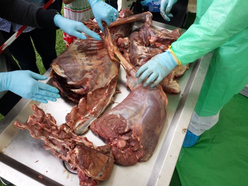
Update 3: Cheetah meat & greet
Ben, Alex, Sophie and I tackled the cheetah dissection today and it went GREAT! Much better than I’d optimistically expected. Rain didn’t scare the crowds off and neither did the gore, which there was some of (gelatinous spinal cords, lumpy tumors and at least one flying tiny bit of cheetah flesh that landed on a good-natured audience member!). Photos will tell the tale:
Peek-a-boo!
Sophie and Alex help us get set up in our tent.
Our initial rough schedule- although we ended up improvising more after lunch.
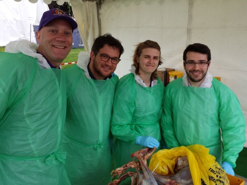
Dissectors assemble!
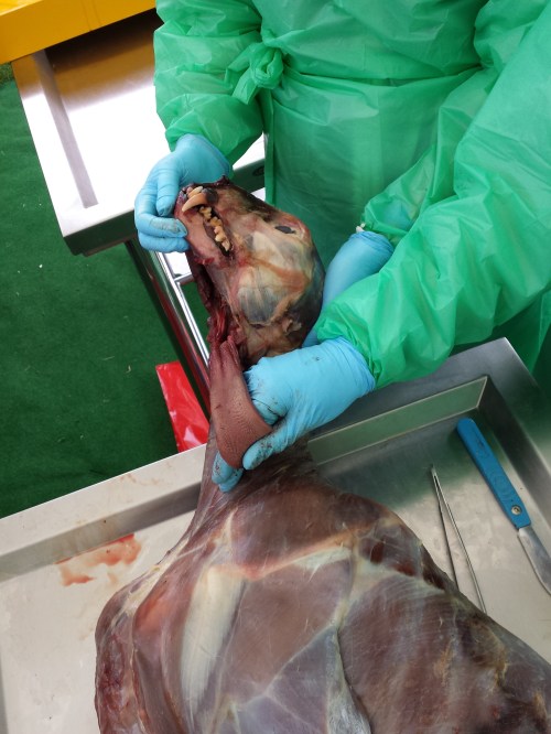
The beast revealed. It was skinned by the museum that loaned it to us.
Alex showing his talent: removing the viscera in one piece from end to end, starting with the tongue.
Impressive finding of a surgical fixture (plate and wires) on the tibia, which had been used to hold the shattered bone back together long enough for it to heal. Added to the kidney disease and liver-spleen-lung cancer, this cheetah was in the sorriest shape of any cadaver I’ve seen yet.
Cheetah coming to pieces: (from bottom) lumbar/pelvic region, hindlimb, thorax, forelimb and other bits.
Dr Adam Rutherford, an eye expert, did a nice dissection of the cheetah’s eye, here showing the tapetum lucidum (reflective membrane), which shows up as light blue colour. Its small size befits the not-very-nocturnal habits of cheetahs.
The lens of the cheetah’s eye. Now cloudy because of dehydration and crystalization, but still fascinating to see.
Ambitious experimental and morphological studies of a modern fish show how a flexible phenotype may have helped early “fishapods” to make the long transition from finned aquatic animals into tetrapods able to walk on land.
Stomach-Churning Rating: 1/10. Cute fish. Good science. Happy stomachs!
Photo by Antoine Morin, showing Polypterus on land.
Napoleon Bonaparte’s military excursions into Egypt in 1798-1799 led a young French naturalist, Ètienne Geoffroy Saint-Hilaire, to cross paths with a strange fish that had paired lungs and could “walk” across land on its stubby, lobelike fins. In 1802, he dubbed this fish “Polyptère bichir”1, today known as the Nile bichir, Polypterus bichir La Cepède 1803. The bichir’s mélange of primitive and advanced traits helped to catapult Geoffroy into scholarly conflict with the reigning naturalist Georges Cuvier back in France and to establish Ètienne as a leading anatomist, embryologist and early evolutionary researcher of repute even today2. Now, on their own excursion under the very “evo-devo” flag that the discoverer of Polypterus helped raise, Canadian scientists Standen et al.3 suggest how the remarkable plasticity of the skeleton of Polypterus (the smaller west African relative of P. bichir, P. senegalus or “Cuvier’s bichir”) reveals a key part of the mechanism that might have facilitated the gradual transition from water to land and thus from “fishapods” to tetrapods (four-limbed vertebrates).
In a bold experiment, the authors raised 149 young bichirs on land and in water for eight months, then studied how they moved on land vs. in water, and also how the ultimate shape of the skeletal elements of the paired front fin bases differed between the land- and water-raised bichirs. Standen et al.3 discovered that both the form and function of the fins’ foundations transformed to better satisfy the constraints of moving on land. Land-acclimated bichirs took faster steps on land, their fins slipped across the substrate less, they held their fins closer to their body, their noses stayed more aloft and their tails undulated less, with less variable motions overall—behaviours that the authors had predicted should appear to enhance walking abilities on land. In turn, the bones of the neck and shoulder region altered their shape to produce a more mobile fin base with greater independence of fin from neck motion, along with improved bracing of the ventral “collarbone” region. These environmentally-induced traits should have fostered the locomotor changes observed in “terrestrialized” fish and aided the animals in resisting gravity, and they represent a common biological phenomenon termed developmental plasticity4,5. Interestingly, the land-reared fish could still swim about as well as the wholly aquatic cohort, so there was not a clear trade-off between being a good swimmer and a good walker, which is surprising.
Considered alone, the developmental plasticity of bichir form and function shows how impressive these amphibious fish are. But Standen et al.’s study3 ventured further, to apply the lessons learned from bichir ontogeny to a phylogenetic context and macroevolutionary question. The phenotypic plasticity during bichir development, they infer, could have been harnessed during the evolutionary transformation of fins for swimming into limbs for walking, in the “fishapod” ancestors of tetrapods. Indeed, bichirs are close to the base of the family tree of fishes6, and other living relatives of tetrapods have reduced or lost their fins (lungfishes) or adapted to strange deep-sea swimming lifestyles, never walking on land (coelacanths). Thus perhaps bichirs and the “fishapod” lineage share what Geoffroy would have called “unity of type”, today termed homology, of their developmental plasticity in response to a land environment. Surveying the fossil record of early “fishapods” and tetrapods, Standen et al.3 found that the macroevolutionary changes of neck and shoulder anatomy in these gradually more land-adapted animals parallel those they observed in terrestrialized Polypterus, providing ancillary support for their hypothesis.
A further test of the application of Polypterus’s plasticity to fossil tetrapods is naturally difficult. However, the “fishapod” lineage has some exceptional examples of fossil preservation. With sufficient sample sizes (e.g. fossil beds that reveal growth series, such as the Late Devonian Miguasha site in Canada7) and palaeoenvironmental gradients in fish or tetrapods, one could imagine performing a rigorous indirect test. Even small samples could be helpful– for example, the early tetrapod Ichthyostega exhibits some developmental changes in its forelimb suggesting that it became more terrestrial as it grew, whereas the related Acanthostega does not evidence such changes8— this hints at some developmental plasticity in the former animal.
During the Devonian period (~360-420 million years ago), were the “fishapod” ancestors of tetrapods floundering about on land now and then, gradually shifting from anatomy and behaviours that were more developmentally plastic (as in bichirs) to ones that were more canalized into the terrestrialized forms and functions that more land-adapted tetrapods retained? An attractive possibility is that the developmental plasticity could have led to fixation (reduction of plasticity), an evolutionary phenomenon called genetic assimilation, which another intellectual descendant of Geoffroy, Conrad Hal Waddington, promoted from the 1950s onwards9, a concept that now enjoys numerous cases of empirical support10 that this one may eventually join.
The nature of the genetic and developmental mechanism that bichirs use to achieve the observed developmental plasticity is still unclear. If it has a high enough degree of heritability, then it could be selected for in cross-generational experiments with bichirs. With sufficient time and luck raising these unusual fish, the hypothesis that their plastic response to a terrestrial environment can become genetically assimilated could be directly tested. This study could thus become an epic exemplar of how genetic assimilation can contribute not only to microevolutionary change but also to major macroevolutionary events, as was presciently suggested in a seminal review of developmental plasticity4.
This genetic assimilation is the Polypterus study’s reasonable speculation, and one that Geoffroy likely would have applauded, all the more for involving his beloved bichirs. Much as Napoleon’s landfall in Egypt was not a lasting success, bichirs never left wholly terrestrial descendants despite their malleable locomotor system. But the same type of plastic developmental mechanism that bichirs use today to make tentative, floppy incursions of the terrestrial realm might have been harnessed by our own “fishapod” forebears, leaving a far more revolutionary dynasty upon the Earth.
References
Geoffroy, E. (1802). Histoire naturelle et description anatomique d’un nouveau genre de poisson du Nil, nommé polyptère. Annales du Muséum d’Histoire Naturelle 1:57-68.
Le Guyader, H., & Grene, M. (2004) Geoffroy Saint-Hilaire: A Visionary Naturalist. Univ. Chicago Press.
Standen, E. M., Du, T. Y., & Larsson, H. C. E. (2014). Developmental plasticity and the origin of tetrapods. Nature, published online.
West-Eberhard, M. J. (1989). Phenotypic plasticity and the origins of diversity. Annual Review of Ecology and Systematics 20:249-278.
Pigliucci, M., Murren, C. J., & Schlichting, C. D. (2006). Phenotypic plasticity and evolution by genetic assimilation. Journal of Experimental Biology 209(12):2362-2367.
Near, T. J., Dornburg, A., Tokita, M., Suzuki, D., Brandley, M. C., & Friedman, M. (2014). Boom and bust: ancient and recent diversification in bichirs (Polypteridae: Actinopterygii), a relictual lineage of ray‐finned fishes. Evolution68:1014-1026.
Cloutier, R. (2013). Great Canadian Lagerstätten 4. The Devonian Miguasha Biota (Québec): UNESCO World Heritage Site and a Time Capsule in the Early History of Vertebrates.Geoscience Canada40:149-163.
Callier, V., Clack, J. A., & Ahlberg, P. E. (2009). Contrasting developmental trajectories in the earliest known tetrapod forelimbs.Science324:364-367.
Waddington, C. H. (1953). Genetic assimilation of an acquired character. Evolution 7:118-126.
Crispo, E. (2007). The Baldwin effect and genetic assimilation: revisiting two mechanisms of evolutionary change mediated by phenotypic plasticity. Evolution 61:2469-2479.
My team had a new technician arrive, Kyle Chadwick from Uni. Virginia, and NSF Postdoctoral Research Fellow, Dr. Ashley Heers (see here for an example of new stuff she’s starting here at the RVC!), started working with me at the RVC, and then these guys showed up…
First a tiger salamander (Ambystoma) paid a visit, for filming an episode of the Windfall Films/PBS documentary “Your Inner Fish” (a la the famous book):
So cute! Tiger salamander, soon to be a TV celebrity.
And that gorgeous salamander was a star performer in strutting his stuff for the camera to demonstrate the locomotion of modern tetrapods, including some lovely slo-mo footage from our lab cameras:
(if that’s too slow for you, try the normal-speed footage. I’ll admit, salamanders don’t really need slo-mo video for normal walking, but I like it)
So cool!
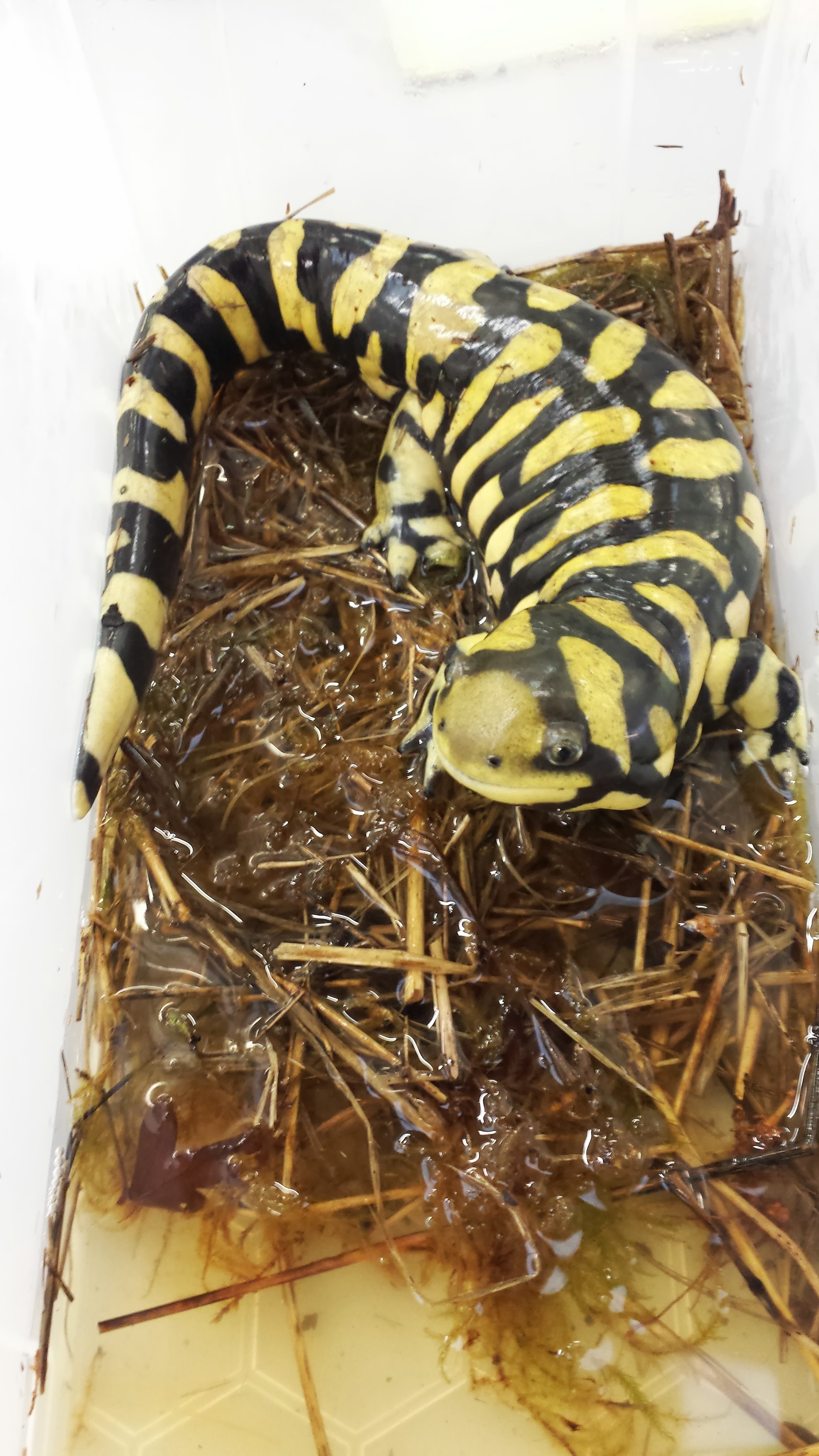
But then we got a special package… with three frozen fire salamanders (Salamandra salamandra) from colleagues in Germany!
Three new occupants of the freezers, for planning our studies of salamander locomotion
This marks the start of an exciting new period in my team’s work in the lab. I’ve always liked salamanders and newts, and we’ve scanned and modelled plenty (e.g. this old post), but now we’re going to work with live fire salamanders (a first for me)! We are using the dead ones to plan the new studies with the live ones– these new studies will involve lots of high speed videos and force platform analysis (as shown above), in conjunction with XROMM (biplanar fluoroscopy/3D skeletal motion analysis) and other techniques including computer simulations. We got initial approval this week to work with these salamanders, and found a reputable source this week too, so it was definitely Salamander Week in my group!
This research all will feed into our upcoming studies of extinct tetrapods: we’re using salamanders to figure out how salamanders move and what limits their speed and gait, and then we’re using the same sorts of computer tools to try to estimate how extinct tetrapods may have moved and how locomotion evolved, in much more specific detail than our prior work had done, which was mainly about using 3D reconstructions of anatomy to show what those animals could not do. More about the project here.
Watch this space for more scampering salamanders!
UPDATE: And here’s one! Not quite scampering, but…
Setting up our two fluoroscopes for a test run of our gait studies– but with one of the deceased salamanders. Gotta get good images before any live animal work begins!
An example of the kind of footage we’re aiming for (single 2D fluoroscope view from Nadja Schilling’s team’s research; see XROMM website for more details on the methodology)
UPDATE 2:
I did a CT scan with a normal medical grade CT scanner at the highest resolution we can manage (0.625 mm slices). Check out the results below, which amuse me:
Looks like a toy; too crude resolution. But we can see major structures, and we can very nicely see the “microchip” (which looks HUGE) that was placed in this animal’s back when in captivity, and then another structure is visible near the pelvis which might be another chip or else remains of some food, pathology, or a really odd pelvis– I am not totally sure!
So this is why we tend to use microCT, which can go down to as low as ~5 micron resolution, to get 3D anatomy of animals this small. It’s no surprise to me, but it is fun to see how far we could push our normal CT machine. The results aren’t horrid but wouldn’t have much scientific value for us. They did confirm for us that this specimen is heavily ossified, so the faint images of bone that we are getting in our x-ray fluoroscopes (above) are due to something going wrong with our camera system, not the animal’s immature skeleton. Stay tuned for more updates as the science happens!
UPDATE 3:
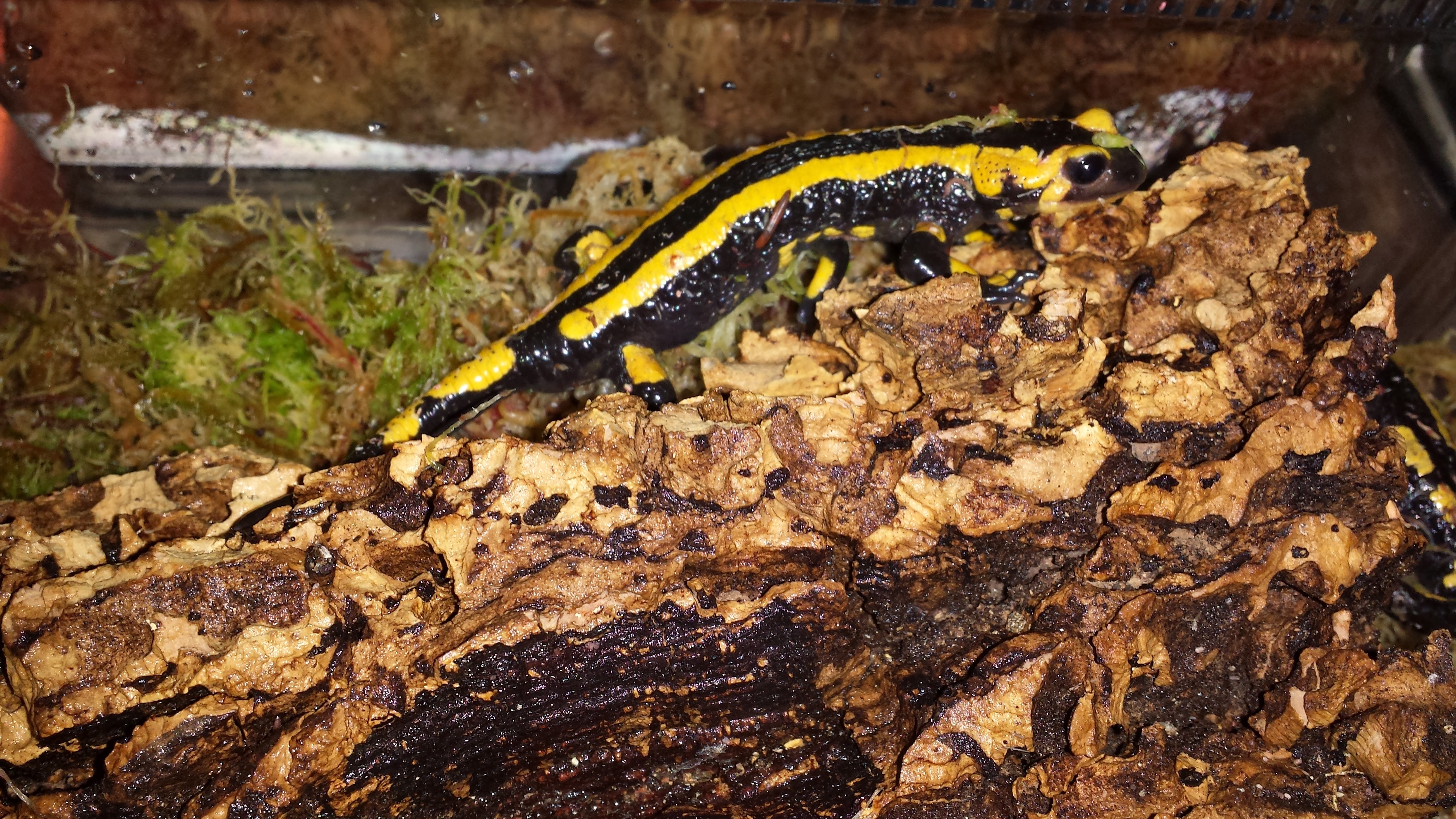
20 wonderful adult Fire Salamanders have joined our team and are relaxing over the coming week before we start taking them for walks. Here is one exploring its new home:
UPDATE 4:
August 11-15, 2014 we are in Jena, Germany using their fancy biplanar radiography system (“x-ray video”) to study our salamanders, at last! Follow the tweets starting here, for more information as it happened! https://twitter.com/JohnRHutchinson/status/500187568416518144
and this video of “Jabba” the corpulent salamander walking-
I’m letting the dogs out today. Science gone barking mad! Hopefully my puns will not screw the pooch.
Stomach-Churning Rating: 4/10; a dog cadaver’s leg (not messy), then just tame digital images of anatomy.
I am working with Rich Ellis, a former MSc student at Univ. Colorado (see his cool new paper here!), for a fun new collaboration this year. He was awarded a prestigious Whitaker Foundation scholarship to do this research, which focuses on how different animals stand up from a squatting position, with the legs about as bent as they can be.
We want to know how animals do this standing up movement, because it is in some ways a very demanding activity. Very flexed/bent limb joints mean that the muscles (and some tendons) are stretched about as far as they ever will be. So this places them at disadvantageous lengths (and leverage, or mechanical advantage) for producing force. We know almost nothing about how any animal, even humans, does this-– how close to their limits of length are their muscles? Which muscles are closest? Does this change in animals with different numbers of legs, postures, anatomy, size, etc? Such fundamental questions are totally unaddressed. It’s an exciting area to blaze a new trail in, as Rich is doing. So far, we’ve worked with quail, humans, and now greyhounds; in the past I did some simple studies with horses and elephants, too. Jeff Rankin from my team and other collaborators have also worked on six species of birds, of varying sizes, to see how their squat-stand mechanics change. Thus we’ve covered a wide diversity of animals, and now we’re learning from that diversity. “Diversity enables discovery,” one of my former PhD mentors Prof. Bob Full always says. Too true.
Greyhounds are interesting because they are medium-sized, long-legged, quadrupedal, quite erect in posture, and very specialized for fast running. Fast runners tend to have big muscles with fairly short fibres. Short fibres are bad for moving the joints through very large ranges of motion. So how does a greyhound stand up? Obviously they can do it, but they might have some interesting strategies for doing so- the demands for large joint motion may require a compromise with the demands for fast running. Or maybe the two demands actually can both be optimized without conflict. We don’t know. But we’re going to find out, and then we’ll see how greyhounds compare with other animals.
To find out, we first have to measure some dogs standing up. We’ve done that for about 8 greyhounds. Here is an example of a cooperative pooch:
Those harmless experiments, if you follow me on Twitter, were live-tweeted under the hashtag #StandSpotStand… I dropped the ball there and didn’t continue the tweeting long after data collection, but we got the point across– it’s fun science addressing useful questions. Anyway, the experiments went well, thanks to cooperative pooches like the one above, and Rich has analyzed most of the data.
Now the next step involves the cadaver of a dog. We could anaesthetize our subjects and do this next procedure to obtain subject-specific anatomy. But it really wouldn’t be ethically justified (and if I were an owner I wouldn’t allow it either!) and so we don’t. A greyhound is a greyhound as far as we’re concerned; they’ll be more like each other than either is like a quail or a human. Individual variation is a whole other subject, and there are published data on this that we can compare with.
We get a dead dog’s leg — we don’t kill them; we get cadavers and re-use them:
We study the hindlimb because birds and humans don’t use their forelimbs much to stand up normally, so this makes comparisons simpler. We’re collecting forelimb data, though, as we work with quadrupeds, for a rainy day.
We then CT scan the leg, getting a stack of slices like this– see what you can identify here:
It’s not so clear in these images, but I was impressed to see that the muscles showed up very clearly with this leg. That was doggone cool! Perhaps some combination of formalin preservation, fresh condition, and freezing made the CT images clearer than I am used to. Anyway, this turned out to be a treat for our research, as follows.
We then use commercial software (we like Mimics; others use Amira or other packages) to “segment” (make digital representations in 3D) the CT scan data into 3D anatomy, partitioning the greyscale CT images into coloured individual objects– two views of one part of the thigh are shown below.
What can you identify as different colours here? There are lots of clues in the images (click to embiggen):
And here is what the whole thigh looks like when you switch to the 3D imaging view:
Quite fetching image, eh?!
The next steps after we finish the limb segmentation are to apply the experimental data we observed for greyhounds of comparable size by importing the model and those data into biomechanics software (SIMM/OpenSim). We’ve done about 40 models like this for various species. I detailed this procedure for an elephant here.
Then, at long last, science will know how a greyhound stands up! Wahoo! Waise the woof! Stay tuned as we hound you with more progress on this research-as-it-happens. Rich just finished the above thigh model this week, and the rest of the leg will be done soon.
Many thanks to Rich Ellis for providing images used here. And thank you for persevering my puns; they will now be cur tailed.
Happy Freezermas! Sing it: “On the fifth day of Freezermas, this blo-og gave to me: one tibiotarsus, two silly Darwins, three muscle layers, four gory hearts, a-and five stages modelling a doggie!” ♪♫
To kick off the New Year just right, our tetrapod team has a new paper in Nature, following up on last year’s Ichthyostega not-so-good-at-walking study (also see here). Yet this paper has a more anatomically descriptive — and also an “evo-devo” — twist to it. For brevity, I’ll let our press release tell the story, since I think it does a good job of it (like I always preach scientists should do, we worked with our PR company to write this together, so we’re happy with how the press release came out). In a nutshell, our study used some very fancy synchotron radiation techniques to image the 3D anatomy of the backbone in early land vertebrates. Our findings surprised even us, and ended up turning around palaeontology/comparative anatomy’s view of how the backbone evolved, giving us a new glimpse into our inner tetrapod.
Stick around for the videos at the end, which are the first four supplementary movies from the paper and are rather pretty (there are two more, for imaging/segmenting afficionados, but they are not as pretty or interesting for most of this blog’s readership). The final figure (Figure 1 from our paper) gives some extra visual context.
The paper is:
Pierce, S.E., Ahlberg, P.E., Hutchinson, J.R., Molnar, J.L., Sanchez, S., Tafforeau, P., Clack, J.A. 2013. Vertebral architecture in the earliest stem tetrapods. Nature, published online [here].
I should note that I’m just 3rd author, so I deserve only modest credit. But I helped. Even though no freezers were involved, or harmed, in the process.
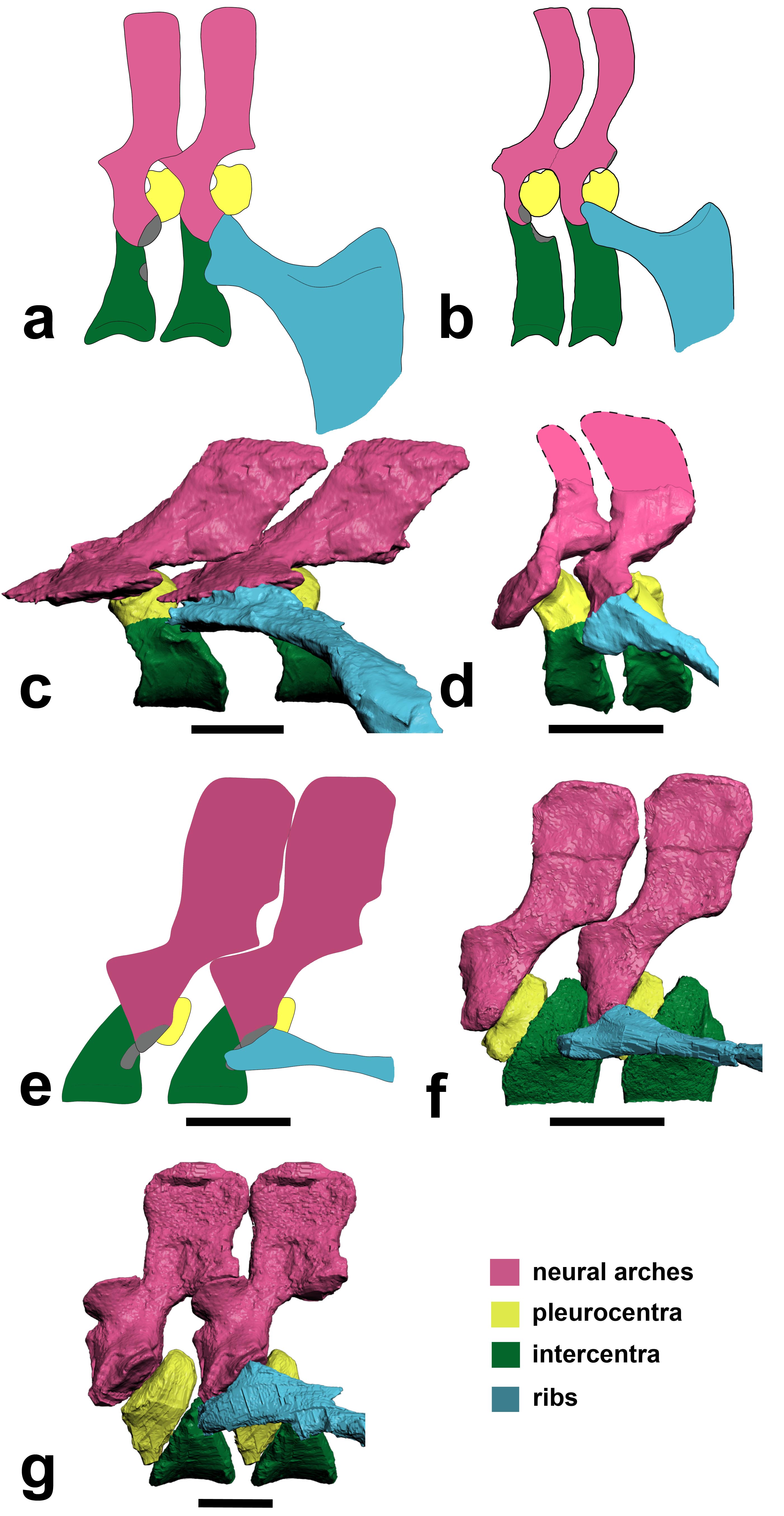
Above image: Julia Molnar‘s illustration of Ichthyostega showing anatomical changes of its spine from front to back, with neural arch/spine in pink, twin pleurocentra in yellow, and intercentrum in green. These four parts, three kinds of bones, made up the backbone of the first land vertebrates. These parts evolved in different ways in later animals, but formed one main bone in all living lineages of vertebrates.
RVC PRESS RELEASE:
Scientists reassemble the backbone of life using a particle accelerator
Research published today (Sunday 13 January 2013) in the journal Nature documents, for the first time, the intricate three-dimensional structure of the backbone in the earliest four-legged animals (tetrapods).
The backbone, also known as the spine or vertebral column, is a bony structure found in all tetrapods, along with other vertebrates such as fish. It is formed from many elements or vertebrae all connected in a row – from head to tail. Unlike the backbone of living tetrapods (e.g. humans), in which each vertebra is composed of only one bone, early tetrapods had vertebrae made up of multiple parts.
Lead author Dr Pierce says: “For more than 100 years, early tetrapods were thought to have vertebrae composed of three sets of bones – one bone in front, one on top, and a pair behind. But, by peering inside the fossils using synchrotron X-rays we have discovered that this traditional view literally got it back-to-front.”
For the analysis, the European Synchrotron Radiation Facility (ESRF) in France, where the three fossil fragments were scanned with X-rays, used a new protocol to reveal tiny details of the fossil bones buried deep inside the rock matrix.
Using this new technology, the team of scientists discovered that what was thought to be the first bone – known as the intercentrum – is actually the last in the series. And, although this might seem like a trivial oversight, this re-arrangement in vertebral structure has over-arching ramifications for the functional evolution of the tetrapod backbone.(see here for a now out-of-date image from Wikipedia)
Dr. Pierce explains: “By understanding how each of the bones fit together we can begin to explore the mobility of the spine and test how it may have transferred forces between the limbs during the early stages of land movement”.
But, the findings didn’t end there. One of the animals – known as Ichthyostega – was also found to have an assortment of hitherto unknown skeletal features including a string of bones extending down the middle of its chest.
Professor Clack says: “These chest bones turned out to be the earliest evolutionary attempt to produce a bony sternum. Such a structure would have strengthened the ribcage of Ichthyostega, permitting it to support its body weight on its chest while moving about on land.”
This unexpected discovery supports recent work done by the same authors that showed Ichthyostega probably moved by dragging itself across flat ground using synchronous ‘crutching’ motions of its front legs – much like that of a mudskipper or seal.
Dr Pierce adds: “The results of this study force us to re-write the textbook on backbone evolution in the earliest limbed animals.”
The next step, the researchers say, is to understand how the backbone aided locomotion in these early tetrapods using sophisticated biomechanical analysis.
These are rotating images of the anatomy, colour-coded, of the four species of early tetrapod that we examined for this study. Each shows the same basic pattern of having a “reverse rhachitomous” (pleurocentra in the front, intercentrum in the back; trying to think of a mullet joke…) anatomy. This is opposite the pattern that essentially all studies since famed evolutionary biologist/palaeontologist Edward Drinker Cope coined the term “rhachitomous” in 1878 have portrayed these and related animals as having. And this realization forces a re-examination of how the backbone structures first evolved in tetrapods and which parts (intercentra? pleurocentra? And where?) formed the spines of later animals.
For once, as authors we all felt that this finding really deserved the painfully hackneyed “rewrite the textbooks” label. It changes a lot of what we thought we knew about this classic evolutionary transition of anatomy. Check a vertebrate palaeontology/comparative anatomy textbook and you’ll likely find rhachitomous vertebrae and/or changes of pleurocentra vs. intercentra told in a way that we now are pretty sure is wrong.
You can also see the “sternebrae” (sternal elements; parts of the sternum that evolved independently in later land animals) in the first movie. This, to my knowledge, is by far the oldest such evidence. I know of ossified sternal plates in Early Permian mesosaurs like Stereosternum, but nothing earlier although perhaps in some synapsid I don’t know, or a basal diapsid of some kind? Chime in in the comments if you know of something I missed. Regardless, the sternebrae in Ichthyostega have nothing to do directly with those convergently evolved in lissamphibians, lepidosaurs, synapsids and archosaurs, although there may be some parallel developmental mechanisms involved and at least similar dermal tissues recruited into ossification patterns. Even so, these sternebrae are further evidence of how that taxon, at least, was beginning to make forays onto land, as they’d have helped it to support its belly on land and breathe.
The segmented PPC-SRµCT of Ichthyostegastensioi MGUH VP 6115 spinning in yaw and roll.
The segmented PPC-SRµCT of Ichthyostegaeigili MGUH VP 29017a spinning in yaw and roll.
The segmented PPC-SRµCT of Acanthostegagunnari MGUH f.n. 1227 spinning in yaw.
The segmented µCT of Pederpes finneyae GLAHMS 100815 spinning in yaw.
FIGURE:
Above: (a,b) How we used to think the vertebrae were composed in early tetrapods like Ichthyostega. (c) How we found that Ichthyostega‘s posterior thoracic vertebrae actually tend to look. (d) Ichthyostega‘s anterior lumbar vertebral morphology. (e) Acanthostega according to Coates’s important description. (f) Our revision of the anatomy of Acanthostega(anterior dorsal). (g) Our new interpretation of Pederpes‘s morphology, from a posterior dorsal. Focus on the yellow vs. green elements. In a,b and e they are in different positions (reversed) compared with our new versions in c,d,f,g.
To put the above figure and movies into broader context, check this Wikipedia image. We think the red/pink bones (pleurocentra) are in the wrong place relative to the blue ones (intercentrum); the ones currently there in this image actually belong to the vertebral unit behind that one, so the pleurocentra should be moved to the front (left end) of each unit. But also look down toward the bottom of the figure. Some of those vertebrae may need to have their blue/pink bits re-examined and interpreted, too. Is it turtles intercentra all the way down?
There you have it! Welcome to your new, revised, irradiated, reverse-rhachitomous inner tetrapod’s vertebrae. Propagation phase-contrast X-ray synchrotron microtomography FTW!!!!
Science media articles arising from this study–
I like to keep track of media stories covering our research, using this blog, so here are some of the stories about this paper. It’s funny… this was one of the most broadly important papers I’ve ever been on, but the coverage was relatively scant. It was too technical. We knew that would be a problem, and really had a hard time putting into words why the study was so surprising even to us! Most writers wanted the “how did the animals move?” angle, which was not what the study was about. I still feel that this angle was not even needed; the study (and again I take minimal credit for it) is exciting without it. To comparative anatomy and evo-devo specialists, anyway. Well, that’s science for you; sometimes it is just too hard to explain its value to the outside world, even when you feel its importance in your very spine… And the press coverage was not terrible by any means; no sour grapes from me. Regardless, we’re glad it has been well received by specialist researcher colleagues we’ve spoken to, and that matters a lot.
NERC’s Planet Earth (nice story from our funder)- “Scientists had fossil backbone backwards”
BBC online (the only story aside from NERC’s that did more than read the press release) “Tetrapod anatomy: Backbone back-to-front in early animals”
Discovery News online– “First Land Animals Shuffled Like Seals” (good, but is sort of mixing up our this study, our 2012 one and Ahlberg et al’s 2005 seal-analogue study; latter two were more about movement. As often happens, a lot of other media stories basically copied this one’s headline/angle.)
Discover 80beats– “Paleontologists Use 3-D Models to Rewrite Evolution” (also in “top stories”)
Popsci– “Particle Accelerator Reveals That First Land Animals Walked Like Seals”
Daily FMail (nice pics)- “Astonishing 3D images reveal the first four-legged land animals in amazing detail – and overturn a century of research” (wins longest headline award)
Red Orbit– “Study Reveals First Ever Images Of Early Tetrapod Backbone And How It Helped In Land Evolution”
Examiner.com– “X-ray study rewrites tetrapod backbone evolution (Photos)”
Business Standard– “Scientists recreate earliest quadraped’s backbone” (Proofread, editors! Quadruped.)
Geekosystem– “Early Land-Dwelling Animals Moved About Like Seals, Probably Didn’t Balance Balls on Their Noses” (scores some pts for humour)
…and the PR-copying, non-spellchecking fail of the week award goes to… Physorg! “Scientists reassemble the backbone of life with a particle acceleratorynchrotron [sic] X-rays”
Warming up the acceleratorynchrotron for our next study… 🙂