Feeds:
Posts
Comments

Archive for the ‘Frozen Mammals’ Category

Today is the 210th anniversary of Charles R. Darwin’s birthday so I put together a quick post. I’d been meaning to blog about some of our latest scientific papers, so I chose those that had an explicit evolutionary theme, which I hope Chuck would like. Here they are, each with a purty picture and a short explainer blurb! Also please check out Anatomy To You’s post by Katrina van Grouw on Darwin’s fancy pigeons.

Stomach-Churning Rating: 1/10 science!

First, Brandon Kilbourne at the Naturkunde Museum in Berlin kindly invited me to assist in a paper from his German fellowship studying mustelid mammals (otters, weasels, wolverines, badgers, etc.; stinky smaller carnivorous mammals). Here we (very much driven by Brandon; I was along for the ride) didn’t just look at how forelimb bone shape changes with body size in this ecologically diverse group. We already knew bigger mustelids would have more robust bones, although it was cool to see how swimming-adapted and digging-adapted mustelids evolved similarly robust bones; whereas climbing ones had the skinniest bones.

The really exciting and novel (yes I am using that much-abused word!) aspect of the paper is that Brandon conjured some sorcery with the latest methods for analysing evolutionary trends, to test how forelimb bone shapes evolved. Was their pattern of evolution mostly a leisurely “random walk” or were there early bursts of shape innovation in the mustelid tree of life, or did shape evolve toward one or more optimal shapes (e.g. suited to ecology/habitat)? We found that the most likely pattern involved multiple rates of evolution and/or optima, rather than a single regime. And it was fascinating to see that the patterns of internal shape change deviated from external shape change such as bone lengths: so perhaps selection sometimes works independently at many levels of bone morphology?

Various evolutionary models applied to the phylogeny of mustelids.

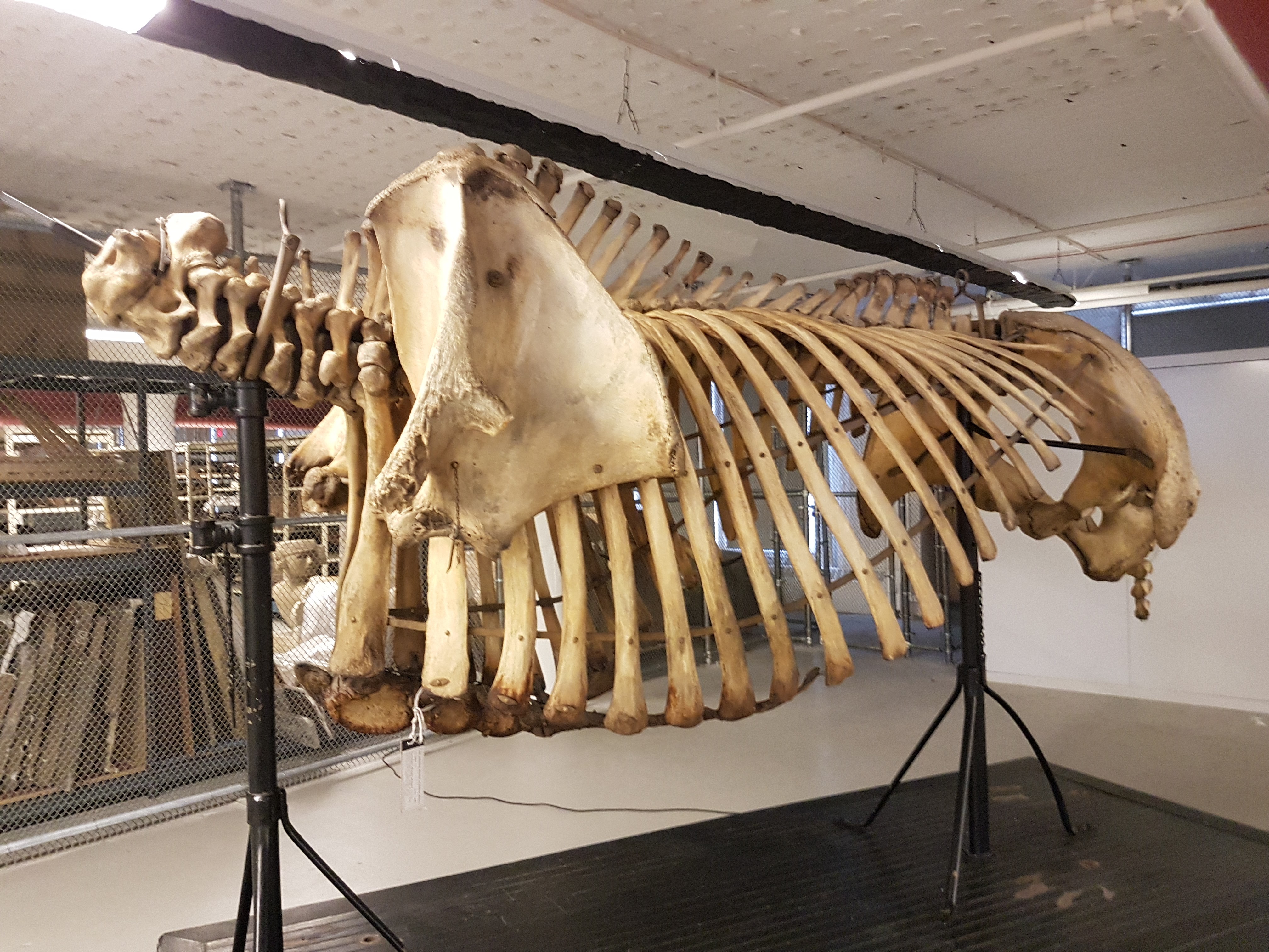
Then there, coincidentally, was another paper originating in part from the same museum group in Berlin. This one I’d been involved in as a co-investigator (author) on a Volkswagen (yes! They like science) grant back about 8 years ago and since. There is an amazing ~290 million year old fossil near-amniote (more terrestrial tetrapod) called Orobates pabsti, preserved with good skeletal material but also sets of footprints that match bones very well, allowing a rare match of the two down to this species level. John Nyakatura’s team had 3D modelled this animal before, so we set out to use digital techniques to test how it did, or did not, move—similar to what I’d tried before with Tyrannosaurus, Ichthyostega and so forth. The main question was whether Orobates moved in a more “ancestral” salamander-like way, a more “derived” lizard-like way (i.e. amniote-ish), or something else.

The approach was like a science sledgehammer: we combined experimental studies of 4 living tetrapods (to approximate “rules” of various sprawling gaits), a digital marionette of Orobates (to assess how well its skeleton stayed articulated in various motions), and two robotics analysis (led by robotics guru Auke Ijspeert and his amazing team): a physical robot version “OroBOT” (as a real-world test of our methods), and a biomechanical simulation of OroBOT (to estimate hard-to-measure things in the other analyses, and matches of motions to footprints). And, best of all, we made it all transparent: you can go play with our interactive website, which I still find very fun to explore, and test what motion patterns do or do not work best for Orobates. We concluded that a more amniote-like set of motions was most plausible, which means such motions might have first evolved outside of amniotes.

OroBOT in tha house!

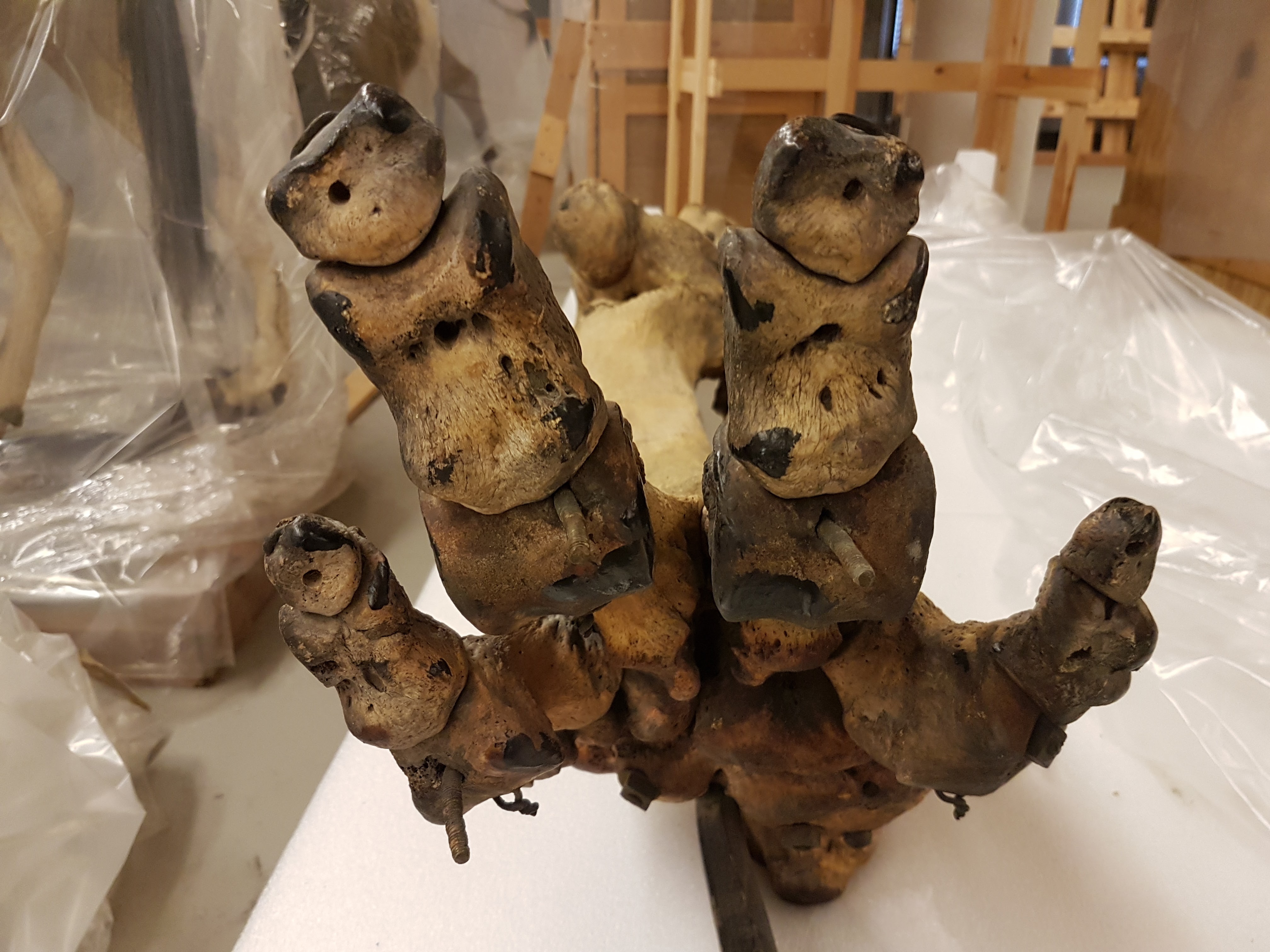
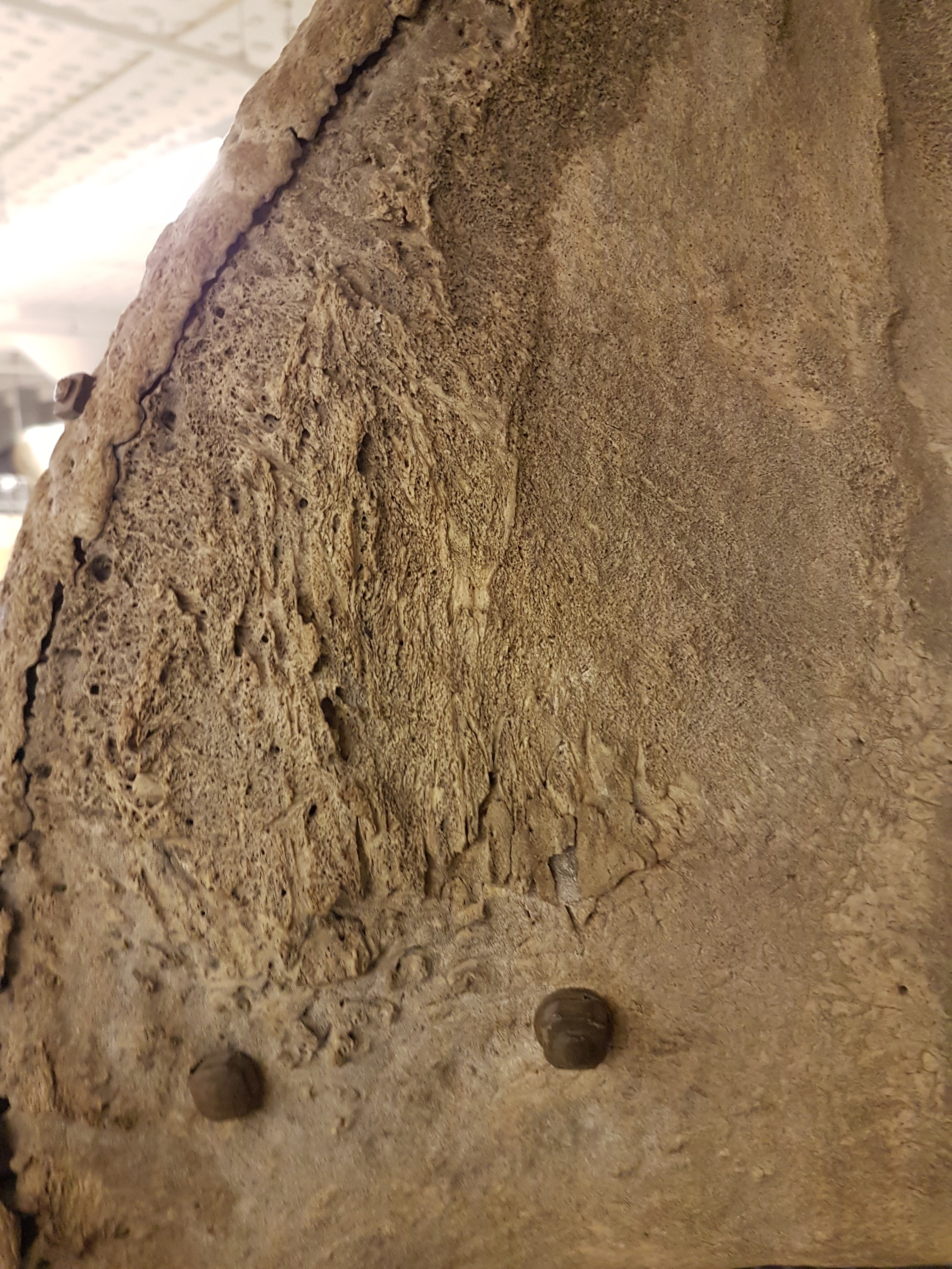
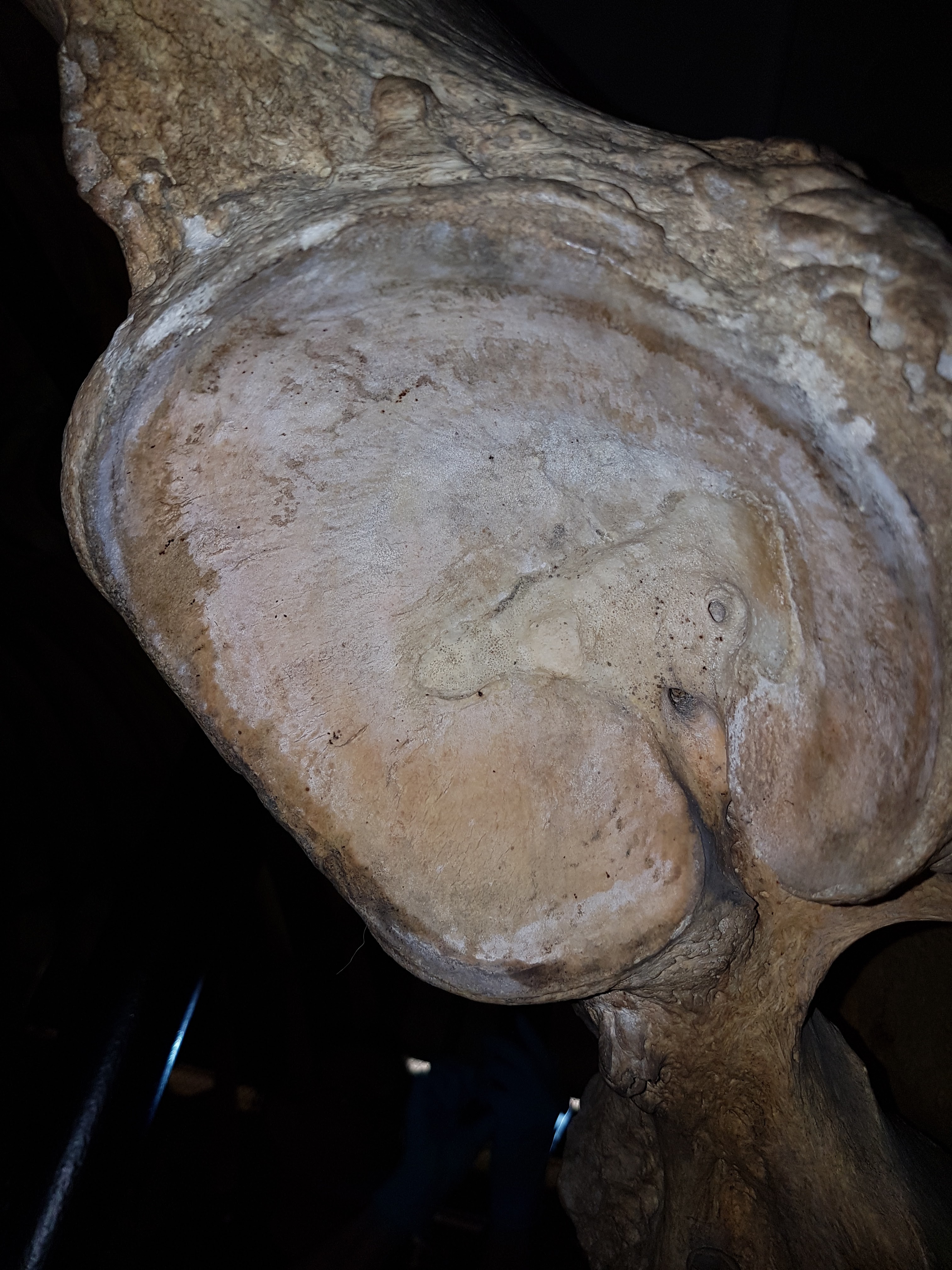
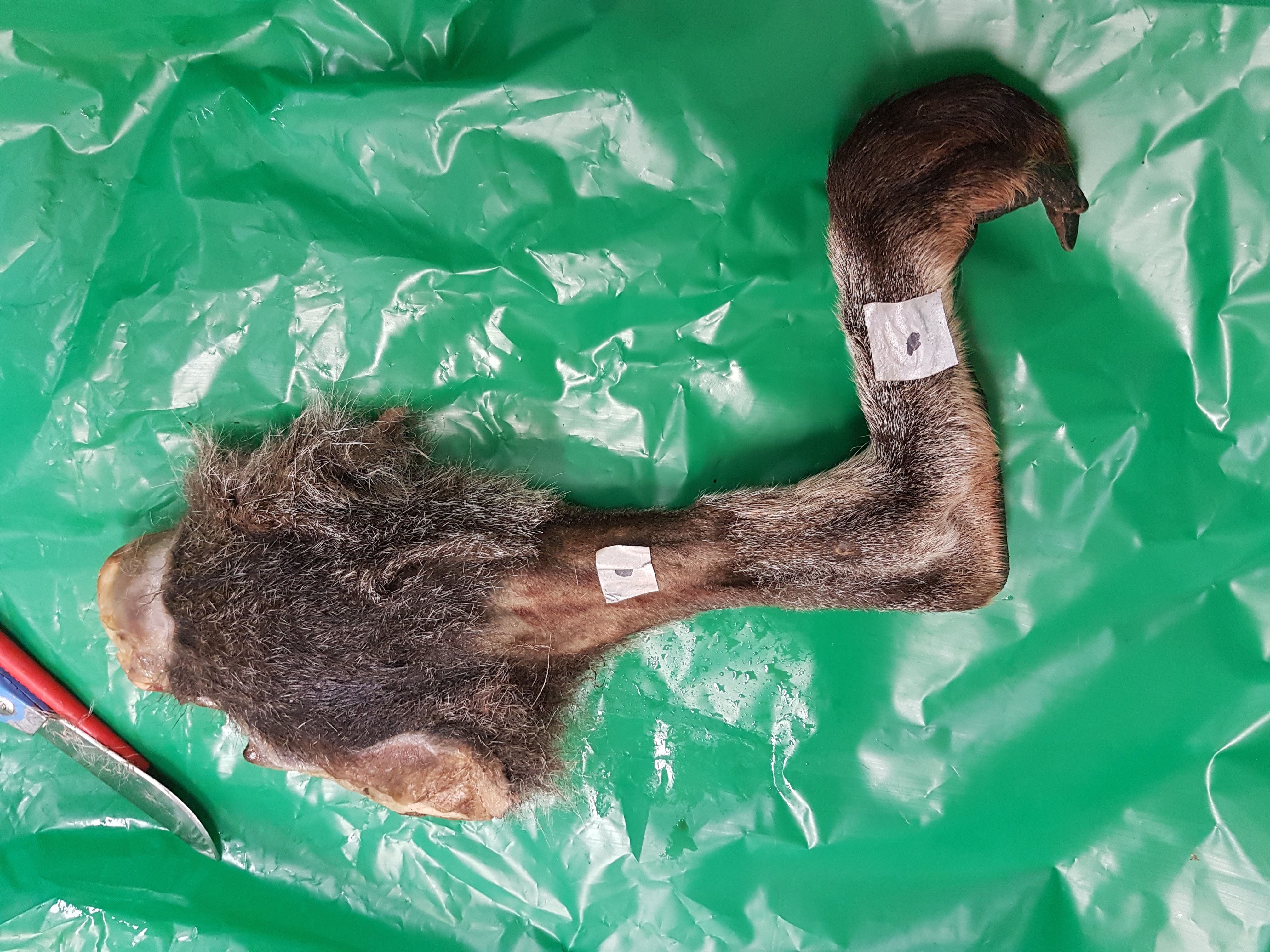
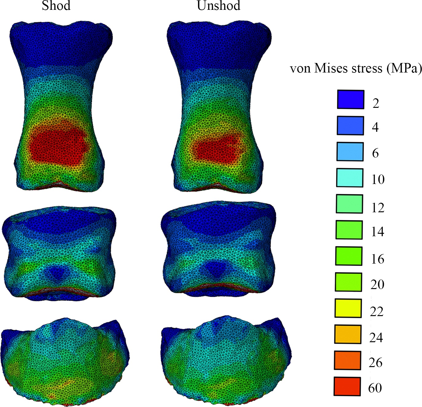
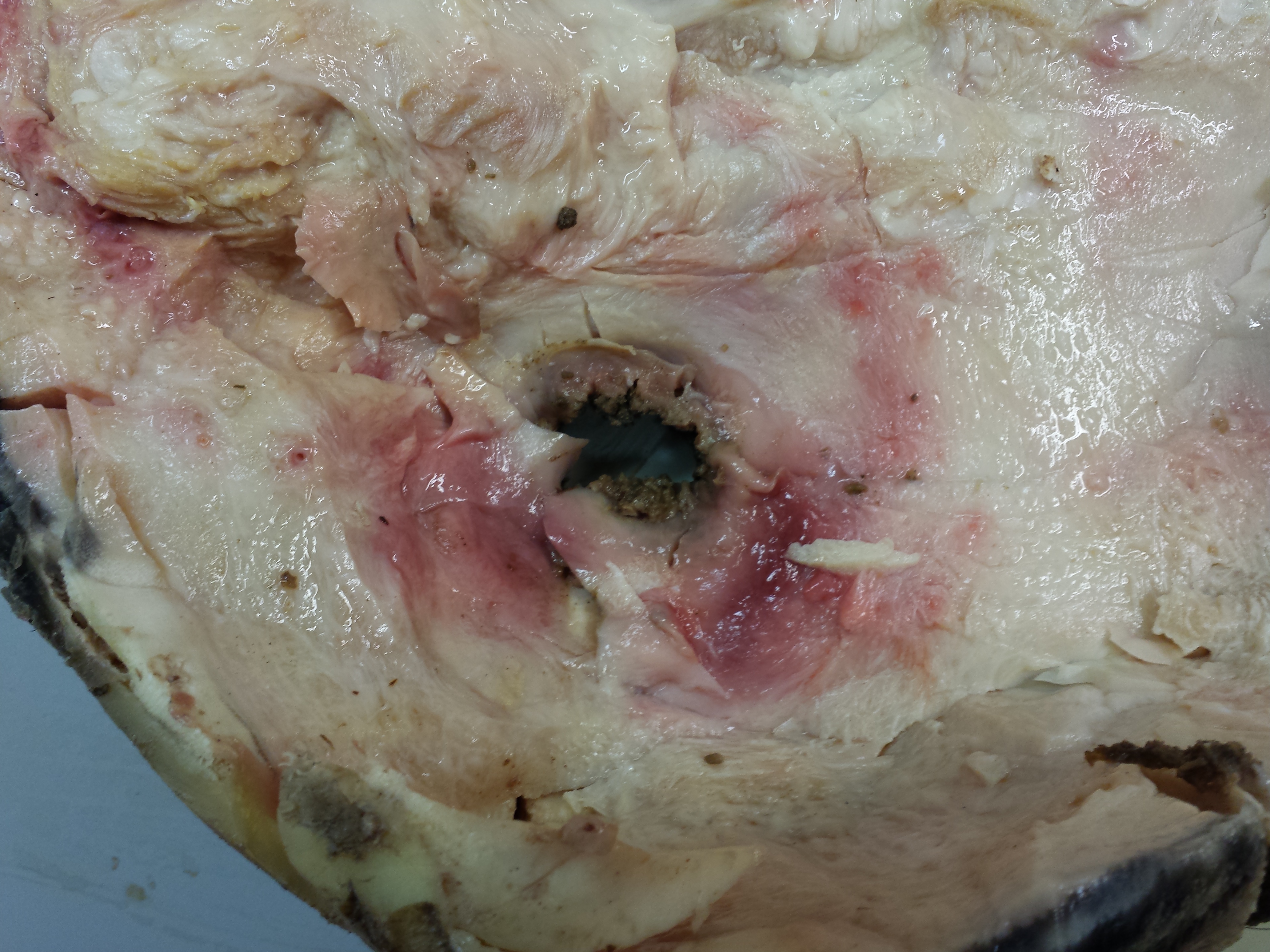
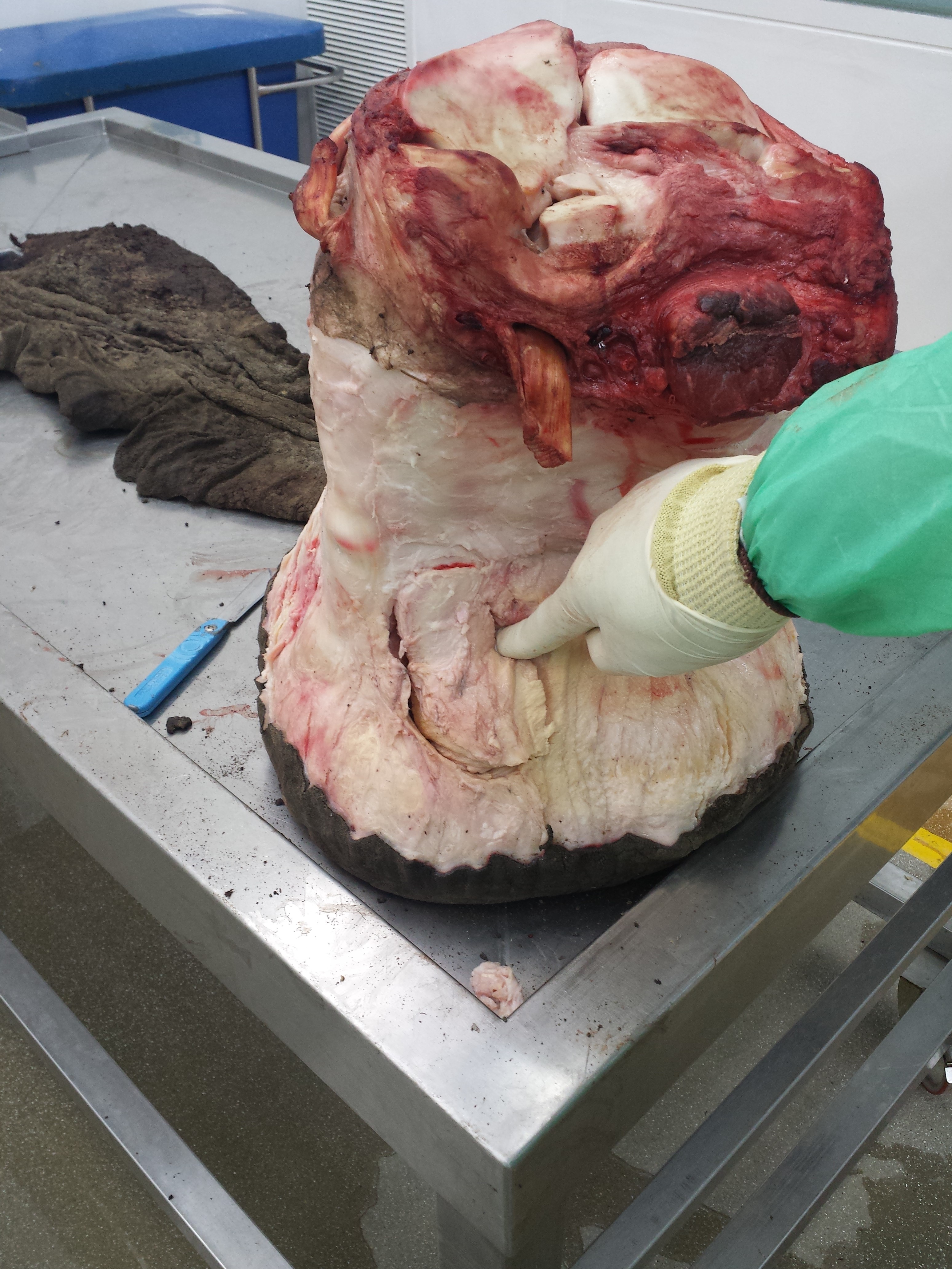
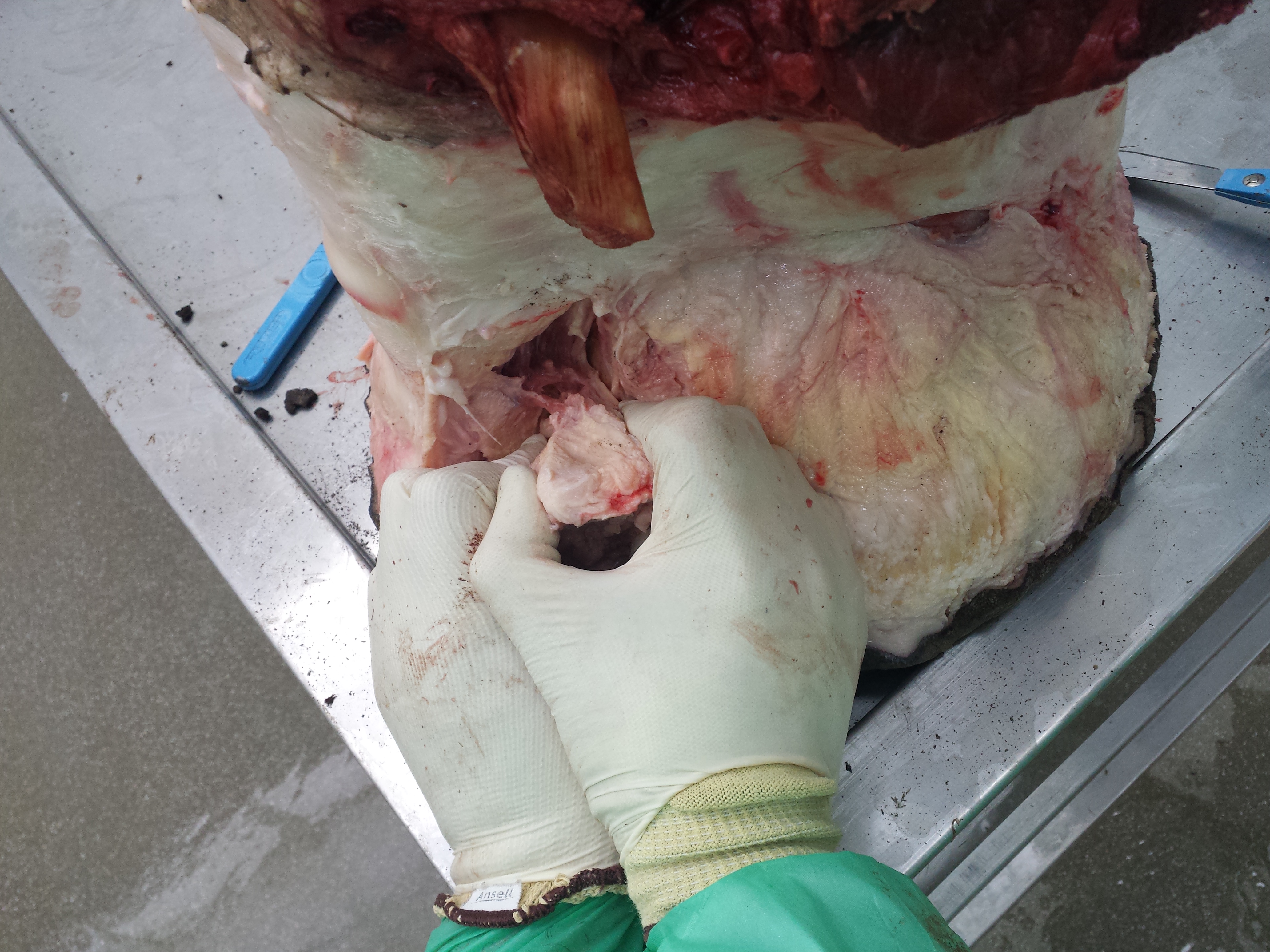
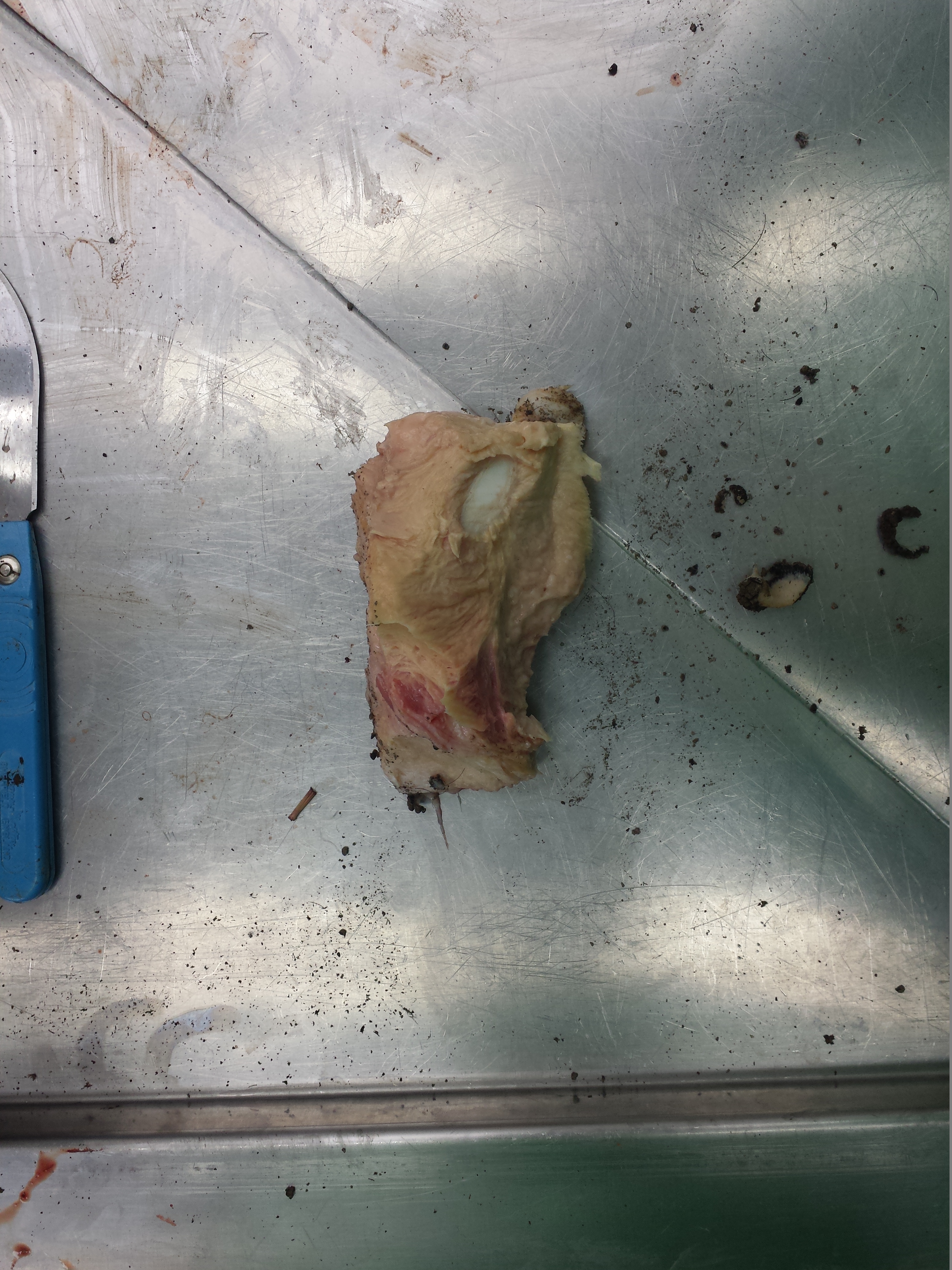
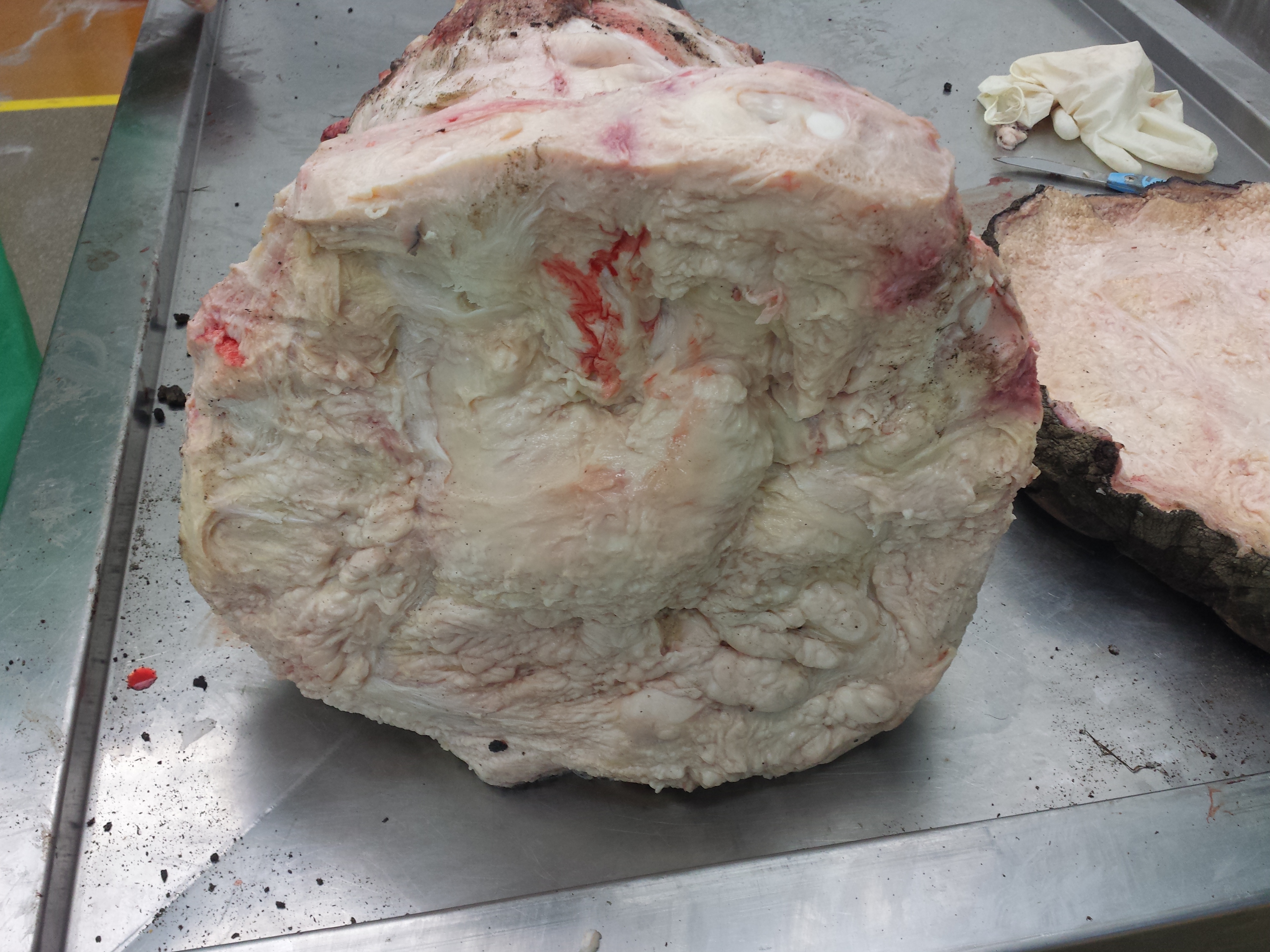
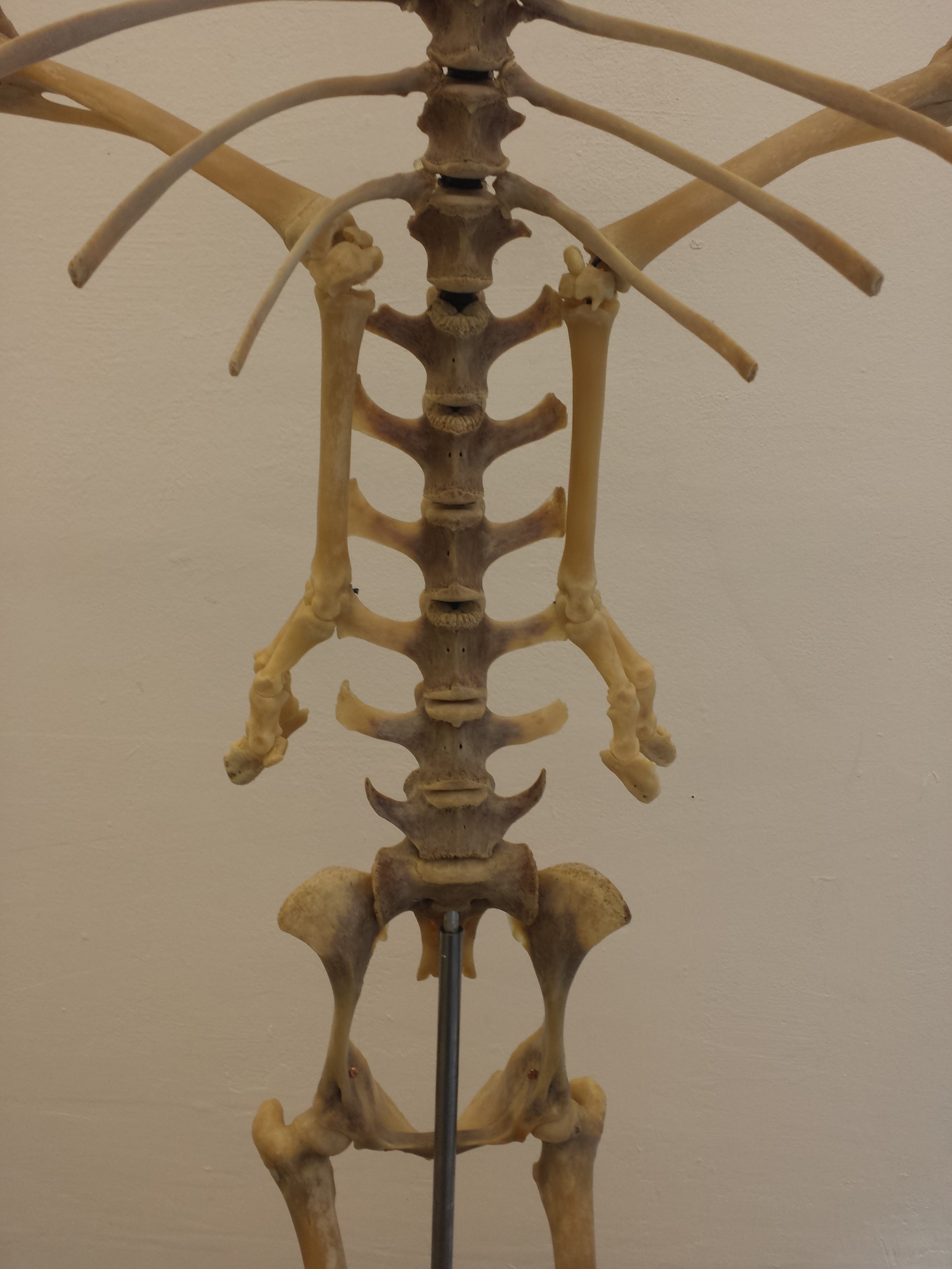
You may remember Crassigyrinus, the early tetrapod, from a prior post on Anatomy To You. My PhD student Eva Herbst finished her anatomical study of the best fossils we could fit into a microCT-scanner and found some neat new details about the “tadpole from hell”. Buried in the rocky matrix were previously unrecognized bones: vertebrae (pleurocentra; the smaller nubbins of what may be “rhachitomous” bipartite classic tetrapod/omorph structure), ribs (from broad thoracic ones to thin rear ones), pelvic (pubis; lower front), and numerous limb bones. One interesting trait we noticed was that the metatarsals (“sole bones” of the foot) were not symmetrical from left-to-right across each bone, as shown below. Such asymmetry was previously used to infer that some early tetrapods were terrestrial, yet Crassigyrinus was uncontroversially aquatic, so what’s up with that? Maybe this asymmetry is a “hangover” from more terrestrial ancestry, or maybe these bones get asymmetrical for non-terrestrial reasons.

The oddly asymmetrical metatarsals of Crassigyrinus.

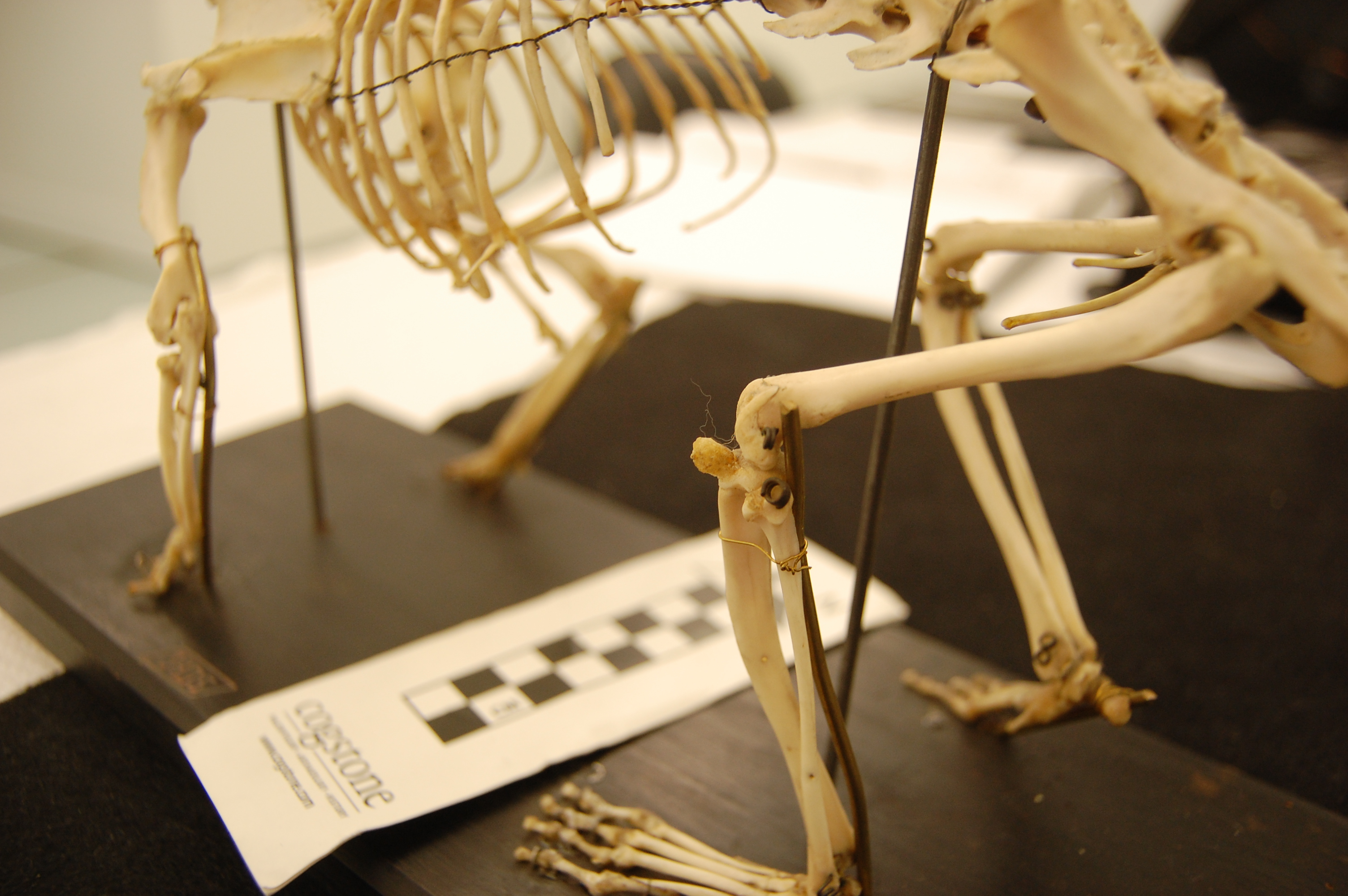
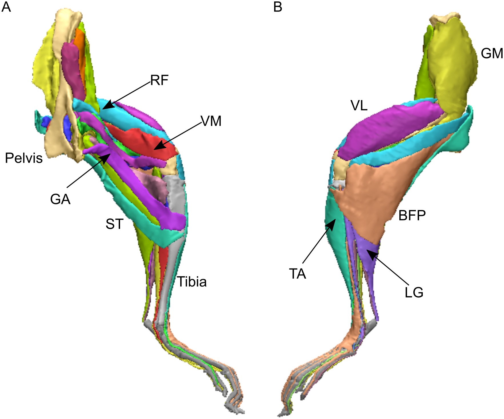
Finally, Dr. Peter Bishop finished his PhD at Griffith University in Australia and came to join us as a DAWNDINOS postdoc. He blasted out three of his thesis chapters (starting here) with me and many others as coauthors, all three papers building on a major theme: how does the inner bone structure (spongy or cancellous bone) relate to hindlimb function in theropod dinosaurs (including birds) and how did that evolve? Might it tell us something about how leg posture or even gait evolved? There are big theories in “mechanobiology” variously named Wolff’s Law or the Trajectorial Theory that explain why, at certain levels, bony struts tend to align themselves to help resist certain stresses, and thus their alignment can be “read” to indicate stresses. Sometimes. It’s complicated!

Undaunted, Peter measured a bunch of theropod limb bones’ inner geometry and found consistent differences in how the “tracts” of bony struts, mainly around joints, were oriented. He then built a biomechanical model of a chicken to test if the loads that muscles placed on the joints incurred stresses that matched the tracts’ orientations. Hmm, they did! Then, with renewed confidence that we can use this in the fossil record to infer approximate limb postures, Peter scanned and modelled a less birdlike Daspletosaurus (smaller tyrannosaur) and more birdlike “Troodon” (now Stenonychosaurus; long story). Nicely fitting many other studies’ conclusions, Peter found that the tyrannosaur had a more straightened hindlimb whereas the troodontid had a more crouched hindlimb; intermediate between the tyrannosaur and chicken. Voila! More evidence for a gradual evolution of leg posture across Mesozoic-theropods-into-modern-birds. That’s nice.

Three theropods, three best-supported postures based on cancellous bone architecture.

If you are still thirsty for more papers even if they are less evolutionary, here’s the quick scoop on ones I’ve neglected until now:

(1) Former PhD student Chris Basu published his thesis work w/us on measuring giraffe walking dynamics with force plates, finding that they move mostly like other quadrupeds and their wobbly necks might cost them a little.

(2) Oh, and Chris’s second paper just came out as I was writing this! We measured faster giraffe gaits in the wilds of South Africa, as zoo giraffes couldn’t safely do them. And we found they don’t normally go airborne, just using a rotary gallop (not trot, pace or canter); unlike some other mammals. Stay tuned: next we get evolutionary with this project!

(2) How do you safely anaesthetize a Nile crocodile? There’s now a rigorous protocol (from our DAWNDINOS work).

(3) Kickstarting my broad interest in how animals do “extreme” non-locomotor motions, we simulated how greyhounds stand up, finding that even without stretchy tendons they should, barely, be able to do it, which is neat. Expect much more about this from us in due time.

(4) Let’s simulate some more biomechanics! Ashley Heers, an NSF research fellow w/me for a year, simulated how growing chukar birds use their wing muscles to flap their way up steeper inclines (“WAIR” for devotees), and the results were very encouraging for simulating this behaviour in more detail (e.g. tendons seem to matter a lot) and even in fossil species; and finally…

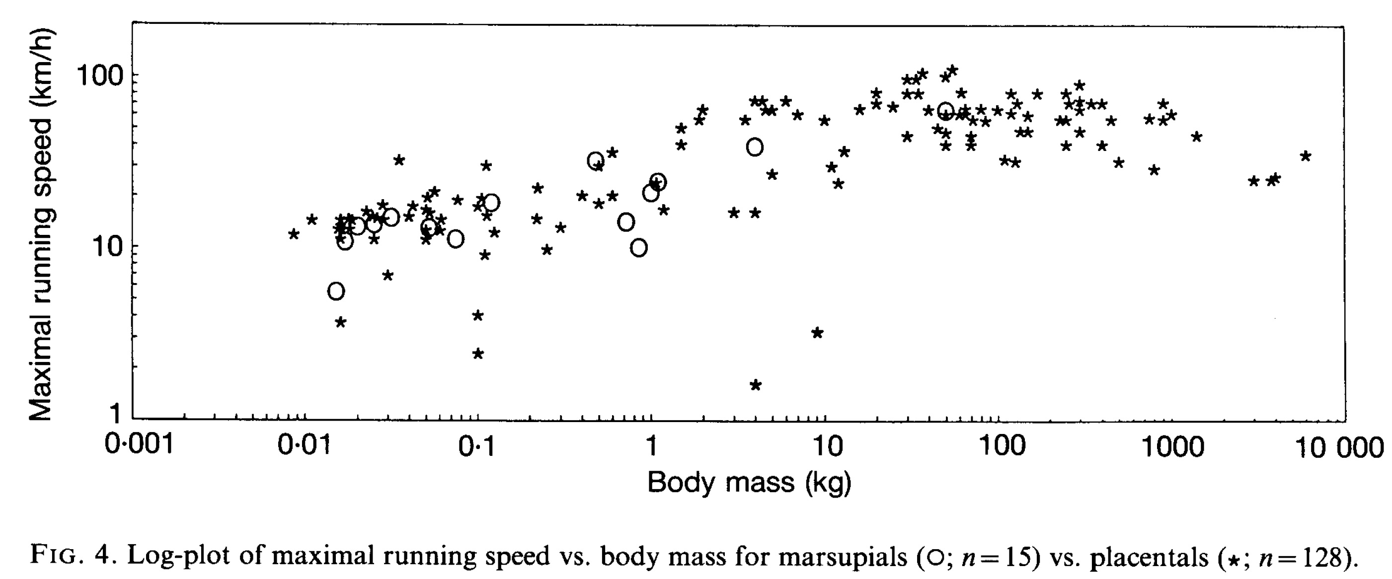
(5) Hey did you ever think about how bone shape differs between hopping marsupials (macropods) and galloping artiodactyl (even-toed) mammals? We did, in long-the-making work from an old BBSRC grant with Michael Doube et al., and one cool thing is that they mostly don’t change shape with body size that differently, even though one is more bipedal at faster speeds—so maybe it is lower-intensity, slower behaviours that (sometimes?) influence bone shape more?

So there you have the skinny on what we’ve been up to lately, messing around with evolution, biomechanics and morphology.

Read Full Post »

Back in November 2016 I got an exciting email from colleague Dr. Richard Thomas, who was building a team of experts for a proposed documentary on Jumbo the elephant; the famed proboscidean of the Victorian era (and arguably most famous elephant of all time, first international celebrity animal, etc.). I knew him from social media and from our mutual interests in chicken anatomy and evolution. And that exciting email, for once, worked out! Over coming months I chatted with the film producers and they could see a place for me in the programme, contributing my expertise in elephant (postcranial) anatomy, locomotion, health/welfare etc. Lo and behold, in May 2017 I met Sir David Attenborough at Heathrow and we flew out to New York City to film with the skeleton at the American Museum of Natural History. And to cap it off, I got to meet another childhood science communication hero: Professor David Suzuki of CBC’s “The Nature of Things“– my adrenaline levels were sky high!

Brooklyn neighborhood by our hotel. Lots to do!

The show has aired in the UK and is coming very soon to Canada and the world (details below). Here’s my part of the story.

Stomach-Churning Rating: 3/10– bad bones but no blood.

We filmed from 15-19 May 2017 at the AMNH’s warehouse of mammalian skeletal remains, which is housed deep in the Brooklyn Army Terminal; a picturesque site in and of itself. And it is a site with a lot of history— WWI and II, Elvis and more.

It was a hectic week of the usual documentary stuff: repeat the same lines and motions again and again from different angles and with different paces and intonations (I cannot help in these cases but think about the Simpsons “Fallout Boy” episode), from ~9am-5pm, with plenty of downtime watching setup or other bits being filmed. I’m used to all that. But having the time to peer around the collection and chat to Richard and colleague Dr. Holly Miller (handling the tissue isotopes side of the story) about Jumbo’s skeleton was a lot of fun during downtime and filming itself. Not to mention the utter joy of studying one of the most famous museum specimens ever, and an animal widely held to be one of the largest of its kind, with much mystery surrounding its history despite its fame. (Wikipedia does a fair job of summarizing some of this)

Here are some photos to tell the story:

Photo of the team, courtesy of Infield Fly Productions (CBC production, “Jumbo: The Life of An Elephant Superstar”.

The Brooklyn Army Terminal, with a view of the harbour beyond.

Inside the terminal: old army staging area and an evocative wooden Liberty/tank artwork.

Army terminal cat. Shipping still comes through the terminal so I guess there are plenty of rats and handouts from cat-lovers to keep it going. I miss our cats when I travel so this moment was appreciated.

Whale skulls and other specimens inside the AMNH warehouse.

First view of Jumbo’s remains.

Photo opp with Sir David.

Photo opp with Prof Suzuki.

That’s the setup. I’ve done ~15 other documentary episodes/shows but this was like nothing else– simply an awesome experience.

Now the delivery: we set to studying those bones. We’d seen photos before, and Henry Fairfield Osborn had illustrated the specimen as his type of “Elephas africanus rothschildi” (Sudanese elephant; no longer valid but those were different times– it’s now just a nicely preserved Loxodonta africana africana), so we knew some of what to expect.

Looking at Osborn’s classic monograph. Oddly he didn’t address the GLARING MASSIVE PROBLEMS WITH THE TEETH!

Skull with terrible tooth pathologies– and let’s play spot Mumbo, my daughter’s toy elephant! He might even appear in some TV footage!

We had noted some serious issues with some bones (pathologies). I won’t spoil the message here but will show some images. I know some experts have voiced issues with how the tooth pathologies/growth were explained in some footage but I can’t address that here; it’s not my expertise. The important point to me is that the teeth are incredibly messed up and that can easily be linked to bad diet and other management/health issues, as the documentary explains.

Jumbo’s torso in left side view. Glorious preservation.

Right forelimb, showing that the “growth plates” (epiphyses”) were not all fused, consistent with Jumbo still growing– as expected for an African male elephant in his 20’s.

Right elbow with some pathologies consistent with degenerative joint disease.

Surprisingly, Jumbo’s feet were not in nasty condition in terms of pathologies. I’d expected to see that. They’d been painted and drilled for mounting, but were not riddled with arthritic changes that I could see.

Strange bony plaque on the left pelvis (hip) region; something I’d never seen before in any elephant (and I’ve seen many). Why? The programme offers a reasonable explanation.

Jumbo’s right hip, with bad erosion of the bone and thus presumably the overlying cartilage. Ouch!

Strange extra prong on one right rib in Jumbo- we didn’t figure that out. It could conceivably be natural variation.

So, poor Jumbo suffered some jumbo-sized problems, and in complex ways. That’s just scratching the surface of what his skeleton tells us, and there’s plenty more in the show plus plenty more we can say later– there’s real science that came out of this programme! I was surprised to find how little had been stated anywhere in the scientific literature about Jumbo’s pathologies.

Sad as Jumbo’s skeletal story is, the broader story of his life and death is sadder still. For purposes of time I don’t think any of the three versions of the show will get to delve into how Jumbo’s mother may have been slashed to death by a broadsword, as the story below describes was the ancient practice:

I’d hate to be “so pestered by a popinjay”, too.

Adding insult to injury, we can reflect on how Jumbo was taken from the Sudan to the east (across the Suez), then on boat to Italy and then overground to Paris, where he lived for a little while until the zoological garden sold him to London. Luckily Jumbo avoided becoming a meal to starving Parisians during the Prussian siege of 1870-1. So he did not become elephant consommé like some of his co-captives did. The more one learns about Jumbo’s life and the life of elephants in captivity in the 1800s, the more harrowing the tale becomes.

Jumbo is THE celebrity elephant. His name has come to mean ‘big’ and ‘bombastic’, from applications to jumbo jets to hot dogs and other (darkly ironic) forms of consumption and extravagance. He has had a jumbo effect on Western culture, but also symbolizes the complex human-elephant relationship, such as the inspiration for “Dumbo’s” own sad story. We love elephants but our fascination with them can also be their undoing, such as poaching for the ivory trade or mistreatment in captivity. Jumbo’s story writ large is also the story of elephants, and our story to learn from. If anything comes out of my participation in the Jumbo documentary for the public’s benefit, I hope it is increased empathy for how we interact with elephants. They are like us in many ways (maybe over-emphasized with anthropomorphism in many accounts), but also unlike us (maybe even unfathomable) in not only their size and anatomy but also in aspects of their prodigious intellect, emotions and social structure. Elephants aren’t just jumbo spectacles. They are jumbo responsibilities for humans now that we dominate the planet so much.

Want to catch a version of the Jumbo show? I’ll try to keep this list up to date:

BBC iplayer now: https://www.bbc.co.uk/iplayer/episode/b09jcxrj/attenborough-and-the-giant-elephant

BBC One: 5:05pm on January 31st

CBC: 8pm on January 7th– trailer is here:

http://www.cbc.ca/player/play/1115035715562

And the international version is coming soon, plus the above versions surely will circulate globally in some ways.

Have a jumbo time (in a good way) in the rest of 2017 and onwards into 2018!

-John

Read Full Post »

This week we conducted wallaby leg dissections for a study of the kneecaps of marsupials (pouched mammals). Placental (non-pouched) mammals like us almost all have bony kneecaps but many marsupials do not. Kneecaps do important things, acting like gears around the knee joints (e.g. this old post), and yet it is unclear why some marsupials have lost, kept or even re-evolved them as bones. So we’re investigating that and already noticed that one of our wallabies has bony kneecap(s) whereas the other doesn’t, so we’re checking out why and taking tissue samples to do histology (sectioning for microscopic imaging of tissue composition and structure) on so we can see what the knee tendon/kneecap tissues are made of. Some marsupials turn their kneecaps into fibrocartilage rather than bone or tendon and that can be impossible to identify without histology.

The wallabies are small, about 20lbs or so and just take a day or so. Like a turkey. And it’s Thanksgiving today, so here I am with a post about thawing specimens for science, rather than for food. Maybe the title will make sense now.

Stomach-Churning Rating: 7/10; thawed wallaby bits from the get-go.

Thawed lower leg and foot of wallaby. The stickers are for an old study that would take too long to explain…

This post was directly inspired by journalist Jason Bittel’s inquiry to me about my tweet on the wallaby thawing; he wondered if there might be a fun story linking thawing-for-science with thawing-for-Thanksgiving. Some highfalutin editors didn’t agree, so no printed/online story came of this, but I am not so highfalutin, hence this blog post.

Thawed wallaby forelimbs. I’m also looking into the “false thumbs” that some marsupials have (“sixth fingers”), much as elephants and other mammals may have.

Thawing is second nature for our lab’s team; we do it all the time. Avid readers will be unsurprised to learn that just about everything I’ve worked on has been frozen at some time, and thus has been thawed out at some time(s). Normally we don’t freeze if we need live tissue or undistorted tissue, e.g. to measure physiology or very fine microstructure– freezing disrupts all of that. We would instead use physiological saline solution or else a preservative like formalin. And you can only freeze and then thaw a specimen for two times or so before it becomes too useless even for anatomical study.

A small specimen like this salamander can be thawed out simply by running it under warm water for a little while or leaving it out for an hour.

We just leave specimens in a cart, or on a table or sometimes in a cold-room shelving area, for slower thawing. Space heaters tend to overdo things. We don’t do any rough calculation from some sort of thermodynamic first principles of time-to-thaw vs. specimen size (I wish we were that smart!); just seat-of-pants guessing and checking (yes, poking specimens to check their thawedness is a method of choice). Cutting things in half along the way, or skinning them, may be used to accelerate the thawing process. But it’s about as unscientific a method as we use.

The hardest specimens to thaw of course have been the largest specimens. Elephant legs can be >2 metres long and hundreds of kilograms (especially when frozen). A week at room temperature tends to work OK for getting them to a dissectable state. One has to balance the outer deterioration with the inner frigidness. We’re not so concerned about microbe growth in most cases, as one would be with a thawing turkey, and not at all about consumption. We just want to be able to dissect it and make observations, mostly via eyeballing the specimens as we dissect them,

Left hindfoot of an Asian elephant. Still frozen; this was bandthawed- I mean bandsawed- to see its internal anatomy nice and clearly. You may see this specimen again somewhere else– stay tuned! 🙂

Moisture and fluids can be a challenge: generally the rooms we thaw in are low humidity so moisture may not be an issue once the ice melts away, and we have drains nearby. We try to remove ice first or have towels to wipe/soak fluids up as thawing progresses. But if a specimen is sitting in a cart or storage bag with too much ice early on, that can thaw first and then turn the specimen into a nasty slurry of the stuff you’re interested in and the less desirable muck. So we try to avoid that.

De-thawing too early is bad. The smell gets progressively worse– and once the interior of the specimen is thawed enough, then bacteria get in there and the interior becomes a brewing ground for heat production (rather than remaining a cooler region), which accelerates decay, so we don’t want that. We have to check on thawing specimens regularly and move them to cooler storage areas, or begin dissection earlier, if the decay process is noticeably getting excessive.

Any insulation affects thawing time- so scales, feathers, thick skin, shells, fat (for a short while until it decays), and other layers will slow thawing—and may keep heat inside, if there begins to be thawing of the core. So sometimes you open up a specimen that seems dry and clean on the outside and the inside is unpleasant. But with experience that is not hard to avoid.

Thawed wallaby patella prepared for histology.

The foulest specimen I’ve thawed by far was a monitor lizard… it was shipped to me in California from Arizona when I was a PhD student. This was in August’s heat and the box of the big lizard sat thawing at the post office for 2 weeks before they contacted me and asked why a smelly box was bleeding. I came and got it and brought it back to our department but the smell was so bad it set off our building health & safety person’s alarm bells (sorry, David!) and they emailed around a “toxic alert” warning, until I bashfully made it clear that my lizard was the cause, not some toxic chemical. I got in some trouble and was very ashamed. But we put the specimen into a big tank of brine solution and the smell was reduced—the specimen may well still be preserved there 20 years later; I do wonder! Anyway, that experience was so horrendous – and I have a strong stomach—that I regularly recall it and seek to avoid a repeat. It was the most disgusting thing I’ve ever experienced. I do not recommend it.

What we tend to want to get from thawed specimens is: (1) descriptive anatomy (what connects where), and maybe (2) quantitative measurements (laborious metrics of “muscle architecture”– how much does each muscle weigh, how long is it, etc; over and over again for many muscles…). These data not only serve to tell us what makes animals different (and how this evolved) but also the data are used to test questions such as how animals work. In the case of things like wallabies, ultimately we’d love to know what their kneecaps do if they are bony or not; what difference does it make and why might there be differences? We’d spotted one wallaby already that seemed to have a bony kneecap on one leg, and a non-bony one on the other leg, so that asymmetry got us excited.

What’s surprising to learn about thawing animals for science? Well, my first thought is that it’s beautiful. I don’t tend to think of it as gross. I’ve rhapsodized about this before. Animals are wonderful inside and out, and I regularly pause during a dissection to marvel at how amazing the anatomical specializations of animals are. Simple details- shapes, colours, configurations- can be gorgeous. (Often the blood is minimal, drained out early, so that doesn’t detract from or hide the detailed imagery) The gentle yet complex path of a tendon around a joint can yield profound visual enchantment in its elegance. This is all the more true once one ponders how these complex structures evolved, and how much diversity of form and function is out there to study—and how little we know about it! We still don’t know well how to fix many problems humans have with their anatomy, and that’s orders of magnitude worst for most animals, because we don’t understand how anatomy works, or even what the anatomy is like in some cases. So that keeps me busy discovering things. Every specimen is different with surprising little variations, or big ones—sometimes there is one muscle, sometimes it is clearly divided into two muscles, in the same species or even the left vs. right legs. I love seeing those intricacies and wondering about them.

Thawed wallaby shank sliced open to show lovely digital flexors and gastrocnemius muscles. So many questions are raised by this!

If you’re thawing for Thanksgiving, or thawing for science, or thawing out family relations during a gathering, or thawing yourself out from the winter’s cold– my best wishes to you! May we all enjoy what we thaw.

Read Full Post »

The early, hippo-like mammal Coryphodon. I didn’t know it had a patella but it does. From Yale Peabody Museum.

I’m not shy about my fondness for the patella (kneecap) of tetrapod vertebrates, and neither are the other members of RVC’s “Team Patella”. We’ve had a fun 3+ years studying these neglected bones, and today we’ve published a new study of them. Our attention has turned from our prior studies of bird and lepidosaur kneecaps to mammalian ones. Again, we’ve laid the groundwork for a lot of future work by focusing on (1) basic anatomy and (2) evolutionary history of these sesamoid bones, with a lot of synthesis of existing knowledge from the literature; including development and genetics. This particular paper is a sizeable monograph of the state of play in the perusal of patellae in placental and other synapsids. Here’s what we did and found, focusing mostly on bony (ossified) patellae because that allowed us to bring the fossil record better to bear on the problem.

Reference: Samuels, M., Regnault, S., Hutchinson, J.R. 2016. Evolution of the patellar sesamoid bone in mammals. PeerJ 5:e3103 https://doi.org/10.7717/peerj.3103

Stomach-Churning Rating: 1/10; bones and more bones.

The short version of the story is that mammals evolved bony kneecaps about five times, with marsupials gaining and losing them (maybe multiple times) whereas monotremes (platypus and echidna) and placentals (us and other mammals) didn’t do much once they gained them, and a couple of other fossil groups evolved patellae in apparent isolation.

Evolution of the patella in mammals: broad overview from our paper. Click to zoom in.

The marsupial case is the most fascinating one because they may have started with a fibrocartilaginous “patelloid” and then ossified that, then reduced it to a “patelloid” again and again or maybe even regained it. There needs to be a lot more study of this group to see if the standard tale that “just bandicoots and a few other oddballs have a bony patella” is true for the Metatheria (marsupials + extinct kin). And more study of the development of patellae in this group could help establish whether they truly do “regress” into fibrocartilage when they are “lost” in evolution, or if other, more flexible patterns exist, or even if some of the cases of apparent “loss” of a bony patella are actually instances of delayed ossification that only becomes evident in older adults. Our paper largely punts on these issues because of an absence of sufficient data, but we hope that it is inspiration for others to help carry the flag forward for this mystery.

The higgledy-piggledy evolution of a patella in Metatheria, including marsupials. Click to zoom in.

Some bats, too, do funky things with their kneecaps, analogous to the marsupial “patelloid” pattern, and that chiropteran pattern also is not well understood. Why do some bats such as Pteropus fruit bats “lose” their kneecaps whereas others don’t, and why do some bats and other species (e.g. various primates) seem to have an extra thing near their kneecaps often called a “suprapatella”? Kneecap geeks need to know.

The short-nosed bandicoot (marsupial) Isoodon, showing a nice bony patella as typifies this group. From Yale Peabody Museum.

Otherwise, once mammals evolved kneecaps they tended to keep them unless they lost their hindlimbs entirely (or nearly so). Witness the chunky patellae of early whales such as Pakicetus and join us in wondering why those chunks persisted. The evolutionary persistence of blocky bits of bone in the knees of various aquatic animals, especially foot-propelled diving birds, may help answer why, as the hindlimbs surely still played roles in swimming early in cetacean evolution. Ditto for sea cows (Sirenia) and other groups.

Early whale Ambulocetus, showing hefty kneecaps.

But I’m still left wondering why so many groups of land vertebrates (and aquatic ones, too) never turned parts of their knee extensor tendons into bone. We know a bit about the benefits of doing that, to add leverage to those joints that enables the knee muscles to act with dynamic gearing (becoming more forceful “low gear” or more speedy “high gear” in function). Non-avian (and most early avian/avialan) dinosaurs, crocodiles, turtles, amphibians, early mammal relatives, and almost all other known extinct lineages except for those noted above got by just fine without kneecaps, it seems, even in cases where a naïve biomechanist would expect them to be very handy, such as in giant dinosaurs.

A quoll, Dasyurus, with what is probably a fibrocartilaginous “patelloid”. From Yale Peabody Museum.

However, tendons don’t turn to bone unless the right stresses and strains are placed upon them, so maybe kneecaps are a “spandrel” or “exaptation” of sorts, to abuse Gould’s ghost, whose adaptive importance is overemphasized. Maybe that adaptive myopia overshadows a deeper ontogenetic story, of how tissues respond to their history of mechanical loading environment. It has been speculated that maybe (non-marsupial) mammals have broadly “genetically assimilated” their kneecaps, fixing them into semi-permanence in their genetic-developmental programmes, whereas in contrast the few studies of birds indicate more responsiveness and thus less assimilation/fixation. That “evo-devo-mechanics” story is what now fascinates me most and we’ve poked at this question a bit now, with some updates to come- watch this space! Regardless, whether an animal has a bony vs. more squishy soft tissue patella must have consequences for how the knee joint and muscles are loaded, so this kind of question is important.

Giant marsupial Diprotodon (at NHM London); to my knowledge, not known to have had kneecaps- why?

In the meantime, enjoy our latest contribution if it interests you. This paper came about when first author Dr. Mark Samuels emailed me in 2012, saying he’d read some of my old papers on the avian musculoskeletal system and was curious about the evolution of patellae in various lineages. Unlike many doctors and vets I’ve run into, he was deeply fascinated by the evolutionary and fossil components of patellae and how those relate to development, genetics and disorders of patellae. We got talking, found that we were kindred kneecap-spirits, and a collaboration serendipitously spun off from that, soon adding in Sophie. It was a blast!

Read Full Post »

(Marcela with some furry friends; photo by Oliver Siddon)

(Marcela with some felid friends; photo by Oliver Siddon)

A guest post by Marcela Randau (m.randau@ucl.ac.uk)

Stomach-Churning Rating: 1/10; just bones and data plots!

It is often said that all cats are very similar in terms of their skeletal morphology (“a cat is a cat is a cat”). But is this really the case? It may be if only gross, qualitative anatomy is taken into consideration, i.e., if you just eyeball the skeletons of tigers and lions you might find yourself not knowing which one is which. But with huge advances in technology that allows for extracting detailed shape information off a structure (e.g., a skull) and for analysing this information (‘Geometric Morphometrics’), it has become more and more possible to distinguish between relatively similar forms – which may be from distinct species, separate sexes, or even just different populations of the same taxon.

And it is reasonable to think that cat skeletons might be a lot more different than what meets the eye, as for a lineage of apparently similarly built animals, with not that much variation in diet  (all cats are hypercarnivores) there is substantial variation in body mass (over 300-fold just in living species!) and in ecology across cat species. From the cursorial cheetah to the arboreal clouded leopard, felids present a wide range of locomotory adaptations. Yes, all cats can climb, but some do it better than others: think lion versus margay (yes, they do descend trees head-first). As hypercarnivores, all cats are meat specialists, but they also change with regards to how big their prey is, with a general and sometimes-not-so-black-and-white three-tier classification into small, mixed and large prey specialists. The rule of thumb is ‘if you are lighter than ~20-25 kg, hunt small stuff. If you are heavier than that, hunt BIG BIG things; bigger than yourself. And if you are in the middle ground, hunt some small-ish things, some big-ish things, and things about your size. Well, -ish’ – their prey size preference has a lot to do with energetic constraints (have a look at Carbone et al. 1999; and Carbone et al. 2007, if you’re interested in this). But the fun bit here is that form sometimes correlates quite strongly with function, so we should be able to find differences in some of their bones that carry this ecological signal.

Indeed, for a while now, we have known that the shape of the skull and limbs of felids can tell us a lot about how they move and how big their prey is (Meachen-Samuels and Van Valkenburgh 2009, 2009), but a large proportion of their skeleton has been largely ignored: we don’t know half as much about ecomorphology and evolution of the vertebral column. Well, it was time we changed this a bit! As the PhD student in the Leverhulme-funded ‘Walking the cat back’ (or more informally, “Team Cat”) project, I’ve spend a big chunk of my first two years travelling around the world (well, ok, mainly to several locations in the USA) carrying a heavy pellet case containing my working tool, a Microscribe, to collect 3-D landmarks (Fig. 1) across the presacral vertebral column of several cat species. And some of first results are just out! Check them out by reading our latest paper, “Regional differentiation of felid vertebral column evolution: a study of 3D shape trajectories” in the Organisms Diversity and Evolution journal (Randau, Cuff, et al. 2016).

cheetah-verts

Fig. 1: Different vertebral morphologies and their respective three-dimensional landmarks. Vertebral images are from CT scans of Acinonyx jubatus (Cheetah, USNM 520539)

Building from results based on our linear vertebral data from the beginning of the year (Randau, Goswami, et al. 2016), the 3-D vertebral coordinates carry a lot more information and we were able to describe how this complex shape-function relationship takes place throughout the axial skeleton (in cats at least) in much better detail than our prior study did. One of the difficulties in studying serial structures such as the vertebral column is that some clades present variation in vertebral count which makes it less straightforward to compare individual vertebrae or regions across species. However, mammals are relatively strongly constrained in vertebral count, and Felidae (cats; living and known fossils) show no variation at all, having 27 presacral vertebrae. So adaptation of the axial skeleton in mammals has been suggested to happen by modification of shape rather than changes in vertebral number.

Using a variety of geometric morphometric analyses, under a phylogenetically informative methodology, we have shown that there is clear shape and functional regionalisation across the vertebral column, with vertebrae forming clusters that share similar signal. Most interestingly, the big picture of these results is a neck region which is either very conservative in shape, or is under much stronger constraints preventing it from responding to direct evolutionary pressures, contrasting with the ‘posteriormost’ post-diaphragmatic tenth thoracic (T10) to last lumbar (L7) vertebral region, which show the strongest ecological correlations.

We were able to analyse shape change through functional vertebral regions, rather than individual vertebrae alone, by making a novel application of a technique called the ‘Phenotypic Trajectory Analysis’, and demonstrated that the direction of vertebral shape trajectories in the morphospace changes considerably between both prey size and locomotory ecomorphs in cats, but that the amount of change in each group was the same. It was again in this T10-L7 region that ecological groups differed the most in vertebral shape trajectories (Fig. 2).

pta-cats

Figure 2: Phenotypic trajectory analysis (PTA) of vertebrae in the T10 – L7 region grouped by prey size (A) and locomotory (B) categories.

So in the postcranial morphology of cats can be distinguished, changing its anatomy in order to accommodate the different lifestyles we see across species. But the distinct parts of this structure respond to selection differently. The next step is figuring out how that might happen and we are working on it.

While Team Cat continues to investigate other biomechanical and evolutionary aspects of postcranial morphology in this interesting family, we’ve been able to discuss some of these and other results in a recent outreach event organised by the University College of London Grant Museum of Zoology and The Royal Veterinary College. We called it “Wild Cats Uncovered: movement evolves”. Check how it went here: (https://blogs.ucl.ac.uk/museums/2016/11/17/cheetah-post-mortem/) and here (http://www.rvc.ac.uk/research/research-centres-and-facilities/structure-and-motion/news/wild-cats-uncovered), with even more pics here (https://www.flickr.com/photos/144824896@N07/sets/72157676695634065/).

References used here:

Carbone, C., Mace, G. M., Roberts, S. C., and Macdonald, D. W. 1999. Energetic constaints on the diet of terrestrial carnivores. Nature 402:286-288.

Carbone, C., Teacher, A., and Rowcliffe, J. M. 2007. The costs of carnivory. PLoS biology 5 (2):e22.

Meachen-Samuels, J. and Van Valkenburgh, B. 2009. Craniodental indicators of prey size preference in the Felidae. Biol J Linn Soc 96 (4):784-799.

———. 2009. Forelimb indicators of prey-size preference in the Felidae. Journal of morphology 270 (6):729-744.

Randau, M., Cuff, A. R., Hutchinson, J. R., Pierce, S. E., and Goswami, A. 2016. Regional differentiation of felid vertebral column evolution: a study of 3D shape trajectories. Organisms Diversity and Evolution Online First.

Randau, M., Goswami, A., Hutchinson, J. R., Cuff, A. R., and Pierce, S. E. 2016. Cryptic complexity in felid vertebral evolution: shape differentiation and allometry of the axial skeleton. Zoological Journal of the Linnean Society 178 (1):183-202.

Read Full Post »

It has been almost three months since my last post here, and things have fallen quiet on our sister blog Anatomy to You, too. I thought it was time for an update, which is mostly a summary of stuff we’ve been doing on my team, but also featuring some interesting images if you stick around. The relative silence here has partly been due to me giving myself some nice holiday time w/family in L.A., then having surgery to fix my right shoulder, then recovering from that and some complications (still underway, but the fact that I am doing this post is itself evidence of recovery).

Stomach-Churning Rating: 4/10; semi-gruesome x-rays of me and hippo bits at the end, but just bones really.

X-ray of my right shoulder from frontal view, unlabelled

X-ray of my right shoulder from frontal view, unlabelled

Labelled x-ray

Labelled x-ray

So my priorities shifted to those things and to what work priorities most badly needed my limited energy and time. I’ve also felt that, especially since my health has had its two-year rough patch, this blog has been quieter and less interactive than it used to be, but that is the nature of things and maybe part of a broader trend in blogs, too. My creative juices in terms of social media just haven’t been at their ~2011-2014 levels but much is out of my control, and I am hopeful that time will reverse that trend. Enough about all this. I want to talk about science for the rest of this post.

My team, and collaborators as well, have published six recent studies that are very relevant to this blog’s theme- how about we run through them quickly? OK then.

  1. Panagiotopoulou, O., Pataky, T.C., Day, M., Hensman, M.C., Hensman, S., Hutchinson, J.R., Clemente, C.J. 2016. Foot pressure distributions during walking in African elephants (Loxodonta africana). Royal Society Open Science 3: 160203.

Our Australian collaborators got five African elephants together in Limpopo, South Africa and walked them over pressure-measuring mats, mimicking our 2012 study of Asian elephants. While sample sizes were too limited to say much statistically, in qualitatively descriptive terms we didn’t find striking differences between the two species’ foot pressure patterns. I particularly like how the centre of pressure of each foot (i.e. abstracting all regional pressures down to one mean point over time) followed essentially the same pattern in our African and Asian elephants, with a variable heelstrike concentration that then moved forward throughout the step, and finally moved toward the outer (3rd-5th; especially 3rd) toes as the foot pushed off the ground, as below.

African elephant foot COP traces vs. time in red; Asian elephant in orange. Left and right forefeet above; hindfeet below.

African elephant foot COP traces vs. time in red; Asian elephant in orange-yellow. Left and right forefeet above; hindfeet below.

Gradually, this work is moving the field toward better ability to use similar techniques to compare elephant foot mechanics among species, individuals, or over time– especially with the potential of using this method (popular in human clinical gait labs) to monitor foot (and broader musculoskeletal) health in elephants. I am hopeful that a difference can be made, and the basic science we’ve done to date will be a foundation for that.

  1. Panagiotopoulou, O., Rankin, J.W., Gatesy, S.M., Hutchinson, J.R. 2016. A preliminary case study of the effect of shoe-wearing on the biomechanics of a horse’s foot. PeerJ 4: e2164.

Finally, about six years after we collected some very challenging experimental data in our lab, we’ve published our first study on them. It’s a methodological study of one horse, not something one can hang any hats on statistically, but we threw the “kitchen sink” of biomechanics at that horse (harmlessly!) by combining standard in vivo forceplate analysis with “XROMM” (scientific rotoscopy with biplanar fluoroscopy or “x-ray video”) to conduct dynamic analysis of forefoot joint motions and forces (with and without horseshoes on the horse), and then to use these data as input values for finite element analysis (FEA) of estimated skeletal stresses and strains. This method sets the stage for some even more ambitious comparative studies that we’re finishing up now. And it is not in short supply of cool biomechanical, anatomical images so here ya go:

fig5-vonmises

Above: The toe bones (phalanges) of our horse’s forefoot in dorsal (cranial/front) view, from our FEA results, with hot colours showing higher relative stresses- in this case, hinting (but not demonstrating statistically) that wearing horseshoes might increase stresses in some regions on the feet. But more convincingly, showing that we have a scientific workflow set up to do these kinds of biomechanical calculations from experiments to computer models and simulations, which was not trivial.

And a cool XROMM video of our horse’s foot motions:

  1. Bates, K.T., Mannion, P.D., Falkingham, P.L., Brusatte, S.L., Hutchinson, J.R., Otero, A., Sellers, W.I., Sullivan, C., Stevens, K.A., Allen, V. 2016. Temporal and phylogenetic evolution of the sauropod dinosaur body plan. Royal Society Open Science 3: 150636.

I had the good fortune of joining a big international team of sauropod experts to look at how the shapes and sizes of body segments in sauropods evolved and how those influenced the position of the body’s centre of mass, similar to what we did earlier with theropod dinosaurs. My role was minor but I enjoyed the study (despite a rough ride with some early reviews) and the final product is one cool paper in my opinion. Here’s an example:

fig6a-bates-sauropod-com-evol

The (embiggenable-by-clicking) plot shows that early dinosaurs shifted their centre of mass (COM) backwards (maybe related to becoming bipedal?) and then sauropods shifted the COM forwards again (i.e. toward their forelimbs and heads) throughout much of their evolution. This was related to quadrupedalism and giant size as well as to evolving a longer neck; which makes sense (and I’m glad the data broadly supported it). But it is also a reminder that not all sauropods moved in the same ways- the change of COM would have required changes in how they moved. There was also plenty of methodological nuance here to cover all the uncertainties but for that, see the 17 page paper and 86 pages of supplementary material…

  1. Randau, M., Goswami, A., Hutchinson, J.R., Cuff, A.R., Pierce, S.E. 2016. Cryptic complexity in felid vertebral evolution: shape differentiation and allometry of the axial skeleton. Zoological Journal of the Linnean Society 178:183-202.

Back in 2011, Stephanie Pierce, Jenny Clack and I tried some simple linear morphometrics (shape analysis) to see how pinniped (seal, walrus, etc) mammals changed their vertebral morphology with size and regionally across their backbones. Now in this new study, with “Team Cat” assembled, PhD student Marcela Randau collected her own big dataset for felid (cat) backbones and applied some even fancier techniques to see how cat spines change their shape and size. We found that overall the vertebrae tended to get relatively more robust in larger cats, helping to resist gravity and other forces, and that cats with different ecologies across the arboreal-to-terrestrial spectrum also changed their (lumbar) vertebral shape differently. Now Marcela’s work is diving even deeper into these issues; stay tuned…

fig2-randau-measurements

Example measurements taken on felid vertebrae, from the neck (A-F) to the lumbar region (G-J), using a cheetah skeleton.

  1. Charles, J.P., Cappellari, O., Spence, A.J., Hutchinson, J.R., Wells, D.J. 2016. Musculoskeletal geometry, muscle architecture and functional specialisations of the mouse hindlimb. PLOS One 11(4): e0147669.

RVC PhD student James Charles measured the heck out of some normal mice, dissecting their hindlimb muscle anatomy, and using microCT scans produced some gorgeous images of that anatomy too. In the process, he also quantified how each muscle is differently specialized for the ability to produce large forces, rapid contractions or fine control. Those data were essential for the next study, where we got more computational!

mouse-mimics

  1. Charles, J.P., Cappellari, O., Spence, A.J., Wells, D.J., Hutchinson, J.R. 2016. Muscle moment arms and sensitivity analysis of a mouse hindlimb musculoskeletal model. Journal of Anatomy 229:514–535.

James wrangled together a lovely musculoskeletal model of our representative mouse subject’s hindlimb in the SIMM software that my team uses for these kinds of biomechanical analyses. As we normally do as a first step, we used the model to estimate things that are hard to measure directly, such as the leverages (moment arms) of each individual muscle and how those change with limb posture (which can produce variable gearing of muscles around joints). James has his PhD viva (defense) next week so good luck James!

mouse-simm

The horse and mouse papers are exemplars of what my team now does routinely. For about 15 years now, I’ve been building my team toward doing these kinds of fusion of data from anatomy, experimental biomechanics, musculoskeletal and other models, and simulation (i.e. estimating unmeasurable parameters by telling a model to execute a behaviour with a given set of criteria to try to perform well). Big thanks go to collaborator Jeff Rankin for helping us move that along lately. Our ostrich study from earlier this year shows the best example we’ve done yet with this, but there’s plenty more to come.

I am incredibly excited that, now that my team has the tools and expertise built up to do what I’ve long wanted to do, we can finally deliver the goods on the aspirations I had back when I was a postdoc, and which we have put enormous effort into pushing forward since then. In addition to new analyses of horses and mice and other animals, we’ll be trying to push the envelope more with how well we can apply similar methods to extinct animals, which brings new challenges– and evolutionary questions that get me very, very fired up.

Here we are, then; time has brought some changes to my life and work and it will continue to as we pass this juncture. I suspect I’ll look back on 2016 and see it as transformative, but it hasn’t been an easy year either, to say the least. “Draining” is the word that leaps to mind right now—but also “Focused” applies, because I had to try to be that, and sometimes succeeded. I’ve certainly benefited a lot at work from having some talented staff, students and other collaborators cranking out cool papers with me.

I still have time to do other things, too. Once in a while, a cool critter manifests in The Freezers. Check out a hippo foot from a CT scan! It’s not my best scan ever (noisy data) but it shows the anatomy fairly well, and some odd pathologies such as tiny floating lumps of mineralized soft tissue here and there. Lots to puzzle over.

Read Full Post »

I still have my original photocopy, from my grad school days circa 1996, of the 1983 Ted Garland classic paper “The relation between maximal running speed and body mass in terrestrial mammals”, festooned with my comments and highlighter pen marks and other scribblings. That paper remains the backbone of many research questions I am interested in today, and I often think about its underlying concepts. Here’s the key scatterplot from that paper, which I could almost replot by hand from memory, it is so full of implications (and can be clicked to embiggen it, perhaps even speedily depending on your internet connection):

Garland 1983- max speed

Stomach-Churning Rating: 1/10; data and their ramifications; offal-free.

The major points (IMO there are less exciting ones about which theoretical scaling model the data best fit) of the paper are: (1) the fastest-running mammals are neither the smallest nor largest, but those around ~100 kg body mass; (2) if you fit a linear equation to the data (see above; hashed line), it seems like speed increases with body mass linearly (with no limit to that increase, within the body mass range of the data), but if you analyze individual groups of mammals they either don’t change speed significantly with size or they get slower– refer back to point #1 and the polynomial regression that is shown in the figure above (curved line). That’s the biological-question-driven science at the core of the paper (with some methods-y questions at their foundation; e.g. should we use a linear or polynomial regression to fit the data? The latter fits best, and gives a different answer from the former, so it matters.).

But what also fascinates me is the question of data. As the author, who taught me Evolution as an undergrad at U Wisconsin (this had a big impact on me), fully admits in the paper, the ~3-page table of data “necessarily sacrifices some accuracy for completeness”. This paper is about a big question, how mammal speed changes with size, and so its big question explicitly allows for some slop in the data (I will return to this issue of slop later). But given that very few of the data points have very accurate measurements for speed, or for body mass for that matter, how much can we trust an x-y plot of those data, no matter what method is used? Oh there is so much opportunity here for geeky pedantry and niggling scrutiny of data points, true, but hold on…

Plenty of follow-up papers have mused over that latter question, and spin-off ones. Here are some of their plots, re-analyzing the same or very similar data in different contexts. A look at how these papers examine these data and related questions/methods leads into some avenues of science that fascinate me:

Garland 1988- max perf

Garland and Baudinette (link to pdf here) checked whether placental (i.e. most; including us) mammals could run/hop faster than marsupial (pouched; e.g. kangaroos) mammals. Their results said “not really”, as the plot intimates. Scatter in the data, especially between 0.01-10 kg, confounds the issue- there’s a lot of specialization going on (notably, animals that are very slow for their size, e.g. sloths). But marsupials are not, as had been suggested before, inferior to placentals in some basic way such as running ability.

GarlandJanis1993-Fig5

Above, Garland and Janis 1993 (link to pdf here) examined how the ratio of metatarsal (“sole bones” of the lower end of the leg/foot) vs. femur (thigh bone) length relate to speed, with evolutionary relationships taken into account. The methods (“independent contrasts” and its conceptual kin; I won’t delve into that morass more here!) did not exist for looking at phylogeny’s effects on the results in Garland’s 1983 paper. Yet “cursoriality” (relative elongation of the lower limb) had been thought to relate to running speed for over 80 years at that time, so that was what they tested: how much does limb-elongation correlate in a positive way with maximal running speed? They found that the answer was “sort of”, but that other things like home range size, energetics, ecology, etc. might explain as much/more, so caveat emptor. And by looking at the plot above, it’s evident that there’s a lot of specialization (scatter, along the x and/or y axes– check out the giraffe/Giraffa and cheetah/Acinonyx outliers, for example). While ungulates seemed to have a better relationship of speed and limb dimensions, their predatory carnivoran relatives did not.Christiansen 2002- max speed

Christiansen was one of two studies in 2002 that looked back on those Garland 1983 data in a new way, and like the 1993 study with Janis considered these data in light of limb lengths too.  The plot above delved into how running speed changes with lengths of forelimb bones, again finding appreciable curvilinearity (indirectly supporting the non-linear scaling idea– even at large sizes, relatively longer-legged mammals aren’t faster). The plot on the right side (b) measured the relative length of the olecranon process; the “funny bone” that acts as a lever for support of the elbow joint against gravity. Again, even mammals that have stouter elbow-supporting processes aren’t faster; there’s a “happy medium” of elbow-osity for optimizing running speed (and huge scatter in the data!). Ultimately, this analysis concluded that it wasn’t speed that animal anatomy seemed to be optimizing overall, especially as size increased, but rather energetic cost, although there was a lot of variation in the data and accounting for phylogeny only muddled things up more (as it tends to do).

diaz2002

Iriarte-Diaz was the other 2002 study to tackle the speed-vs-size issue. It focused primarily on whether mammal speeds showed “differential” (i.e. non-linear) scaling with size, as per the polynomial regression in Garland’s 1983 study. It showed that smaller mammals seemed to either get slightly slower with increasing size or else not change maximal speed (depending on detailed methods/data stuff that don’t matter here), whereas bigger mammals exhibited very strong declines of speed with size past a threshold (optimal) body mass.

So, repeated analyses of Garland’s 1983 data (and modifications of those data) at least uphold the fundamental conclusion that big land mammals cannot move quickly, in an absolute sense (meters/sec or kph or mph) — and much more so in a relative sense (e.g. body lengths/second or other normalized metrics). We might then ask why, and my research scrutinizes this issue in terms of the fundamental mechanisms of movement biomechanics and anatomy that might help to explain why, but for brevity I won’t go there in this post. I want to wander elsewhere.

I want to wander back to those data used in the above (and other) studies. All of the studies discuss the quality of the data and bemoan the lack of quality. I’d agree with them that it’s hard to imagine most of the data being consistently off in a biased way that would fundamentally alter their conclusions. But I still worry. We should worry about the data points for the extreme animals- the fastest, slowest, largest and smallest. We should worry about subjectively removing “outliers” such as hippos or cheetahs, as they do change some of the results.

I worry about elephants, for example: my work has shown that they can “run” about 7 meters/second or ~25 kph; not the 35 kph used as data for African elephants (from speedometer-y anecdotal estimates)– ~1.4 times the speed we’ve been able to measure for both species. See this old “blog post” (sort of) for more information on the tortuous history of characterizing elephant speeds and gaits. And are a white rhino and hippo able to run at this same 25 kph speed as the original data in the 1983 study state, or faster/slower? No one has really nicely measured this so we can’t be sure, but I can imagine it being off by a similar 40% or so. On the other hand, if the bigger animals in the dataset are slower than the original data, that actually strengthens the conclusion that bigger animals are slower, so who cares that much, in the grand scheme of things?

We could worry about plenty of other maximal speed data points, and the “average” adult body masses assumed (although I doubt those would change the results as much as the speed errors). Maybe another question is, in doing such broad-scale analyses should we only include data points that have maximal precision (e.g. elephants, horses, cheetahs, greyhounds, humans and a few others)? We’d maybe be able to do a study of 20 or so species. I doubt it would show much that is different if we did, although I expect that sample size and noise would begin to dampen out the signal. See below.

However, a double standard begins to become evident here. In modern biomechanics (and probably the rest of biology/science), there’s a strong emphasis on data quality and technologically precise measurement. Garland’s 1983 study might be hard to get past peer review today (or maybe not). We agonize over single-species studies trying hard to measure animals’ maximal speeds (a very hard thing to be sure of in terms of motivation, but not intractable unless one takes an almost antiscientific/overly cynical view that animals could always be holding back some critical reserve unless they run for their lives– is that reserve 1%, 10% or 100%? Probably closer to the middle, in good studies). We measure multiple animals and many trials, in field and/or lab conditions, with documented video footage at high resolution and frame rate, with GPS tracking or other tools to maximize precision. We take pride in these high standards today. That’s what makes scientists wriggle uncomfortably when we look back at the data in those older maximal speed papers and ponder how few data points are verified, documented, precise and essentially trustworthy.

So should broad studies be working by the same standards as narrow studies? (I’m far, far, far from the first scientist to think about this but it’s interesting for me at least to think about it in this case and others) There is potential tension here between empiricists who want precise data and theoreticians who want to tackle those Big Questions, and that’s a pattern one can see throughout much of science. I sit on the fence myself, doing both approaches. I can think of plenty of similar examples, in “big data” palaeobiology, morphometrics, genomics, physics and so on. Some of those fields have nice databases with quality control over the data; they’ve maybe solved this problem to a large degree. This tiny area of mammalian maximal speeds hasn’t solved it, but how urgent is the need to?

On the flip side, even if the data points have some error of 10-20% or even 40% that error will probably be largely random, not biased toward assuming that bigger or smaller animals are slower than they truly are, or medium-sized animals faster. We still have the reliable cheetah data point (and racehorses, and greyhounds) showing >100 kph (and 70 kph) speeds for ~100 (and ~40, 400ish) kg animals, so there is evidence for a peak of maximal speed (the cheetah outlier, and one might also throw in pronghorn antelope or others that are pretty damn fast but not yet well measured) at medium body size. I expect there would be incremental overall progress if we did improve the data quality, and that would still be nice (comforting!) but it would be a tough, tough slog. Indeed, my team is doing its share of that, already tackling the data point for giraffes this year (stay tuned!). The potential gains are still there, especially for understanding the unique biology of individual species– that noise in the data (or specialization, if you prefer) is interesting!!! We need that kind of work, partly because the big questions, sexy as they are, still depend on having data quality as a foundation, and old questions still need revisiting from time to time as data quality is improved by those in the trenches of gathering it.

My team’s journal club has gone over the Garland paper lately and we’re hitting the others later this summer, but I wanted to throw these thoughts out there on this blog now to see if they generated any fun discussion, or they might introduce others to the science of maximal speeds and what we do/don’t know. One thing we don’t know much about is what kinds of patterns non-mammalian groups exhibit today. Chris Clemente did some great work on this with lizards, finding a pattern similar to the mammal one. I’ve struggled in my work to move toward trying to address similar questions for extinct groups, but there the data quality presents a challenge I find exciting rather than depressing, although I still have to shrug when I see limb lengths or proportions being used as a proxy for speed. We can do better.

So I’d love to hear your thoughts on any of the points here. Maybe some of the old-timers have stories from ye olden days when Garland’s work was originally published; I’d love to hear those, or other points/questions/favourite papers.

Read Full Post »

I was recently featured on Daily Planet, a great Canadian science show on TV that lamentably is not broadcast more globally. It is always high quality science communication, aided by the superb hosts Ziya Tong and Dan Riskin (and a talented crew!). What were we doing? Dissecting an elephant’s foot, of course!

Stomach-Churning Rating: 9/10; no-holds-barred dismantling of elephant feet, from the video onwards, and this post is heavy on moist, goopy photos afterwards, with some nasty pathologies. Not nice at all. I’ll give you a chance to turn around while contemplating the cart that we use to carry elephant feet around campus (each foot is 20-30kg; up to 70lbs; so we need the help!), before the video.

no_poo

Here is a snippet of the full segment from Daily Planet:

And here is more of some of my recent dissections. I’ll walk you through two dissections, via photos. This goes back to the roots of this blog: unflinching, gritty examinations of real anatomy! Of course, no elephants were harmed for this work. They died at EU zoos/parks and were sent to me for postmortem examination and research, so we hope that this benefits the future care of elephants. We’re currently finishing up a grand overview paper that describes all of the odd pathologies we’ve observed in elephant feet, for the benefit of zoo keepers and vets who are trying to detect, diagnose and monitor any foot problems.

As the post’s title alludes, elephant feet (and more proximal parts of the limbs) are no stranger to this blog. If you’ve forgotten or are unfamiliar, here are some of my past proboscidean-posts: on elephant foot pathologies (a close sister post to this one), our “six-toed” elephants paper, how to make a computer simulation of an elephant’s limb (umm, paper yet to come!), how we boil and bleach bones to clean them up, and a few others. Last but not least, there was the post that went viral in the early #JohnsFreezer/WIJF days: dissecting an elephant with the “Inside Nature’s Giants” show.

There are two feet in this post, both front right feet (manus is the technical term; singular and plural). The first one is the messier (unhealthy and bloodier, less fresh and clean) one, from the show/video. It is an Asian elephant (Elephas maximus). I kick off with photos I took after the filming, so the foot is already deconstructed:

Skinned foot, oblique front/inside view.

Skinned foot, oblique front/inside view. The wrist is on the right side of the photo; the toes on the left.

Sole ("slipper"), with a hole on the fourth toe showing where the abscess is that let infection in/pus drain out.

Sole (“slipper”), with a hole on the fourth toe showing where the abscess is that let infection in/pus drain out. The slipper here is upside-down.

Top-down view of the sole of the foot, once the slipper is removed.

Top-down view of the sole of the foot, once the slipper is removed; flipped over and rotated 90 degrees clockwise from the above photo. Some of the fat pad of the foot is on the right side of the image; it’s very hard to separate from the keratinous sole of the foot.

Looking down into the fourth toe's abscess on the other side of the above view.

Looking down into the fourth toe’s (ring finger) abscess on the other side of the above view.

Looking down into the third (middle) toe, same view as above. Some redness and greyness where this toe had some of its own pathological issues.

Looking down into the second toe (index finger), same view as above. Some redness and greyness where this toe had some of its own pathological issues like infection and a smaller abscess.

Looking up from the slipper at the fat pad and toes of the foot, where they interface with the sole/slipper. The fat pad is toward the bottom and left side; the five toes are on the upper/right side (knobby subcircular regions on the perimeter of the foot).

Looking up from the slipper (removed) at the fat pad and toes of the foot, where they interface with the sole/slipper. The fat pad is toward the bottom and left side; the five toes are on the upper/right side (knobby subcircular regions on the perimeter of the foot). The very bad infection on the fourth toe is visible on the bottom right.

The sproingy fat pad is worth a video!

And one good wiggle deserves another!

A view down onto the wrist joint. The carpal (wrist) bones are visible at the bottom of the image, whereas the flexor (palmar) tendons and muscles on the back of the "hand" are at the top. There is a LOT of musculotendinous tissue on the back side of an elephant's foot.

A view down onto the wrist joint. The carpal (wrist) bones are visible at the bottom of the image, whereas the flexor (palmar) tendons and muscles on the back of the “hand” are at the top. There is a LOT of musculotendinous tissue on the back side of an elephant’s foot. As you will see in my dissection of the second foot, further below!

Looking down onto the medial (inner/"thumb") border of the foot, where I've exposed the prepollex, or false "sixth finger" by removing the first metacarpal (knuckle) bone.

Looking down onto the medial (inner/”thumb”) border of the foot, where I’ve exposed the prepollex, or false “sixth finger”, by removing the first metacarpal (knuckle) bone.

Removed the prepollex from the foot. The white oval structure is the top of the prepollex; white is cartilage, whereas the red "islands" are blood vessels that have invaded the cartilage and are starting to turn it into patches of bone. So this prepollex is at a very early stage of bone formation, still almost entirely cartilaginous, whereas some older elephants have the prepollex largely formed of bone.

I’ve removed the prepollex from the foot. The white oval structure (bottom right) is the top of the conical prepollex, where it connected to the rest of the foot. White is cartilage, whereas the red “islands” are blood vessels that have invaded the cartilage and are starting to turn it into patches of bone. So this prepollex is at a very early stage of bone formation, still almost entirely cartilaginous, whereas some older elephants have the prepollex largely formed of bone. The fleshy pink tissue adhering to the surface of the prepollex here is a remnant of “abductor” muscle that connects it to the thumb and thus could allow some active control of the prepollex’s mobility.

Well, that was one very pathological elephant’s foot; one of the worst I have ever seen. Every foot I dissect is different and tells me a unique story about that animal’s development, history and health. This one told a very sad tale. What does a somewhat normal elephant’s foot look like? I thawed one out for comparison, and to thin out my overstuffed freezer stock. This one starts off from an intact (if severed) foot so you can witness the stages of dissection:

Whole foot. African elephant (Loxodonta africana).

Whole foot. African elephant (Loxodonta africana). You may spot in later photos that the second and fourth toes’ nails are cracked longitudinally. This happens sometimes in elephants without any obvious health problems such as infection, but if it lasts long enough and conditions are bad enough (e.g. unsanitary conditions getting bacteria into the crack; spreading the crack to let them into the foot tissue), it could worsen.

Nice clean sole.

Nice clean sole. No abscesses or other problems. You can faintly see the cracked toenails here.

Gorgeous white cartilage surfaces of the wrist joints. Nice and healthy-looking. A young animal, in this case.

Gorgeous white cartilage surfaces of the wrist joints. Nice and healthy-looking. A young animal, in this case.

Removing the skin; nice soft whitish connective tissue underneath.

Removing the skin; nice soft whitish connective tissue underneath.

Skinned foot; rear view. The yellowish fat pad is wonderfully visible through the connective tissue sheath.

Skinned foot; rear view. The yellowish fat pad is wonderfully visible through the connective tissue sheath.

Skinned foot; front view. The thin, broad extensor tendons that would draw the fingers forward in life are visible here as longitudinal lines along the foot's surface, running to the toes.

Skinned foot; front view. The thin, broad extensor tendons that would draw the fingers forward in life are visible here as longitudinal lines along the foot’s surface, running to the toes.

Ahh, my favourite thing! I've cut around the prepollex and am pointing at it. It's almost impossible otherwise to see through all the fatty tissue of the fat pad that surrounds it.

Ahh, my favourite thing! I’ve cut around the prepollex and am pointing at it. It’s almost impossible otherwise to see through all the fatty tissue of the fat pad that surrounds it.

Removing the prepollex. It's tiny and enmeshed in connective tissue; harder to see than in the first elephant (photos above).

Removing the prepollex. It’s tiny and enmeshed in connective tissue; harder to see than in the first elephant (photos above).

There is the prepollex! Maybe 12cm long. A little bit of cartilage (white) visible where it connected to the foot. These "sesamoid bones" vary tremendously in elephants I've inspected. I am still getting my head around that, after >10 years of staring at them in >75 feet!

There is the prepollex! Maybe 12cm long. A little bit of cartilage (white) visible where it connected to the foot. These “sesamoid bones” vary tremendously in elephants I’ve inspected. I am still getting my head around that, after >10 years of staring at them in >75 feet!

Gap left by removal of the prepollex, on the median border of the foot; thumb region. Imagine having a little extra thumb growing off the base of your thumb and sticking toward your palm. That's what elephants have.

Gap left by removal of the prepollex, on the median border of the foot; thumb region. Imagine having a little extra thumb growing off the base of your thumb and sticking toward your palm. That’s what elephants have.

Here, removing the slipper/sole of the foot, from the back side forwards. Hard work!

Here, removing the slipper/sole of the foot, from the back side forwards. Hard work!

The slipper. Compare with the image above (same orientation). Nothing wrong here that I could see.

The slipper. Compare with the image above (same orientation). Nothing wrong here that I could see.

Front view of the toes, where they connect to the toenails. This specimen was so fresh that they were surprisingly easy to cut through and remove the foot from the sole.

Front view of the toes, where they connect to the toenails. This specimen was so fresh that they were surprisingly easy to cut through and remove the foot from the sole.

Looking up at the palm. You can see the bulbous fat pad (yellower tissue) bulging out in the centre of the palm, and segments of it extending between each finger, separated by fibrous tracts. I love this anatomy. I can stare at it for hours and still be fascinated after all these years. So complex!

Looking up at the palm. You can see the bulbous fat pad (yellower tissue) bulging out in the centre of the palm, and segments of it extending between each finger, separated by fibrous tracts. I love this anatomy. I can stare at it for hours and still be fascinated after all these years. So complex!

Looking down onto the inside of the toenails, toes 3 and 4. Healthy, relatively intact tissue; no swelling or bleeding or other pathology.

Looking down onto the inside of the toenails, toes 3 and 4. Healthy, relatively intact tissue; no swelling or bleeding or other pathology.

Skinned foot, oblique front/inside view again, as above.

Skinned foot, oblique front/inside view again, as above.

Fat pad removed, looking up through where it was at the palm of the "hands", where the tendons and ligaments connect to the five toes. Each arc-like structure is a toe; the "thumb" (first toe) is on the upper left.

Fat pad removed, looking up through where it was at the palm of the “hands”, where the tendons and ligaments connect to the five toes. Each arc-like structure is a toe; the “thumb” (first toe) is on the upper left.

Elephant's-eye-view looking down onto the fat pad, where the palm of the foot in the image below would be placed in life.

Elephant’s-eye-view looking down onto the fat pad, where the palm of the foot in the image below would be placed in life (i.e. the limb would be coming down vertically, perpendicular to the plane of the image). The fat pad of the foot is visibly thicker toward the back of the foot (bottom of the image), as you’d expect, because the toes occupy most of the front parts.

Palmar tendons and muscles; the common digital extensor muscle group. Clenches the toes. Not a small muscle, either!

Palmar tendons and muscles; the common digital extensor muscle group, which clenches the toes. Not a small muscle, either!

Tendons of the digital flexor muscle exposed.

Tendons of the digital flexor muscle exposed.

Removed the digital flexor muscle so the three major tendons can be seen (the two short side branches to the first and fifth toes have been cut off).

I removed the digital flexor muscle so the three major tendons can be seen (the two short side branches to the first and fifth toes have been cut off).

Forefoot with flexor tendons removed, revealing the channels that they coursed through.

Forefoot with flexor tendons removed, revealing the channels that they coursed through.

Closeup of the glistening channels for the flexor tendons. They are lined with lubricative tissue to help the tendons glide through them. And the tendons do need to be able to glide- although elephant feet look very solid from the outside, and are to an extent, but we've done studies showing that they do move if you apply even a moderate load to them in a cadaver, and thus would move in life, too.

Closeup of the glistening channels for the flexor tendons. They are lined with lubricative tissue to help the tendons glide through them. And the tendons do need to be able to glide- although elephant feet look very solid from the outside, and are to an extent, but we’ve done studies showing that they do move if you apply even a moderate load to them in a cadaver, and thus would move in life, too.

Let’s finish off with some osteology, shall we? First the unhealthy Asian elephant, then the healthy African elephant; same front right feet, just the bones (from my CT scans):

Ouch, indeed!

Much better. And that’s the end!

Wow, that was an elephantine post! I wanted to take yet another opportunity to share the amazing anatomy of elephant feet with you. You’re all now qualified experts if you made it this far!

Any questions?

Read Full Post »

Goat morphology is cool! (from work with local artist)

I posted the above photo once before, but didn’t explain any of the fun details of artist-designer Thomas Thwaites‘s visit to the RVC to dissect a goat with us. Now his show has just finished in London, celebrating the end of his project and the near-completion of his book about his experience trying to live life as a goat. This week, I went to his east side gallery and had some time to chat with Thomas about his transhuman experiences. Because the project has a strong biomechanics, anatomy, art and science theme to it, I’m posting a photo-blog post about all of that. It’s goat to be seen to be believed! I for one wouldn’t mind being a goat right now; I could use a break from my decrepit body…

Stomach-Churning Rating: Too late, there’s the goat pic above and more like it below. I’d give those a 8/10; no kidding. The puns make it worse, too.

The context

The context. Thomas never did get to gallop (sorry, spoiler!) but he did manage a trot, and some other capricious behaviours. I forgot to ask him if he’d tried the Goat Simulator. I have; it’s good for an hour of fun hircosity.

Starting the dissection at the RVC.

Starting the dissection at the RVC, to get inside a goat.

Hide.

Hide.

Fore- and hindlimbs.

Fore- and hindlimbs; comparative design for inspiring prosthetics.

Dissections!

Dissections on display!

Prototype goat-suits. Their mobility was too limited.

Prototype goat-suits. Their mobility was too limited.

The prototype in the foreground could not move without falling down.

The prototype in the foreground could not move without falling down.

Goat-suit shots.

Inhabited-goat-suit shots.

The Goat-Suit: custom made prothetics, a helmet, and some form-fitting casts.

The final Goat-Suit: custom prosthetics, a helmet, and some form-fitting casts.

Thomas Thwaites with the goat-suit.

Thomas Thwaites with the goat-suit.

The forelimb prosthesis. I was worried it would hurt his wrists but apparently it transferred the loads mainly to the forearms.

The forelimb prosthesis. I was worried it would hurt his wrists but apparently it transferred the loads mainly to the forearms. It was made by a prosthetics clinic up in Salford.

Showroom

Photos from rambling around the Swiss Alps in the goat-suit with goats.

Trip-trap-trip-trap...

Trip-trap-trip-trap… (but no trolls)

Goat-suit in action!

Goat-suit in action! With Goat-Pro camera, I see.

Acceptance?

Acceptance?

And the goat that we had dissected, skeletonized at RVC and re-articulated by Thomas:

Do goats wish they were human?

Do goats wish they were human?

What are you looking at?

What are you looking at?

Close-up of goat head.

Close-up of goat head and shoulders.

Goat hooves-on-hips

Goat hooves-on-hips; a gruff pose.

So like us.

So like us.

Read Full Post »

Coddling Caudals

Think of a region of general mammalian anatomy. You’re probably not thinking of the tail. We’re mammals and yet have reduced ours to a puny coccyx embedded in muscle and fat. It’s an alien organ to us. Let’s face it, the tail gets the short shrift when it comes to morphological, functional and evolutionary studies in tetrapod vertebrates. There are notable exceptions such as in studies of prehensile tails or the role of the tail in cetacean locomotion, but broadly we know far less about the caudal vertebrae of mammals than we do about heads or limbs or some other bits. It is timely to coddle caudals: to talk about tails, not turn tail and run rostrally. This post wags its tail affectionately at the topic.

No blog post on tails is complete without a photo of the business end of Euoplocephalus's (Ankylosauria) caudals.

No blog post on tails is complete without a photo of the business end of Euoplocephalus’s (Ankylosauria) caudals.

Thagomizer.

Thagomizer of Stegosaurus. But of course!

Stomach-Churning Rating: well, there are some rancid emu butts, so I’m giving a 7/10; but otherwise mostly just line drawings.

My team has done a bit of work trying to better appreciate the tail as part of our expanded emphasis on the evolutionary morphology and biomechanics of the vertebral column; not just limbs as I was used to. Former PhD student Michael Pittman did his whole thesis on the evolution of tail form and function in dinosaurs, publishing a nice PLOS paper on theropod tail evolution, with other cool stuff in the pipeline. Pittman visited our lab recently with collaborator Heinrich Mallison to gather new data on the tails of archosaurs, focusing on Nile crocodiles and emus as an initial phylogenetic bracket. In my team’s prior work, we’d dabbled with theropod tails, focusing on the caudofemoralis muscle of theropod dinosaurs like T. rex, and on the size of the tail and its effects on the body’s centre of mass, following up on super-duper classic work by my mentor Steve Gatesy. So we have some caudal street cred, but we don’t have the tiger by the tail. (Hang in there, I’m going to milk the tail puns here!)

Don’t be afraid to touch the tail pics- they lead to bigger versions.

Mallison and Pittman know how to coddle caudals!

Mallison and Pittman know how to coddle caudals!

"Emu butt"- the tail is hidden in the smelly bulb of fat on the left side.

“Emu butt”- the tail is hidden in the smelly bulb of fat on the left side.

Bending an emu tail to measure its stiffness.

Bending an emu tail to measure its mobility.

Emu tail bones: our collection

Emu tail bones: our collection

I was inspired to write this post because of Michael’s visit, which gave me the opportunity to shoot some deliriously disgusting images of “emu butts” during the dissections and CT scans, but also got me thinking more about tails. And as usual, I poked around the literature looking for tall tales of tails.

I ran across one of those great review papers that is fodder for a hundred or more research projects: “The mammalian tail: a review of functions” (1979) by Graham C. Hickman (Mammal Review 9(4): 143-157. The rest of this post reviews his review.

Hickman, like I do here, starts off by reminding us of the tail’s neglect in science; e.g. “modifications of caudal vertebrae such as lengthened zygapophyses and neural spines are not as striking as the flexibility shown in the changing length and fusing of limb bones.” True that, but Hickman adds the great turn of phrase “A rodent chewing off its leg to escape a trap seems much more of an extreme than chewing off the tail, though it has four legs and but one tail.” Then he runs through a general overview of the diversity of tail forms and functions in mammals, with plenty of citations of older literature (there’s bound to be much to find in the tailings from the goldmine of 1800s German morphology papers, too).

What would a giant anteater look like without its tail? Odd indeed.

HickmanFig7

Mammalian tails range from four caudals in us freakish humans (does no mammal naturally have fewer, or have truly lost the tail? I wonder if anything has been missed) to fifty in pangolins (huzzah!). Breeds of dogs seem not to vary as much in terms of tail bone count as I’d expect: 20-23. But Hickman’s mention of Thorington’s (1970) study showing that mouse embryos raised at higher temperatures develop longer tails grabbed me… and reminded me of groundbreaking work that RVC PhD student Andrea Pollard is doing with temperature effects on bird and crocodile limbs (stay tuned).

Figure 2 in Hickman (1979) was what grabbed me most, depicting tail disparity in mammals. It’s a figure that gets your tail thumping. Check it out:

TO ADD

Anatomical disparity of mammalian tails! A, Black Rat; B, 9-banded Armadillo; C, Grey Squirrel; D, Horse; E, Fallow Deer; F, Wooly Spider Monkey; G, Coatimundi; H, Beaver; I, Bottle-nosed Whale; J, Manatee; K, Flying Squirrel; L, Fat-tailed Gerbil; M, Scaly-tailed Squirrel, N, Plains Pocket Gopher; O, Porcupine; P, Grey Kangaroo; Q, Naked Sand-rat; R, Big Brown Bat; S, Merriam’s Kangaroo Rat; T, anonymous Glyptodont; U, Ceylon Shrew.

Boom!

Hickman continues on to consider tail functions and behaviours, commenting that most bipedal mammals have long tails whereas humans buck the trend. Pangolins and anteaters get due mention here, but I really liked the factoid that “Beavers occasionally walk bipedally with an armload of mud” (p.145).

Mammals, like other vertebrates, that have substantial tails tend to use them for locomotor support at least when moving slowly, and Hickman lists kangaroos+kin, anteaters, pangolins and beavers as examples of mammals that thus use their tails as “fifth limbs”. But there are stranger tail functions in mammals than this ancestral tail-prop role. The bat Nycteris has a singular tail that ends in a “T”, bracing the uropatagium (tail-leg membrane).

Lovely kangaroo sculpt/skeleton from the incomparable comparative anatomy museum in Paris.

Lovely kangaroo sculpt/skeleton from the incomparable comparative anatomy museum in Paris.

However, some mammals also don’t use their tails the way we might expect- the platypus (Ornithorhynchus) doesn’t power its swimming with its tail so much as it uses it for stabilization, according to Hickman; paddling with the limbs seems more important (but this could use some modern scientific study using proper hydrodynamic testing). Yet they do use their tails to tamp the earth of their burrows and, curling them up to their belly, to bring in vegetation and such to provision their nests, as well as using their tails as energy stores (like many animals do). In contrast, beavers don’t transport much with their flat tails, whereas the more prehensile tails of pangolins may be used for carrying their babies.

HickmanFig5

Hickman notes how few mammals use their tails as weapons to harm others, although he properly brings up glyptodonts as a counter-example.  And then comes the striking description of how, by a “grinding motion of the tail against the body” a pangolin “almost severed the fore paw of a dog.” (p.148) And then, other mammals do the opposite of tail weaponry: Hickman cites that some 15 species of rodents can shed their tails (autotomy) as a defense, and like salamanders or lizards, regenerate them. Autophagy (self tail-cannibalism), however, Hickman rightly infers is a pathological, desperate condition, not a normal adaptation in mammals.

Big glyptodont tail club!

Big Glyptodon tail club!

More glyptodont tail clubs!

More glyptodont tail clubs! Neosclerocalyptus

Giant armadillo, showing glyptodont-lite version of the tail.

Giant armadillo Priodontes, showing glyptodont-lite version of the tail.

Need to motivate a rat to solve a maze puzzle or eat food? Pinching the tail had been shown to help, Hickman explains. This fits with the more obvious role of the tail in mammalian communication, including scent-marking. Here, Hickman notes that rather than using scent glands, hippos take the feces way out and just whip their tails around while pooping to spread their perfume. Which the internet knows well…

And then, finally, Hickman gets to the Rat Kings, which had me incredulous at first… but there are a bunch of references, so… What’s a Rat King? A “ball” of rats (from 3 to 32 of them!) with their tails tangled together for “group cohesion”, fabled in European stories for centuries but possibly “a frequent phenomenon” (p.152). An explanation for this phenomenon, Hickman explains, is confinement of rat in enclosed spaces where their tails do get entangled, only to be “found during a cold part of year, usually as a result of loud squealing noises which drew attention to the hide-away.”(p.153) In surveying the amusing range of explanations through history for Rat Kings (“itchy tails”?), Hickman relents and concludes “perhaps the tails of Rat Kings function best as cocktail discussion.” I concur—and append blogging discussion to that!

HickmanFig9

Tails you win, pre-caudals you lose, but Hickman’s review article is full of win! There’s plenty more of interest in there. I hope you enjoyed the look back at this classic paper, and at the tales that tails tell. This is the tale end.

I’ll let Ray get your caudals shakin’ as we depart:

Read Full Post »

Older Posts »