This is the mammoth image I remember, from a 1971 book, with no artist credited. It’s actually not as good as I remember, by modern standards at least.
Mammoths and I go way back, not quite to the Ice Age but at least to the late 1970s with my family’s visits to the University of Wisconsin Geology Museum, and Milwaukee Public Museum, to name two prominent places that inspired me. And one of my favourite science books had a colourful mammoth painting on the cover (above), an image that has stayed with me as awesomely evocative.
Stomach-Churning Rating: 3/10. But there’s a butt below, but that’s too late for you now. And there’s poo and other scatological (attempts at) humour. Otherwise, bones and a baby mammothsicle.
Fast forward to the 2000’s and I’m studying mammoths, along with their other kin amongst the Proboscidea (elephants and relatives). I even bumped into a frozen mammoth in Sapporo, Japan, nine years ago–

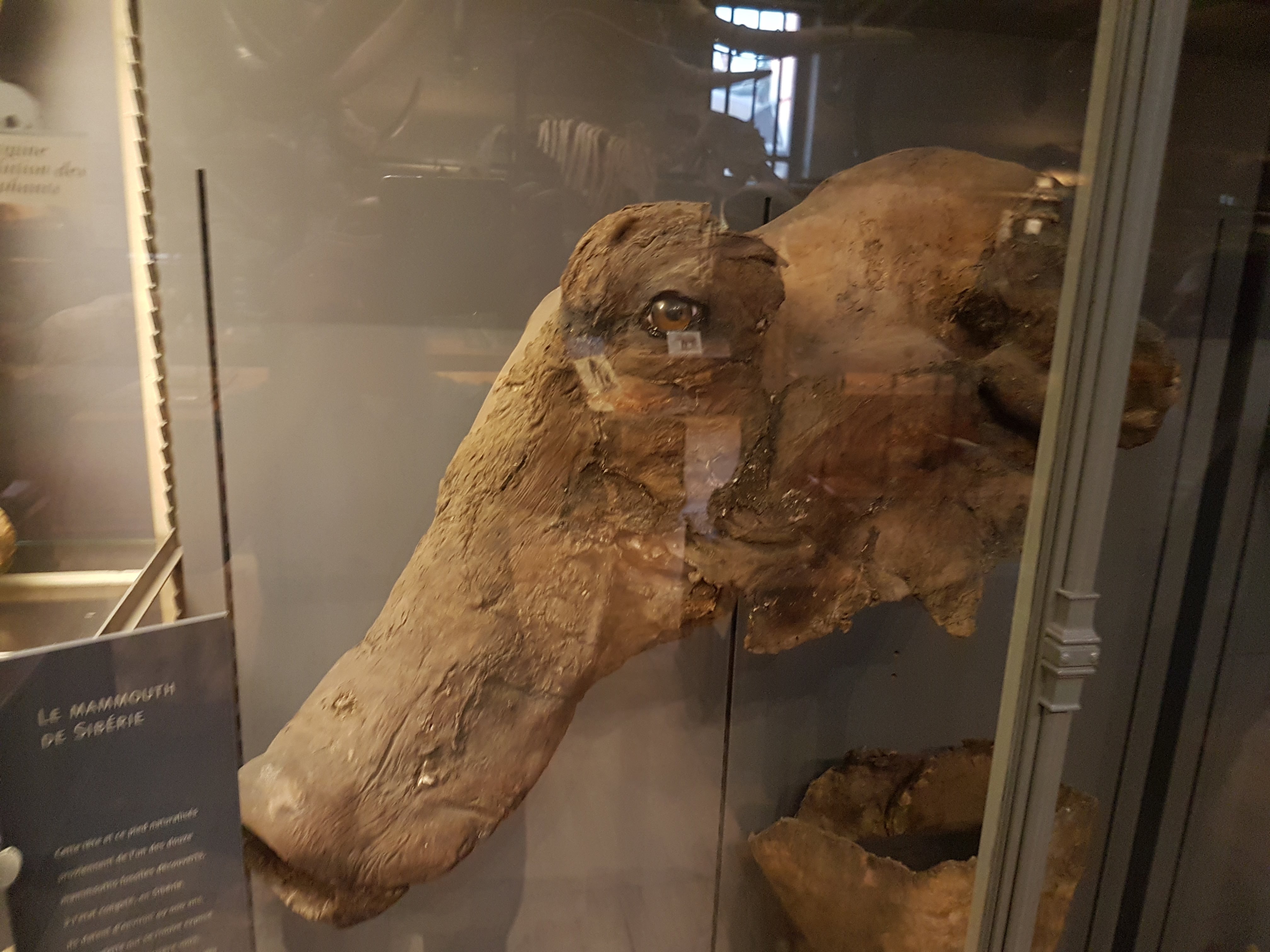
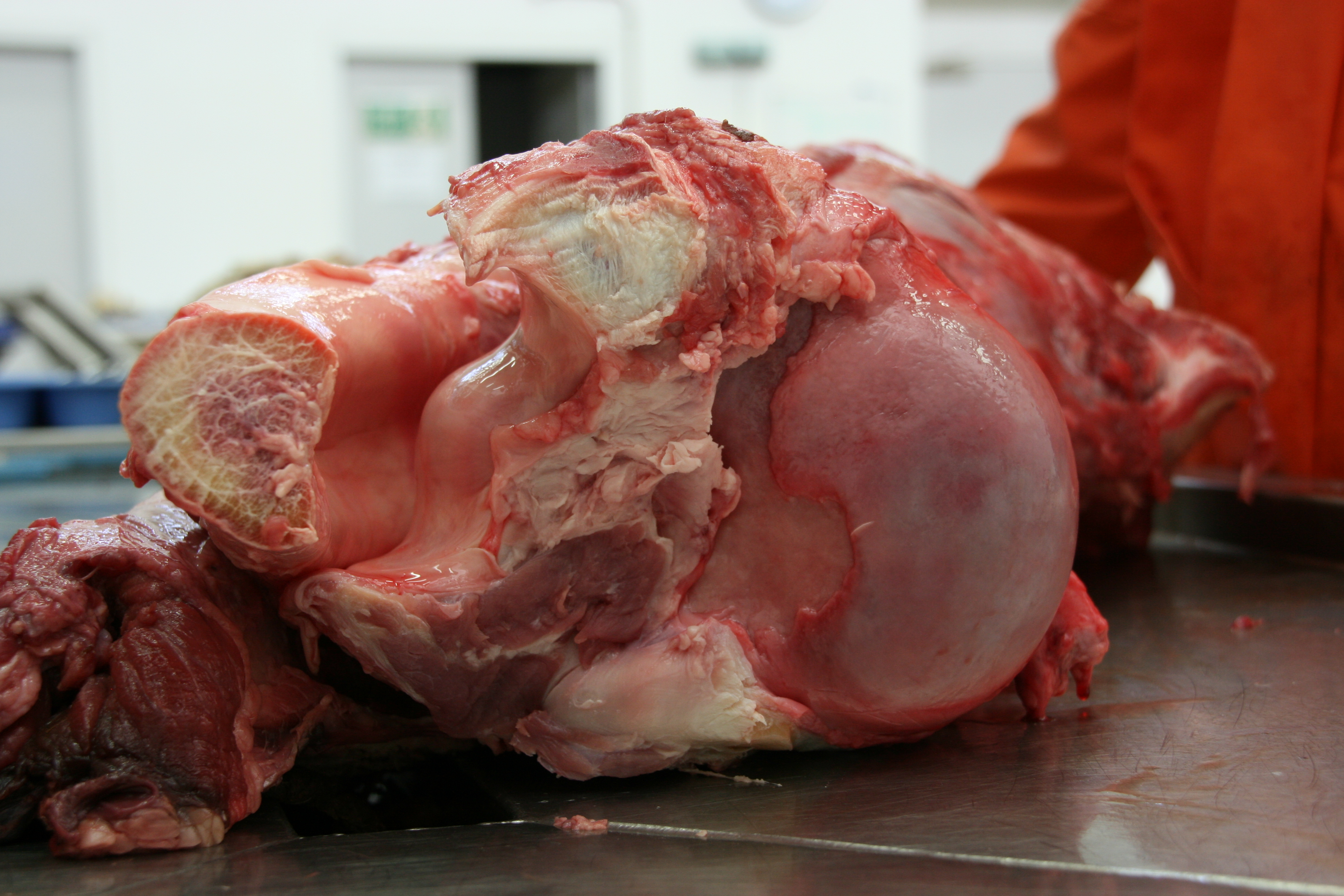
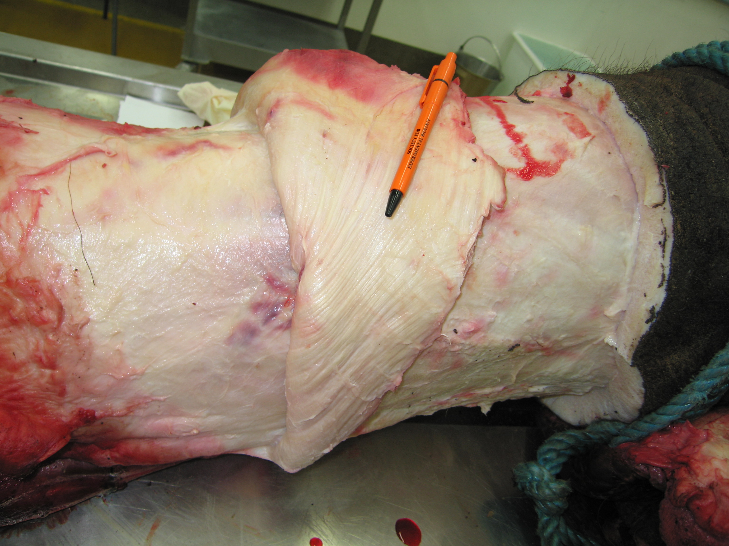
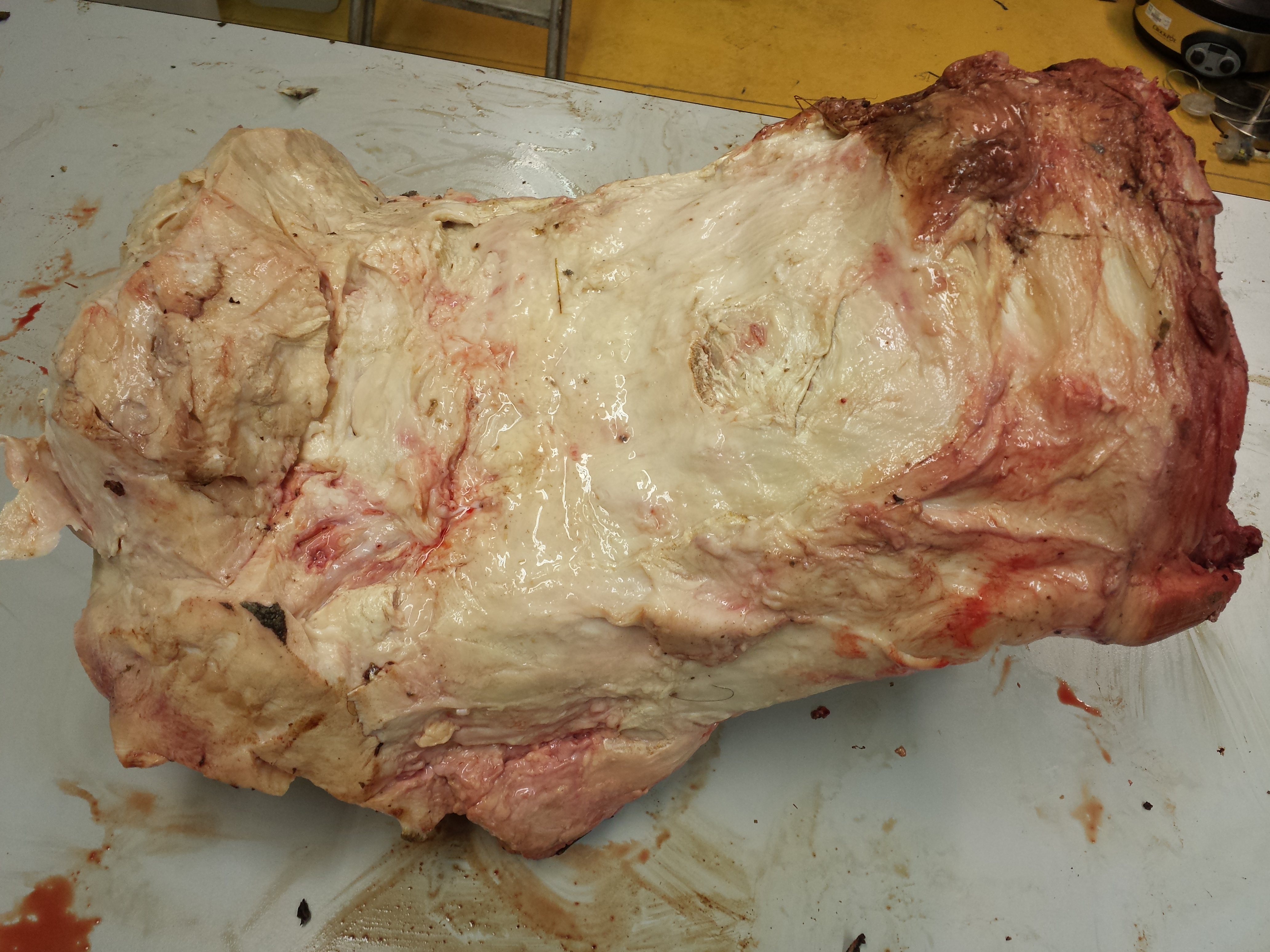
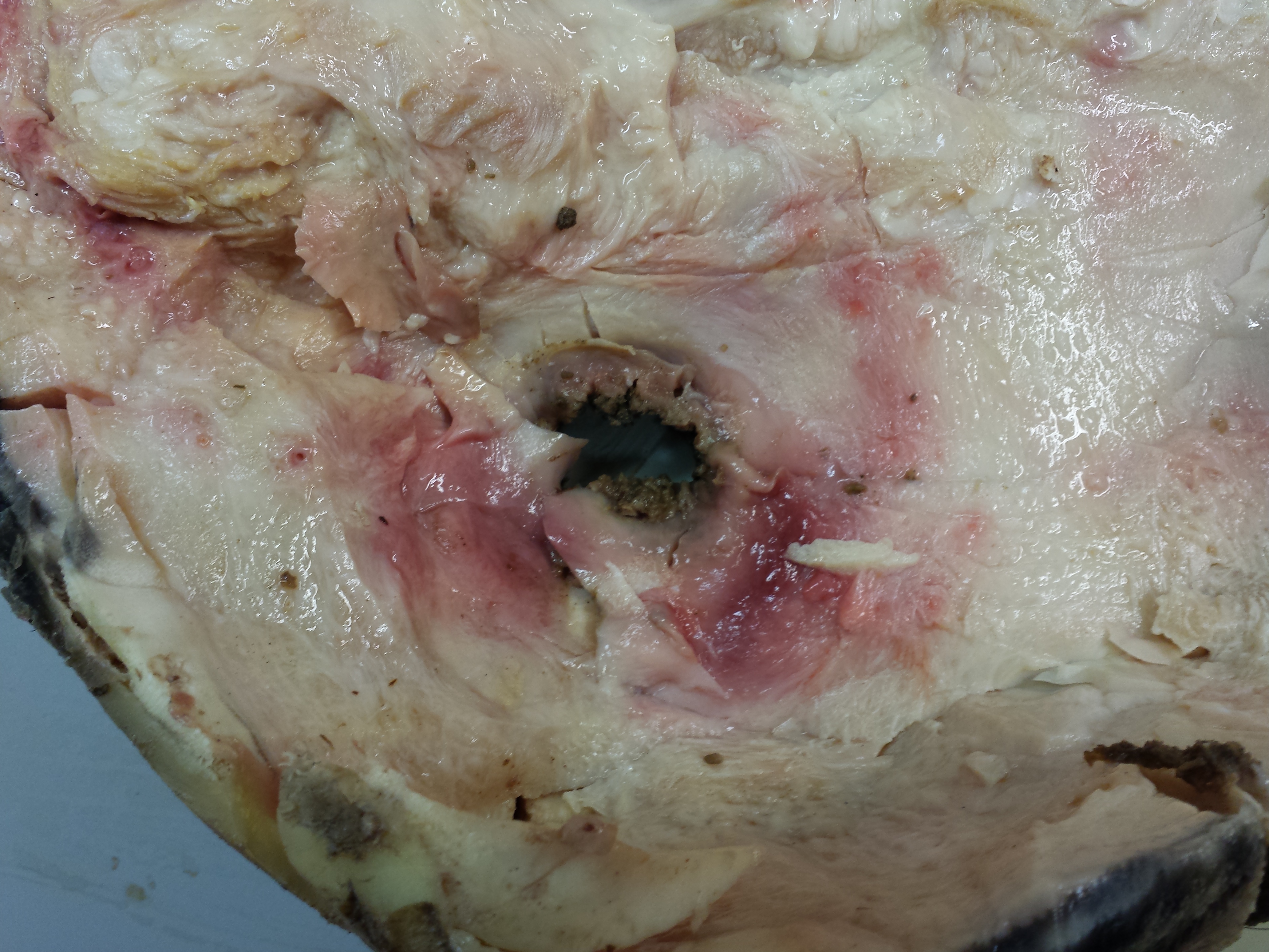
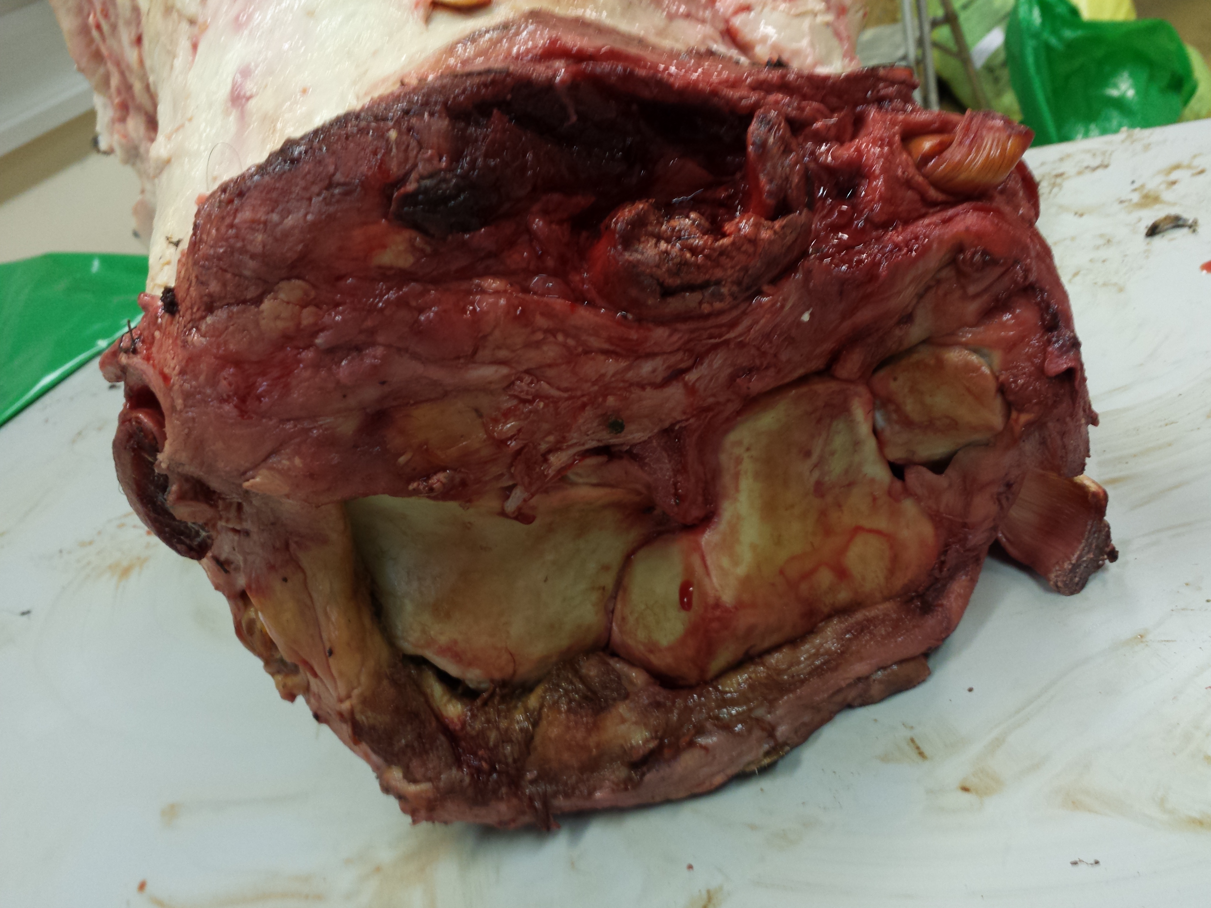
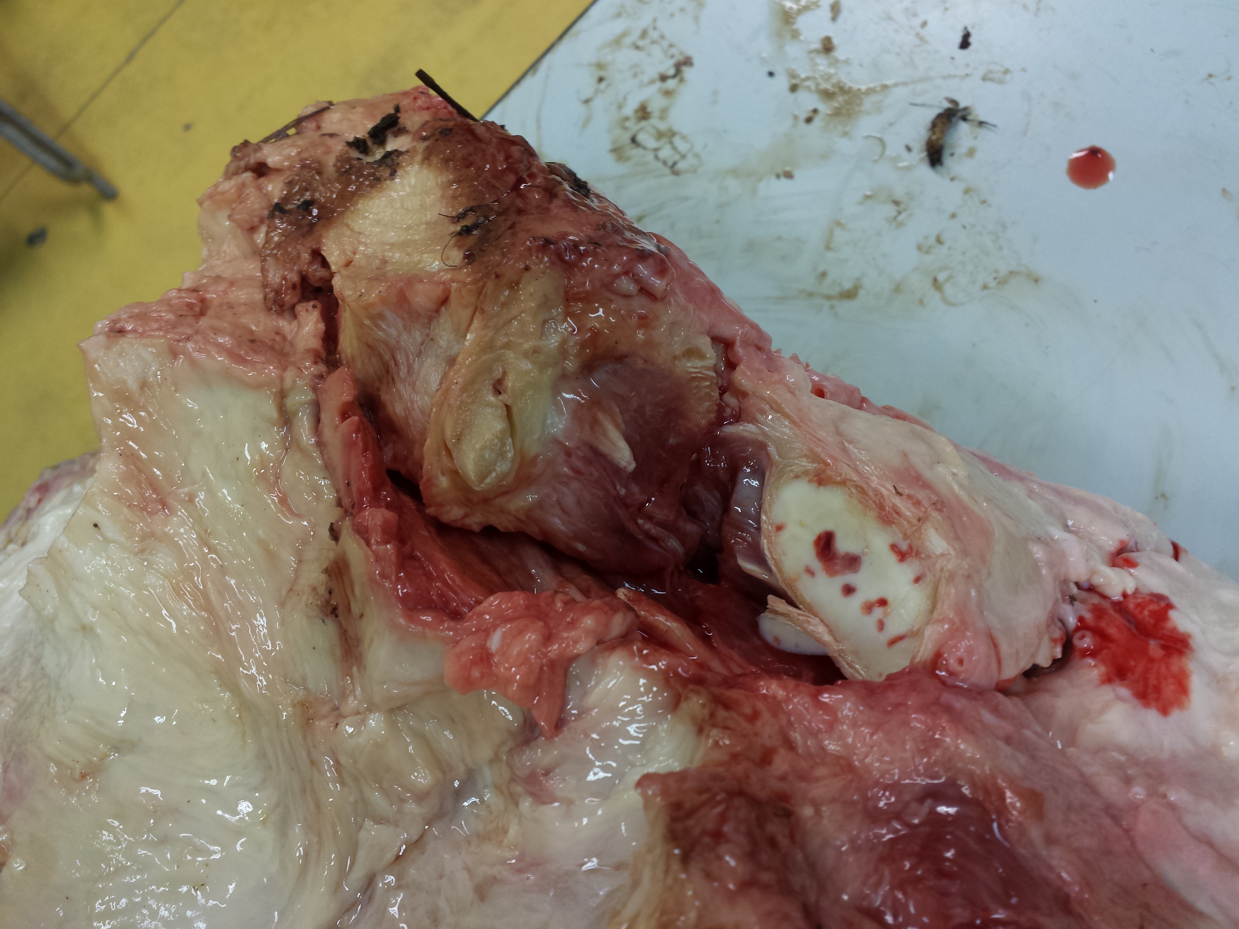
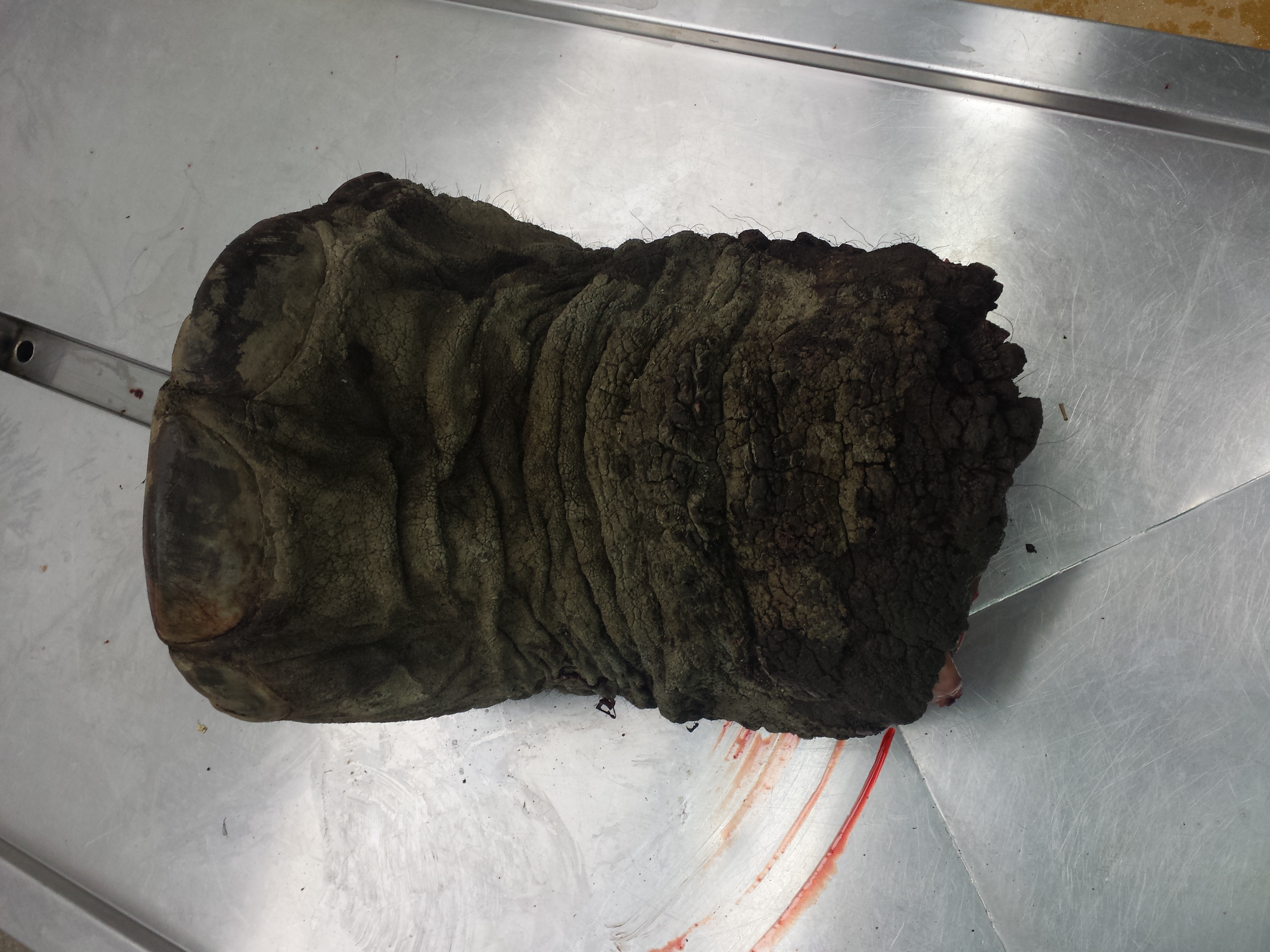
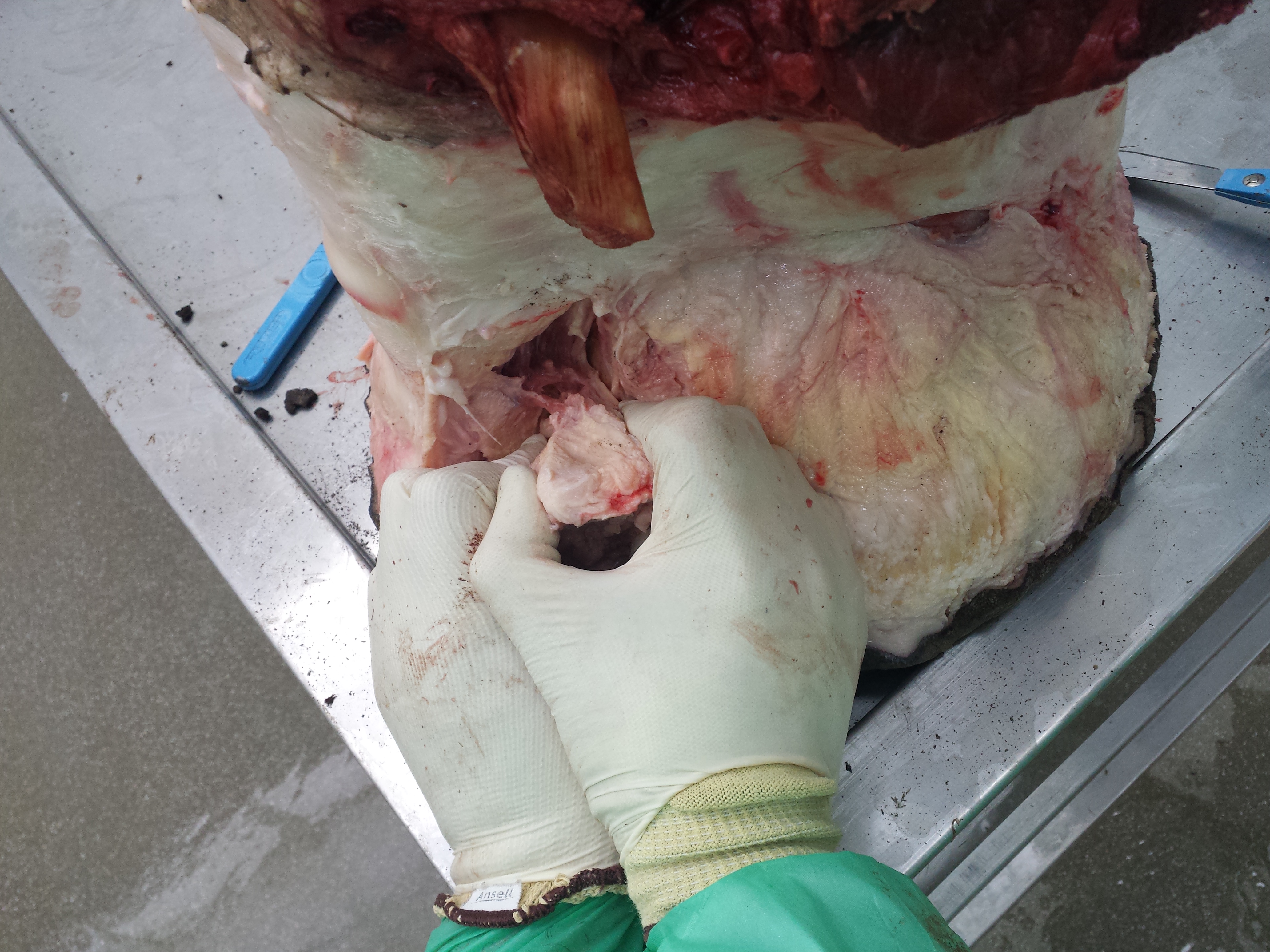
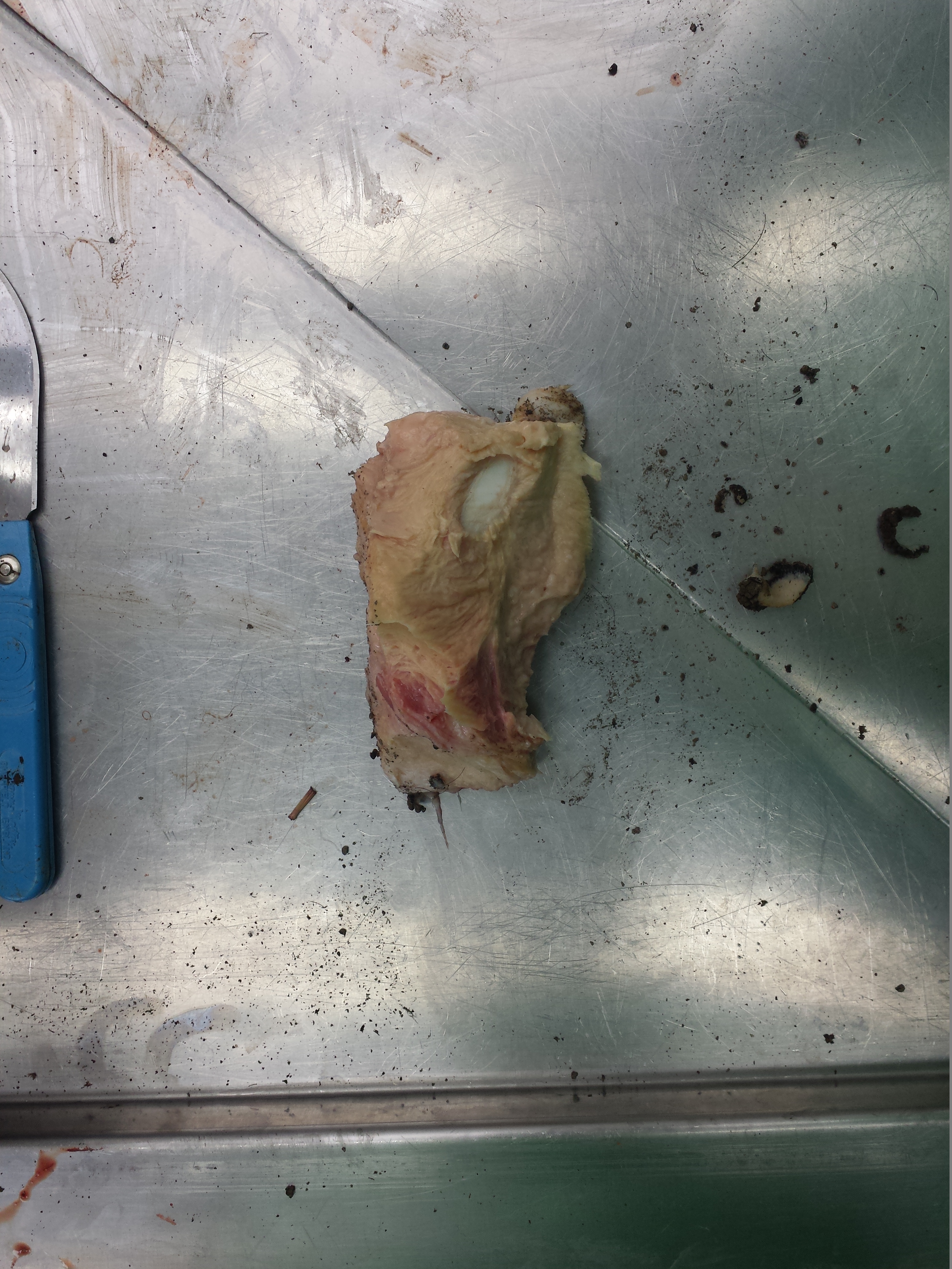
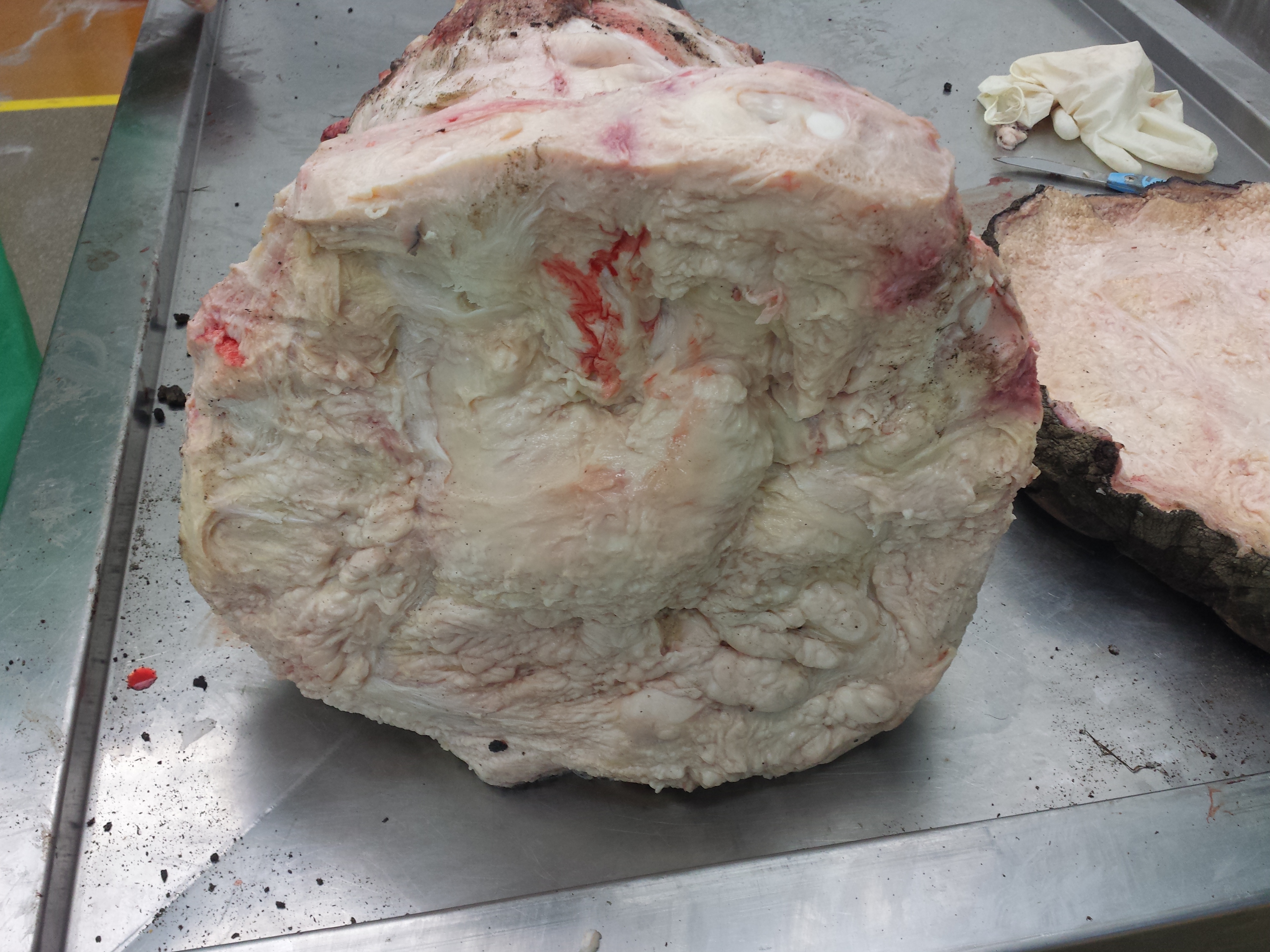
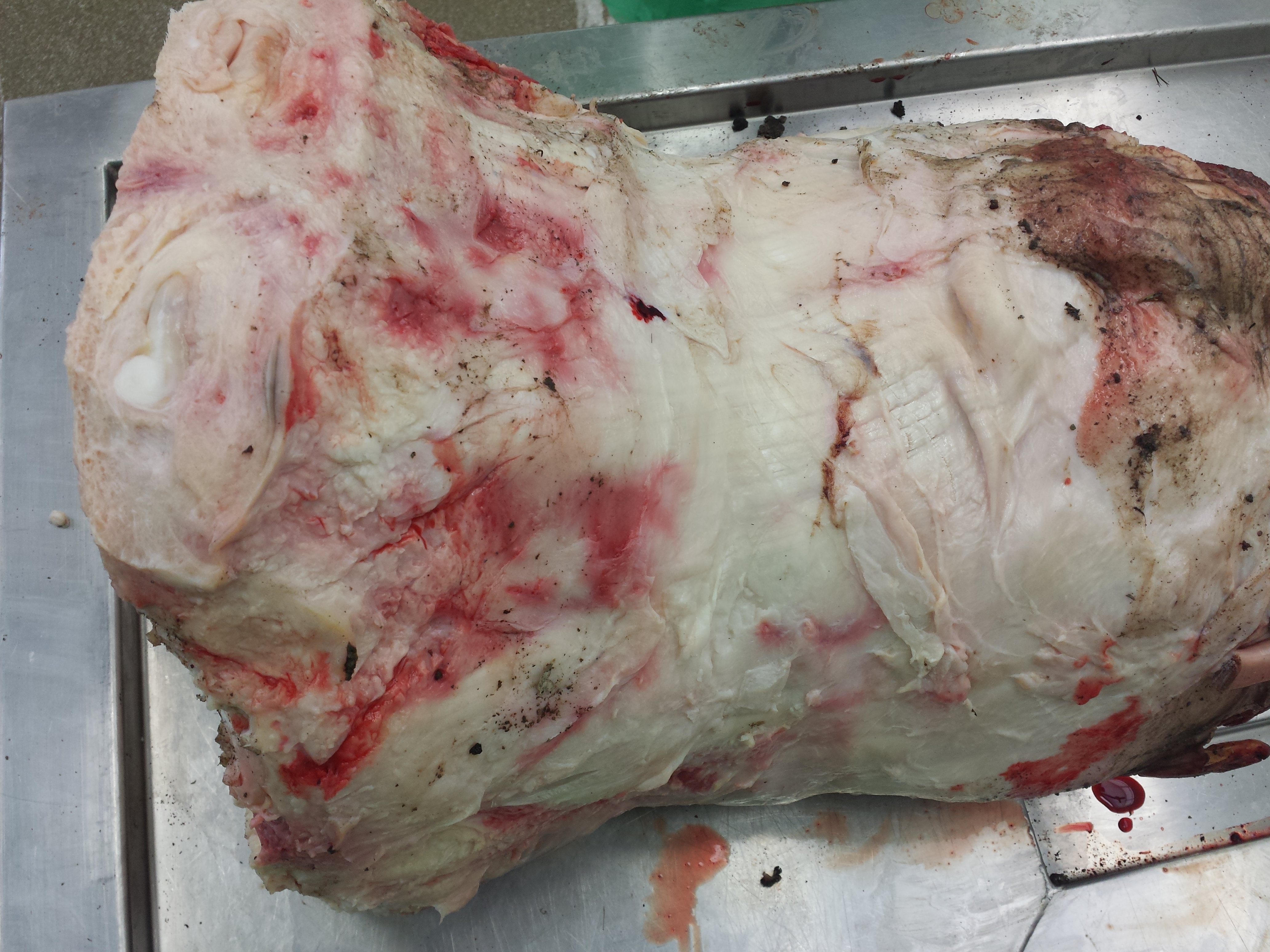
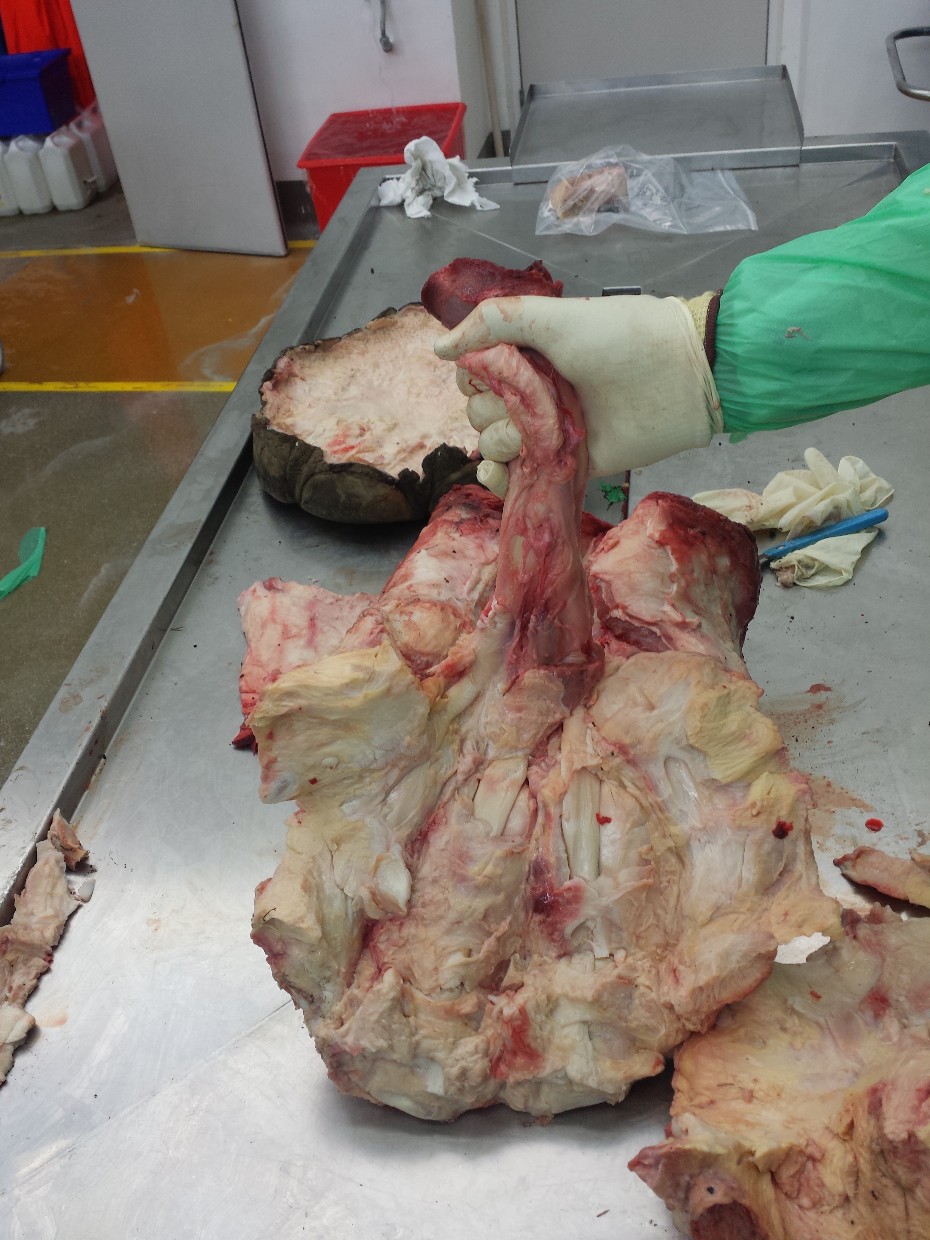
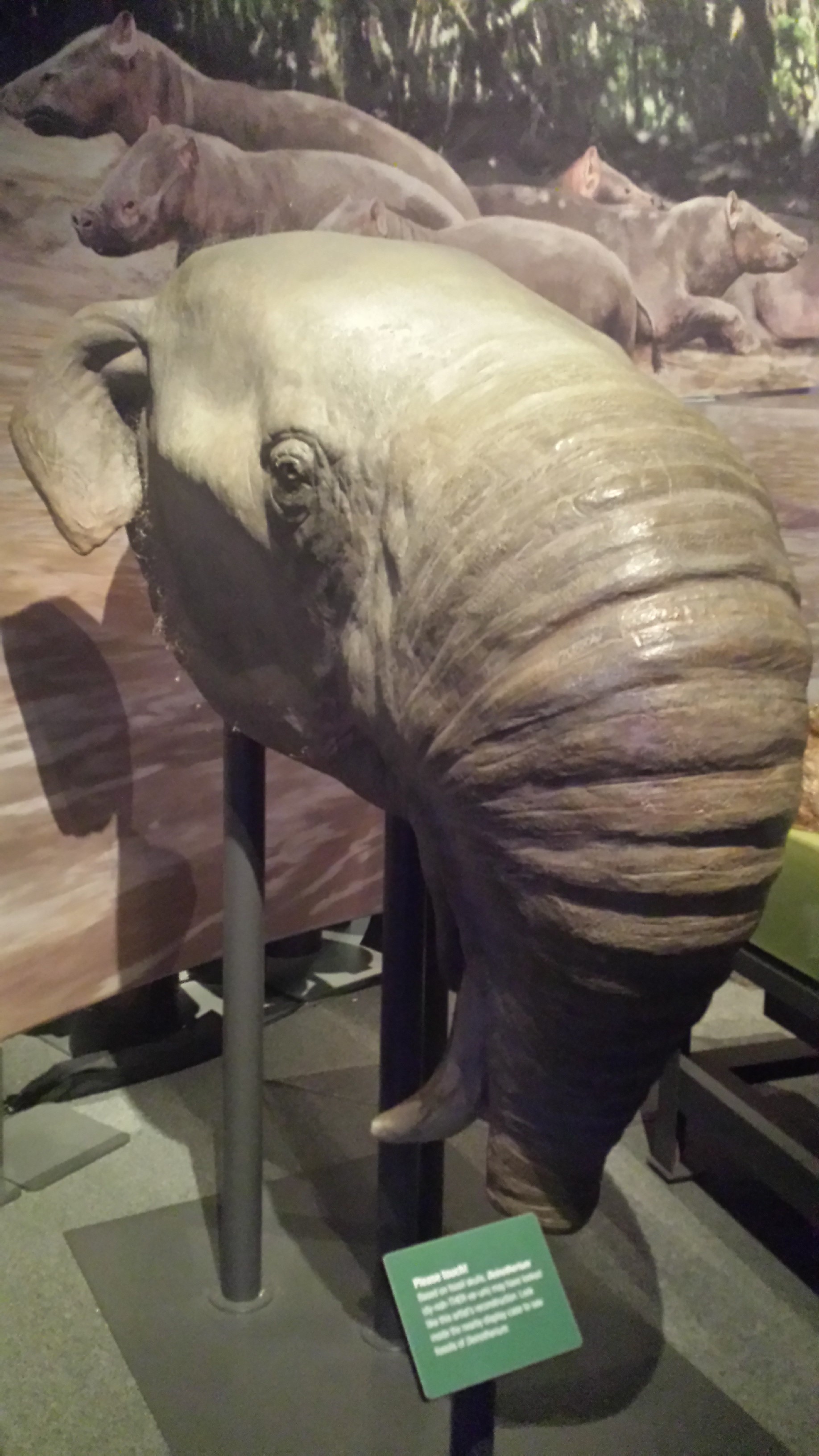
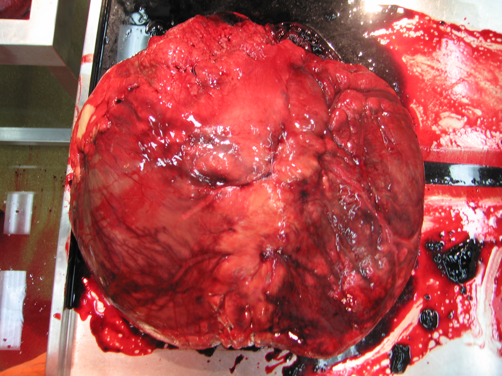
Yep. That’s what it looks like. Nope, not the front end. That dark orifice is not the mouth. This is a mammoth that was found on Bolshoi Lyakhovsky island, in the east Siberian arctic (New Siberian Islands archipelago), in 2003. Just think of finding this and being all excited then realizing, “Jackpot! Wait… Oh man, I just found the ass. I’ve discovered a mammoth bunghole, dammit.” Still, it’s pretty damn amazing, as frozen Ice Age buttocks go. I’d love to find one. I would not be bummed.
found on Bolshoi Lyakhovskiy island in 2003
What I know now that I didn’t realize as a kid, is that a mammoth is an elephant in all but name. Mammoths are more closely related to Asian elephants than either is to African elephants, and all of these elephants are members of the group Elephantidae. If we saw a smallish Columbian mammoth, we’d probably mostly look upon it as similar to a slightly hairy Asian elephant (but a scientist would be able to spot the distinctive traits that each has). Only woolly mammoths adopted the uber-hirsute state that we tend to think of as a “mammoth” trait. Think about it: a big animal would benefit most from a thick hairy insulation in an extremely cold habitat, and Columbian mammoths ranged further south than Woolly ones. No mammoths were radically different from living elephants, unless you count the dwarf ones. But as a kid, like most people do, I saw them as something else: an exotic monster of the past, eerily unlike anything today, and bigger too. And mammoths have the added mystique of the extinct.
Now I see mammoths as neither exotic nor that far in the past. Giant ground sloths, now those are still alien and exotic to me. I don’t get them. I know elephants pretty well, and I can understand mammoths in their light and in light of mammoth fossils. Various mammoth species persisted as late as maybe 10,000 (for the Woolly and Columbian species; the latter seeming to vanish earlier) to <4000 (for isolated Siberian forms) years ago, into quasi-historic times. And only some mammoths got larger than African elephants (Loxodonta) do, such as Columbian mammoths (~10,000 kg or more maximal body mass; Loxodonta is closer to 7-10 tonnes at best).
Lately, coincidence has brought me new knowledge of – and even greater interest in – mammoths.
First, a fortunate last-minute visit to Waco, Texas’s “Mammoth Site” (see my Flickr photo tour here) two weeks ago during a short visit to give a talk in that fine central Texan city.
Second, the subject of today’s post: the Natural History Museum’s new special exhibit “Mammoths: Ice Age Giants“, which is open until 7 September. The exhibit was created by the Field Museum in Chicago, but the NHM has given it a special upgrade under the expert guidance of mammoth guru Prof. Adrian Lister of the NHM, who was very kind to give me a tour of the exhibit.
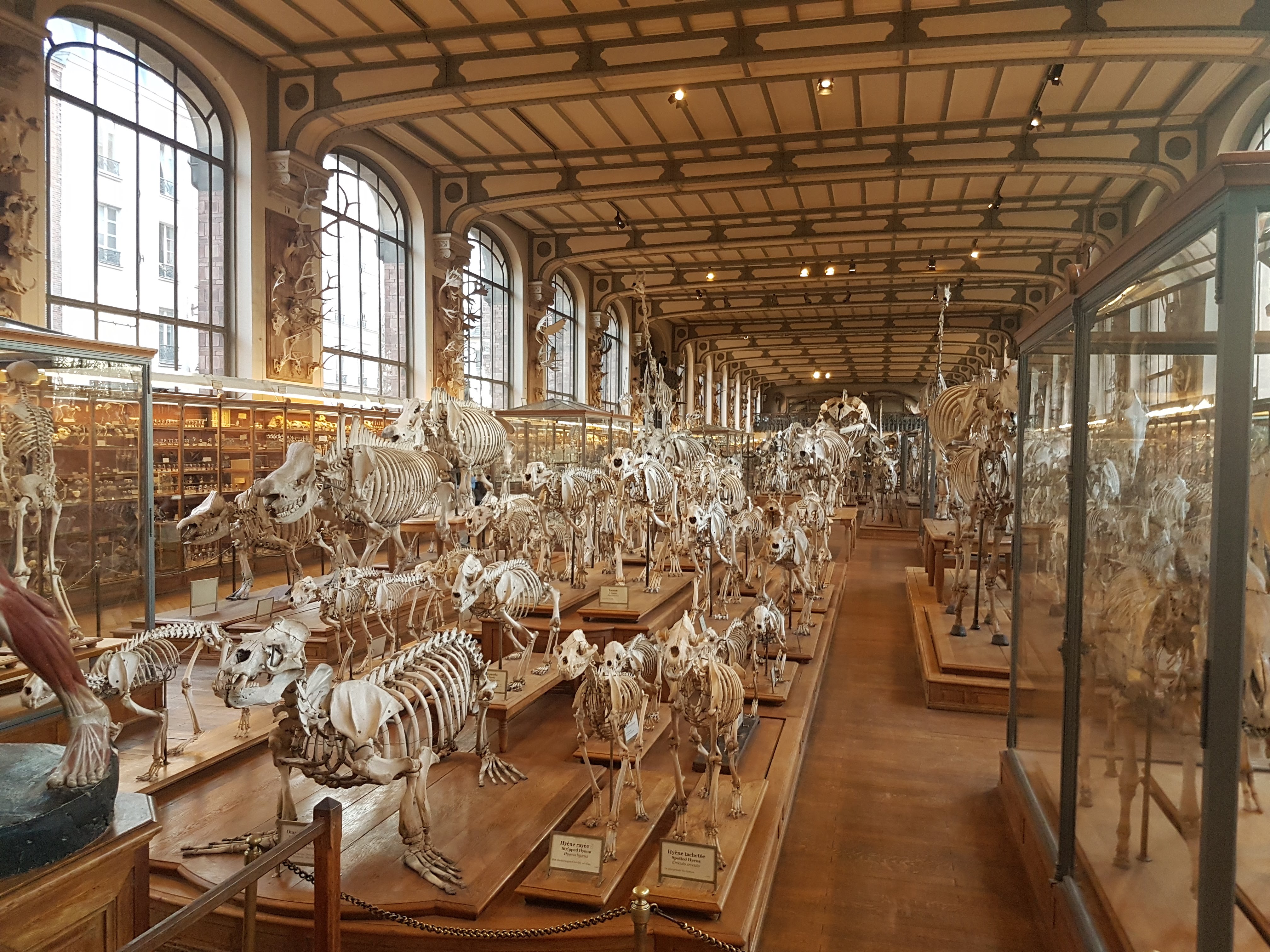
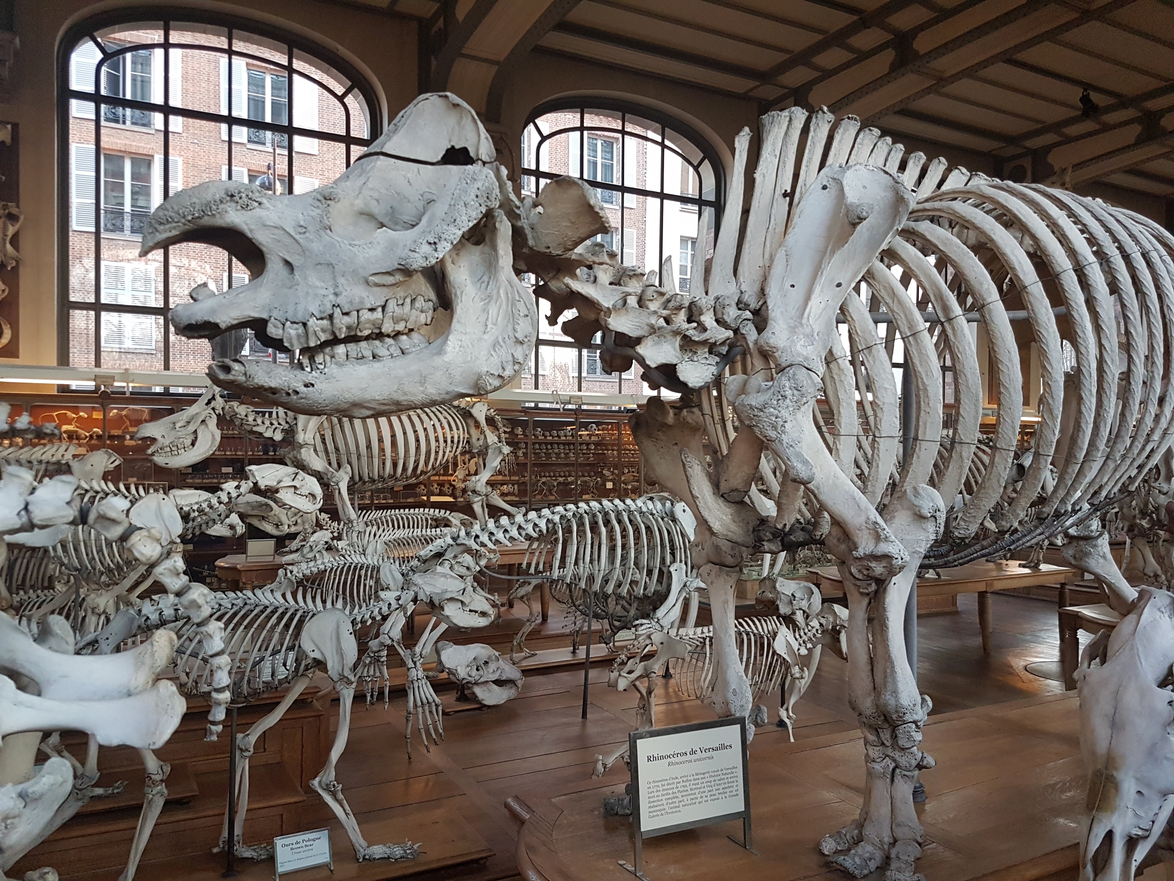
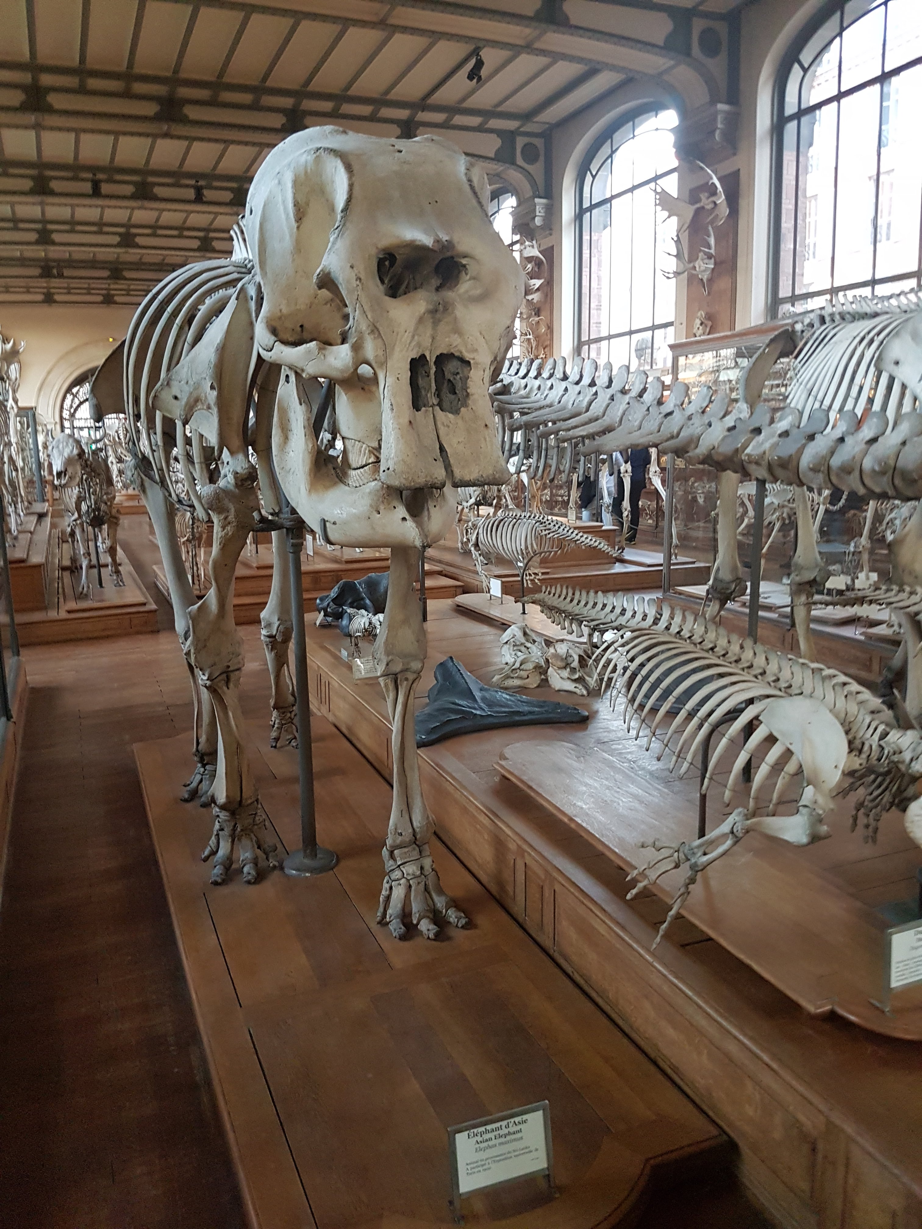
What follows is primarily a photo-blog post and review of the exhibit, but with some thoughts and facts and anecdotes woven through it. Dark setting, glass cases, caffeination, crowds, and mobile phone camera rather than nice SLR in hand means that the quality isn’t great in my images– but all the more reason to go see the exhibit yourself! All images can be clicked to em-mammoth them.

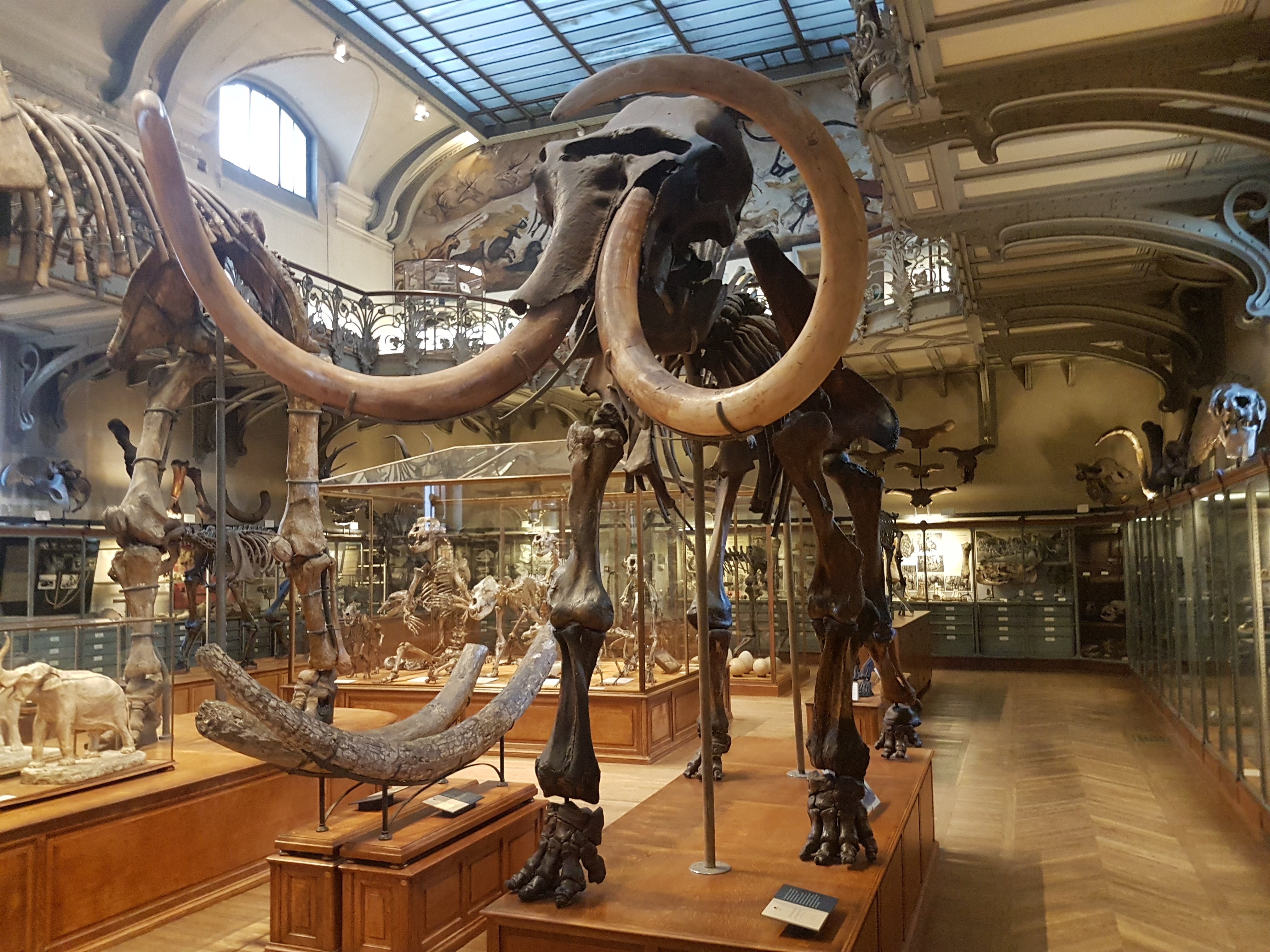
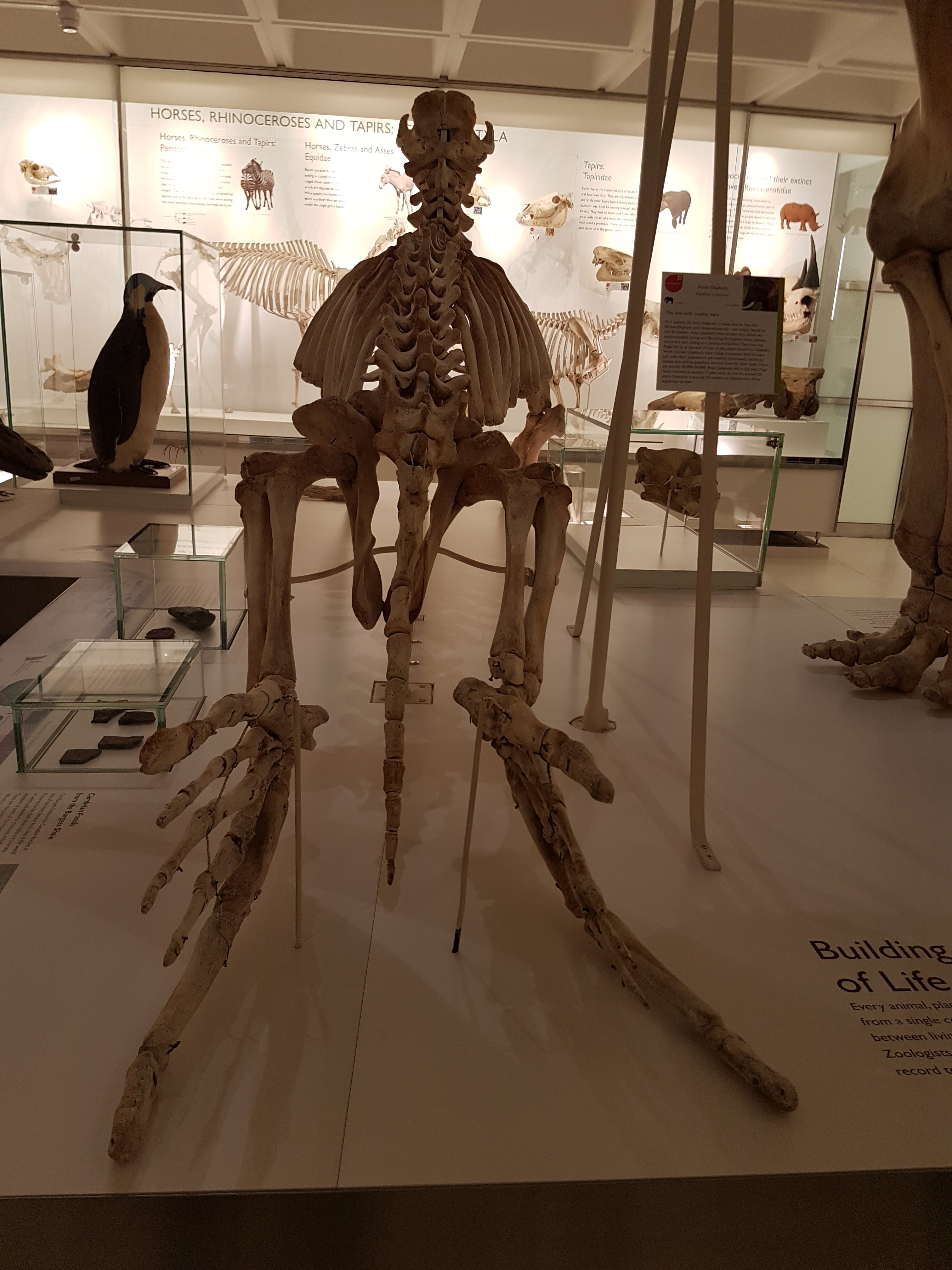
On entry, one views a mammoth skeleton with a timelapse video backdrop that shows how the landscape (somewhere in USA) has changed since ~10,000 BCE.
The first part of the exhibit does a nice job of introducing key species of Proboscidea (elephants and their closest extinct relatives), with a phylogeny and timescale to put them into context, starting with the earliest forms:

from species like the tapir-sized Moeritherium…

Skull of Moeritherium, reconstructed. Not that different from an early sirenian (seacow) in some ways, and general shape, whereas still quite a long way from a modern elephant in form– but the hints of tusks and trunk are already there.

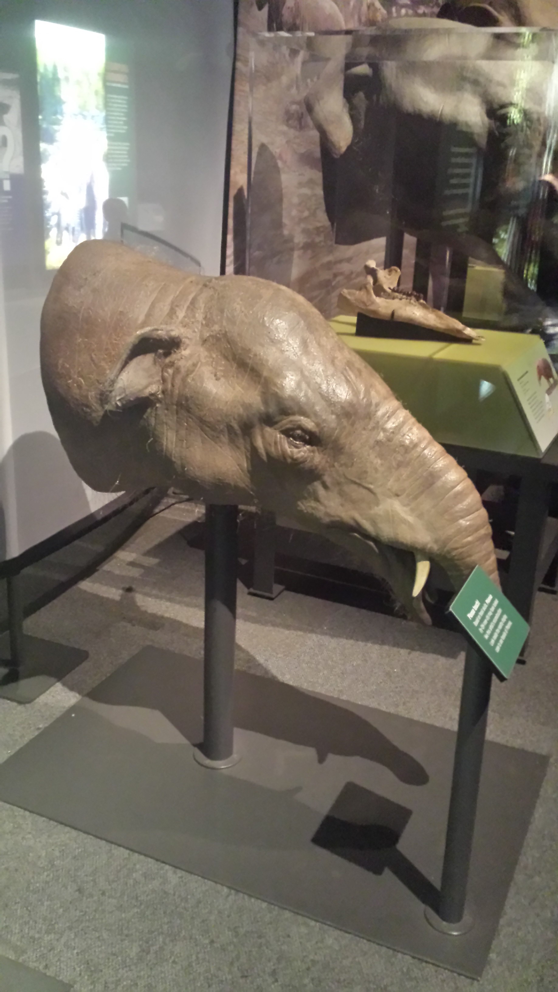
…To the early elephantiform Phiomia, here shown as a smallish animal but I’m told it actually got quite large. And continuing with giant terrestrial taxa…

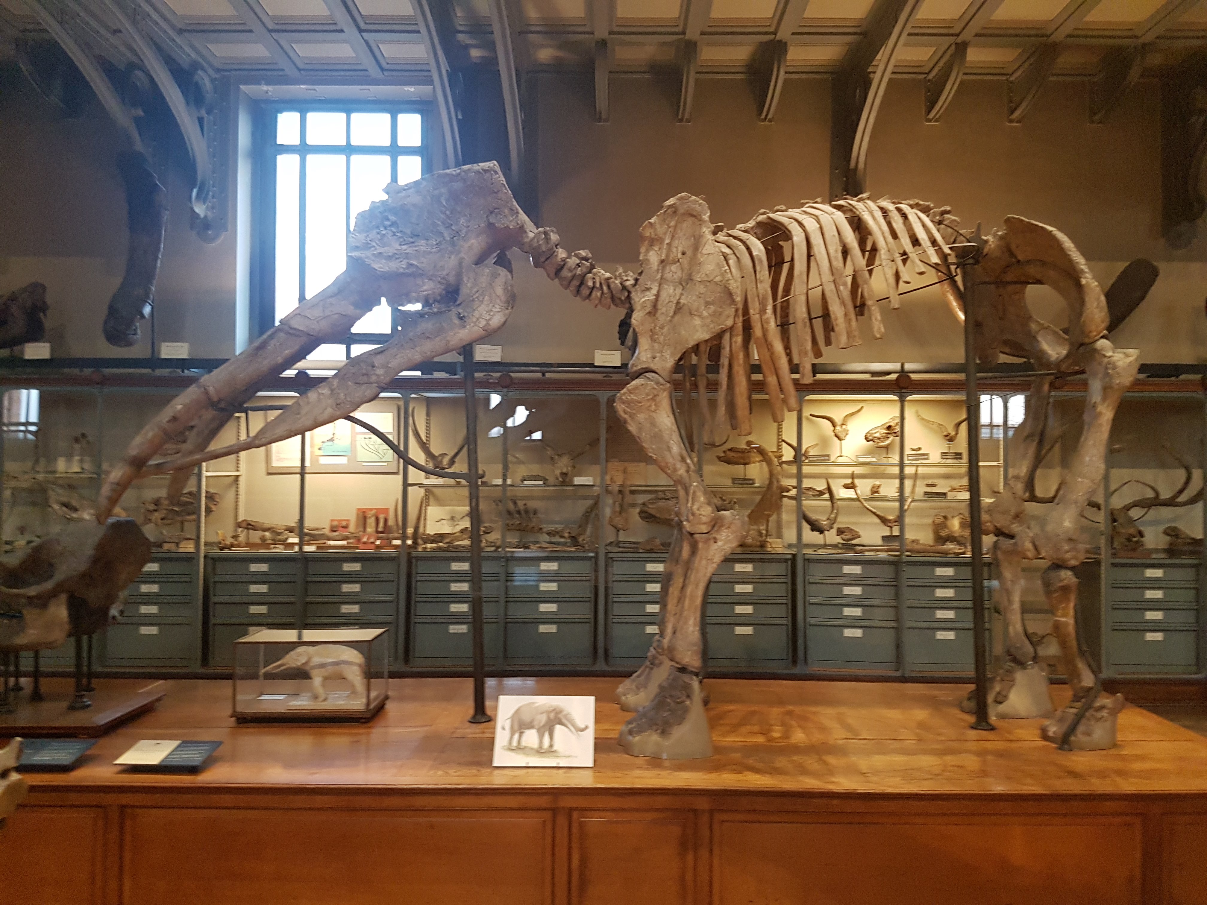
I was awed by this reconstruction of the huge early elephantiform-relative Deinotherium, with the short, swollen trunk and downturned tusks– so bizarre!

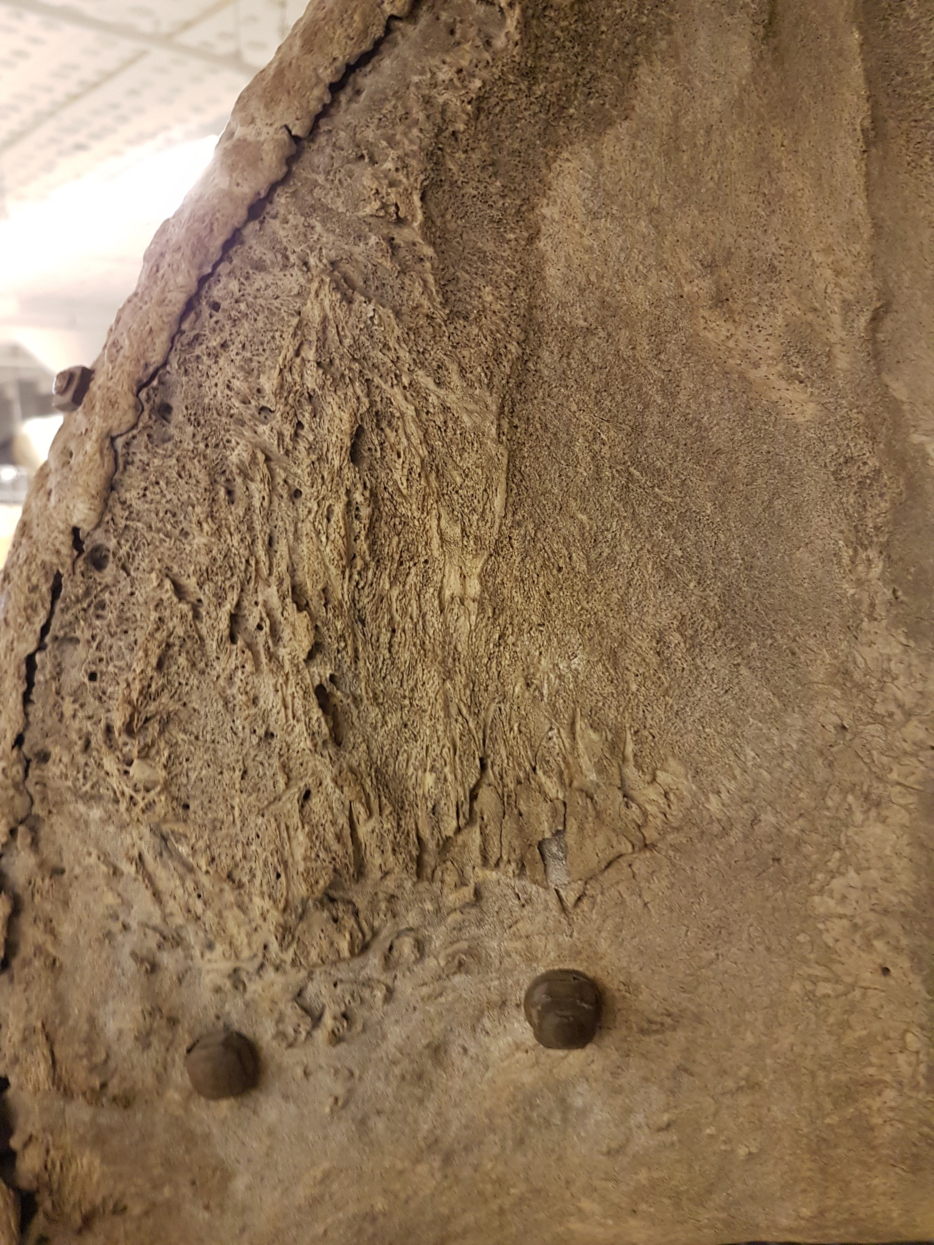
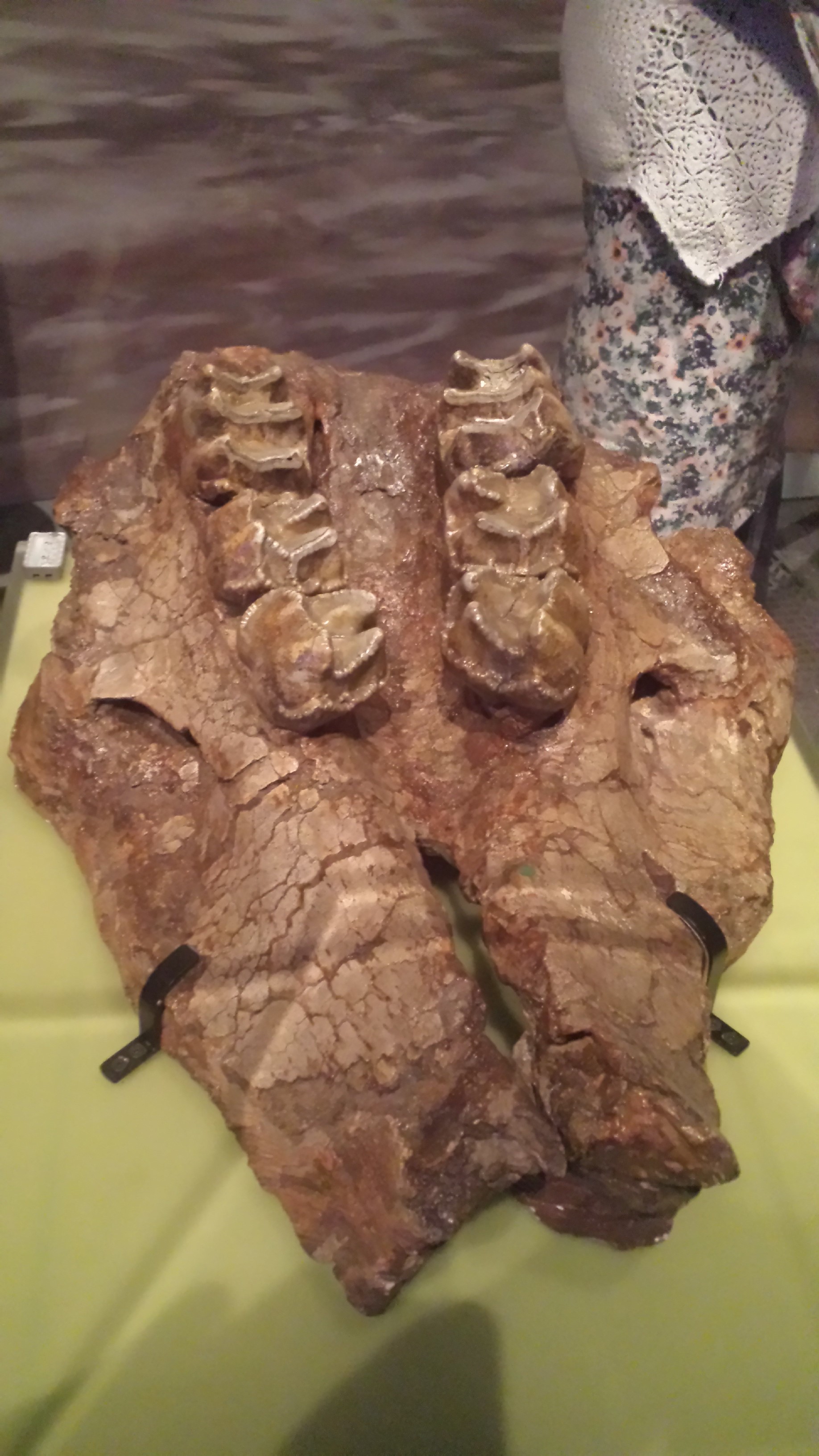
Looking down onto the roof of the mouth of an NHM specimen of Deinotherium. Big, sharper-edged, almost rhino-like teeth; far from the single mega-molars of modern elephants.

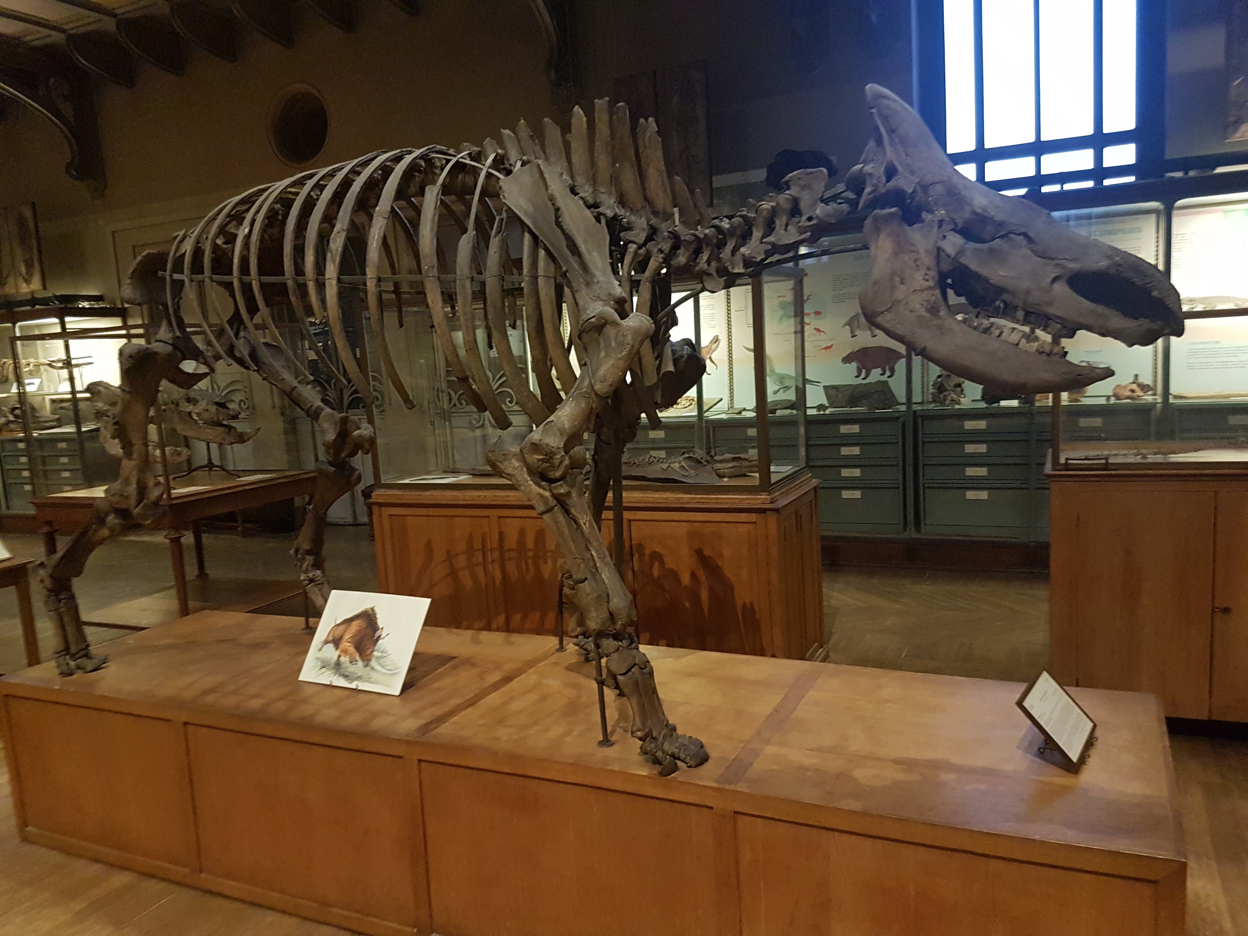
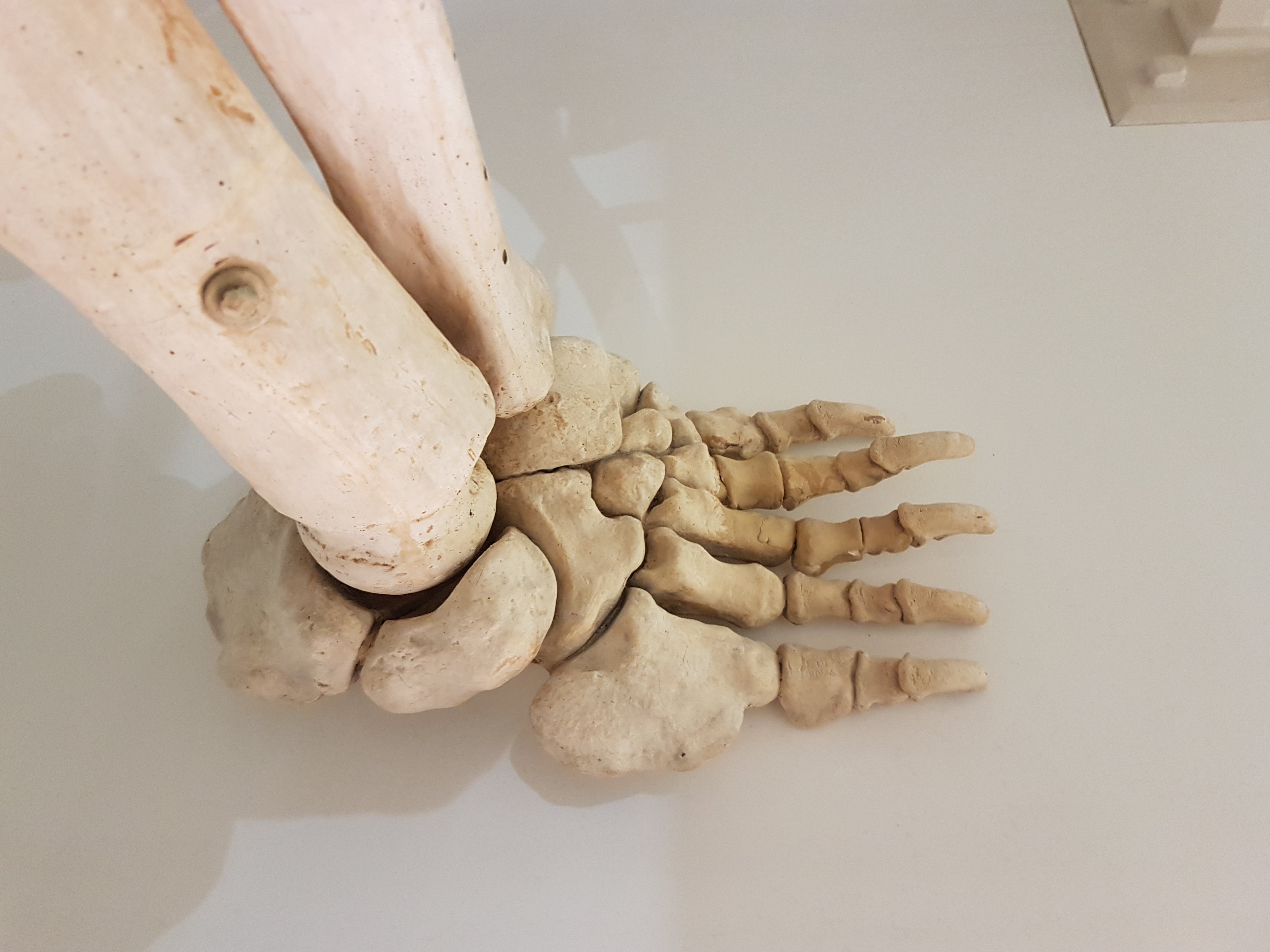
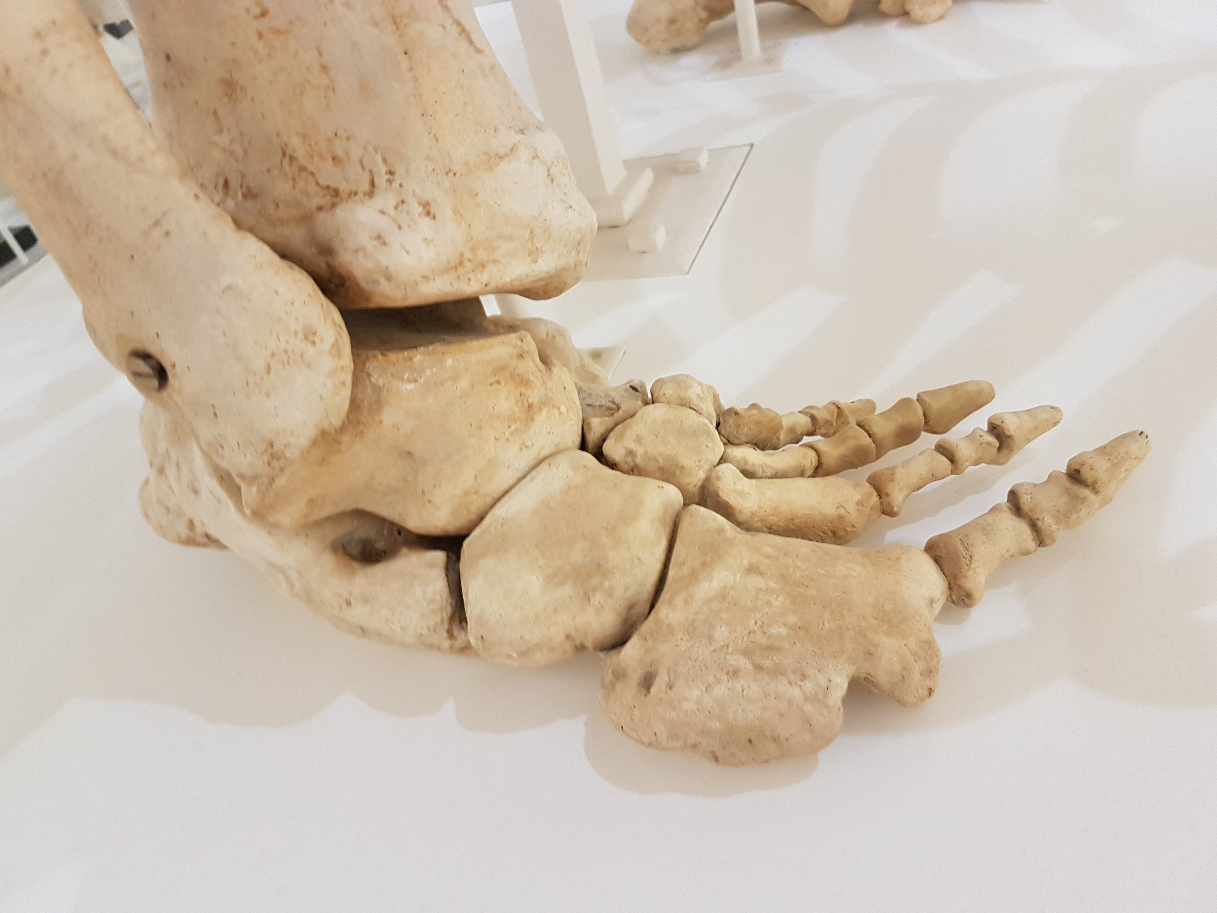
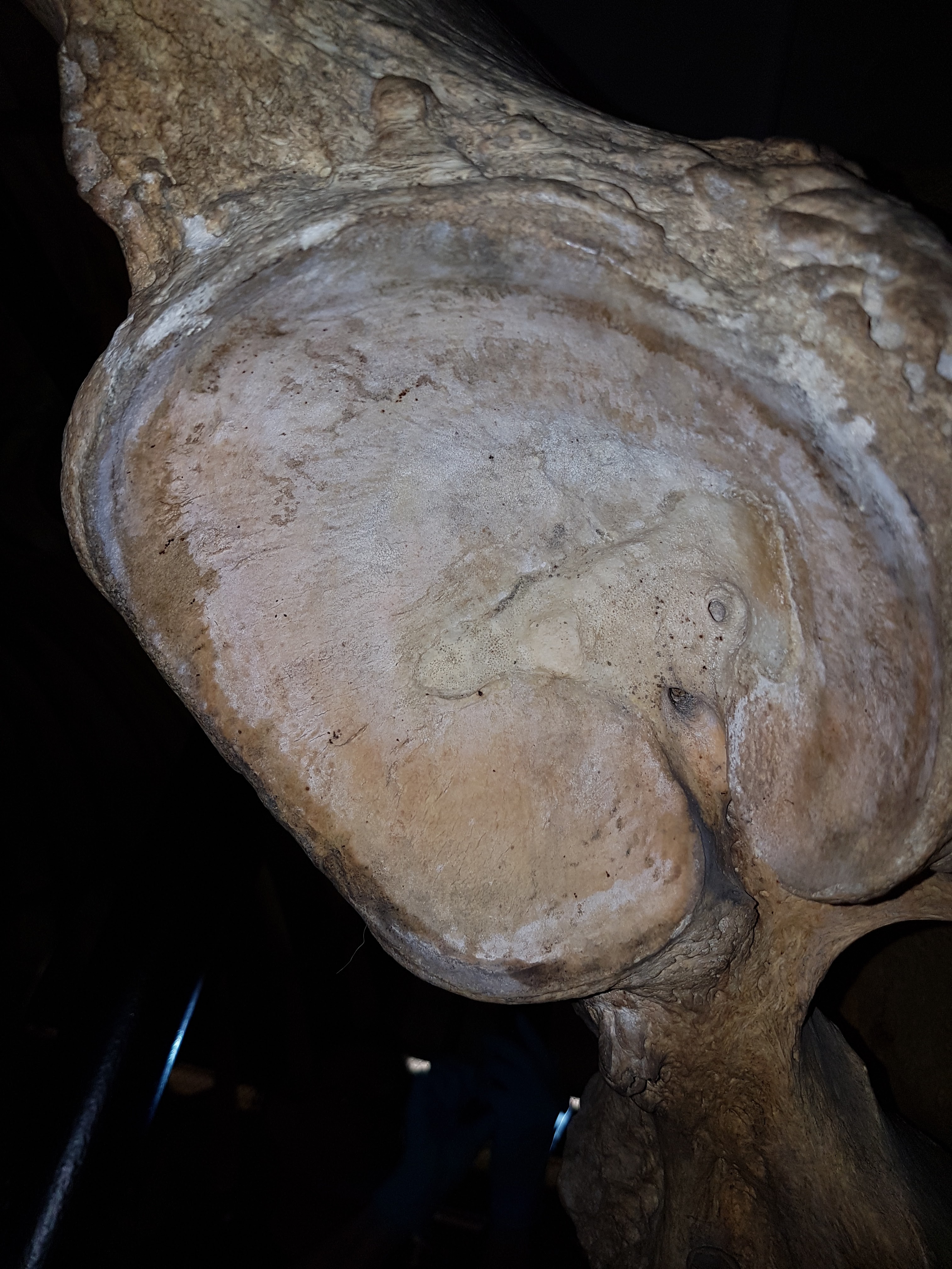
The lower jaw (top) and fairly straight tusk (bottom) of the widespread, early elephantiform Gomphotherium.

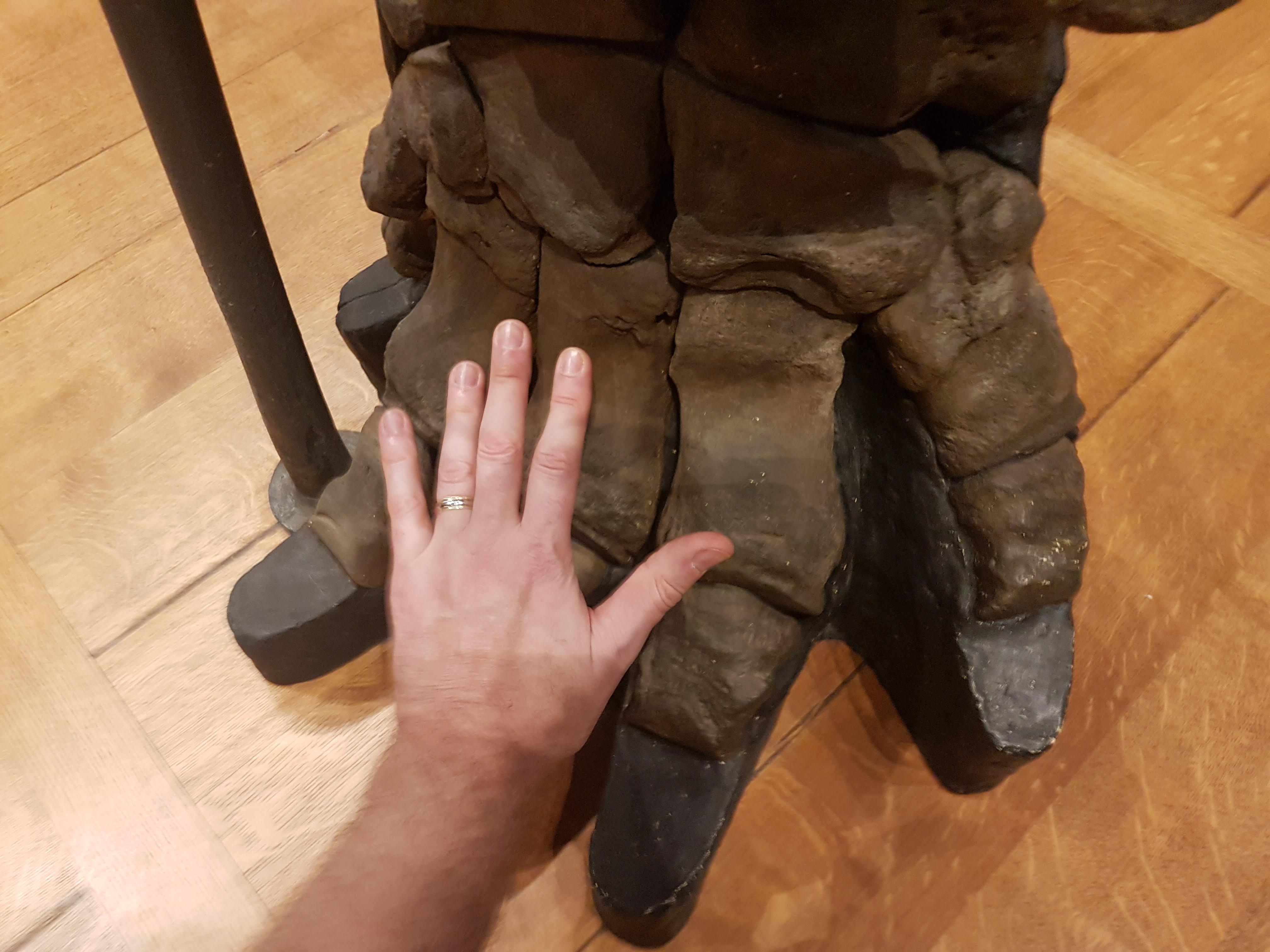
The big “shovel-tusked” elephantiform Amebelodon. This was one of the earliest stem elephants I learned of as a kid; the odd tusks still stir wonder in me.

Amebelodon lower jaw, sans shovel tusks. Extended chin looks like some sort of childrens’ fun-slide. To me, anyway.
Next, there are some fun interactive displays of elephant biomechanics!

How would a mammoth hold up its head? This lever demonstration shows how a nuchal ligament helps. Tension on the nuchal ligament is a force that acts with a large lever (represented by the big neural spines on the vertebrae around the shoulders, forming the mammoths’ “hump” there), creating a large moment (i.e. torque; rotational force) that holds the head aloft.

I love this robotic elephant trunk demonstration. It captures some of the weirdness of having a muscular hydrostat attached to your lip and nostrils. Not so easy for a human to control!
But forget the myths about elephants having 40,000 to 150,000 muscles in their trunk. They have three muscle layers: a circumferential one, an oblique one and a longitudinal one. Like any muscles, especially ones this large, the layers each consist of many muscle fibres. That’s where the 40-150k myth comes from, but muscle fibres (cells) are at a more microscopic level than whole muscles (organs). Elephants do have excellent control of their trunks, but it’s not magical. It’s just different.
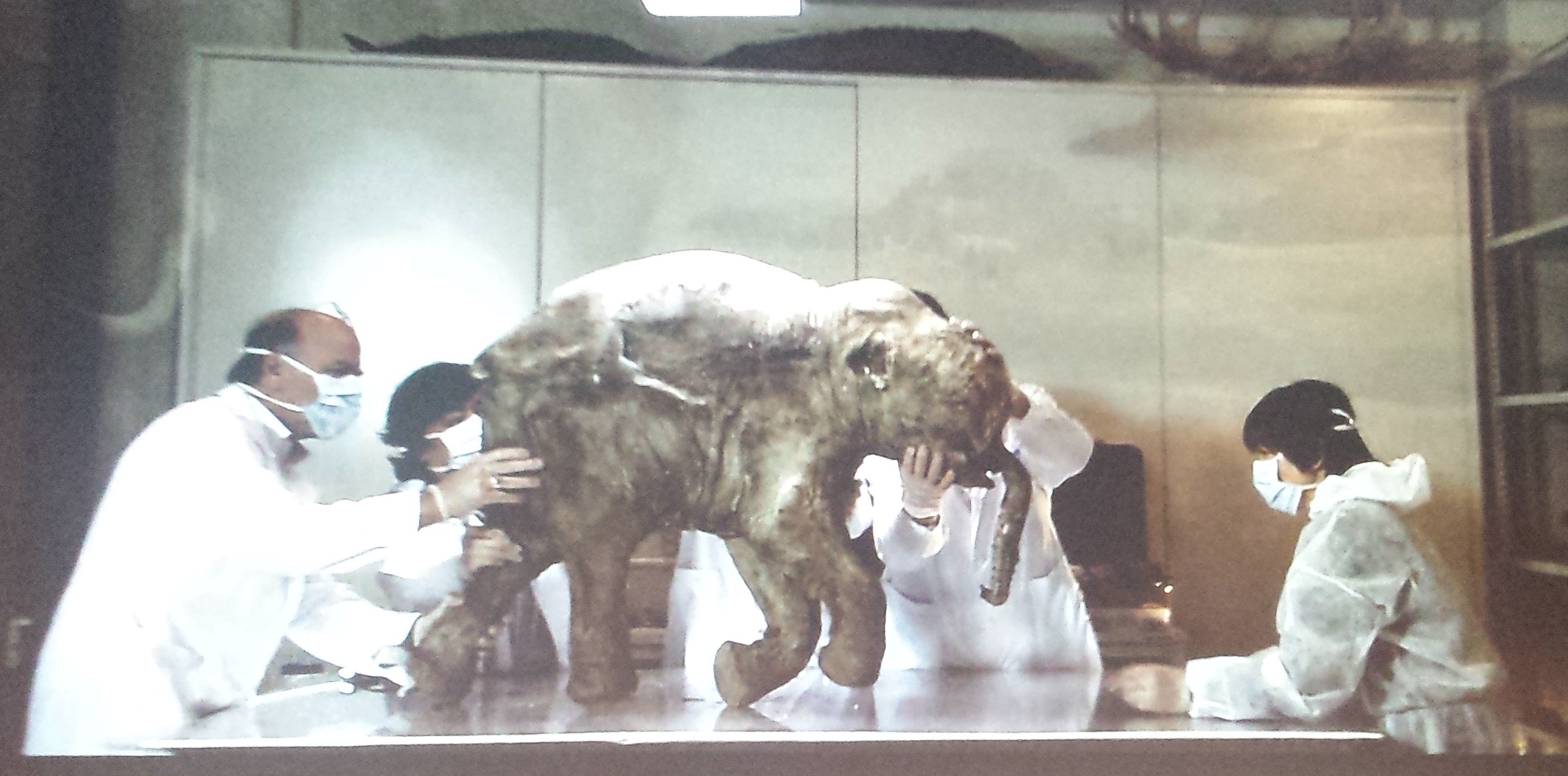
Then we come to the centrepiece of the exhibit, the ~42,000 year old Woolly mammoth (Mammuthus primigenius) baby “Lyuba“, which the NHM added to the original exhibit in this new version, as a star attraction — and a big win. Adrian Lister related to me how he’d never seen Lyuba in person before (access to it was tightly guarded for years). So when the NHM received the crate and held a press event to open it and reveal Lyuba, a journalist asked Adrian to act excited, to which he responded something like, “I don’t need to act! I’m very excited!” I would be, too! Full story on Lyuba’s arrival, by NHM site here. A key paper on Lyuba by Fisher et al. is here.

Studies of tooth growth in Lyuba reveal her gestation period (like living elephants, ~22 months), season of birth (early spring), and age at death (~1 month), among other information.

Here we can see the right ear, which was gnawed off along with the tail by dogs of the reindeer herders that found and retrieved Lyuba in 2006. Regardless, there’s loads of anatomy preserved!
A hump of juvenile “brown fat” sits atop the head and neck of Lyuba. This probably was metabolized during growth to warm the baby; brown fat is packed with mitochondria and thereby conducts what is called “non-shivering thermogenesis”. Furthermore, Lyuba has very strange flanges on the trunk (also visible in 1 other frozen mammoth specimen, but here preserved very clearly! What were they used for?). More details are visible postcranially…
The body was naturally “freeze-dried”, with the addition of later rounds of soaking in formalin and ethanol, leaving the body dessicated and stiff, permanently stuck in a lifelike pose as seen below:

Whole view from an exhibit panel (you cannot photograph the specimen but these are fair game!). Here we see hair on the right forearm and remnant of the ear, and the labia and nipples showing it is a female mammoth are also preserved. The head-hump is lost during growth, and the shoulder changes to change the Asian elephant-like convex curvature of the back into the characteristic humped-shoulder form of a mammoth. But ontogeny still reveals the evolutionary connection of Elephas and Mammuthus.

Lyuba and scientists studying her, which also shows how rigid the carcass is; one can almost stand it up. Inside the digestive tract, researchers found chewed up plant material that was probably dung eaten by the baby to gain vital bacterial digestive flora, and Lyuba had plenty of body fat and ingested milk, indicating that she did not starve to death. Rather, vivianite in the respiratory tract indicates drowning as the cause of her demise. Perfusion of the body by these vivianites may have helped to preserve the body.

Answering a question the public may be wondering about: is the hype about cloning a mammoth very soon true? Nope. Well addressed, including what to me is the urgent question: would cloning a mammoth be ethical?
The fourth part of the exhibit takes on a largely North American focus to first illustrate what mammoths were like biologically, and second to wow the visitor with some huge beasts in full body, full scale glory, as we shall see!

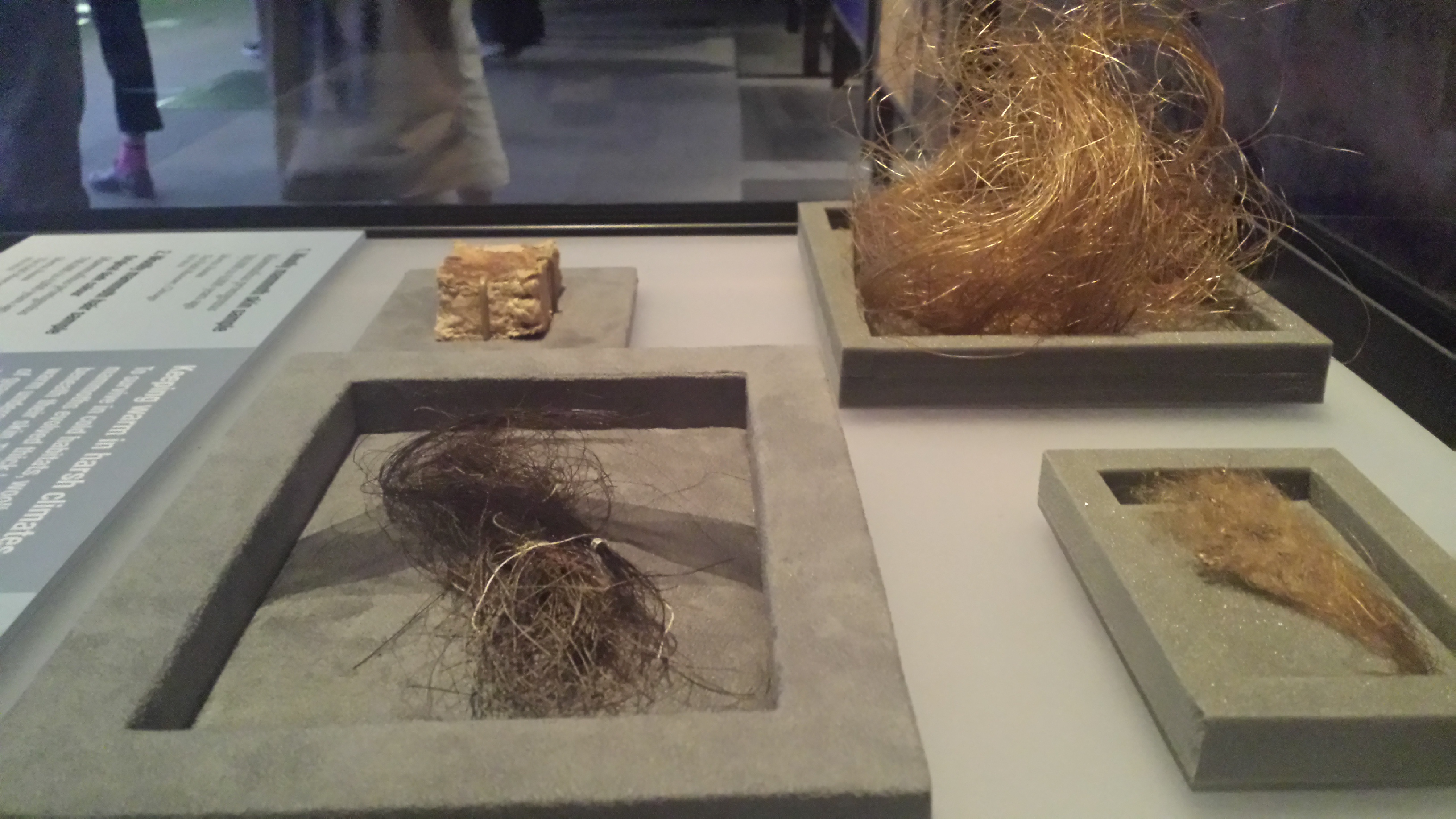
Mammoth hair! These samples and recent molecular studies show that mammoths were not ginger-coloured as we long thought, but rather the ginger color comes as the dark grey-brown-black colour fades postmortem, as a preservational artefact (story here). I didn’t know that; cool.

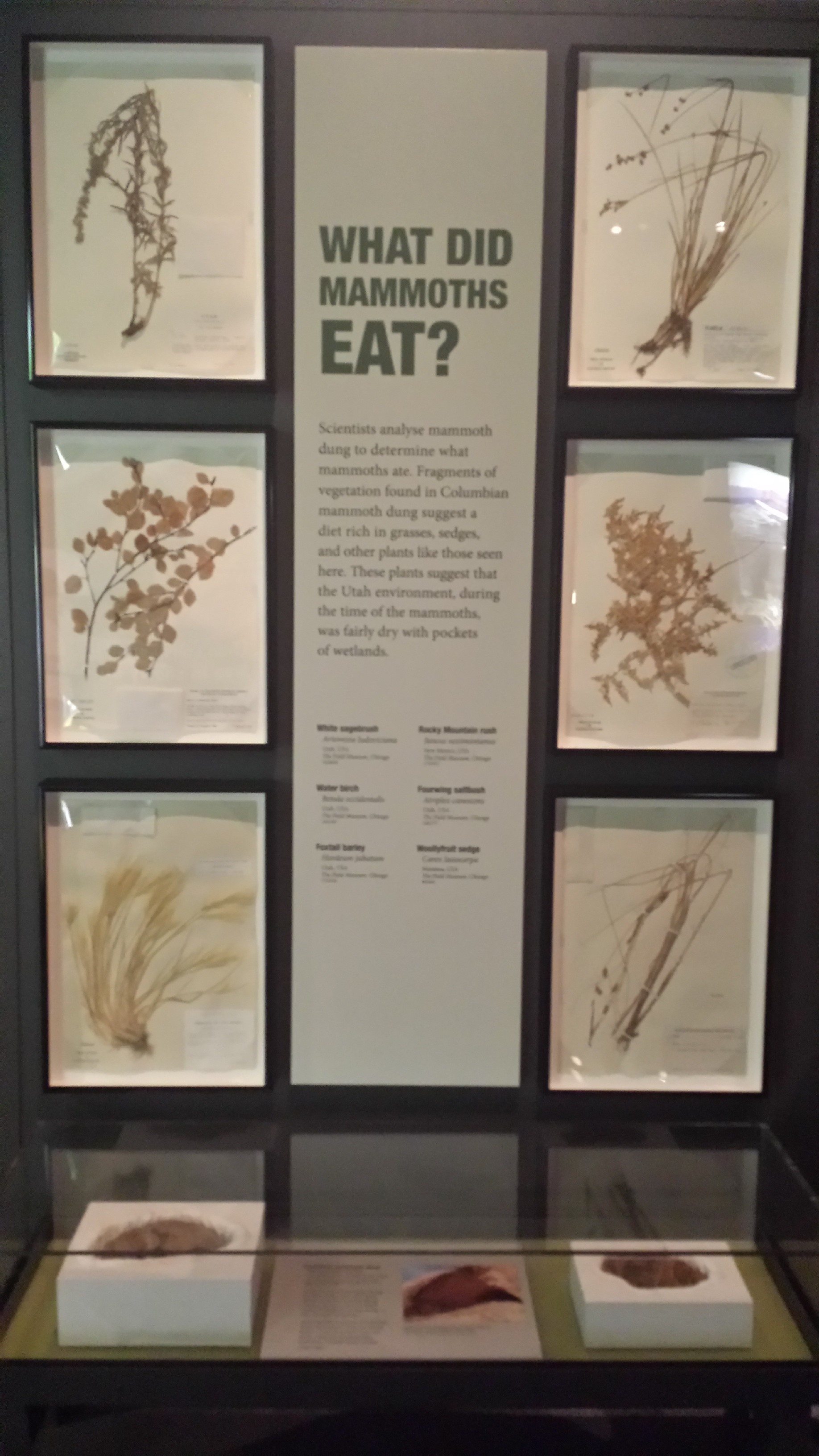
Mammoth chow! I liked this addition to the exhibit. This brought mammoth ecology closer to home for me.

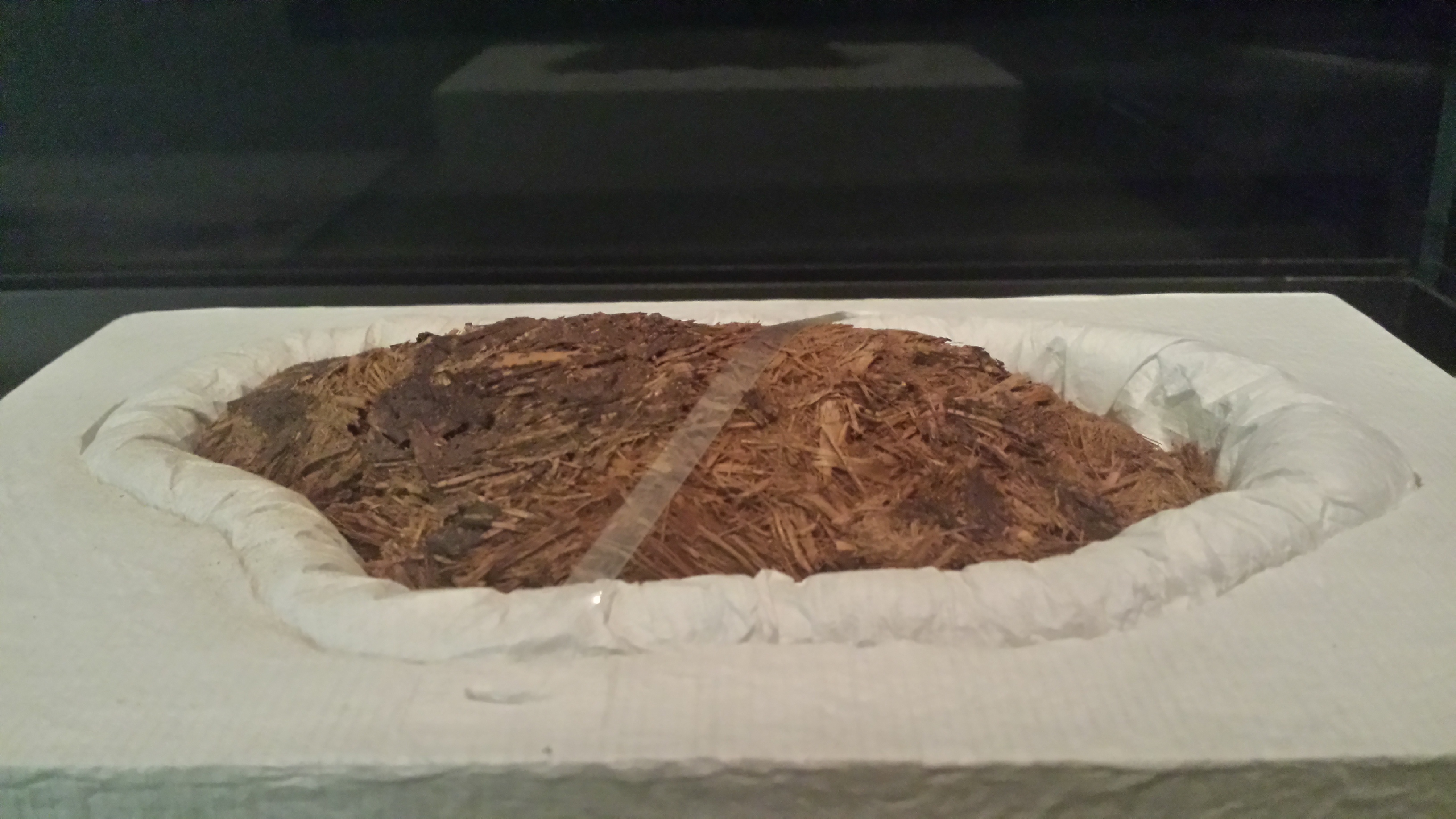
Mammoth poop!
After the biology explanations, let there be megafauna!

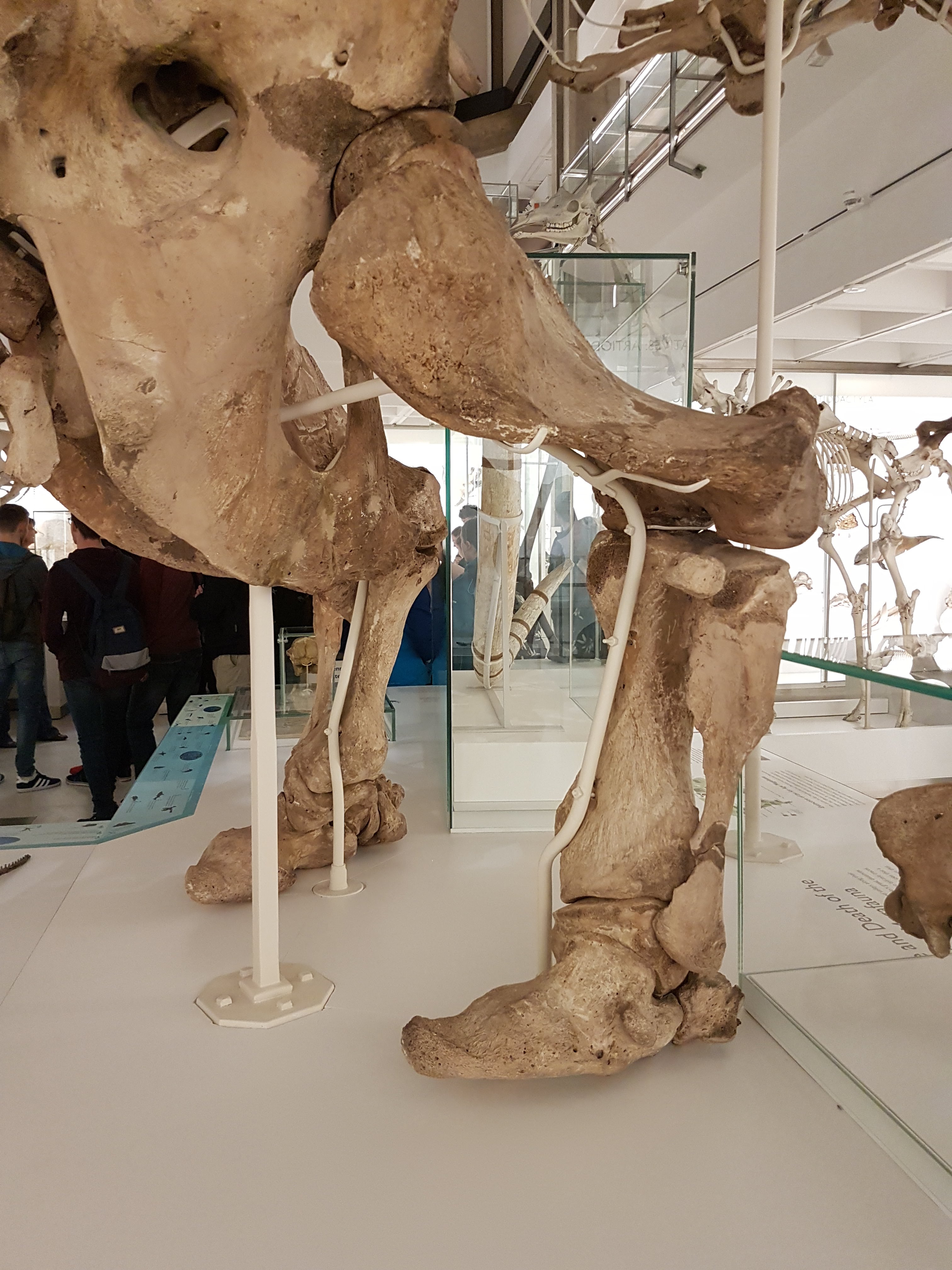
Mammoth skull! A nice one, too.

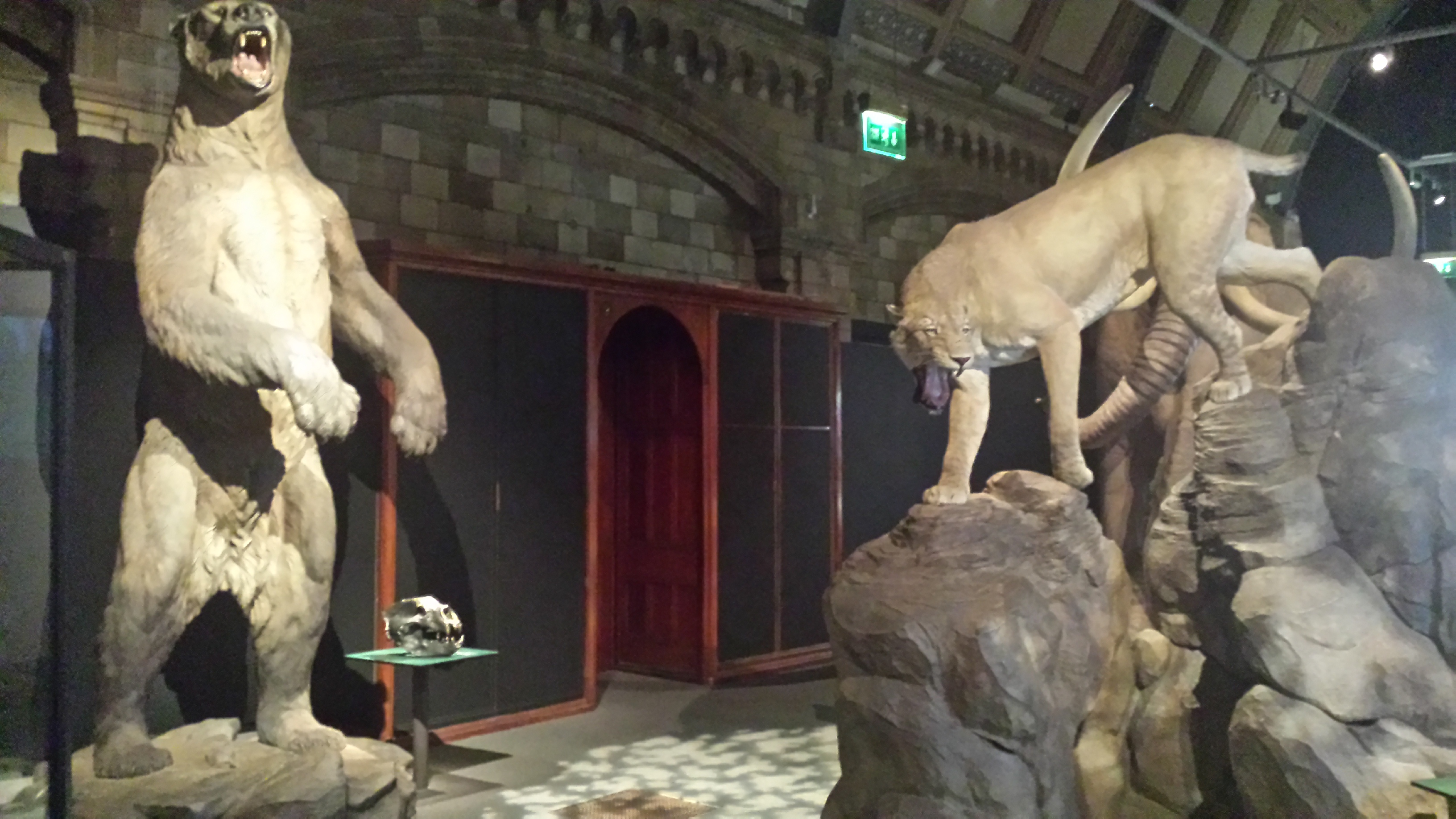
Top predators of Ice Age North America: Arctodus (short-faced bear– does the short face mean they were happy, unlike a long face? Sorry but they never are shown as very happy, unless it is the joy of whupass) and Homotherium (the other sabre-toothed cat; not the longer-toothed Smilodon).

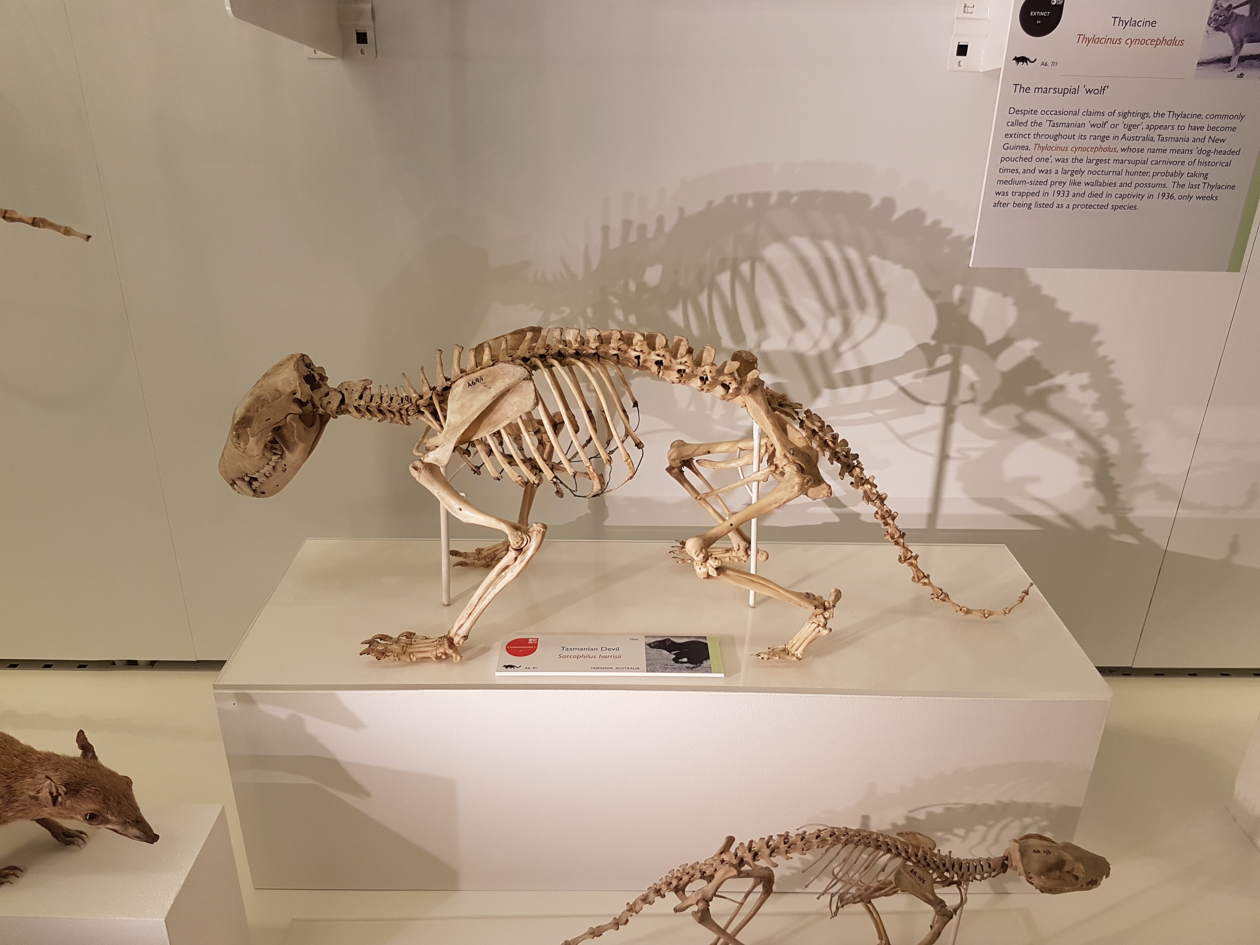
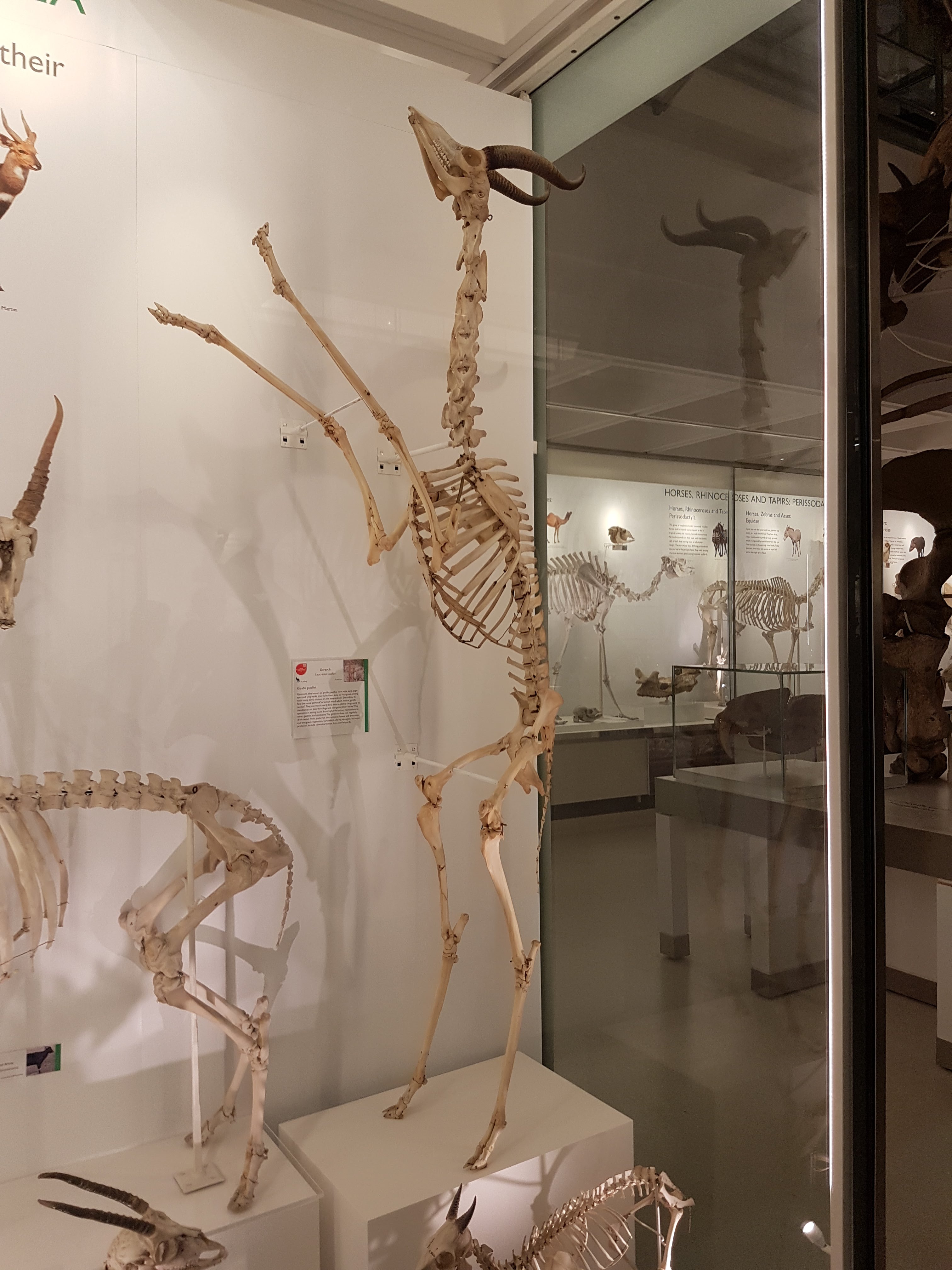
Skulls of North American (mega)fauna: left to right, top to bottom: horse, short-faced bear, giant ground sloth, then camel, sabretooth cat, rabbit, direwolf (viva Ned Stark!), and pronghorn antelope.

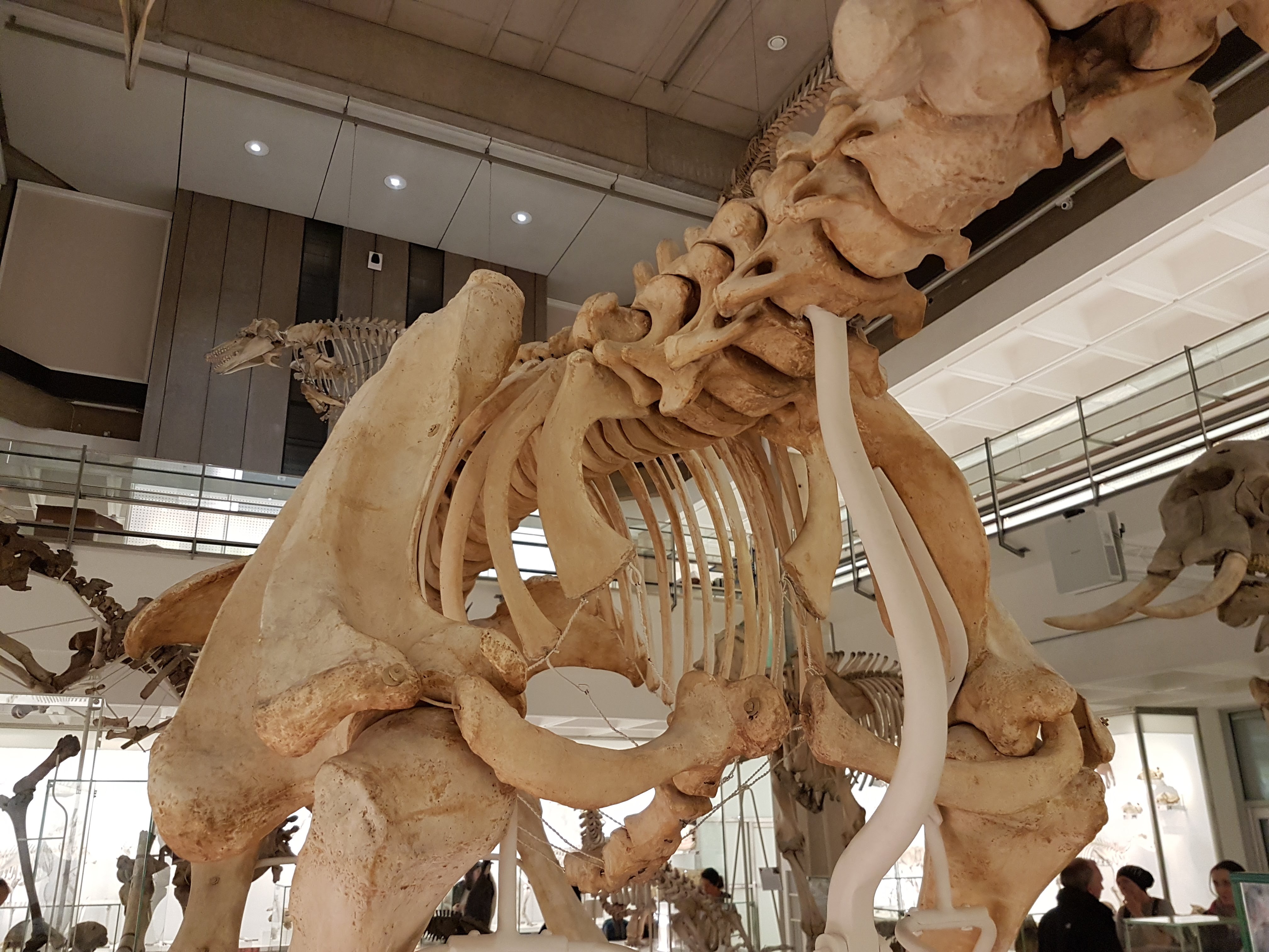
Mastodon (Mammut americanum) skeleton!

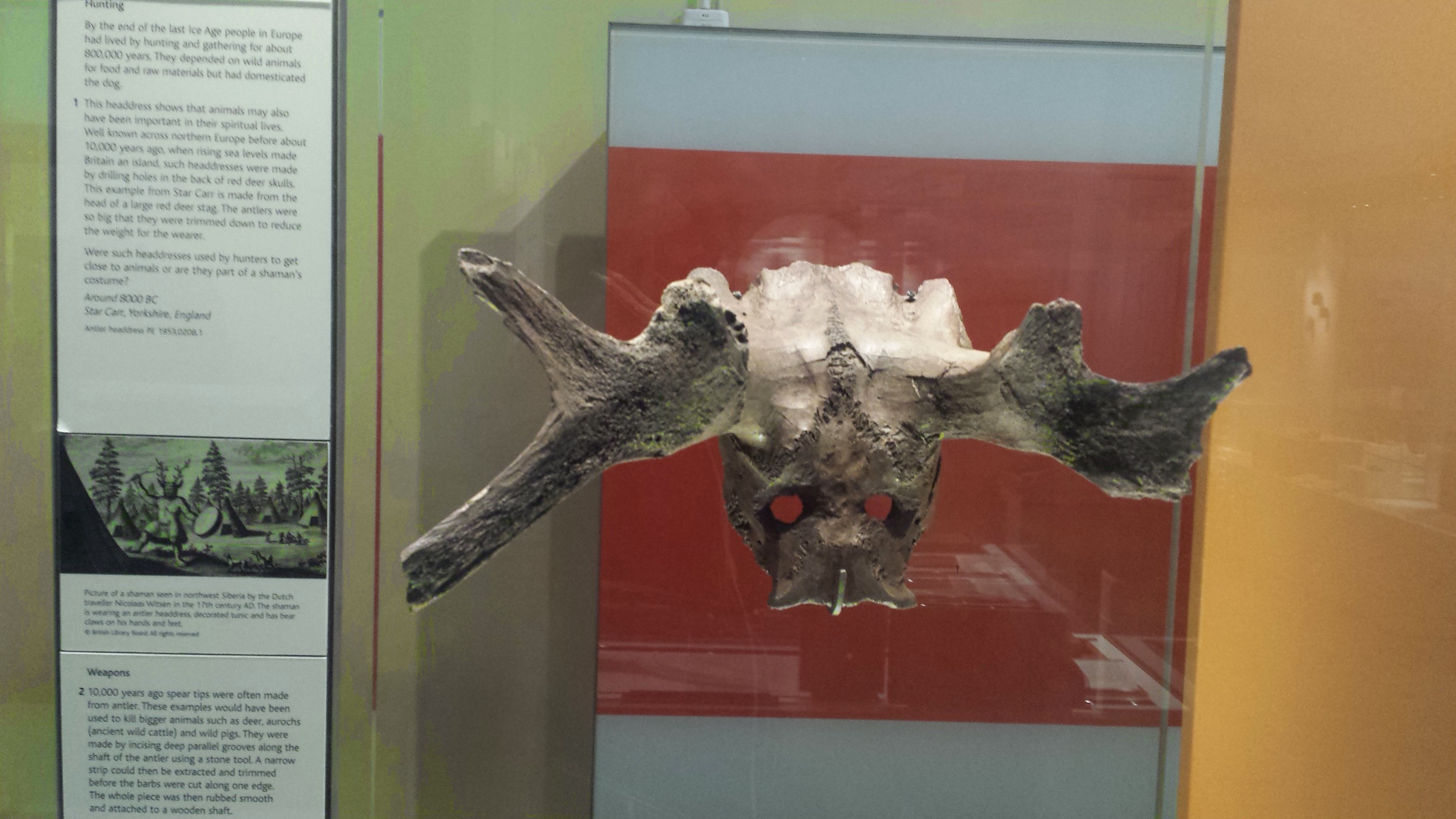
Mammoths (and perhaps mastodons, etc.) seem to have been wiped out by a combination of climate change and habitat fragmentation, combined with what this item symbolizes: human hunting. This beautiful piece is the main part of an atlatl, or javelin-hurling lever. It would have given Ice Age hunters the extra power they’d need to penetrate mammoth hide and cause mortal injuries. It is also a great tie-in to my recent post on the British Museum’s odd-animals-in-art.
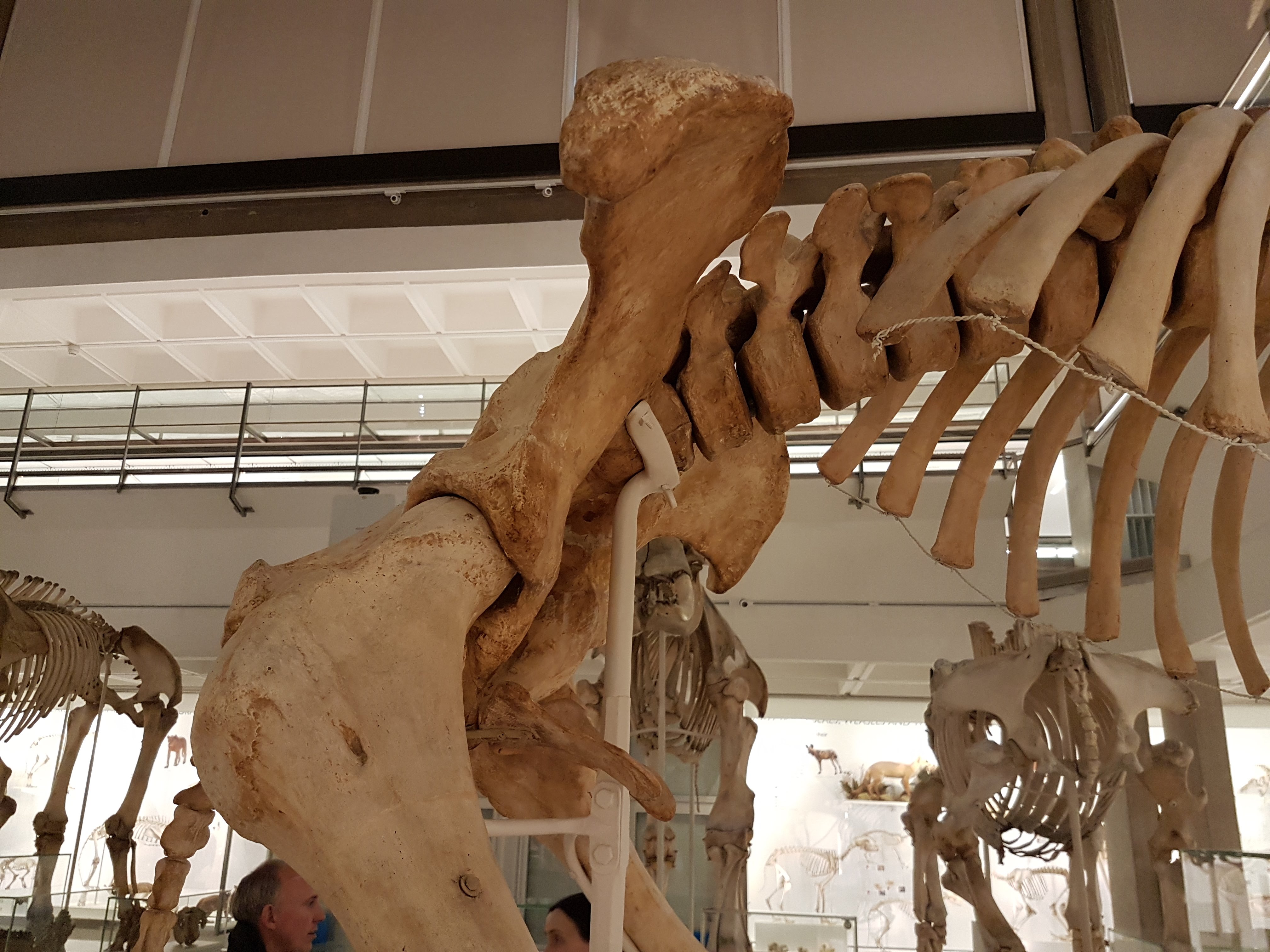
Finally, the exhibit surveys the kinds of mammoths that existed- there is a huge reconstruction of a Columbian mammoth near the mastodon (above), then smaller kinds and discussions of dwarfism, which is another strength of NHM mammoth research:

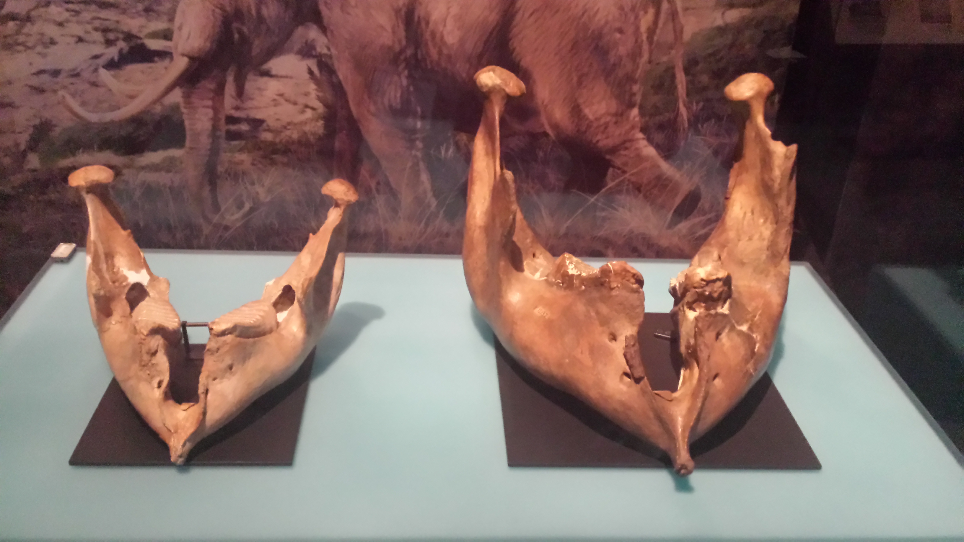
Woolly mammoth lower jaw (right) and its likely descendant, the pygmy mammoth of the Californian coastline, Mammuthus exilis.

The world’s smallest mammoth (left), molar tooth compared with that of its much larger ancestor Palaeoloxodon. The status of Mammuthus creticus as a dwarf mammoth from Crete was cemented by Victoria Herridge and colleagues, including Adrian Lister at the NHM.

Pygmy mammoth reconstruction. Shorter than me. I want one!

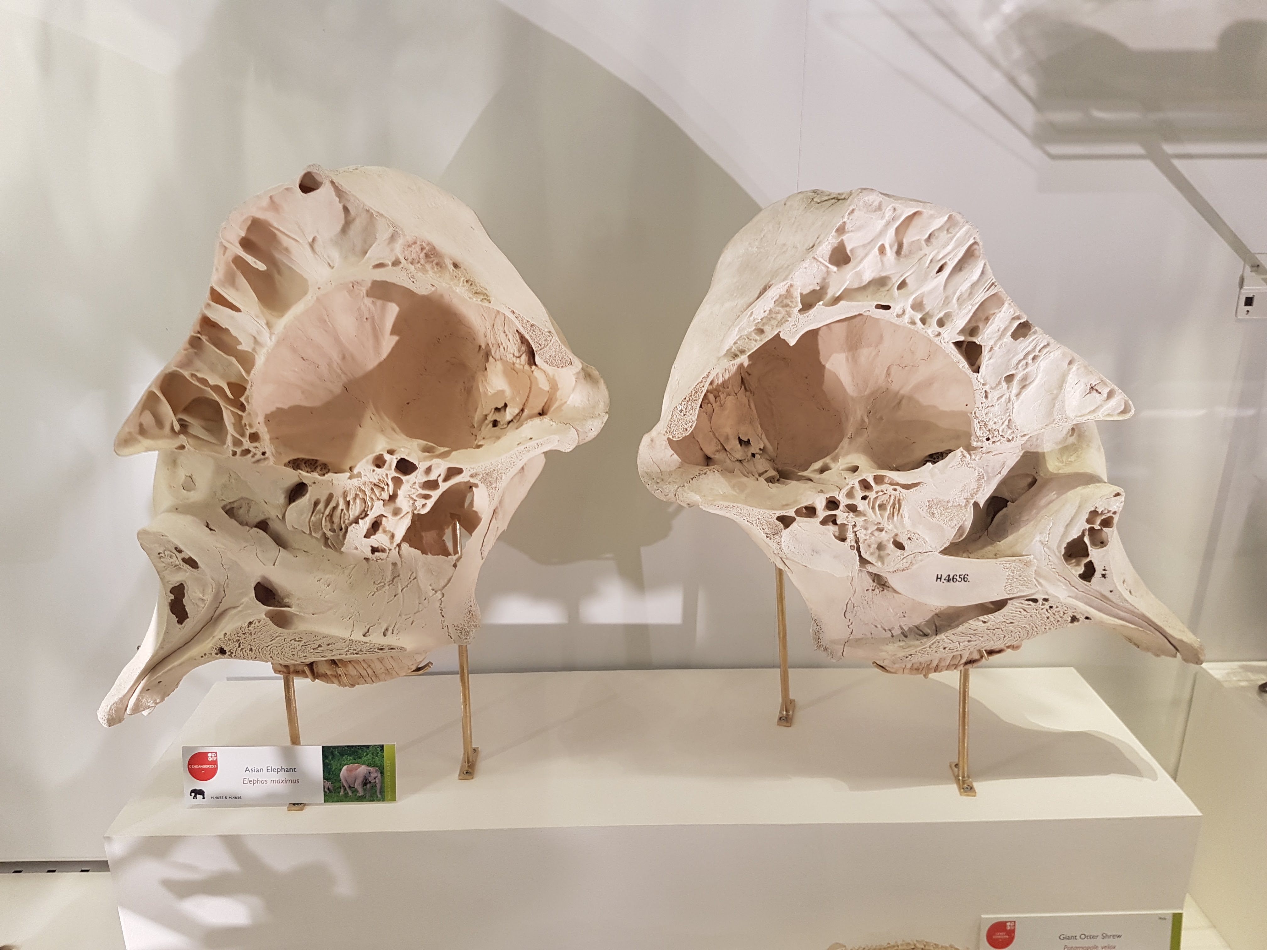
In the end, from all that glorious proboscidean diversity we were left with just 2 or 3 species of elephantids today (depending on your species concepts; it’s probably worth calling the African forest elephant its own species, Loxodonta cyclotis). The exhibit closes with a consideration of their conservation and fate. Ironically, this elephant skull could not be mounted with its tusks on display, because that would be commercializing ivory usage– even though the whole point of the exhibit’s denouement is to explain why elephants need protection!
Reactions to the exhibit: the photos tell the tale. It’s undeniably great, in terms of showing off the coolness of mammoths, other proboscideans and Ice Age beasties, to the general public. I felt like the factual content and learning potential was good. It didn’t feel at all like pandering to the lowest common denominator like some other exhibits I’ve seen (cough, Dino Jaws, cough). I loved the reconstructions, which were top quality in my opinion. I could have done with some more real skeletons, yet more realistically the exhibit hall was already large and full of cool stuff. But give me a break: Lyuba. This trumps everything. Going to see a real friggin’ frozen mammoth baby buries the needle of the awesomeness meter on the far right. That’s pretty much all I need to say. The spectacle was a spectacle.
This exhibit shows a lot of work, a lot of thought, and a personalized NHM touch that reflects the actual research (even very recent work!) that NHM staff like Prof. Lister are doing with collaborators around the globe. What more could we want, a herd of cloned mammoth babies frolicking around and tickling guests with their flanged trunks? Don’t hold your breath.
You’ve got just over 2 months to see the exhibit. Don’t come complaining on September 8 “BBBBBbbbut I didn’t know, I didn’t think it would be that cool! I just thought there’d be a guy in a Snuffleupagus suit signing autographs!” You have a duty as a Freezerino to go bask in the frozen glory of these Ice Age critters. There may be an exam at the end. 🙂
Is the exhibit kid-friendly? More or less. The text is more targeted at teenager-level or so, but the visual impact is powerful without it. I’d warn a sensitive child about the withered baby mammoth body before showing it to them, so they aren’t caught off guard and scarred by the experience. I saw plenty of kids in the exhibit and they all seemed happy. Parents may want to linger longer and absorb all the interesting information, whereas kids may blitz through or goof around, so plan accordingly if you’re inbound with sprogs.

You know what I was eyeing up in the gift shop…
Aside: The frozen mammoths get me wondering- what else does the Siberian (or extreme northern Canadian/Scandinavian) permafrost conceal? There are a lot of awesome Ice Age megafauna I’d cut my left XXXXX off to study quasi-intact… think about how amazing it would be to find a giant ground sloth (not bloody likely), sabretooth cat, or other species. There’s a lot of north up north. A lot of space and ice. A lot could happen. And climate change will make discoveries like this more likely, while the melting (and humanity) lasts…

Wool we ever find the Lyuba of woolly rhinos (Coelodonta)? Cast of a mummified woolly rhino from the NHM’s entry hall. More of these finds are likely, I’d say.
Read Full Post »