Happy Darwin Day from the frozen tundra sunny but muddy, frosty lands of England! I bring you limb muscles as peace offerings on this auspicious day. Lots of limb muscles. And a new theme for future blog posts to follow up on: starting off my “Better Know A Muscle” (nod to Stephen Colbert; alternative link) series. My BKAM series intends to walk through the evolutionary history of the coolest (skeletal/striated) muscles. Chuck Darwin would not enjoy the inevitable blood in this photo-tour, but hopefully he’d like the evolution. Off we go, in search of better knowledge via an evolutionary perspective!
There is, inarguably, no cooler muscle than M. caudofemoralis longus, or CFL for short. It includes the largest limb muscles of any land animal, and it’s a strange muscle that confused anatomists for many years– was it a muscle of the body (an axial or “extrinsic” limb muscle, directly related to the segmented vertebral column) or of the limbs (an “abaxial” muscle, developing with the other limb muscles from specific regions of the paraxial mesoderm/myotome, not branching off from the axial muscles)? Developmental biologists and anatomists answered that conclusively over the past century: the CFL is a limb muscle, not some muscle that lost its way from the vertebral column and ended up stranded on the hindlimb.
The CFL is also a muscle that we know a fair amount about in terms of its fossil record and function, as you may know if you’re a dinosaur fan, and as I will quickly review later. We know enough about it that we can even dare to speculate if organisms on other planets would have it. Well, sort of…
Stomach-Churning Rating: 8/10. Lots of meaty, bloody, gooey goodness, on and on, for numerous species. This is an anatomy post for those with an appetite for raw morphology.
Let’s start from a strong (and non-gooey) vantage point, to which we shall return. The CFL in crocodiles and most other groups is (and long was) a large muscle extending from much of the front half or so of the tail to the back of the femur (thigh bone), as shown here:
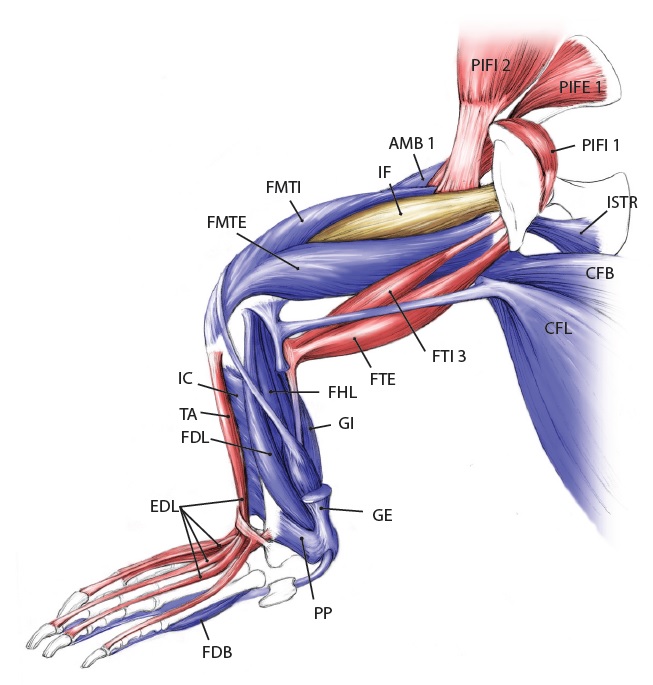
Julia Molnar’s fabulous illustration of Alligator‘s limb muscles, from our 2014 paper in Journal of Anatomy. Note the CFL in blue at the bottom right.
As the drawing shows, the CFL has a friend: the CFB. The CFB is a shorter, stumpier version of the CFL restricted to the tail’s base, near the hip. The “B” in its name means “brevis”, or runty. It gets much less respect than its friend the CFL. Pity the poor CFB.
But look closer at the CFL in the drawing above and you’ll see a thin blue tendon extending past the knee to the outer side of the lower leg. This is the famed(?) “tendon of Sutton“, or secondary tendon of the CFL. So the CFL has two insertions, one on the femur and one (indirectly) onto the shank. More about that later.
Together, we can talk about these two muscles (CFL and CFB) as the caudofemoralis (CF) group, and the name is nice because it describes how they run from the tail (“caudo”) to the femur (“femoralis”). Mammal anatomists were late to this party and gave mammal muscles stupidly unhelpful names like “gluteus” or “vastus” or “babalooey”. Thanks.
But enough abstract drawings, even if they rock, and enough nomenclature. Here is the whopping big CFL muscle of a real crocodile:
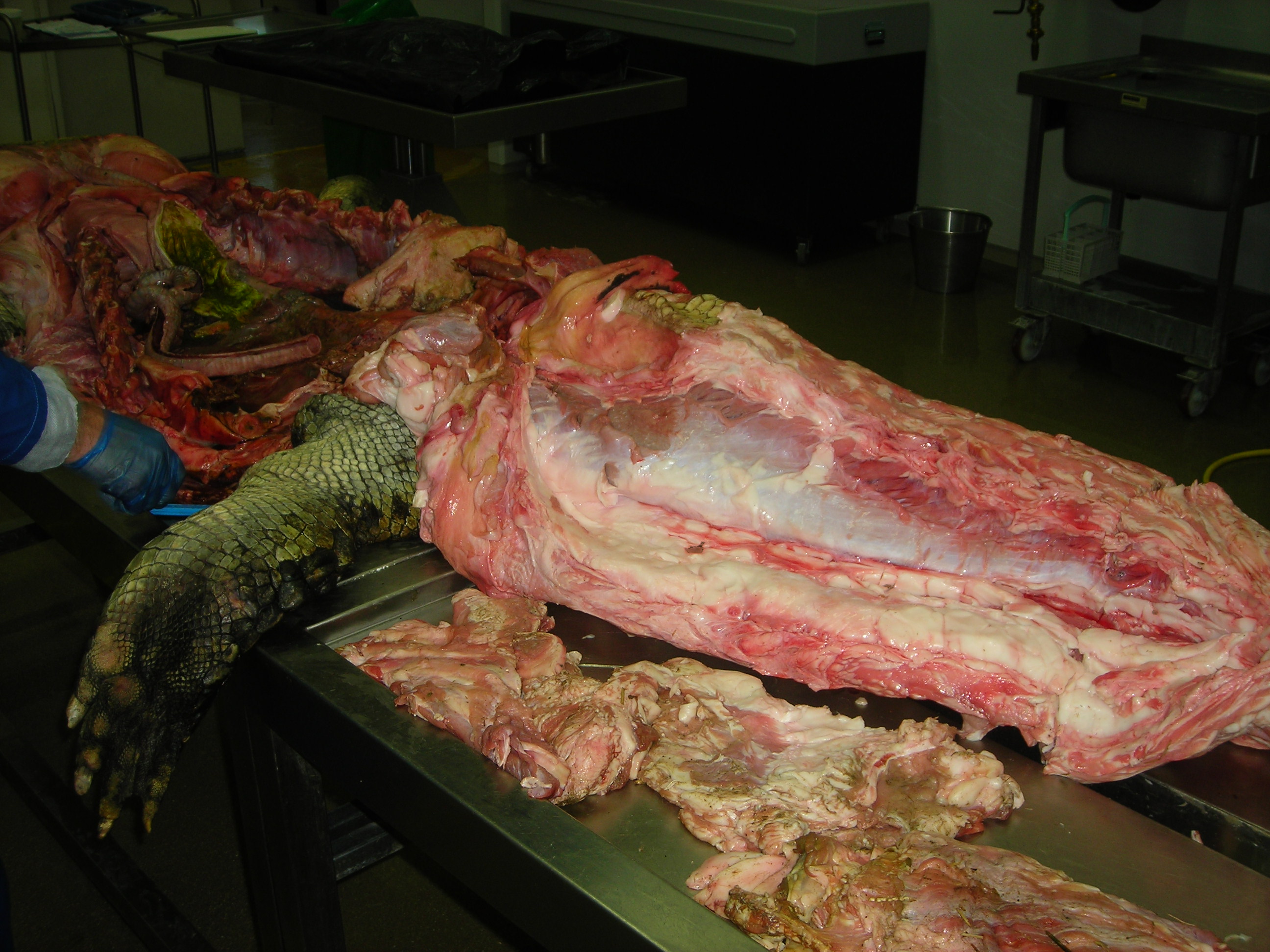
Huge Nile crocodile, but a relatively small CFL.

Bigger crocs have smaller legs and thus smaller leg muscles, relatively speaking. CFL at the top, curving to the left.

The giant Nile croc’s CFL muscle removed for measurements. 2.35 kg of muscle! Not shabby for a 278 kg animal.
However, maybe crocodile and other archosaur CFL muscles are not “average” for leggy vertebrates? We can’t tell unless we take an evolutionary tack to the question.
Where did the CFL come from, you may ask? Ahh, that is shrouded in the fin-limb transition‘s mysteries. Living amphibians such as salamanders have at least one CF muscle, so a clear predecessor to the CFL (and maybe CFB) was present before reptiles scampered onto the scene.
But going further back through the CF muscles’ history, into lobe-finned fish, becomes very hard because those fish (today) have so few fin muscles that, in our distant fishy ancestors, would have given rise eventually to the CF and other muscle groups. With many land animals having 30+ hindlimb muscles, and fish having 2-8 or so, there obviously was an increase in the number of muscles as limbs evolved from fins. And because a limb has to do lots of difficult three-dimensional things on land while coping with gravity, more muscles to enable that complex control surely were needed.
OK, so there were CF muscles early in tetrapod history, presumably, anchored on that big, round fleshy tail that they evolved from their thin, finned fishy one — but what happened next? Lizards give us some clues, and their CFL muscles aren’t all that different from crocodiles, so the CFL’s massive size and secondary “tendon of Sutton” seems to be a reptile thing, at least.

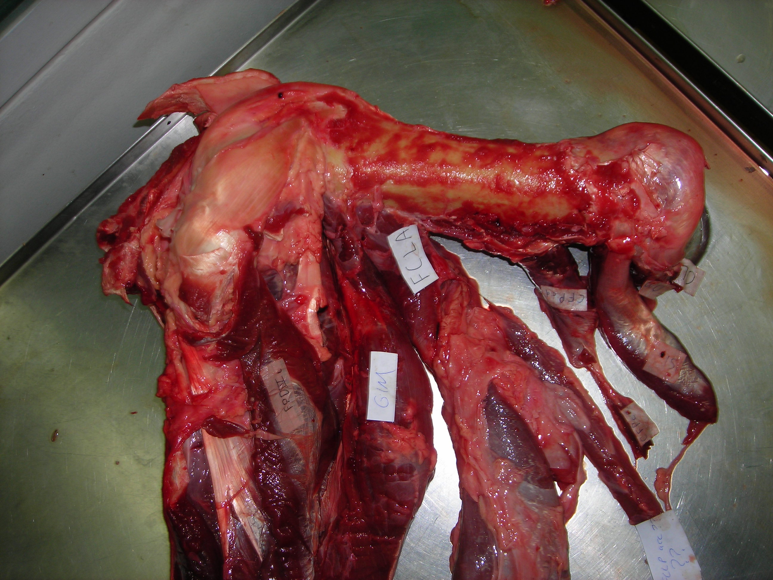
Courtesy of Emma Schachner, a large varanid lizard’s very freshly preserved CFL and other hindlimb muscles.
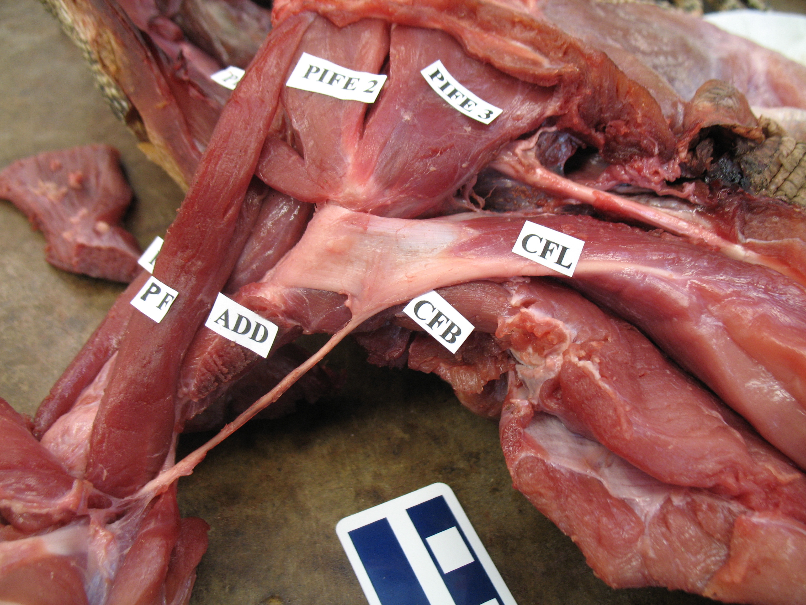
Courtesy of Emma Schachner, zoomed in on the tendons and insertions of the CFL muscle and others. Beautiful anatomy there!
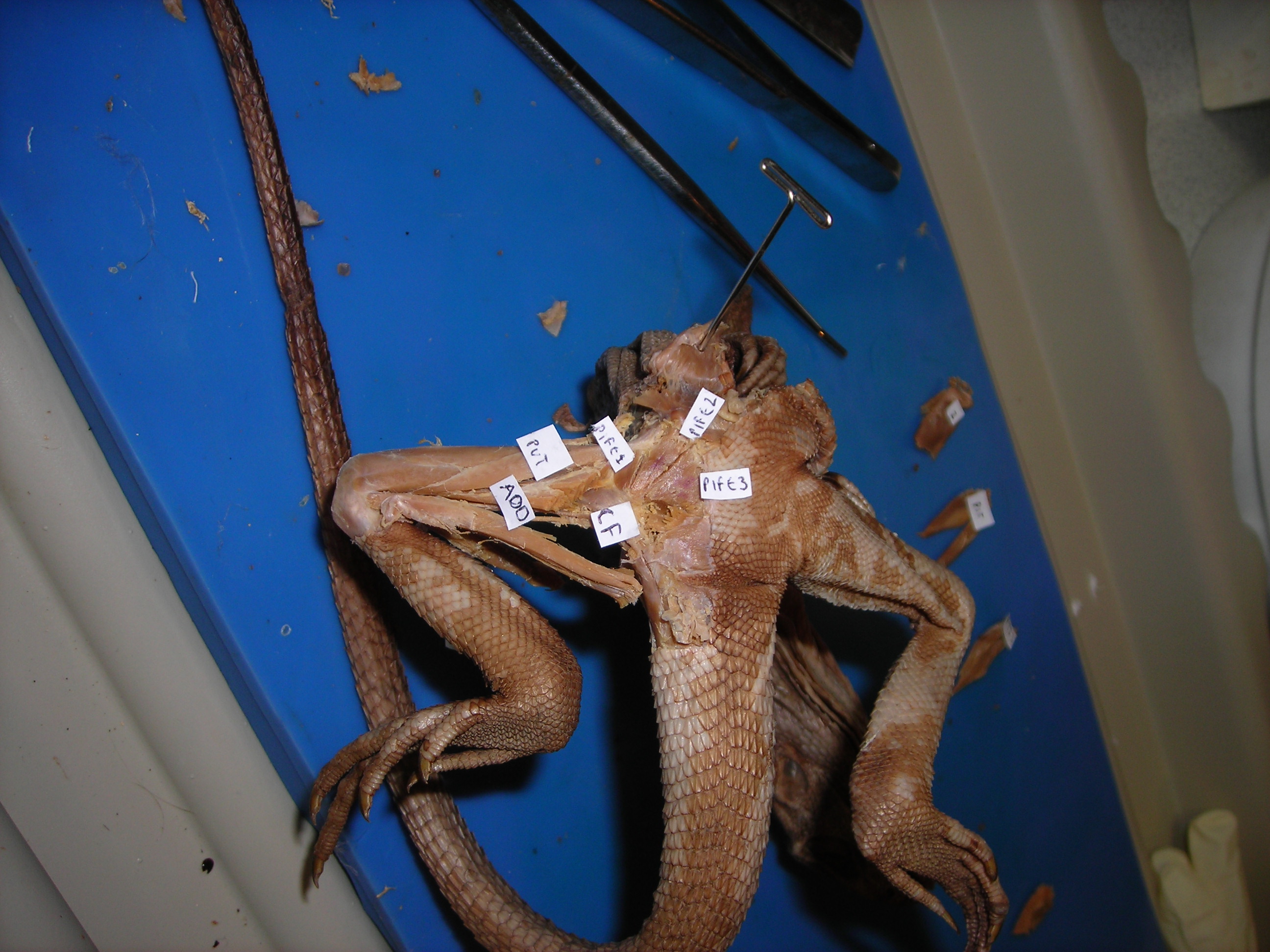
Looking up at the belly of a basilisk lizard and its dissected right leg, with the end of the CFL labelled. It’s not ideally dissected here, but it is present.

An unspecified iguanid(?) lizard, probably a juvenile Iguana iguana, dissected to reveal its CFL muscle near its attachment to the femur. The muscle would extend further, about halfway down the tail, though.
Let’s return to crocodiles, for one because they are so flippin’ cool, and for another because they give a segue into archosaurs, especially dinosaurs, and thence birds:

A moderate-sized (45kg) Nile crocodile with its CFL muscle proudly displayed. Note the healthy sheath of fat (cut here) around the CFL.

American alligator’s CFL dominates the photo [by Vivian Allen].
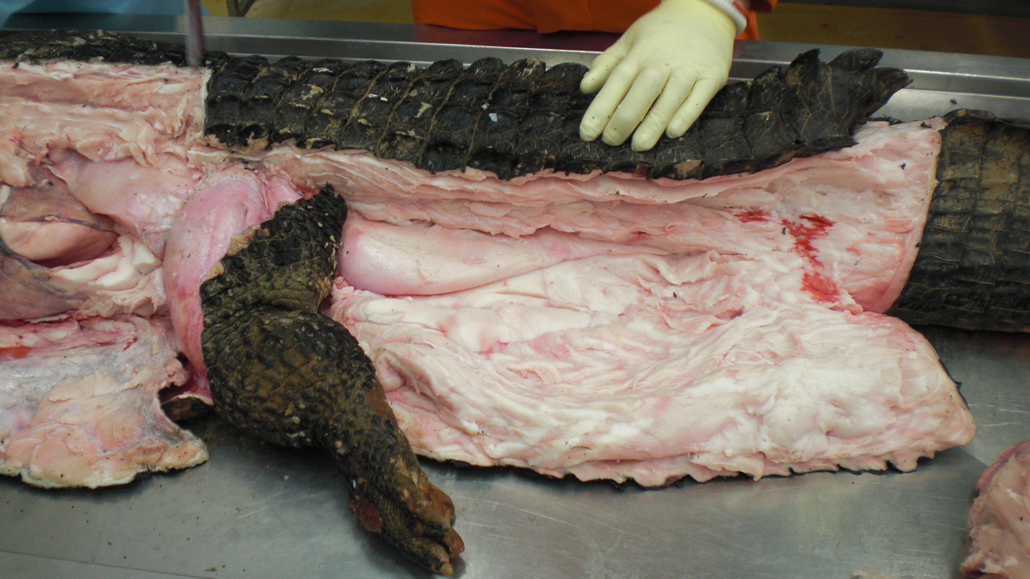
Black caiman, Melanosuchus, showing off its CFL muscle (pink “steak” in the middle of the tail near the leg), underneath all that dark armour and fatty superficial musculature.

A closer look at the black caiman’s thigh and CFL muscle.
Like I hinted above, crocodiles (and the anatomy of the CFL they share with lizards and some other tetrapods) open a window into the evolution of unusual tail-to-thigh muscles and locomotor behaviours in tetrapod vertebrates.
Thanks in large part to Steve Gatesy’s groundbreaking work in the 1990s on the CFL muscle, we understand now how it works in living reptiles like crocodiles. It mainly serves to retract the femur (extend the hip joint), drawing the leg backwards. This also helps support the weight of the animal while the foot is on the ground, and power the animal forwards. So we call the CFL a “stance phase muscle”, referring to how it mainly plays a role during ground contact and resisting gravity, rather than swinging the leg forwards (protracting the limb; i.e. as a “swing phase muscle”).
The “tendon of Sutton” probably helps to begin retracting the shank once the thigh has moved forward enough, facilitating the switch from stance to swing phase, but someone really needs to study that question more someday.
And thanks again to that same body of work by Gatesy (and some others too), we also understand how the CFL’s anatomy relates to the underlying anatomy of the skeleton. There is a large space for the CFL to originate from on the bottom of the tail vertebrae, and a honking big crest (the fourth trochanter) on the femur in most reptiles that serves as the major attachment point, from which the thin “tendon of Sutton” extends down past the knee.

Femur bones (left side; rear view) from an adult ostrich (left) and Nile crocodile (right). Appropriate scale bar is appropriate. The fourth trochanter for the CFL is visible in the crocodile almost midway down the femur. Little is left of it in the ostrich but there is a bumpy little muscle scar in almost the same region as the fourth trochanter, and this is where the same muscle (often called the CFC; but it is basically just a small CFL) attaches.
That relationship of the CFL’s muscular anatomy and the underlying skeleton’s anatomy helps us a lot! Now we can begin to look at extinct relatives of crocodiles; members of the archosaur group that includes dinosaurs (which today we consider to include birds, too), and things get even more interesting! The “tendon of Sutton”, hinted at by a “pendant” part of the fourth trochanter that points down toward the knee, seems to go away multiple times within dinosaurs. Bye bye! Then plenty more happens:

A large duckbill dinosaur’s left leg, with a red line drawn in showing roughly where the CFL would be running, to end up at the fourth trochanter. Many Mesozoic dinosaurs have skeletal anatomy that indicates a similar CFL muscle.
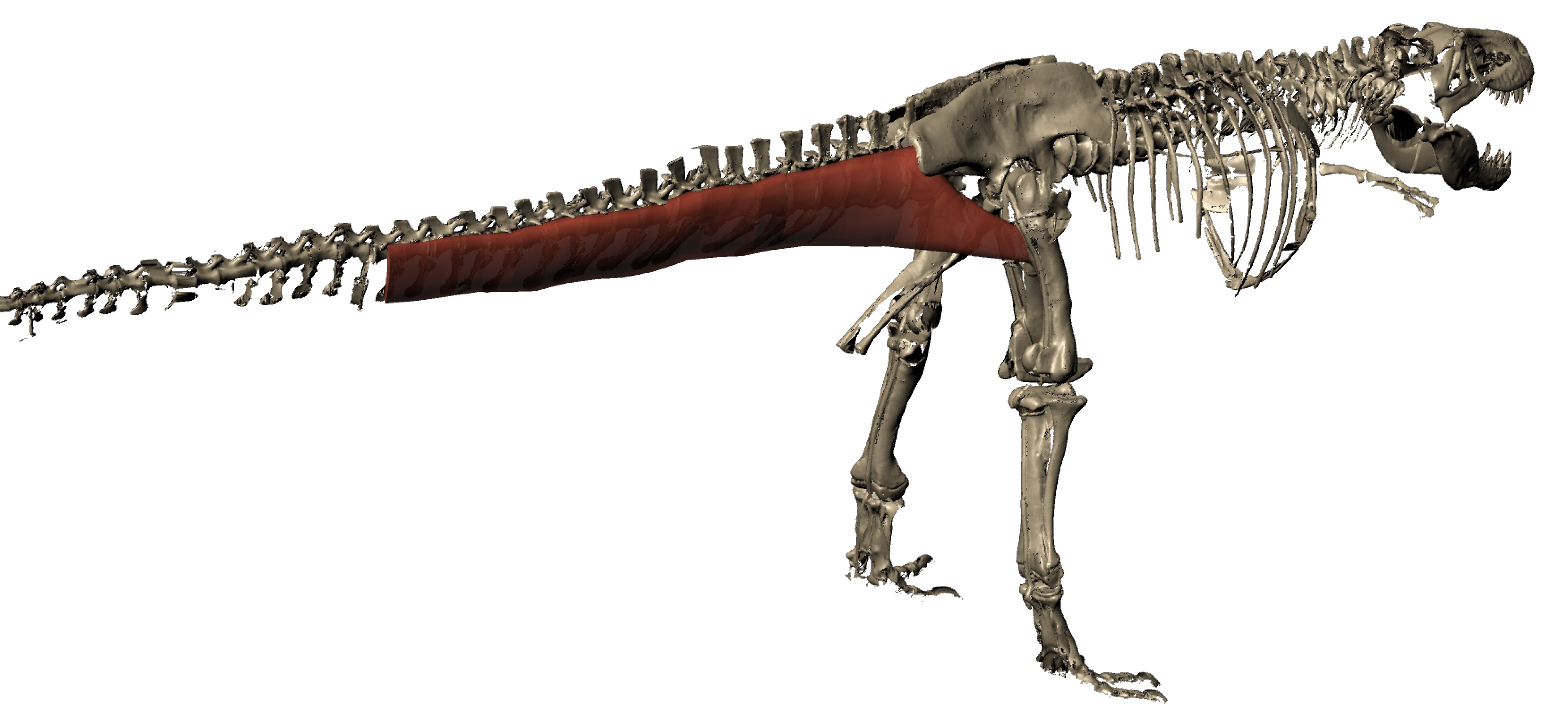
We can even go so far as to reconstruct the 3D anatomy of the CFL in a dinosaur such as T. rex (“Sue” specimen here; from Julia Molnar’s awesome illustration as part of our 2011 paper), with a fair degree of confidence. >180kg steak, anyone?
As we approach birds along the dinosaur lineage, the tail gets smaller and so does the fourth trochanter and thus so must the CFL muscle, until we’re left with just a little flap of muscle, at best. In concert, the hindlimbs get more crouched, the forelimbs get larger, flight evolves and voila! An explosion of modern bird species!
Left femur of an ostrich in side view (hip is toward the right side) showing many muscles that attach around the knee (on the left), then the thin strap of CF muscle (barely visible; 2nd from the right) clinging near the midshaft of the femur.

Another adult ostrich’s CF muscle complex, removed for study. Not enough ostrich myology for you yet? Plenty more in this old post! Or this one! Or this one… hey maybe I need to write less about ostriches? The CF muscle complex looks beefy but it’s no bigger than any other of the main hindlimb muscles, unlike the CFL in a crocodile or lizard, which puts everything else to shame!
STILL not enough ostrich for you yet? Take a tour of the major hindlimb muscles in this video:
And check out the limited mobility of the hip joint/femur here. No need for much femur motion when you’re not using your hip muscles as much to drive you forwards:
But I must move on… to the remainder of avian diversity! In just a few photos… Although the CF muscles are lost in numerous bird species, they tend to hang around and just remain a long, thin, unprepossessing muscle:
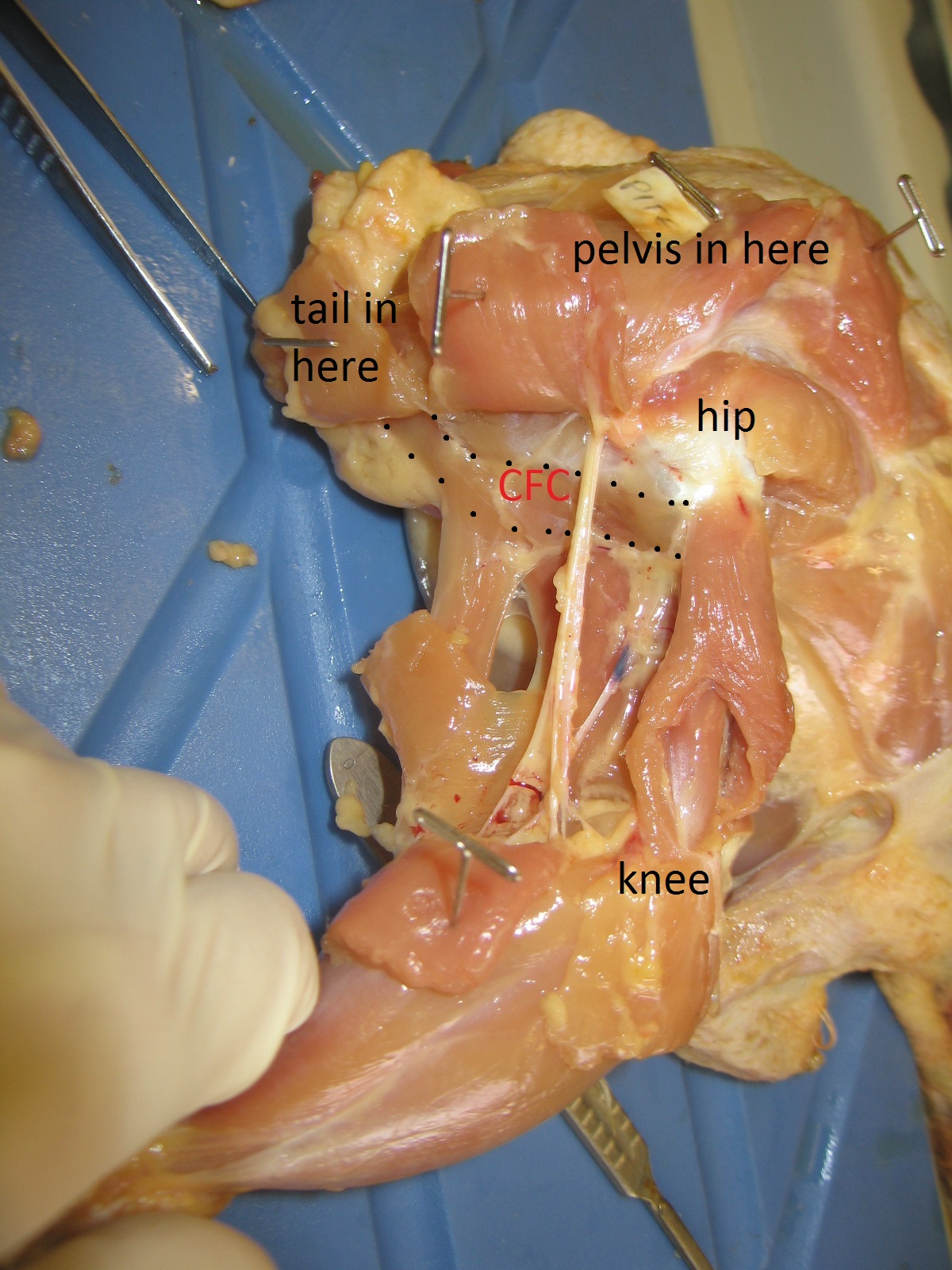
Chicken’s right leg in side view. CFC muscle (equivalent of CFL; the ancestral CFB is confusingly called the CFP in birds, as it entirely resides on the pelvis) outlined and labelled.

A jay (species? I forget) dissected to show some of the major leg muscles, including the CFL-equivalent muscle; again, smallish. [Photo by Vivian Allen]
But many mammals do still have something that is either called the M. caudofemoralis or is likely the same thing, albeit almost always fairly modest in size. This evolutionary reduction of the CF muscle along the mammal (synapsid) lineage hasn’t gotten nearly as much attention as that given to the dinosaur/bird lineage’s CFL. Somebody should give it a thoroughly modern phylogenetic what-for! Science the shit outta that caudofemoralis…
Yet, oddly, to give one apparent counter-example, cats (felids) have, probably secondarily, beefed up their CF muscle a bit:
Cats have a pretty large CF muscle in general, and this jaguar is no exception! But mammals still tend to have fairly wimpy tails and thus CF muscles, or they even lose them (e.g. us?). [photo by Andrew Cuff, I think]

Better Know A Muscle: the evolution of M. caudofemoralis (longus).
I hope you enjoyed the first BKAM episode!
I am willing to hear requests for future ones… M. pectoralis (major/profundus) is a serious contender.
P.S. It was Freezermas this week! I forgot to mention that. But this post counts as my Freezermas post for 2016; it’s all I can manage. Old Freezermas posts are here.


Thank you John! I will really be following this series!
This is a much-welcome post. The caudofemoralis definitely deserves some spotlight.
Question: I had been under the impression that the fourth trochanter is an archosaur feature. Your text seems to imply that it is also present in other reptiles. Is that the case, or have I misunderstood?
Thanks David! It’s complicated, but yes the fourth trochanter is technically only present in archosaurs and their closest kin. However, what did it come from? That’s a paper we’ve been meaning to write. 🙂
Neat, enjoyed this very much. I wonder if the ‘unspecified iguanid’ is an agamid, perhaps Calotes?
Dangit, I was wondering… I am not good with my agamids/iguanids/gekkonids, and it came in a jar w/no info on the specimens inside. Calotes does sound plausible!
Thank you, fine!
If birds came from crocs, they would fly with the tail driven by CFL instead of pectoralis. Wow, wouldn’t they fly on a side, like a flounder? Left or right? – that is the question!
And if lizards came from crocs, the predators who ate them would be happy with their autotomy.
But one note: the “CF” of jaguar is femorococcygeus in fact, which is part of the dorsal musculature of the thigh (together with gluteus). The true mammalian CF belongs to ventral musculature, it is buried deeper, originates from transverse tail processes (contrary to superficial femorococcygeus, which runs from neural spines), was often called “praesemimembranosus” and corresponds to caudofemoralis brevis of crocs and lizards. Mammalian caudofemoralis longus is problematic. The best idea was suggested by Romer, who noticed similarity between the vertebral head of mammalian semitendinosus and urodelan caudalipuboischiotibialis, which is definitely homologous to reptilian CFL.
Ahh, very interesting (and fun) points, thanks Alex! 🙂 I don’t know the mammal side of the CF literature as well but that makes a lot of sense to me- e.g. in cats the CF is so superficial and anterior that it does seem strange as a homologue to CF in reptiles. I’ll have to read up more on that sometime but it certainly seems like the “CF” in mammals (and amphibians) needs some renewed study, like I wrote at the end of the post. Regardless, whatever the CF is in mammals, it is small relative to reptiles– but what the primitive condition of the CF for tetrapods/amniotes was still appears open to interpretations, too.
[…] in how much they rely on their hamstring muscles to power locomotion (at the hip) rather than their caudofemoral muscles, which are reduced. Zooming in on some particular muscles such as parts of the hip or knee extensors, the functions […]
Pumped to see more in-depth muscle posts like this! It triggered many random thoughts…
– I watched that ostrich hip motion video several times… mesmerising fresh anatomy!
– I was interested in Alex’s comment… So in mammals, is CF dorsal or ventral in origin? And so what action would CF have on the tail if the lower limb is fixed; lateral flexion + depression? Is it absent in cetaceans?
– This post reminded me of this paper [http://www.publish.csiro.au/?paper=ZO13085] describing kangaroo tail muscles, although CF isn’t dwelt on, I guess because it’s a limb muscle as you mentioned. Still, kind of interesting to think about, as kangaroos use their tail a bit more than most mammals.
Thanks for that paper, and nice to see “old” blog posts here still getting some love! It has been quiet lately in the comments sections– my fault, I assume! On the tail muscles, thanks for that kangaroo paper, I had missed it! I will have to read it and see what it says. An important point when inferring whether kangaroo tail muscles are adaptations or otherwise would be to compare their anatomy to those of related, non-hopping species and see how that differs; but also to test how the anatomy functions in a biomechanical context (e.g. does it actually help or does it just look like it might?). But I would not be surprised if it played some role, although the question of whether it would help 1% or 10% vs. other possible anatomies would be interesting (and hard) to test. As for Alex’s comment, as I recall the “CF” in mammals is dorsal in origin. I think there still needs to be more work on the homology of mammal vs. other tetrapod limb muscles. A lot of studies, including recent ones, cite a small amount of literature (often the same few studies) and scant developmental data, much of it very old (but some of it very good, too!). New study might change things around by contributing new observations with high-resolution data. Cetaceans: no idea! But if lower limb stays still, then CF action should involve extending (retracting; drawing backwards) the thigh and probably a bit of abduction (drawing the thigh laterally).
I believe the jay to be a eurasian jay 😉
[…] 6. Bat muscles (http://pixgood.com/bat-muscle-anatomy.html) Picture 7. Lizard’s legs muscles (https://whatsinjohnsfreezer.com/2016/02/12/bkam1_cfl/) Picture 8. Building form side view Picture 9. Building form back view Picture 10. Building form […]
What is its equivalent in humans? Do they have one?
Not really an equivalent but quadratus femoris and similar short muscles around the hip are close.
Thank you!