Feeds:
Posts
Comments

Archive for the ‘Exalting Archosauria’ Category

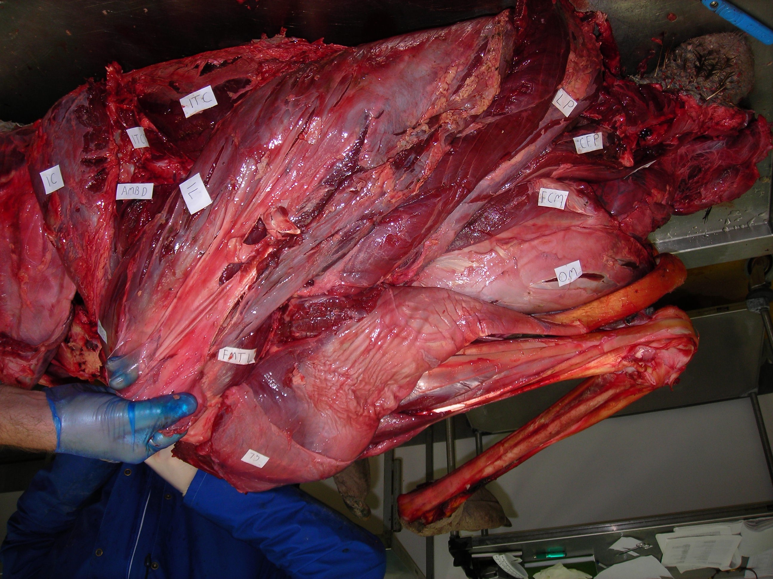
We released a publication that, for me, comes full circle with research that started my career off. Back in 1995 when I started my PhD, I thought it would be great to use biomechanical models and simulations to test how extinct dinosaurs like Tyrannosaurus rex might have moved (or not), taking Jurassic Park CGI animations (for which the goal was to look great) into a more scientific realm (for which the goal is to be “correct”, even at the cost of beauty). “It would be great”, or so I thought, haha. I set off on what has become a ~26 year journey where I tried to build the evidence needed to do so, at each step trying to convince my fairly sceptical mind that it was “good enough” science. For my PhD I mainly focused on reconstructing the hindlimb muscles and their evolution, then using very simple “stick figure”, static biomechanical models of various bipeds to test which could support fast running with their leg muscles, culminating in a 2002 Nature paper that made my early career. I since wrote a long series of papers with collaborators to build on that work, studying muscle moment arms, body/segment centre of mass, and finally a standardized “workflow” for making 3D musculoskeletal models. And gradually we worked with many species, mostly living ones, to simulate walking and running and estimate how muscles controlled observed motions and forces from experiments. This taught us how to build better models and simulations. Now, in 2021, our science has made the leap forward I long hoped for, and the key thing for me is that I believe enough of it is “good enough” for me, which long held me back. This is thus my personal perspective. We have a press release that gives the general story for public consumption; here I’ve written for more of a sciencey audience.

Skeleton of the extinct theropod Coelophysis in a running pose, viewed side-on. Image credit: Scott Hocknull, Peter Bishop, Queensland Museum.

Stomach-Churning Rating: 1/10: just digital muscles.

Earlier in 2021, we simulated tinamou birds in two papers (first one here), the second one revealing our first ever fully predictive simulations, of jumping and landing; detailed here and with a nice summary article here. That research was led by DAWNDINOS postdoc Peter Bishop and featuring new collaborators from Belgium, Dr. Antoine Falisse and Prof. Friedl De Groote. Thanks to the latter duo’s expertise, we used what is called direct collocation (optimal control) simulation; which is faster than standard “single-shooting” forward dynamic simulation. The simulations also were fully three-dimensional, although with some admitted simplifications of joints and the foot morphology; much as even most human simulations do. The great thing about predictive simulations is that, unlike tracking/inverse simulations (all of my prior simulation research), it generates new behaviours, not just explaining how experimentally observed behaviours might have been generated by neuromuscular control.

OK, so what’s this new paper really about and why do I care? We first used our tinamou model to predict how it should walk and sprint, via some basic “rules” of optimal control goals. We got good results, we felt. That is the vital phase of what can be called model “validation”, or better termed “model evaluation”; sussing out what’s good/bad about simulation outputs based on inputs. It was good enough overall to proceed with a fossil theropod dinosaur, we felt.

Computer simulation of modern tinamou bird running at maximum speed. Grey tiles = 10 cm.

And so we returned to the smallish Triassic theropod Coelophysis, asking our simulations to find optimal solutions for maximal speed running. We obtained plausible results for both, including compared against Triassic theropod footprints and our prior work using static simulations. Leg muscles acted in ways comparable with how birds use them, for example, and matching some of my prior predictions (from anatomy and simple ideas of mechanics) of how muscle function should have evolved. The hindlimbs were more upright (vertical; and stiff) as we suspect earlier theropods were; unlike the more crouched, compliant hindlimbs of birds.

TENET: Thou shalt not study extinct archosaur locomotion without looking at extant archosaurs, too!
Computer simulation of extinct theropod dinosaur Coelophysis running at maximum speed. Grey tiles = 50 cm.

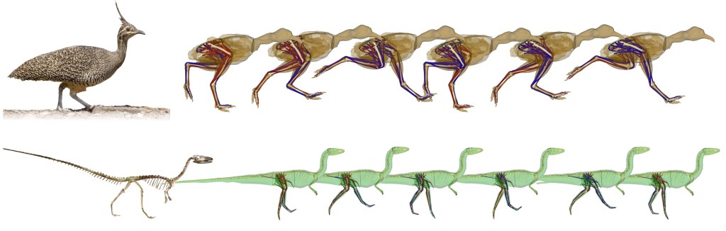
We observed that the simulations did clever things with the tail, swinging it side-to-side (and up-down) with each step in 3D; and in-phase with each leg: as the leg moved backward, the tail moved toward that leg’s side. With deeper analyses of these simulations, we found that this tail swinging conserved angular momentum and thus mechanical energy; making locomotion effectively cheaper, analogous to how humans swing their arms when moving. This motion emerged just from the physics of motion (i.e., the “multi-body dynamics”); not being intrinsically linked to muscles (e.g. the big caudofemoralis longus) or other soft tissues/neural control constraints (i.e., the biology). That is a cool finding, and because Coelophysis is a fairly representative theropod in many ways (bipedal, cursorial-limb-morphology, big tail, etc.), these motions probably transfer to most other fully bipedal archosaurs with substantial tails. Curiously, these motions seem to be opposite (tail swings left when right leg swings backward) in quadrupeds and facultatively bipedal lizards, although 3D experimental data aren’t abundant for the latter. But then, it seems beavers do what Coelophysis did?

Tail swings this-a-way (by Peter Bishop)
Computer simulation of extinct theropod dinosaur Coelophysis running at maximum speed, shown from behind to exemplify tail lateral flexion (wagging). Grey tiles = 50 cm.

The tail motions, and the lovely movies that our simulations produce, are what the media would likely focus on in telling the tale of this research, but there’s much more to this study. The tinamou simulations raise some interesting questions of why certain details didn’t ideally reflect reality: e.g., the limbs were still a little too vertical, a few muscles didn’t activate at the right times vs. experimental data, the foot motions were awkward, and the forces in running tended to be high. Some of these have obvious causes, but others do not, due to the complexity of the simulations. I’d love to know more about why they happen; wrong outputs from such models can be very interesting themselves.

Computer simulation of modern tinamou bird (brown) and extinct theropod dinosaur Coelophysis (green) running at maximum speed. Grey tiles = 10 cm for tinamou, 50 cm for Coelophysis.

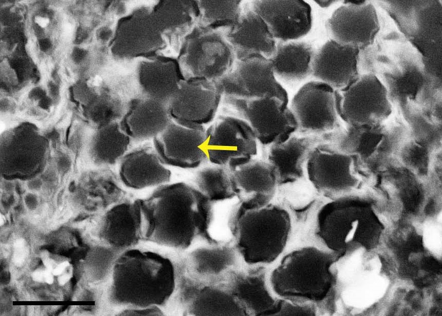
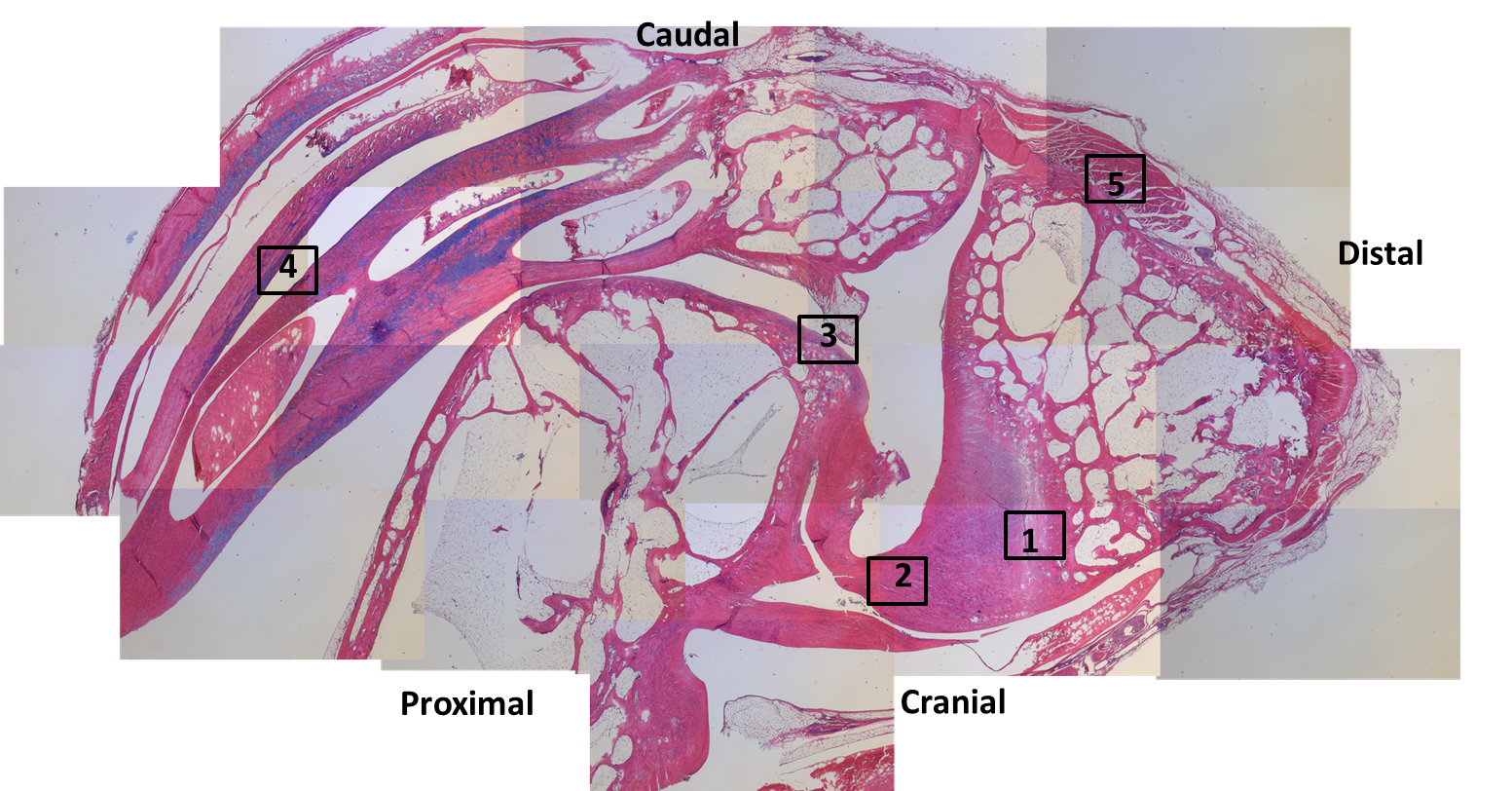
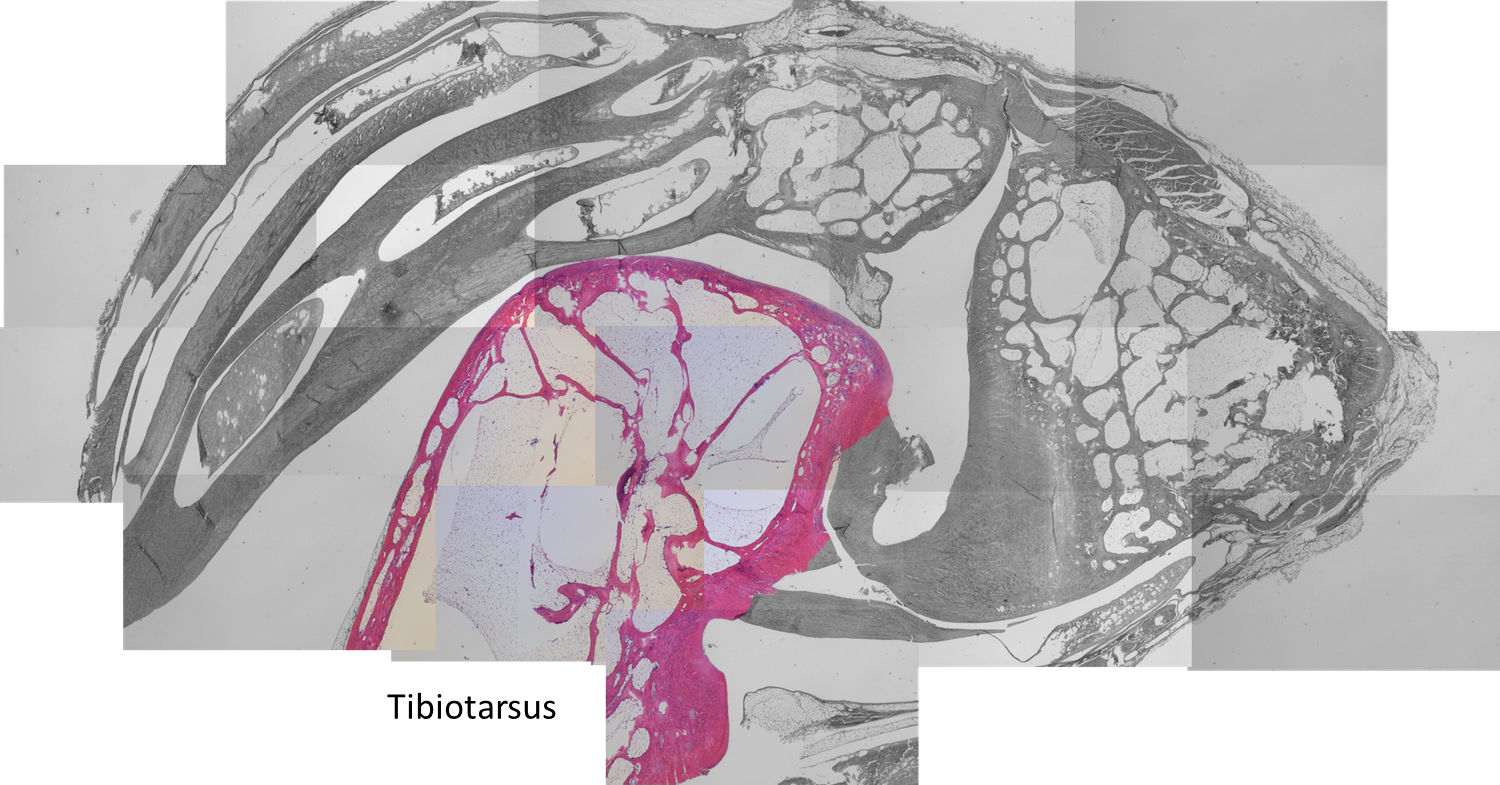
Speaking of wrong, in order to make our Coelophysis walk and run, we had to take two major shortcuts in modelling the leg muscles. The tinamou model had standard “Hill-type” muscles that almost everyone uses, and they’re not perfect models of muscle mechanics but they are a fair start; it also had muscle properties (capacity for force production, length change, etc.) that were based on empirical (dissection, physiology) data. Yet for our fossil, because we don’t know the lengths of the muscle fibres (active contractile parts) vs. tendons (passive stretchy bits), we adopted a simplified “muscle” model that combined both into one set of properties rather than more realistically differentiating them. It was incredibly important, then, that we try this simple muscle model with our tinamou to see how well it performed; and it did OK but still not “perfect”, and that simple muscle model might not work so well in other behaviours. That was the first major shortcut. Second, again because we don’t know the detailed architecture of the leg muscles in Coelophysis, we had to set very simple capacities for muscular force production: all muscles could only produce at most 2.15 body weights of force. This assumption worked OK when we applied it to our simulation of sprinting in the tinamou (vs. average 1.95 body weights/muscle in the real bird), so it was sufficiently justifiable for our purposes. In current work, we’re examining some alternative approaches to these two shortcuts that hopefully will improve outputs while maximising realism and objectivity.

Computer simulation of extinct theropod Coelophysis running at maximum speed, shown alongside running human (at 4 m/s) for scale and context. Image credit: Peter Bishop.

If you pay close attention, our simulations of Coelophysis output rather high leg-forces, and it’s unclear if that’s due to the simple muscle model, the simple foot modelling, or is actually realistic due to the more vertical (hence stiff) hindlimbs; or all of these. Another intriguing technical finding was that shifting the body’s centre of mass forwards slowed down the simulation’s running speed, as one might expect from basic mechanics (greater leg joint torques), but unlike some prior simulations by other teams.

Computer simulation of extinct theropod Coelophysis shown alongside running human for scale and context. Shown from above to illustrate tail wagging behaviour. Image credit: Peter Bishop.

Users of models and simulations are very familiar with catchphrases like “all models are wrong, but some are useful” or the much more cynical (or ignorant) “garbage in, garbage out”; or the very dangerous attitude that “if the mathematics is correct, then the models can’t be that wrong” (but if the biology is wrong, fuggedaboutit!). These are salutary cautionary tales and catechisms that keep us on our toes, because the visual realism that realistic-looking simulations produce can seduce you into thinking that the science is better than it is. It’s not a field that’s well-suited for those fearful of being wrong. I’ll never think these outputs are perfect; that is a crazy notion; but today I feel pretty good. This was a long time coming for me, and it is satisfying to get to this stage where we can push forwards in some new directions such as comparing simulations of different species to address bigger evolutionary questions.

The wrestling with scepticism never ends, but we can make progress while the match goes on.

from WWE… I could not resist

Read Full Post »

The blog is back! Briefly. With dinosaurs. Back in 2005, I published a paper in which I used a “SIMM” 3D musculoskeletal biomechanical model of Tyrannosaurus rex to analyse its muscle actions and infer a relatively upright hindlimb pose. This was an outcome from my NSF-funded postdoctoral research at Stanford University, in which engineers kindly taught me how to use SIMM (handing me a loaded gun?). Part of my plan all along was to build multiple such models along a rough evolutionary sequence to revisit old questions I had with past, qualitative functional morphology papers from 2000 onwards, and see if biomechanics could quantitatively reveal more about the functional evolution of dinosaur hindlimb muscles. So I got data for modelling some extinct dinosaurs (theropods Dilophosaurus, Allosaurus, Velociraptor) and living birds (Struthio, others) and published nuggets of that but held others back…

Stomach-Churning Rating: -1/10; dinosaurs!

I handed these 3D model data off to my PhD student Vivian Allen in ~2007, charging him with the task of making more models to flesh out the phylogeny and finish what I’d started. And he sure did. He graduated, did a couple of postdocs with me, and we gradually massaged his thesis chapter on this topic into a draft paper. Easier said than done, though! That’s why 14 more years have passed.

Viv came up with some clever tools in MATLAB software code (from which he became a very competent programmer and went on to a successful career in that!) to boil complex data on muscle leverages (moment arms) across a wide range of joint motion for the hindlimbs for each taxon.

These data then were fed into further code that took the results from all models, ultimately 13 of them from an Australian freshwater crocodile to two living birds and 10 extinct dinosaurs plus close cousin Mara/Lagosuchus (Figure 1). The code expressed these leverages as changes in ancestral values along the main branch of the evolutionary tree from early (Triassic) “ruling reptile” Archosauria (represented here just by the croc as a proxy) to modern birds, and 9 main ancestral “nodes” in between. Our code tracked both how each of 35 hindlimb muscles we modelled evolved in its leverage, as well as overall “average” leverage of functional groups around the hip, knee and ankle joints.

So, back and forth we went for some 10 years playing with the models (see Video below), data and code, and the paper describing the whole thing, slowly closing in on a final version but also sometimes distracted by our other projects and Real Life Stuff like health and children, and concerns about how we conducted this study (i.e. a lot of fiddling).

Figure 1: Evolutionary tree of dinosaurs and their relatives as used in the study, showing all 13 models, species names, and names of groups along the bottom (red nodes) of the tree. Averostra and Avetheropoda were ancestral groups of theropod dinosaurs that the study inferred had particular specialisations of the hindlimb muscles. Right hindlimbs in side view. The limbs are all straightened vertically into a baseline reference posture but the study investigated variation in muscle function across a wide range of limb poses.

Then I got a new grant “DAWNDINOS” that changed the scene for me, refocusing my team’s energies onto the Triassic (and early Jurassic) and the evolutionary biomechanics of diverse archosaurs’ locomotion, assessed with both LOTS of experimental studies of living crocs and birds, and LOTS of predictive simulations of locomotion. Stay tuned for much more on that from our team, but we’ve already published some key steps here. Most notably, we developed an improved protocol for modelling and simulating our animals, as shown by Bishop et al.’s 2021 study of the early theropod Coelophysis bauri (also appearing in the current paper). Awkwardly for me, that new method rendered our old models and methods a bit obsolete (although still fine), so I pushed to publish this current paper with Viv, and brought collaborator Dr. Brandon Kilbourne on board to aid in some final stats, figures and more. That finally did it, and now we’ve published the paper in Science Advances. Deep breath.

Video: Rotating movies of 3 musculoskeletal models from this study. Models have been posed into representative limb orientations illustrating a gradual or stepwise transformation from more upright to more crouched.

Well what’s the paper about, then? We used our 13 models and processed evolutionary functional patterns to test three main questions (hypotheses) about muscle leverage, making educated guesses at what might prevail from early Archosauria to Aves:

  1. Hip extensor / flexor (i.e. femur retractor/protractor) moment arm ratios remained constant. We weren’t sure what to expect, as these antagonists both seem to change a lot on the whole lineage, so we went with this prediction.
  2. Knee flexor / extensor ratios decreased; i.e. the flexors (“hamstrings” etc.) weakened and/or extensors (equivalent of our quadriceps) strengthened their leverage. Anatomy of the knee joint and muscles around it suggests this, plus since Gatesy’s 1990-onwards studies we’ve expected archosaurs to shift from more ‘hip-based’ to more ‘knee-based’ locomotion as we get closer to avian ancestry.
  3. Hip medial (internal) long-axis rotator / abductor (i.e. pronators of the limb vs. those that draw the leg away from the body) ratios increased. This idea comes right from my paper w/Gatesy in 2000, where we surmised that archosaurs shifted from relying on hip adductors (in crocs/other quadrupeds) to abductors (in bipedal dinosaurs; like humans) to medial rotators (‘torsional control’ as in birds today) during weight support.

Moreover, we reconstructed the evolution of 35 muscles’ actions across ~250 million years, which was a new step.

Here’s a summary of what we found (Figure 2):

Figure 2: Short visualization/explanation of the study’s main insights. Pictures by palaeoartist Jaime Headden: https://qilong.wordpress.com/about/ in left side view, including “muscled” and silhouette images. Right side images include representative hip, knee and ankle muscles from the study. Changes such as the enlargement of muscles in front of the hip that straighten the knee, and reduction of the caudofemoralis longus muscle that runs from the tail to the back of the thigh, are evident.

So, overall hypothesis 1 about hip extensors/flexors ended up complicated; rejected because hip flexor leverage actually increased. Furthermore, we found that around the ancestral nodes for early theropod dinosaurs (Neotheropda through Avetheropoda; around 200 Mya), there were peaks in muscle leverage (size-normalized) that surprised us, and persisted despite many different analyses we threw at them over the years. As far as we could tell, these peaks that kept appearing for various muscles’ actions were “real” (estimates). Which meant these ancestors may have had specialised high leverage relative to both their own ancestors and descendants; the peaks got reversed in evolution. These ancestors had some other weird anatomical and functional traits, such as tightly articulated hip joints early on (which they lost later), increased body size in the later forms, more ‘macropredatory’ ecology (e.g. eating sauropods?), and a centre of mass of the body that was shifted forwards (due to big arms and heads/necks). This weirdness is a cool unexpected finding that showed up for the other hypotheses too, and it needs some more investigating. A ‘failed’ hypothesis test led to neat insights.

Figure 3. From the paper– showing our main results for changes in moment arm ratios across archosaurian ancestors. Hip extensors/flexors decreased then increased; knee flexors/extensors decreased; and hip medial rotators/abductors decreased then had a series of increases.

Hypotheses 2 and 3 found good support, on the contrary, overall (Figure 3). We seem to have been able to quantify the shifts from hip-based to knee-based, and abductor-based to medial-rotator-based, muscle actions. I find that very satisfying. Ankle weight support (extension) capacity also increased, which fits morphological changes fairly well. If you’re into archosaur limb muscle form and function, there’s a lot more food for thought in the paper.

Funnily enough, ~20 years has been sufficient time that we could have had plenty more models in this study if we’d delayed it even longer and re-re-re-analysed our data. But we had to draw the line somewhere and not infinitely revise with every new model we’ve been creating. With the current state of musculoskeletal modelling in my group, we could have more than doubled our sample size and fleshed out the most important gaps such as in the crocodile-lineage (extinct Pseudosuchia) and other Triassic forms plus elsewhere. A big challenge remains having some nice 3D-preserved early fossil birds beyond Archaeopteryx; e.g. so many nice Chinese ones are too flat (e.g. joints we need) to reliably model here. It’s something that can still be done and is worth doing, but I suspect the general trends we’ve found along the dinosaur lineage are “correct”.

What’s personally important to me about this paper is (1) how it not only bridges a huge morphofunctional gap across archosaur evolution in scientific terms, and (2) how we’ve completed a long-delayed project with stubbornness (and during a pandemic!), but also (3) how it bridges my past career from my PhD and postdoc to the present work with DAWNDINOS. We’re now forging well beyond what this new paper has done in terms of truly testing, as best we can (estimate) so far, how limb muscles of archosaurs functioned and evolved, and how these contributed to particular behaviours and performance (maybe even palaeoecology and evolutionary success/extinction?). The current paper is just simple modelling of muscle leverage, but leverage is only one (very important!) piece of muscle function and performance. With fully dynamic, anatomically integrative, physiologically and physically representative biomechanical computer simulations that predict what living and extinct archosaurs could or could not do, we can do even better. So watch for that! Hopefully it won’t all take 20 years, or 250 million.

Read Full Post »

Ho ho ho! The vagaries of the scientific publication system today brings forth TWO open access papers on crocodylian functional anatomy, evolution and biomechanics, from my team with others’; including our DAWNDINOS project in part. Get ready to bite down on the science! I’ve loved crocodylians throughout my life– “dacadile” was among my first words, for a beloved stuffed croc toy, and “Alligators All Around” was an early favourite song (it’s still GREAT).

One of the many large adult alligators in St. Augustine, Florida.

Stomach-Churning Rating: 1/10; bones and movies of awesome behaviours.

First, I am so relieved and pleased to finally publish an experimental study I began over 17 years ago. This is my most-delayed paper ever, due to my own perfectionism, overcommitment and failures at funding it more broadly. But published is published and I’m glad to see it out. We collected a large experimental dataset from 15 species of Crocodylia at the St Augustine Alligator Farm Zoological Park (a conservation/education centre) in Florida. (No matter how you species-ify them, that’s a good chunk of diversity; roughly half or more.) This was a non-invasive study of 42 individuals ranging from 0.5 to 43 kg in body mass (hatchlings to adults). Larger adults were too dangerous or too slow to work with. It took 3 years (2002, 2004, 2005) of data collection to assemble this, with some twists and turns (including a close brush with Hurricane Katrina), and then a lot of analysis and reanalysis; and I’d do it all very differently if I did it today but that’s a moot point. So what’s the paper about?

Adorable Siamese crocodile family “cuddling”. Crocs are great parents! IIRC, that is the father shown.

Some Crocodylia (the inclusive modern name for all crocs, caimans, gharials, gators) are known to use what we call asymmetrical gaits: “mammal-like” footfall patterns in which the left and right limbs do not move as mirror images of each other. In particular, these gaits include galloping (rotary or transverse; either way a “4-beat” pattern with left-right hind- followed by right/left forefoot contacts) and bounding or half-bounding (the former being the most extreme, with left-right hind- and then forefoot contacts as synchronous pairs). Often people just say that crocs can “gallop” but this confuses/conflates the issue and omits that they can use these faster bounding gaits. Regardless, we’ve known about these gaits at least since HB Cott’s 1961 photographic documentation of them in Nile crocodiles; and more detailed studies of Australian freshwater and saltwater crocodiles in the 1970s-2000s. But very often, scientists and popular natural history accounts ascribe the asymmetrical gaits to only a few species or young individuals.

“Freshie” croc bounding in the wilds of Australia; credit Kent Vliet.

Osteolaemus dwarf African crocodile getting marked up for study.

That’s where we came in. We had access to a huge collection of captive Crocodylia and a very supportive institution (with coauthors from there as a result). I wanted to know which Crocodylia do use asymmetrical gaits, having a very strong suspicion from the literature that Alligatoroidea, the alligator and caiman lineage, don’t use them, whereas their cousins the “true crocodiles” in Crocodyloidea do. And I wanted to test how body size interacted with this ability, as prior accounts hinted that asymmetrical gaits got lost with increasing size or in adults. Finally, I was interested in what the benefits of asymmetrical gaits were– did they give those that used them marked boosts in performance, especially maximal speed? Answering that would help understand why these gaits are used.

Cuban crocodile Crocodylus rhombifer in preparation. A gorgeous but aggressive species that we handled carefully.

So we walked and ran our subjects across some platforms past video cameras and collected about 184 useful trials or strides of gait across level ground at a wide range of speeds; and a LOT of not-so-useful data (mostly subjects just sitting and pouting). We found that, yes, most Crocodyloidea we studied could bound or gallop; and no Alligatoroidea did. In the latter case, we didn’t use as large a sample of subjects as we could have, partly because it already seemed evident that alligators did not use asymmetrical gaits, and partly because those alligatoroids we did try to coax to move quickly either only used symmetrical gaits (e.g. trotting) or would only sit and fight or hiss. And we found that bigger animals moved at least relatively more slowly and less athletically, and perhaps even more slowly in absolute terms (metres/second).

Most intriguingly to me, it didn’t matter what gait alligatoroids or crocodyloids used. They all could move at roughly similar top speeds if they wanted to; less than 5 m/s or 11 mph. It’s just that crocodyloids tended to use asymmetrical gaits, especially bounding, at top speeds– but not always: some even chose to trot at their top speeds. We don’t know why, and we still don’t know why asymmetrical gaits are chosen but they likely have other benefits such as acceleration and manoeuvrability.

It’s a thrill to finally be able to share the huge dataset, including a gigantic file of videos (with some highlights shown here), with the paper, closing this study at last. It should be very useful to anyone studying Crocodylia or wanting to educate people about locomotion. I’m a bit tired of hearing that galloping is a mammalian behaviour when we know so well that many species of animals do it, or something like it. And it was absolutely thrilling to see five species of Crocodylia bound or gallop when they hadn’t been properly documented to do it before– enough anecdotes, here’s cold hard facts from video on what happens. What remains is a mystery: did Crocodylia have this ability to use asymmetrical gaits as an ancestral trait, as almost everyone assumes (and thus alligators and caimans have lost or essentially never express the ability), or did crocodiles uniquely evolve this ability more recently? I would join most scientists in wagering on the former; and there are good reasons to suspect the ability goes deeper into extinct Crocodylomorpha.

(my favourite video is below!)

Want more cool videos? Try my Youtube channel— or if you want ALL of the videos, go here!


Next, Torsten Scheyer was kind enough to invite me to join his team in studying a fossil I’ve long been fascinated by: the “giant caiman” Purussaurus mirandai, from the Miocene (~6 million years ago?) of Venezuela, in the Urumaco Formation‘s very weird biota. Purussaurus has been known of for >125 years but Torsten’s team noticed that Purussaurus (mirandai) specimens tended to add one of their trunk vertebrae to their hip girdles (sacrum; normally only two vertebrae in Crocodylia but here three), and that the shoulder and hip girdles had unusual bone morphology (straighter, more vertical relative to the body). So they asked me to help interpret these features. And here’s the paper!

Infographic by Torsten Scheyer’s team– click to emcroccen!

Three-vertebra sacrum and other traits of Purussaurus; with living caiman bones for comparison. E (bottom): inwards-facing femur head. (see paper for more info)

It became evident that, together, those odd traits conveyed a signal that the skeleton was transformed to aid in supporting the huge body against gravity. For example, I found it quite interesting how the head of the femur (thigh bone) was oriented more directly into the hip socket in multiple specimens, more like a dinosaur’s hip, and specialised for support and fore-aft motions. I used Haley O’Brien et al’s data to estimate just how big P. mirandai might have been and it came out as perhaps 3000 kg and 8 metres total length; as we’d thought, among the largest Crocodylia (and there are larger Purussaurus known, too).

Reconstruction of Purussaurus and morphology of the girdles. (see paper for more info)

The team also put a cool “evo-devo-biomechanics” spin on the study. It is well known that the regional identities of vertebrae (e.g. neck, trunk, sacrum, tail) are largely determined by Hox (homeobox) regulatory genes, early in development. So changes of vertebral identity intimate changes of genetic controls. Crocodylia don’t normally add a trunk vertebra to their sacrum, and only a few fossil crocodyliforms (extinct cousins) ever did either, but we noticed that some specimens of Crocodylia would at least partially make this transformation in pathological states (below). Hence the controls to make these changes exist and sometimes manifest in living crocs, but it’s probably not an “easy” transformation to achieve. One could speculate that under intense selection, such as that imposed by giant body size and some degree of activity on land, that transformation could more easily get permanently “fixed” in a species.

Palaeosuchus palpebrosus (Cuvier’s dwarf caiman) with pathological partial-three-vertebra-sacrum; and lots more morphology. (see paper for more info)

As a nice tie-in to the asymmetrical gait study above, we can safely infer that the giant Purussaurus wasn’t a fast animal on land, by any means. But its skeleton is consistent with it having found novel ways to maintain the ability to stand and move on land, even if slowly.

Happy holidays! Santa Jaws is watching you– be good!

Read Full Post »

Today is the 210th anniversary of Charles R. Darwin’s birthday so I put together a quick post. I’d been meaning to blog about some of our latest scientific papers, so I chose those that had an explicit evolutionary theme, which I hope Chuck would like. Here they are, each with a purty picture and a short explainer blurb! Also please check out Anatomy To You’s post by Katrina van Grouw on Darwin’s fancy pigeons.

Stomach-Churning Rating: 1/10 science!

First, Brandon Kilbourne at the Naturkunde Museum in Berlin kindly invited me to assist in a paper from his German fellowship studying mustelid mammals (otters, weasels, wolverines, badgers, etc.; stinky smaller carnivorous mammals). Here we (very much driven by Brandon; I was along for the ride) didn’t just look at how forelimb bone shape changes with body size in this ecologically diverse group. We already knew bigger mustelids would have more robust bones, although it was cool to see how swimming-adapted and digging-adapted mustelids evolved similarly robust bones; whereas climbing ones had the skinniest bones.

The really exciting and novel (yes I am using that much-abused word!) aspect of the paper is that Brandon conjured some sorcery with the latest methods for analysing evolutionary trends, to test how forelimb bone shapes evolved. Was their pattern of evolution mostly a leisurely “random walk” or were there early bursts of shape innovation in the mustelid tree of life, or did shape evolve toward one or more optimal shapes (e.g. suited to ecology/habitat)? We found that the most likely pattern involved multiple rates of evolution and/or optima, rather than a single regime. And it was fascinating to see that the patterns of internal shape change deviated from external shape change such as bone lengths: so perhaps selection sometimes works independently at many levels of bone morphology?

Various evolutionary models applied to the phylogeny of mustelids.

Then there, coincidentally, was another paper originating in part from the same museum group in Berlin. This one I’d been involved in as a co-investigator (author) on a Volkswagen (yes! They like science) grant back about 8 years ago and since. There is an amazing ~290 million year old fossil near-amniote (more terrestrial tetrapod) called Orobates pabsti, preserved with good skeletal material but also sets of footprints that match bones very well, allowing a rare match of the two down to this species level. John Nyakatura’s team had 3D modelled this animal before, so we set out to use digital techniques to test how it did, or did not, move—similar to what I’d tried before with Tyrannosaurus, Ichthyostega and so forth. The main question was whether Orobates moved in a more “ancestral” salamander-like way, a more “derived” lizard-like way (i.e. amniote-ish), or something else.

The approach was like a science sledgehammer: we combined experimental studies of 4 living tetrapods (to approximate “rules” of various sprawling gaits), a digital marionette of Orobates (to assess how well its skeleton stayed articulated in various motions), and two robotics analysis (led by robotics guru Auke Ijspeert and his amazing team): a physical robot version “OroBOT” (as a real-world test of our methods), and a biomechanical simulation of OroBOT (to estimate hard-to-measure things in the other analyses, and matches of motions to footprints). And, best of all, we made it all transparent: you can go play with our interactive website, which I still find very fun to explore, and test what motion patterns do or do not work best for Orobates. We concluded that a more amniote-like set of motions was most plausible, which means such motions might have first evolved outside of amniotes.

OroBOT in tha house!

You may remember Crassigyrinus, the early tetrapod, from a prior post on Anatomy To You. My PhD student Eva Herbst finished her anatomical study of the best fossils we could fit into a microCT-scanner and found some neat new details about the “tadpole from hell”. Buried in the rocky matrix were previously unrecognized bones: vertebrae (pleurocentra; the smaller nubbins of what may be “rhachitomous” bipartite classic tetrapod/omorph structure), ribs (from broad thoracic ones to thin rear ones), pelvic (pubis; lower front), and numerous limb bones. One interesting trait we noticed was that the metatarsals (“sole bones” of the foot) were not symmetrical from left-to-right across each bone, as shown below. Such asymmetry was previously used to infer that some early tetrapods were terrestrial, yet Crassigyrinus was uncontroversially aquatic, so what’s up with that? Maybe this asymmetry is a “hangover” from more terrestrial ancestry, or maybe these bones get asymmetrical for non-terrestrial reasons.

The oddly asymmetrical metatarsals of Crassigyrinus.

Finally, Dr. Peter Bishop finished his PhD at Griffith University in Australia and came to join us as a DAWNDINOS postdoc. He blasted out three of his thesis chapters (starting here) with me and many others as coauthors, all three papers building on a major theme: how does the inner bone structure (spongy or cancellous bone) relate to hindlimb function in theropod dinosaurs (including birds) and how did that evolve? Might it tell us something about how leg posture or even gait evolved? There are big theories in “mechanobiology” variously named Wolff’s Law or the Trajectorial Theory that explain why, at certain levels, bony struts tend to align themselves to help resist certain stresses, and thus their alignment can be “read” to indicate stresses. Sometimes. It’s complicated!

Undaunted, Peter measured a bunch of theropod limb bones’ inner geometry and found consistent differences in how the “tracts” of bony struts, mainly around joints, were oriented. He then built a biomechanical model of a chicken to test if the loads that muscles placed on the joints incurred stresses that matched the tracts’ orientations. Hmm, they did! Then, with renewed confidence that we can use this in the fossil record to infer approximate limb postures, Peter scanned and modelled a less birdlike Daspletosaurus (smaller tyrannosaur) and more birdlike “Troodon” (now Stenonychosaurus; long story). Nicely fitting many other studies’ conclusions, Peter found that the tyrannosaur had a more straightened hindlimb whereas the troodontid had a more crouched hindlimb; intermediate between the tyrannosaur and chicken. Voila! More evidence for a gradual evolution of leg posture across Mesozoic-theropods-into-modern-birds. That’s nice.

Three theropods, three best-supported postures based on cancellous bone architecture.

If you are still thirsty for more papers even if they are less evolutionary, here’s the quick scoop on ones I’ve neglected until now:

(1) Former PhD student Chris Basu published his thesis work w/us on measuring giraffe walking dynamics with force plates, finding that they move mostly like other quadrupeds and their wobbly necks might cost them a little.

(2) Oh, and Chris’s second paper just came out as I was writing this! We measured faster giraffe gaits in the wilds of South Africa, as zoo giraffes couldn’t safely do them. And we found they don’t normally go airborne, just using a rotary gallop (not trot, pace or canter); unlike some other mammals. Stay tuned: next we get evolutionary with this project!

(2) How do you safely anaesthetize a Nile crocodile? There’s now a rigorous protocol (from our DAWNDINOS work).

(3) Kickstarting my broad interest in how animals do “extreme” non-locomotor motions, we simulated how greyhounds stand up, finding that even without stretchy tendons they should, barely, be able to do it, which is neat. Expect much more about this from us in due time.

(4) Let’s simulate some more biomechanics! Ashley Heers, an NSF research fellow w/me for a year, simulated how growing chukar birds use their wing muscles to flap their way up steeper inclines (“WAIR” for devotees), and the results were very encouraging for simulating this behaviour in more detail (e.g. tendons seem to matter a lot) and even in fossil species; and finally…

(5) Hey did you ever think about how bone shape differs between hopping marsupials (macropods) and galloping artiodactyl (even-toed) mammals? We did, in long-the-making work from an old BBSRC grant with Michael Doube et al., and one cool thing is that they mostly don’t change shape with body size that differently, even though one is more bipedal at faster speeds—so maybe it is lower-intensity, slower behaviours that (sometimes?) influence bone shape more?

So there you have the skinny on what we’ve been up to lately, messing around with evolution, biomechanics and morphology.

Read Full Post »

As 2017 approaches its end, there have been a few papers I’ve been involved in that I thought I’d point out here while I have time. Our DAWNDINOS project has been taking up much of that time and you’ll see much more of that project’s work in 2018, but we just published our first paper from it! And since the other two recent papers involve a similar theme of muscles, appendages and computer models of biomechanics, they’ll feature here too.

Stomach-Churning Rating: 0/10; computer models and other abstractions.

Mussaurus patagonicus was an early sauropodomorph dinosaur from Argentina, and is now widely accepted to be a very close relative of the true (giant, quadrupedal) sauropods. Here is John Conway’s great reconstruction of it:

We have been working with Alejandro Otero and Diego Pol on Mussaurus for many years now, starting with Royal Society International Exchange funds and now supported by my ERC grant “DAWNDINOS”. It features in our grant because it is a decent example of a large sauropodomorph that was probably still bipedal and lived near the Triassic-Jurassic transition (~215mya).

In our new study, we applied one of my team’s typical methods, 3D musculoskeletal modelling, to an adult Mussaurus’s forelimbs. This is a change of topic from the hindlimbs that I’ve myopically focused on before with Tyrannosaurus and Velociraptor [in an obscure paper that I should never have published in a book! pdf link], among other critters my team has tackled (mouse, elephant [still to be finished…], ostrich, horse, Ichthyostega… dozens more to come!). But we also modelled the forelimbs of Crocodylus johnstoni (Australian “freshie”) for a key comparison with a living animal whose anatomy we actually knew, rather than reconstructed.

Mussaurus above; Crocodylus below; forelimb models in various views; muscles are red lines.

The methods for this biomechanical modelling are now standard (I learned them from their creator Prof. Scott Delp during my 2001-2003 postdoc at Stanford): scan bones, connect them with joints, add muscle paths around them, and then use the models to estimate joint ranges of motion and muscle moment arms (leverage) around joints. I have some mixed feelings about developing this approach in our 2005 paper that is now widely used by the few teams that study appendicular function in extinct animals. As a recent review paper noted and I’ve always cautioned, it has a lot of assumptions and problems and one must exercise extreme caution in its design and interpretation. Our new Mussaurus paper continues those ruminations, but I think we made some progress, too.

On to the nuts and bolts of the science (it’s a 60 page paper so this summary will omit a lot!): first, we wanted to know how the forelimb joint ranges of motion in Mussaurus compared with those in Crocodylus and whether our model of Mussaurus might be able to be placed in a quadrupedal pose, with the palms at least somewhat flat (“pronated”) on the ground. Even considering missing joint cartilage, this didn’t seem very plausible in Mussaurus unless one allowed the whole forearm to rotate around its long axis from the elbow joint, which is very speculative—but not impossible in Crocodylus, either. Furthermore, the model didn’t seem to have forelimbs fully adapted yet for a more graviportal, columnar posture. Here’s what the model’s mobility was like:

So Mussaurus, like other early sauropodomorphs such as Plateosaurus, probably wasn’t quadrupedal, and thus quadrupedalism must have evolved very close to in the Sauropoda common ancestor.

Second, we compared the muscle moment arms (individual 3D “muscle actions” for short) in different poses for all of the main forelimb muscles that extend (in various ways and extents) from the pectoral girdle to the thumb, for both animals, to see how muscle actions might differ in Crocodylus (which would be closer to the ancestral state) and Mussaurus. Did muscles transform their actions in relation to bipedalism (or reversal to quadrupedalism) in the latter? Well, it’s complicated but there are a lot of similarities and differences in how the muscles might have functioned; probably reflecting evolutionary ancestry and specialization. What I found most surprising about our results was that the forelimbs didn’t have muscles well-positioned to pronate the forearm/hand, and thus musculoskeletal modelling of those muscles reinforced the conclusions from the joints that quadrupedal locomotion was unlikely. I think that result is fairly robust to the uncertainties, but we’ll see in future work.

You like moment arms? We got moment arms! 15 figures of them, like this! And tables and explanatory text and comparisons with human data and, well, lots!

If you’re really a myology geek, you might find our other conclusions about individual muscle actions to be interesting—e.g. the scapulohumeralis seems to have been a shoulder pronator in Crocodylus vs. supinator in Mussaurus, owing to differences in humeral shape (specialization present in Mussaurus; which maybe originated in early dinosaurs?). Contrastingly, the deltoid muscles acted in the same basic way in both species; presumed to reflect evolutionary conservation. And muuuuuuch more!

Do you want to know more? You can play with our models (it takes some work in OpenSim free software but it’s do-able) by downloading them (Crocodylus; Mussaurus; also available: Tyrannosaurus, Velociraptor!). And there will be MUCH more about Mussaurus coming soon. What is awesome about this dinosaur is that we have essentially complete skeletons from tiny hatchlings (the “mouse lizard” etymology) to ~1 year old juveniles to >1000kg adults. So we can do more than arm-wave about forelimbs!

But that’s not all. Last week we published our third paper on mouse hindlimb biomechanics, using musculoskeletal modelling as well. This one was a collaboration that arose from past PhD student James Charles’s thesis: his model has been in much demand from mouse researchers, and in this case we were invited by University of Virginia biomechanical engineers to join them in using this model to test how muscle fibres (the truly muscle-y, contractile parts of “muscle-tendon units”) change length in walking mice vs. humans. It was a pleasure to re-unite in coauthorship with Prof. Silvia Blemker, who was a coauthor on that 2005 T. rex hindlimb modelling paper which set me on my current dark path.

Mouse and human legs in right side view, going through walking cycles in simulations. Too small? Click to embiggen.

We found that, because mice move their hindlimb joints through smaller arcs than humans do during walking and because human muscles have large moment arms, the hindlimb muscles of humans change length more—mouse muscles change length only about 48% of the amount that typical leg muscles do in humans! This is cool not only from an evolutionary (mouse muscles are probably closer to the ancestral mammalian state) and scaling (smaller animals may use less muscle excursions, to a point, in comparable gaits?) perspective, but it also has clinical relevance.

Simulated stride for mouse and human; with muscles either almost inactive (Act=0.05) or fully active (Act=1). Red curve goes through much bigger excursions (along y-axis) than blue curve), so humans should use bigger % of their muscle fibre lengths in walking. Too small? Click to embiggen.

My coauthors study muscular dystrophy and similar diseases that can involve muscle stiffness and similar biomechanical or neural control problems. Mice are often used as “models” (both in the sense of analogues/study systems for animal trials in developing treatments, and in the sense of computational abstractions) for human diseases. But because mouse muscles don’t work the same as human muscles, especially in regards to length changes in walking, there are concerns that overreliance on mice as human models might cause erroneous conclusions about what treatments work best to reduce muscle stiffness (or response to muscle stretching that causes progressive damage), for example. Thus either mouse model studies need some rethinking sometimes, or other models such as canines might be more effective. Regardless, it was exciting to be involved in a study that seems to deliver the goods on translating basic science to clinical relevance.

Muscle-by-muscle data; most mouse muscles go through smaller excursions; a few go through greater; some are the same as humans’.

Finally, a third recent paper of ours was led by Julia Molnar and Stephanie Pierce (of prior RVC “Team Tetrapod” affiliation), with myself and Rui Diogo. This study tied together a bunch of disparate research strands of our different teams, including musculature and its homologies, the early tetrapod fossil record, muscle reconstruction in fossils, and biomechanics. And again the focus was on forelimbs, or front-appendages anyway; but turning back the clock to the very early history of fishes, especially lobe-finned forms, and trying to piece together how the few pectoral fin muscles of those fish evolved into the many forelimb muscles of true tetrapods from >400mya to much more recent times.

Humerus in ventral view, showing muscle attachments. Extent (green) is unknown in the fossil but the muscle position is clear (arrow).

We considered the homologies for those muscles in extant forms, hypothesized by Diogo, Molnar et al., in light of the fossil record that reveals where those muscles attach(ed), using that reciprocal illumination to reconstruct how forelimb musculature evolved. This parallels almost-as-ancient (well, year 2000) work that I’d done in my PhD on reconstructing hindlimb muscle evolution in early reptiles/archosaurs/dinosaurs/birds. Along the way, we could reconstruct estimates of pectoral muscles in various representative extinct tetrapod(omorph)s.

Disparity of skeletal pectoral appendages to work with from lobe-fins to tetrapods.

Again, it’s a lengthy, detailed study (31 pages) but designed as a review and meta-analysis that introduces readers to the data and ideas and then builds on them in new ways. I feel that this was a synthesis that was badly needed to tie together disparate observations and speculations on what the many, many obvious bumps, squiggles, crests and tuberosities on fossil tetrapods/cousins “mean” in terms of soft tissues. The figures here tell the basic story; Julia, as usual, rocked it with some lovely scientific illustration! Short message: the large number of pectoral limb muscles in living tetrapods probably didn’t evolve until limbs with digits evolved, but that number might go back to the common ancestor of all tetrapods, rather than more recently. BUT there are strong hints that earlier tetrapodomorph “fishapods” had some of those novel muscles already, so it was a more stepwise/gradual pattern of evolution than a simple punctuated event or two.

Colour maps of reconstructed right fin/limb muscles in tetrapodomorph sarcopterygian (~”fishapod”) and tetrapod most recent common ancestors. Some are less ambiguous than others.

That study opens the way to do proper biomechanical studies (like the Mussaurus study) of muscle actions, functions… even locomotor dynamics (like the mouse study)– and ooh, I’ve now tied all three studies together, tidily wrapped up with a scientific bow! There you have it. I’m looking forward to sharing more new science in 2018. We have some big, big plans!

Read Full Post »

Putting my morphologist hat back on today, I had an opportunity to dissect an Elegant-crested tinamou (Eudromia elegans) for the second time in my life. The last time was during my PhD work ~20 years ago. In today’s dissection I was struck by another reminder of how studying anatomy is a lifelong learning experience and sometimes it’s really fun and amazing even when it’s stinky.

Tinamou foot. I did know that tinamous don’t have a hallux; big “perching toe” (1st/”big toe” in us); true of ratites/palaeognaths more generally. Unlike a chicken or many other birds. Just the three main toes (2, 3 and 4) are here.

Stomach-Churning Rating: 7/10; you gotta have guts to learn about intestine-churning stuff.

Tinamous are neat little partridge-like ground birds but they are not close cousins of partridges or guineafowl at all. Their closest cousins are other ratites/palaeognaths such as ostriches and kiwis. And hence they are found in South America, especially Patagonia in Argentina. I’ve seen them there, much to my enjoyment.

Said tinamou.

What struck me today was that, as I delved into the digestive system of this bird, I saw features that were unfamiliar to me even after having dissected many species of birds from many lineages. The intestinal region was very lumpy, with little bud-like pockets full of dense droppings. Furthermore, on separating the tubes of the small and large intestines I realized that most of the intestinal volume itself was caecum (normally a modest side-pocket near the juncture of the small and large intestines). Indeed, that caecum was caeca (plural): it had two massive horns; it was a double-caecum, feeding back into the short rectum and cloaca. Birds have variable caeca and it is typical to see subdivision into two parts, but I’d never seen it to this degree.

Oh why not, here’s the gizzard/stomach showing its grinding pebbles and bits of food, plus the strong outer muscle layers (pink) for driving that grinding. Small intestine heads toward the bottom of the image. Yes, we do need a better dissection light…

I had to question my anatomical knowledge at this point, wondering if I was identifying things incorrectly—did I really screw up somehow and these were other organs, like giant ovaries? But no, they were clearly full of faecal matter; they were digestive organs. I finished the dissection, still puzzled, and hit the literature. Right away, Google-Scholaring for “tinamou caecum” I found the answer, here (free pdf link):

“at least one species (Elegant Crested Tinamou, [Eudromia elegans]), the ceca contain multiple sacculations, resulting in structures that look much like two bunches of fused grapes.”

The caeca in question.

OK buddy, those are the little lumpy buds I saw. Bunches of grapes—exactly.

And later:

“The paired ceca of the Elegant Crested Tinamou are extraordinary and probably unique within Aves (Fig. 3): long and wide (12.5-13.0 X 2.2- 2.5 cm; Wetmore 1926) and internally honeycombed by many small diverticula. These outpocketings gradually diminish in size and organization from the base to the tip of the organ, apically showing a more spiral form of internal ridges like ratite ceca. Externally, the basal diverticula protrude from the ceca as pointed lobes, gradually becoming flatter but still clearly apparent toward the organ’ s tip.”

Whoa! I never knew that! So I happened to be dissecting a bird, fairly common in its homeland, that has a really bizarre and singular form of caeca/ceca! That hit my morphologist sweet-spot so I was very pleased and decided to share with you. It is one of those many examples of times when you quickly go from confusion to illumination as a scientist, emerging with a neat fact about animal biology. And journal articles help you get there!

The bare “brood patch” on the back end of the tinamou’s belly; a nicely hotspot for keeping eggs warm. Perhaps for brooding bad puns, too.

Read Full Post »

Hi, sorry for the social media spam but this is important to me: I got EU money to study dinosaur movement and we made a website for the project. There will be some fun stuff posted there and nowhere else, such as new palaeo-art that we commissioned specifically for this project. Oh, and science, too! Five years of science!

So please have a look at it now that it is live!

https://dawndinos.com/

I love our logo (by Andrew Bourne) so I will spray paint it everywhere I can.

Work from the DAWNDINOS project won’t be featured here much, so either watch that new website or me on other social media to find out what’s up!

And coming up on John’s Freezer: another episode of “Better Know A Muscle”! Yeah, baby!

Read Full Post »

Uh oh, a “why?” question in biology! There are many potential, and not mutually exclusive, answers to such questions. Ultimately there is a historical, evolutionary answer that underpins it all (“ostriches evolved two kneecaps because…”). But we like ostrich knees and their funky double-kneecaps (patellae; singular = patella) so we wanted to know why they get so funky. One level of addressing that question is more like a “how?” they have them. So we started there, with what on the surface is a simple analysis. And we published that paper this week, with all of the supporting data (CT, MRI, FEA).

Stomach-Churning Rating: 6/10 because there is a gooey image of a real dissection later in the post, not just tidy 3D graphics.

First author Kyle Chadwick was my research technician for 2 years on our sesamoid evolution grant, and we reported earlier on the detailed 3D anatomy of ostrich knees (this was all part of his MRes degree with me, done in parallel with his technician post). Here, in the new paper with Sandra Shefelbine and Andy Pitsillides, we took that 3D anatomy and subjected it to some biomechanical analysis in two main steps.

Ostrich (right) knee bones. The patellae are the two knobbly bits in the knee.

First, we used our previous biomechanical simulation data from an adult ostrich (from our paper by Rankin et al.) to estimate the in vivo forces that the knee muscles exert onto the patellar region during moderately large loading in running (not maximal speed running, but “jogging”). That was “just” (Kyle may laugh at the “just”– it wasn’t trivial) taking some vectors out of an existing simulation and adding them into a detailed 3D model. We’ve done similar things before with a horse foot’s bones (and plenty more to come!), but here we had essentially all of the soft tissues, too.

Ostrich knee with muscles as 3D objects.

Second, the 3D model that the muscular forces were applied to was a finite element model: i.e., the original 3D anatomical model broken up into a mesh, whose voxels each had specific properties, such as resistance to shape change under loading in different directions. The response of that model to the loads (a finite element analysis; FEA) gave us details on the stresses (force/area) and strains (deformations from original shape) in each voxel and overall in anatomical regions.

Finite element model setup for our study. If you do FEA, you care about these things. If not, it’s a pretty, sciencey picture.

The great thing about a computer/theoretical model is that you can ask “what if?” and that can help you understand “how?” or even “why?” questions that experiments alone cannot address. Ostriches aren’t born with fully formed bony kneecaps; indeed those patellae seem to mature fairly late in development, perhaps well after hatching. We need to know more about how the patellae form but they clearly end up inside the patellar (knee extensor) tendon that crosses the knee. So we modelled our adult ostrich without bony patellae; just with a homogeneous patellar tendon (using the real anatomy of that tendon with the bony bits replaced by tendon); and subjected it to the loading environment for “jogging”.

The right knee of an ostrich hatchling. The patellae have yet to form; indeed there is little bone around the knee region at all, yet.

We then inspected our FEA’s results in light of modern theory about how tissues respond to loading regimes. That “mechanobiology” theory, specifically “tissue differentiation”, postulates that tendon will tend to turn into fibrocartilage if it is subjected to high compression (squishing) and shear (pushing). Then, the fibrocartilage might eventually be reworked into bone as it drops the compression and shear levels. So, according to that theory (and all else being equal; also ignoring the complex intermediate states that would happen in reality), the real ostrich’s kneecaps should be located in the same positions where the FEA, under the moderately large loads we applied, predicts the homogeneous tendon to have high compression and shear. But did the real anatomy match the mechanical environment and tissue differentiation theory’s predictions?

Tissue differentiation diagram displaying the theoretical pathways for transformation of tissues. If tendon (red) experiences high shear (going up the y-axis) and high compression (going toward the left), it should turn into fibrocartilage (purple). Transformation into bone (diagonally to the bottom right) would reduce the shear and compression.

Well, sort of. The image below takes some unpacking but you should be able to pick out the red areas on the bottom row where the patellae actually are, and the yellow shaded regions around some of those patellar regions are where the compression and shear regimes are indeed high and overlapping the actual patellar regions. The upper two rows show the levels of compression (or tension; pulling) and shear, but the bottom row gets the point across. It’s not a bad match overall for the first (“real”; common to all living birds) patella, located on top of the upper knee (femur). It’s not a good match overall for the second (unique to ostriches) patella, located below the first one (and attached to the tibia bone).

FEA results! (click to embiggen)

Kyle says, “Being a part of this project was exciting because of the application of engineering concepts to interesting biological (including evolutionary) questions. Also, it never gets old seeing people’s reactions when I tell them I study ostrich knees.

The study had a lot of nuances and assumptions. We only looked at one instant in slow running and only at one adult ostrich, not at the full development of ostrich anatomy and loading. That’s harder. We started simple. The tissue differentiation theory is used more for fracture healing than for sesamoid bone formation but there’s some reason to suspect that similar mechanisms are at play in both. And there’s much more; if you want the gory details see the paper.

So did we solve why, or how, ostriches have two kneecaps? We felt that the mechanical environment of our FEA was a good theoretical explanation of where the first patella forms. We originally expected the second patella, which evolved more recently and might be more mechanically sensitive as a result, to be a better match than the first one, but it was the opposite. C’est la science!

Enough models, let’s have some reality! I warned you this post would get messy, and here it is. Left leg (skinned) of an ostrich showing the muscles around the knee. The patellar region would be in the gloved hand of the lucky individual shown.

This study, for me, was a fun experience in moving toward more fusion of “evo-devo” and biomechanical analyses, a research goal of mine lately– but there’s still a ways to go with the “how?” and “why?” questions even about ostrich kneecaps.

We felt that the best conclusion supported by our analyses was that, rather than have homogeneous stresses and strains throughout their knee tissues (e.g. the patellar tendon), ostriches have a lot of regional diversity in how those tissues are loaded (in the condition we modelled, which is adequately representative of some athletic exertion). Look at the complex FEA coloured results above again, the top two rows: there are a lot of different shades of compression/tension and shear; not homogeneous strains. That diversity of regional loading sets those tissues up for potential transformation throughout growth and development. And thus ONE of the reasons why ostriches might have two kneecaps is that the heterogeneous loading of their knee tendon favours formation of heterogeneous tissue types.

Another, compatible, explanation is that these different tissues might have consequences for how the muscles, tendon and joint operate in movement behaviours. In due time there will be more about that. In the meantime, enjoy the paper if this post makes you want to know more about the amaaaaaazing knees of ostriches!

Read Full Post »

A Confuciusornis fossil; not the one from our study but prettier (more complete).

Today almost three years of collaboration come together in a publication that is a fun departure from my normal research, but also makes sense in light of it. Professor Baoyu Jiang from Nanjing University in China has been being working on the taphonomy of the Early Cretaceous Jehol biota from northeastern China (Manchuria) for a while, and he found a lovely Confuciusornis (early bird) fossil; one of thousands of them; from the volcanic pyroclastic flow-based lake deposits there.

Although at first glance the skeletal remains of that fossil are not fabulous compared with some other Confuciusornis, what makes this one lovely is that, on peering at it with multiple microscopic and other imaging techniques, he (and me, and a China-UK collaboration that grew over the years) found striking evidence of very well-preserved fossil soft tissues. Our paper reporting on these findings has gone live in Nature Communications so I can blog about it now.

Reference: Jiang, B., Zhao, T., Regnault, S., Edwards, N.P., Kohn, S.C., Li, Z., Wogelius, R.A., Benton, M., Hutchinson, J.R. 2017. Cellular preservation of musculoskeletal specializations in the Cretaceous bird Confuciusornis. Nature Communications 8:14779. doi: 10.1038/NCOMMS14779

Stomach-Churning Rating: 3/10; gooey, but fossil gooey, except for some colourful, gastrically-tolerable histology of bird tissue.

Front view of the ankle/foot of our specimen.

Back view of the ankle/foot of our specimen.

What has been fun about this collaboration is that, for one, it fits in perfectly with my prior work. Ever since my PhD thesis I’d been wondering about odd bones in the legs of birds, including a very puzzling and very, very neglected bit of bone called the tarsal sesamoid, on the outside of the upper end of the ankle joint. Furthermore, a tunnel of tissue called the tibial cartilage sits next to that sesamoid bone, and then across the ankle joint there is a bony prominence with grooves and tunnels that vary highly among bird species; that is called the hypotarsus. These structures are all known in living birds and, to a degree, in extinct fossil cousins. Our specimen seems to reveal an earlier stage in how these little features of bird ankles originated, which we concluded to be a step along the transition to the more crouched legs that modern birds have.

This study has also challenged me to broaden my horizons as a scientist. Although this was a big collaboration and thus we had several specialists to apply supercharged technological techniques to our fossil, I had to learn something about what all that meant. My kind colleagues helped me learn more about tissue histology, scanning electron microscopy, synchrotron mapping, FTIR and mass spectrometry and more. I won’t go through all of these techniques but there are some pretty pictures sprinkled here and in the paper, and a lot more detail in the paper for those who want the gory techno-detail. Basically we threw the kitchen sink of science at the fossil to crack open some of its secrets, and what we found inside was nifty.

Scanning electron micrograph image of probable tendon or ligament fibres (arrow) in cross-section, from near the ankle joint.

We found preserved cells and other parts of connective tissues including tendons and/or ligaments, fibrocartilage (the tougher kind) and articular cartilage (the softer joint-padding kind). That’s great, although not unique, but the kitchen sink also flushed out even more reductionist data: those tissues included some organic residues, including what appear to be bits of proteins (amino acids); particularly the collagen that makes up tendons.

Fibrocartilage (“fc”) from the ankle joint region.

Hopefully we’re right, and we included as much of the data as we could manage so that others can look at our findings. The specimen is crushed into nearly two dimensions, like all Jehol biota organisms, so its anatomy was hard to interpret but we think we got it right. All of the other kitchen-sinky tools have their own nuances and pitfalls but we did our best with a superb team of experts. We’ve had to wait 125 million years to uncover this specimen and a few more years to find out if we’ve looked at the right way is no greater test of patience.

I thank my coauthors, especially Baoyu Jiang for the kind invitation to participate and the very fun experience of collaborating. I think I’ll remember this study for a long time because, for me, it takes a step beyond just describing Another Case of Jaw-Dropping Fossilization (can you hear the hipsters recounting the excitement and cynicism of the 1990s when this all was dawning? I was there and maybe now I’m one of them). By combining all of those methods we learned new things about the palaeobiology of birds and the evolution of traits within birds. Confuciusornis, not shockingly, had ankles that should have functioned in ways intermediate between those of bog-standard non-avian theropods and modern birds.

Same anatomical regions in an extant bird as in the main fossil specimen. Left distal tibiotarsus (TT; below) and proximal tarsometatarsus (TMT; above) from an adult helmeted guineafowl (Numida meleagris) after formalin fixation. (from our paper’s Supp Info)

I’m hopeful that more synthesis of molecular/cellular, imaging, biomechanical and other tools (not to mention good old palaeontology and anatomy!) can wash away some more of this mystery. And it was fun to be a part of a study that adds to overwhelming evidence that was heretical ~25 years ago: some hardy biomolecules such as collagen and keratin can survive hundreds of millions of years, not just thousands. Pioneers such as Prof. Mary Schweitzer led the original charge that made reporting on discoveries like ours much easier today.

I know how the birds fly, how the fishes swim, how animals run. But there is the Dragon. I cannot tell how it mounts on the winds through the clouds and flies through heaven. Today I have seen the Dragon.“– Confucius, ca. 500 BCE.

Let’s finish with some images of a living bird’s ankle region, by co-author and PhD student Sophie Regnault. We considered these for inclusion in the paper but they didn’t fit quite right. I love them anyway so here they are:

Patchwork of histology slide images, from a guineafowl’s ankle (as per photo above). The numbered squares correspond to zoomed-in images below. The tibiotarsus is on the proximal end (bottom left); the tarsometatarsus is on the distal end (right side); and the enigmatic tarsal sesamoid is at the top. Magnification: 20x overall.

Region 1. nice (fibro)cartilage-bone inferface at ligament insertion.

Region 2: longitudinal slice through ligaments connecting the tibiotarsus to the tarsometatarsus across the ankle joint.

Region 3: front (bottom) of the tibiotarsus/upper ankle.

Region 4: tendon fibres in longitudinal section; on the back of the tibiotarsus. Some show mineralization into ossified tendons (“metaplasia”); another curious feature of modern birds.

Region 5: muscle attachment to the back of the upper tarsometatarsus bone. Small sesamoid fragment visible.

Read Full Post »

Short post here– I have 4 jobs now opened on my team, 1 short-term one (~4 months or less) and 3 long-term ones (5 years; negotiable down to 2-3 minimum) as follows:

Stomach-Churning Rating: -10/10 Let’s do some SCIENCE!

  1. Research Technician in Vertebrate Anatomical Imaging; until ~1 December 2016 (some flexibility), on our Leverhulme Trust sesamoid bone grant. Lots of flexibility here and on a super fun, established project! Deadline to apply: 11 August (interviews will be 22 August)
  2. Part-time (50%) Research Administrator, on our ERC dinosaur evolution/locomotion grant until 2021. I’m hunting for someone that’s super organized and enthusiastic and not afraid of paperwork (it is EU funding, after all), but there is sure to be some involvement in science communication, too. Deadline to apply: 11 August  (interviews will be 31 August)
  3. Research Technician in Biomechanics; until 2021 as above. This post will not “just” be technical support but hands-on doing science. Some vital experience in biomechanics will be needed as the research will begin very quickly after starting. If the right person applies, we could agree for them to do a part-time PhD or MRes related to the grant research (but that’s not guaranteed in advance). Deadline to apply: 26 August (interviews will be 7/8 September)
  4. Postdoctoral Researcher in Biomechanics; until 2021 as above. This second postdoc on the project will join Dr. Vivian Allen and the rest of my team to push this project forward! I am keenest on finding someone who is good at biomechanical computer simulation, i.e., has already published on work in that general area. But the right person with XROMM (digital biplanar fluoroscopy), other digital imaging and biomechanics experience might fit. Deadline to apply: 23 August (interviews will be 7/8 September)

Update: all jobs have closed for applications.

Update 2: BUT not all the jobs are 5-year contracts. Some may open up again for new people in the future (but not very soon). Stay tuned…

Note that on the bottom of each page linked above, there are Person Specification and Job Description documents that explain more what the jobs are about and what skills we’re looking for in applicants. I strongly encourage any applicants to read these before applying. If those documents don’t describe you reasonably well, it is probably best not to apply, but you can always contact me if you’re not sure.

The project for jobs 2-4 is about testing the “locomotor superiority hypothesis”, an old idea that dinosaurs gained dominance in the Triassic-Jurassic transition because something about their locomotion was better in some way than other archosaurs’. That idea has been dismissed, embraced, ignored and otherwise considered by various studies over the past 40+ years but never really well tested. So in we go, with a lot of biomechanical and anatomical tools and ideas to try to (indirectly) test it! As usual for projects that I do, there is a healthy mix of empirical (e.g. experiments) and theoretical (e.g. models/simulations) research to be done.

Please spread the word if you know of someone right for any of these roles. I am casting a broad net. The next year (and beyond) is going to be a very exciting time on my team, with this big ~£1.9M ERC Horizon 2020 grant starting and lots of modelling, simulation, experiments, imaging and more. Non-EU/EEA/UK people are very welcome to apply– “Brexit” is not expected to affect this project. If you’re not familiar with my team, check out my “mission statement” for what we stand for professionally and as a team. Join us!

Read Full Post »

Older Posts »