Feeds:
Posts
Comments

Posts Tagged ‘biomechanics’

Our special guest post this week comes from Dr. Liz Clark of Yale University in New Haven, Connecticut, USA. She continues to bring biomechanics-fu to echinoderms– the weird marine critters like seastars and sea urchins. Including fossils, as you’ll see today! You may remember her from blog posts such as “Guest Post: Brittle Star Arms Are Weird“.

Stomach-Churning Rating: 1/10; echinoderms are inoffensive.

Imagine that you’re stuck in a cardboard box on the beach, holding a small stick. Could you use the stick to move yourself forward? What would you do? You could try digging into the sediment ahead of you to pull yourself along. You could try rowing side to side, as if you were in a rowboat. Or maybe it’s not possible and you’d give up, decide to stay put, and wave your stick in the air for help.

Believe it or not, this is a strange-but-important dilemma that some paleobiologists- like me!- have been wrestling with for generations. My research specialty is in the biomechanics of locomotion– how organisms use their bodies to get from one place to the next (through walking or swimming, for instance). We can learn a lot about an animal by studying their locomotion, such as why their body is shaped the way that it is, or what role they occupy in their ecosystem. Animal motion is a major inspiration for robotic design, and I work with engineers to apply the novel insights on animal locomotion from my research to create new kinds of devices.

Studying the biomechanics of motion in living organisms is (relatively) straightforward. We can use high-speed cameras, motion capture software, and 3D imaging tools to visualize and understand how organisms move in real-time, informing our inferences about how they perform certain tasks. Inferring locomotion in fossil organisms, on the other hand, is tricky since we can’t observe the organism’s behavior like we could if the organism were alive. Instead of being able to watch the organism move, we’re left with a snapshot of the animal frozen in place in a rock. We’re also missing a lot of physical information: locomotion in most animals requires soft tissue and hard skeletal structures, but typically with fossils, only the hard structures get preserved.

However, we can often garner some insights from living organisms to determine the locomotion strategies that fossil organisms use. Most organisms in the fossil record look at least somewhat similar to organisms alive today. If our fossil has four legs, for instance, we can study locomotion in living tetrapods (four-legged animals) to help us create a framework for deriving inferences about locomotion in our extinct tetrapod fossil animal. But for some really strange-looking animals- ones without obvious modern analogues- we’re not so lucky. For me, this is where the fun begins.

Figure 1: Stylophorans! Here are four fossilized stylophorans from the Helderberg Group of the Early Devonian (YPM 036413)

So getting back to the cardboard box and the stick. These are metaphorical examples of the different locomotion strategies that have been proposed for a group of fossil animals known as stylophorans (Figure 1). Stylophorans are extinct organisms related to sea stars and sea urchins, but with a body structure unlike any organism on the planet today. They have a large, relatively flat body called a theca (i.e., the cardboard box), and a long, thin segmented tail known as the aulacophore (i.e. the stick) (Figure 2). They’re known in the paleontological community as some of “the strangest-looking animals of all time.”

Figure 2: Stylophoran anatomy. The “theca” is the body cavity, and the “aulacophore” comprises of the proximal aulacophore, the stylocone, and the distal aulacophore.

By reconstructing stylophoran locomotion, we can unlock the mechanics of a unique system for motion and its potential applications to engineering. We can also understand more about how this organism lived and functioned in its ancient ecosystem. And, by developing a new approach to understand locomotion in stylophorans, we can apply this strategy to analyze locomotion and movement in other unusual fossil animals as well!

For years, scientists have been documenting the incredible array of stylophoran diversity in the fossil record and making their best predictions about how they would have been able to move (or not!). These predictions are based on their morphology– the structure of an organism’s body. For stylophorans, that means the shape and structure of the theca and aulacophore. There are a variety of stylophoran thecal shapes, ranging from ovoid in Enopleura to trapezoidal in Ceratocystis to almost crescent-shaped in Cortnurnocystis. There’s a similarly wide array of aulacophore morphologies as well.

Figure 3: Left: One half of the concretion within which the stylophoran fossil we analyzed is preserved. Right: The 3D digital image of the stylophoran fossil, created by micro-CT scanning the fossil specimen.

We developed a new approach using 3D imaging (Figure 3) to create a digital model of a stylophoran specimen. We used the model to test if several different locomotion strategies that had been proposed before were physically possible or impossible for a stylophoran to actually perform.

First, we used a micro-CT scanner to image a fossil stylophoran. This outputs a digital 3D picture of the stylophoran fossil that we can look at and analyze on a computer. Next, we developed a program to calculate the joint centers- the point at which one skeletal structure rotates relative to another-within the digitized stylophoran’s aulacophore (Figure 4). This created a digital marionette– a rig of our stylophoran fossil that flexes at the junctures between aulacophore segments as it would have in life. We then rotated each segment at the joint center to calculate the aulacophore’s total range of motion– a reconstruction of how far the aulacophore could flex in each direction (Figure 5).

Figure 4: A look into some of the nuts and bolts of the 3D model we created. Tri-colored axes demarcate where the joint centers are in the proximal aulacophore. 

We used this 3D range of motion model to evaluate several locomotion strategies that had been previously hypothesized for this group of stylophorans. One hypothesis suggested that these stylophorans dug their aulacophores into the substrate– sediment on the ocean floor- to pull themselves forward. Another suggested that they moved the aulacophore side to side in order to push themselves along. We found that the first hypothesis would have been impossible to conduct based on the range of motion we calculated, but the second strategy was theoretically possible! We’ll need to do more work to see how likely it was that stylophorans would have actually used this technique. Nevertheless, through this investigation, our team produced the first objective, data-driven methodology for analyzing locomotion in fossil invertebrates, which is a big step in the right direction for the study of fossil invertebrate biomechanics! Our technique can be applied to study other organisms with rigid skeletons as well, like crabs, insects, or sea stars, for instance, and we’re looking forward to seeing our technique used to uncover more interesting locomotion strategies!

Figure 5: A snapshot of the 3D model where we can observe how dorsal and ventral range of motion compare to the originally preserved orientation of the aulacophore (highlighted in green).

Do you want to know more? You can! We published a paper on this topic here!

Read Full Post »

Ho ho ho! The vagaries of the scientific publication system today brings forth TWO open access papers on crocodylian functional anatomy, evolution and biomechanics, from my team with others’; including our DAWNDINOS project in part. Get ready to bite down on the science! I’ve loved crocodylians throughout my life– “dacadile” was among my first words, for a beloved stuffed croc toy, and “Alligators All Around” was an early favourite song (it’s still GREAT).

One of the many large adult alligators in St. Augustine, Florida.

Stomach-Churning Rating: 1/10; bones and movies of awesome behaviours.

First, I am so relieved and pleased to finally publish an experimental study I began over 17 years ago. This is my most-delayed paper ever, due to my own perfectionism, overcommitment and failures at funding it more broadly. But published is published and I’m glad to see it out. We collected a large experimental dataset from 15 species of Crocodylia at the St Augustine Alligator Farm Zoological Park (a conservation/education centre) in Florida. (No matter how you species-ify them, that’s a good chunk of diversity; roughly half or more.) This was a non-invasive study of 42 individuals ranging from 0.5 to 43 kg in body mass (hatchlings to adults). Larger adults were too dangerous or too slow to work with. It took 3 years (2002, 2004, 2005) of data collection to assemble this, with some twists and turns (including a close brush with Hurricane Katrina), and then a lot of analysis and reanalysis; and I’d do it all very differently if I did it today but that’s a moot point. So what’s the paper about?

Adorable Siamese crocodile family “cuddling”. Crocs are great parents! IIRC, that is the father shown.

Some Crocodylia (the inclusive modern name for all crocs, caimans, gharials, gators) are known to use what we call asymmetrical gaits: “mammal-like” footfall patterns in which the left and right limbs do not move as mirror images of each other. In particular, these gaits include galloping (rotary or transverse; either way a “4-beat” pattern with left-right hind- followed by right/left forefoot contacts) and bounding or half-bounding (the former being the most extreme, with left-right hind- and then forefoot contacts as synchronous pairs). Often people just say that crocs can “gallop” but this confuses/conflates the issue and omits that they can use these faster bounding gaits. Regardless, we’ve known about these gaits at least since HB Cott’s 1961 photographic documentation of them in Nile crocodiles; and more detailed studies of Australian freshwater and saltwater crocodiles in the 1970s-2000s. But very often, scientists and popular natural history accounts ascribe the asymmetrical gaits to only a few species or young individuals.

“Freshie” croc bounding in the wilds of Australia; credit Kent Vliet.

Osteolaemus dwarf African crocodile getting marked up for study.

That’s where we came in. We had access to a huge collection of captive Crocodylia and a very supportive institution (with coauthors from there as a result). I wanted to know which Crocodylia do use asymmetrical gaits, having a very strong suspicion from the literature that Alligatoroidea, the alligator and caiman lineage, don’t use them, whereas their cousins the “true crocodiles” in Crocodyloidea do. And I wanted to test how body size interacted with this ability, as prior accounts hinted that asymmetrical gaits got lost with increasing size or in adults. Finally, I was interested in what the benefits of asymmetrical gaits were– did they give those that used them marked boosts in performance, especially maximal speed? Answering that would help understand why these gaits are used.

Cuban crocodile Crocodylus rhombifer in preparation. A gorgeous but aggressive species that we handled carefully.

So we walked and ran our subjects across some platforms past video cameras and collected about 184 useful trials or strides of gait across level ground at a wide range of speeds; and a LOT of not-so-useful data (mostly subjects just sitting and pouting). We found that, yes, most Crocodyloidea we studied could bound or gallop; and no Alligatoroidea did. In the latter case, we didn’t use as large a sample of subjects as we could have, partly because it already seemed evident that alligators did not use asymmetrical gaits, and partly because those alligatoroids we did try to coax to move quickly either only used symmetrical gaits (e.g. trotting) or would only sit and fight or hiss. And we found that bigger animals moved at least relatively more slowly and less athletically, and perhaps even more slowly in absolute terms (metres/second).

Most intriguingly to me, it didn’t matter what gait alligatoroids or crocodyloids used. They all could move at roughly similar top speeds if they wanted to; less than 5 m/s or 11 mph. It’s just that crocodyloids tended to use asymmetrical gaits, especially bounding, at top speeds– but not always: some even chose to trot at their top speeds. We don’t know why, and we still don’t know why asymmetrical gaits are chosen but they likely have other benefits such as acceleration and manoeuvrability.

It’s a thrill to finally be able to share the huge dataset, including a gigantic file of videos (with some highlights shown here), with the paper, closing this study at last. It should be very useful to anyone studying Crocodylia or wanting to educate people about locomotion. I’m a bit tired of hearing that galloping is a mammalian behaviour when we know so well that many species of animals do it, or something like it. And it was absolutely thrilling to see five species of Crocodylia bound or gallop when they hadn’t been properly documented to do it before– enough anecdotes, here’s cold hard facts from video on what happens. What remains is a mystery: did Crocodylia have this ability to use asymmetrical gaits as an ancestral trait, as almost everyone assumes (and thus alligators and caimans have lost or essentially never express the ability), or did crocodiles uniquely evolve this ability more recently? I would join most scientists in wagering on the former; and there are good reasons to suspect the ability goes deeper into extinct Crocodylomorpha.

(my favourite video is below!)

Want more cool videos? Try my Youtube channel— or if you want ALL of the videos, go here!


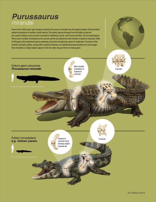
Next, Torsten Scheyer was kind enough to invite me to join his team in studying a fossil I’ve long been fascinated by: the “giant caiman” Purussaurus mirandai, from the Miocene (~6 million years ago?) of Venezuela, in the Urumaco Formation‘s very weird biota. Purussaurus has been known of for >125 years but Torsten’s team noticed that Purussaurus (mirandai) specimens tended to add one of their trunk vertebrae to their hip girdles (sacrum; normally only two vertebrae in Crocodylia but here three), and that the shoulder and hip girdles had unusual bone morphology (straighter, more vertical relative to the body). So they asked me to help interpret these features. And here’s the paper!

Infographic by Torsten Scheyer’s team– click to emcroccen!

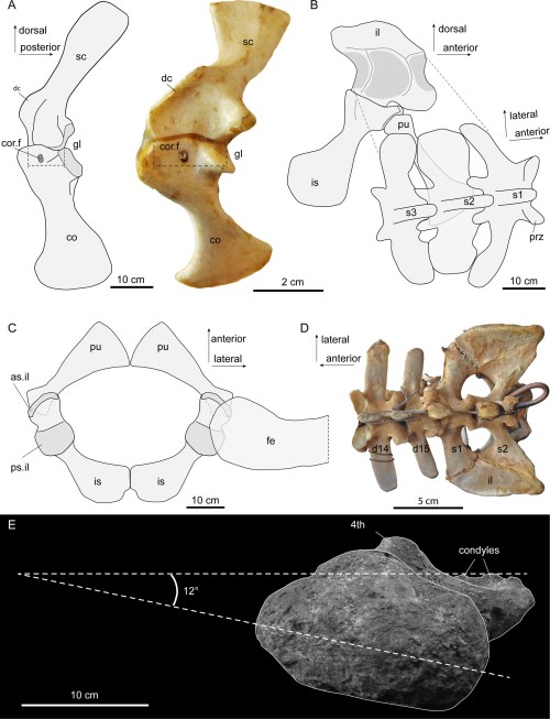
Three-vertebra sacrum and other traits of Purussaurus; with living caiman bones for comparison. E (bottom): inwards-facing femur head. (see paper for more info)

It became evident that, together, those odd traits conveyed a signal that the skeleton was transformed to aid in supporting the huge body against gravity. For example, I found it quite interesting how the head of the femur (thigh bone) was oriented more directly into the hip socket in multiple specimens, more like a dinosaur’s hip, and specialised for support and fore-aft motions. I used Haley O’Brien et al’s data to estimate just how big P. mirandai might have been and it came out as perhaps 3000 kg and 8 metres total length; as we’d thought, among the largest Crocodylia (and there are larger Purussaurus known, too).

Reconstruction of Purussaurus and morphology of the girdles. (see paper for more info)

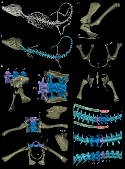
The team also put a cool “evo-devo-biomechanics” spin on the study. It is well known that the regional identities of vertebrae (e.g. neck, trunk, sacrum, tail) are largely determined by Hox (homeobox) regulatory genes, early in development. So changes of vertebral identity intimate changes of genetic controls. Crocodylia don’t normally add a trunk vertebra to their sacrum, and only a few fossil crocodyliforms (extinct cousins) ever did either, but we noticed that some specimens of Crocodylia would at least partially make this transformation in pathological states (below). Hence the controls to make these changes exist and sometimes manifest in living crocs, but it’s probably not an “easy” transformation to achieve. One could speculate that under intense selection, such as that imposed by giant body size and some degree of activity on land, that transformation could more easily get permanently “fixed” in a species.

Palaeosuchus palpebrosus (Cuvier’s dwarf caiman) with pathological partial-three-vertebra-sacrum; and lots more morphology. (see paper for more info)

As a nice tie-in to the asymmetrical gait study above, we can safely infer that the giant Purussaurus wasn’t a fast animal on land, by any means. But its skeleton is consistent with it having found novel ways to maintain the ability to stand and move on land, even if slowly.

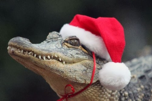
Happy holidays! Santa Jaws is watching you– be good!

Read Full Post »

Today is the 210th anniversary of Charles R. Darwin’s birthday so I put together a quick post. I’d been meaning to blog about some of our latest scientific papers, so I chose those that had an explicit evolutionary theme, which I hope Chuck would like. Here they are, each with a purty picture and a short explainer blurb! Also please check out Anatomy To You’s post by Katrina van Grouw on Darwin’s fancy pigeons.

Stomach-Churning Rating: 1/10 science!

First, Brandon Kilbourne at the Naturkunde Museum in Berlin kindly invited me to assist in a paper from his German fellowship studying mustelid mammals (otters, weasels, wolverines, badgers, etc.; stinky smaller carnivorous mammals). Here we (very much driven by Brandon; I was along for the ride) didn’t just look at how forelimb bone shape changes with body size in this ecologically diverse group. We already knew bigger mustelids would have more robust bones, although it was cool to see how swimming-adapted and digging-adapted mustelids evolved similarly robust bones; whereas climbing ones had the skinniest bones.

The really exciting and novel (yes I am using that much-abused word!) aspect of the paper is that Brandon conjured some sorcery with the latest methods for analysing evolutionary trends, to test how forelimb bone shapes evolved. Was their pattern of evolution mostly a leisurely “random walk” or were there early bursts of shape innovation in the mustelid tree of life, or did shape evolve toward one or more optimal shapes (e.g. suited to ecology/habitat)? We found that the most likely pattern involved multiple rates of evolution and/or optima, rather than a single regime. And it was fascinating to see that the patterns of internal shape change deviated from external shape change such as bone lengths: so perhaps selection sometimes works independently at many levels of bone morphology?

Various evolutionary models applied to the phylogeny of mustelids.

Then there, coincidentally, was another paper originating in part from the same museum group in Berlin. This one I’d been involved in as a co-investigator (author) on a Volkswagen (yes! They like science) grant back about 8 years ago and since. There is an amazing ~290 million year old fossil near-amniote (more terrestrial tetrapod) called Orobates pabsti, preserved with good skeletal material but also sets of footprints that match bones very well, allowing a rare match of the two down to this species level. John Nyakatura’s team had 3D modelled this animal before, so we set out to use digital techniques to test how it did, or did not, move—similar to what I’d tried before with Tyrannosaurus, Ichthyostega and so forth. The main question was whether Orobates moved in a more “ancestral” salamander-like way, a more “derived” lizard-like way (i.e. amniote-ish), or something else.

The approach was like a science sledgehammer: we combined experimental studies of 4 living tetrapods (to approximate “rules” of various sprawling gaits), a digital marionette of Orobates (to assess how well its skeleton stayed articulated in various motions), and two robotics analysis (led by robotics guru Auke Ijspeert and his amazing team): a physical robot version “OroBOT” (as a real-world test of our methods), and a biomechanical simulation of OroBOT (to estimate hard-to-measure things in the other analyses, and matches of motions to footprints). And, best of all, we made it all transparent: you can go play with our interactive website, which I still find very fun to explore, and test what motion patterns do or do not work best for Orobates. We concluded that a more amniote-like set of motions was most plausible, which means such motions might have first evolved outside of amniotes.

OroBOT in tha house!

You may remember Crassigyrinus, the early tetrapod, from a prior post on Anatomy To You. My PhD student Eva Herbst finished her anatomical study of the best fossils we could fit into a microCT-scanner and found some neat new details about the “tadpole from hell”. Buried in the rocky matrix were previously unrecognized bones: vertebrae (pleurocentra; the smaller nubbins of what may be “rhachitomous” bipartite classic tetrapod/omorph structure), ribs (from broad thoracic ones to thin rear ones), pelvic (pubis; lower front), and numerous limb bones. One interesting trait we noticed was that the metatarsals (“sole bones” of the foot) were not symmetrical from left-to-right across each bone, as shown below. Such asymmetry was previously used to infer that some early tetrapods were terrestrial, yet Crassigyrinus was uncontroversially aquatic, so what’s up with that? Maybe this asymmetry is a “hangover” from more terrestrial ancestry, or maybe these bones get asymmetrical for non-terrestrial reasons.

The oddly asymmetrical metatarsals of Crassigyrinus.

Finally, Dr. Peter Bishop finished his PhD at Griffith University in Australia and came to join us as a DAWNDINOS postdoc. He blasted out three of his thesis chapters (starting here) with me and many others as coauthors, all three papers building on a major theme: how does the inner bone structure (spongy or cancellous bone) relate to hindlimb function in theropod dinosaurs (including birds) and how did that evolve? Might it tell us something about how leg posture or even gait evolved? There are big theories in “mechanobiology” variously named Wolff’s Law or the Trajectorial Theory that explain why, at certain levels, bony struts tend to align themselves to help resist certain stresses, and thus their alignment can be “read” to indicate stresses. Sometimes. It’s complicated!

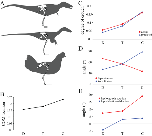
Undaunted, Peter measured a bunch of theropod limb bones’ inner geometry and found consistent differences in how the “tracts” of bony struts, mainly around joints, were oriented. He then built a biomechanical model of a chicken to test if the loads that muscles placed on the joints incurred stresses that matched the tracts’ orientations. Hmm, they did! Then, with renewed confidence that we can use this in the fossil record to infer approximate limb postures, Peter scanned and modelled a less birdlike Daspletosaurus (smaller tyrannosaur) and more birdlike “Troodon” (now Stenonychosaurus; long story). Nicely fitting many other studies’ conclusions, Peter found that the tyrannosaur had a more straightened hindlimb whereas the troodontid had a more crouched hindlimb; intermediate between the tyrannosaur and chicken. Voila! More evidence for a gradual evolution of leg posture across Mesozoic-theropods-into-modern-birds. That’s nice.

Three theropods, three best-supported postures based on cancellous bone architecture.

If you are still thirsty for more papers even if they are less evolutionary, here’s the quick scoop on ones I’ve neglected until now:

(1) Former PhD student Chris Basu published his thesis work w/us on measuring giraffe walking dynamics with force plates, finding that they move mostly like other quadrupeds and their wobbly necks might cost them a little.

(2) Oh, and Chris’s second paper just came out as I was writing this! We measured faster giraffe gaits in the wilds of South Africa, as zoo giraffes couldn’t safely do them. And we found they don’t normally go airborne, just using a rotary gallop (not trot, pace or canter); unlike some other mammals. Stay tuned: next we get evolutionary with this project!

(2) How do you safely anaesthetize a Nile crocodile? There’s now a rigorous protocol (from our DAWNDINOS work).

(3) Kickstarting my broad interest in how animals do “extreme” non-locomotor motions, we simulated how greyhounds stand up, finding that even without stretchy tendons they should, barely, be able to do it, which is neat. Expect much more about this from us in due time.

(4) Let’s simulate some more biomechanics! Ashley Heers, an NSF research fellow w/me for a year, simulated how growing chukar birds use their wing muscles to flap their way up steeper inclines (“WAIR” for devotees), and the results were very encouraging for simulating this behaviour in more detail (e.g. tendons seem to matter a lot) and even in fossil species; and finally…

(5) Hey did you ever think about how bone shape differs between hopping marsupials (macropods) and galloping artiodactyl (even-toed) mammals? We did, in long-the-making work from an old BBSRC grant with Michael Doube et al., and one cool thing is that they mostly don’t change shape with body size that differently, even though one is more bipedal at faster speeds—so maybe it is lower-intensity, slower behaviours that (sometimes?) influence bone shape more?

So there you have the skinny on what we’ve been up to lately, messing around with evolution, biomechanics and morphology.

Read Full Post »

As 2017 approaches its end, there have been a few papers I’ve been involved in that I thought I’d point out here while I have time. Our DAWNDINOS project has been taking up much of that time and you’ll see much more of that project’s work in 2018, but we just published our first paper from it! And since the other two recent papers involve a similar theme of muscles, appendages and computer models of biomechanics, they’ll feature here too.

Stomach-Churning Rating: 0/10; computer models and other abstractions.

Mussaurus patagonicus was an early sauropodomorph dinosaur from Argentina, and is now widely accepted to be a very close relative of the true (giant, quadrupedal) sauropods. Here is John Conway’s great reconstruction of it:

We have been working with Alejandro Otero and Diego Pol on Mussaurus for many years now, starting with Royal Society International Exchange funds and now supported by my ERC grant “DAWNDINOS”. It features in our grant because it is a decent example of a large sauropodomorph that was probably still bipedal and lived near the Triassic-Jurassic transition (~215mya).

In our new study, we applied one of my team’s typical methods, 3D musculoskeletal modelling, to an adult Mussaurus’s forelimbs. This is a change of topic from the hindlimbs that I’ve myopically focused on before with Tyrannosaurus and Velociraptor [in an obscure paper that I should never have published in a book! pdf link], among other critters my team has tackled (mouse, elephant [still to be finished…], ostrich, horse, Ichthyostega… dozens more to come!). But we also modelled the forelimbs of Crocodylus johnstoni (Australian “freshie”) for a key comparison with a living animal whose anatomy we actually knew, rather than reconstructed.

Mussaurus above; Crocodylus below; forelimb models in various views; muscles are red lines.

The methods for this biomechanical modelling are now standard (I learned them from their creator Prof. Scott Delp during my 2001-2003 postdoc at Stanford): scan bones, connect them with joints, add muscle paths around them, and then use the models to estimate joint ranges of motion and muscle moment arms (leverage) around joints. I have some mixed feelings about developing this approach in our 2005 paper that is now widely used by the few teams that study appendicular function in extinct animals. As a recent review paper noted and I’ve always cautioned, it has a lot of assumptions and problems and one must exercise extreme caution in its design and interpretation. Our new Mussaurus paper continues those ruminations, but I think we made some progress, too.

On to the nuts and bolts of the science (it’s a 60 page paper so this summary will omit a lot!): first, we wanted to know how the forelimb joint ranges of motion in Mussaurus compared with those in Crocodylus and whether our model of Mussaurus might be able to be placed in a quadrupedal pose, with the palms at least somewhat flat (“pronated”) on the ground. Even considering missing joint cartilage, this didn’t seem very plausible in Mussaurus unless one allowed the whole forearm to rotate around its long axis from the elbow joint, which is very speculative—but not impossible in Crocodylus, either. Furthermore, the model didn’t seem to have forelimbs fully adapted yet for a more graviportal, columnar posture. Here’s what the model’s mobility was like:

So Mussaurus, like other early sauropodomorphs such as Plateosaurus, probably wasn’t quadrupedal, and thus quadrupedalism must have evolved very close to in the Sauropoda common ancestor.

Second, we compared the muscle moment arms (individual 3D “muscle actions” for short) in different poses for all of the main forelimb muscles that extend (in various ways and extents) from the pectoral girdle to the thumb, for both animals, to see how muscle actions might differ in Crocodylus (which would be closer to the ancestral state) and Mussaurus. Did muscles transform their actions in relation to bipedalism (or reversal to quadrupedalism) in the latter? Well, it’s complicated but there are a lot of similarities and differences in how the muscles might have functioned; probably reflecting evolutionary ancestry and specialization. What I found most surprising about our results was that the forelimbs didn’t have muscles well-positioned to pronate the forearm/hand, and thus musculoskeletal modelling of those muscles reinforced the conclusions from the joints that quadrupedal locomotion was unlikely. I think that result is fairly robust to the uncertainties, but we’ll see in future work.

You like moment arms? We got moment arms! 15 figures of them, like this! And tables and explanatory text and comparisons with human data and, well, lots!

If you’re really a myology geek, you might find our other conclusions about individual muscle actions to be interesting—e.g. the scapulohumeralis seems to have been a shoulder pronator in Crocodylus vs. supinator in Mussaurus, owing to differences in humeral shape (specialization present in Mussaurus; which maybe originated in early dinosaurs?). Contrastingly, the deltoid muscles acted in the same basic way in both species; presumed to reflect evolutionary conservation. And muuuuuuch more!

Do you want to know more? You can play with our models (it takes some work in OpenSim free software but it’s do-able) by downloading them (Crocodylus; Mussaurus; also available: Tyrannosaurus, Velociraptor!). And there will be MUCH more about Mussaurus coming soon. What is awesome about this dinosaur is that we have essentially complete skeletons from tiny hatchlings (the “mouse lizard” etymology) to ~1 year old juveniles to >1000kg adults. So we can do more than arm-wave about forelimbs!

But that’s not all. Last week we published our third paper on mouse hindlimb biomechanics, using musculoskeletal modelling as well. This one was a collaboration that arose from past PhD student James Charles’s thesis: his model has been in much demand from mouse researchers, and in this case we were invited by University of Virginia biomechanical engineers to join them in using this model to test how muscle fibres (the truly muscle-y, contractile parts of “muscle-tendon units”) change length in walking mice vs. humans. It was a pleasure to re-unite in coauthorship with Prof. Silvia Blemker, who was a coauthor on that 2005 T. rex hindlimb modelling paper which set me on my current dark path.

Mouse and human legs in right side view, going through walking cycles in simulations. Too small? Click to embiggen.

We found that, because mice move their hindlimb joints through smaller arcs than humans do during walking and because human muscles have large moment arms, the hindlimb muscles of humans change length more—mouse muscles change length only about 48% of the amount that typical leg muscles do in humans! This is cool not only from an evolutionary (mouse muscles are probably closer to the ancestral mammalian state) and scaling (smaller animals may use less muscle excursions, to a point, in comparable gaits?) perspective, but it also has clinical relevance.

Simulated stride for mouse and human; with muscles either almost inactive (Act=0.05) or fully active (Act=1). Red curve goes through much bigger excursions (along y-axis) than blue curve), so humans should use bigger % of their muscle fibre lengths in walking. Too small? Click to embiggen.

My coauthors study muscular dystrophy and similar diseases that can involve muscle stiffness and similar biomechanical or neural control problems. Mice are often used as “models” (both in the sense of analogues/study systems for animal trials in developing treatments, and in the sense of computational abstractions) for human diseases. But because mouse muscles don’t work the same as human muscles, especially in regards to length changes in walking, there are concerns that overreliance on mice as human models might cause erroneous conclusions about what treatments work best to reduce muscle stiffness (or response to muscle stretching that causes progressive damage), for example. Thus either mouse model studies need some rethinking sometimes, or other models such as canines might be more effective. Regardless, it was exciting to be involved in a study that seems to deliver the goods on translating basic science to clinical relevance.

Muscle-by-muscle data; most mouse muscles go through smaller excursions; a few go through greater; some are the same as humans’.

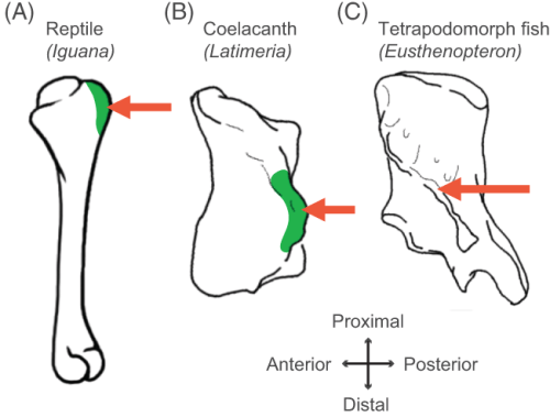
Finally, a third recent paper of ours was led by Julia Molnar and Stephanie Pierce (of prior RVC “Team Tetrapod” affiliation), with myself and Rui Diogo. This study tied together a bunch of disparate research strands of our different teams, including musculature and its homologies, the early tetrapod fossil record, muscle reconstruction in fossils, and biomechanics. And again the focus was on forelimbs, or front-appendages anyway; but turning back the clock to the very early history of fishes, especially lobe-finned forms, and trying to piece together how the few pectoral fin muscles of those fish evolved into the many forelimb muscles of true tetrapods from >400mya to much more recent times.

Humerus in ventral view, showing muscle attachments. Extent (green) is unknown in the fossil but the muscle position is clear (arrow).

We considered the homologies for those muscles in extant forms, hypothesized by Diogo, Molnar et al., in light of the fossil record that reveals where those muscles attach(ed), using that reciprocal illumination to reconstruct how forelimb musculature evolved. This parallels almost-as-ancient (well, year 2000) work that I’d done in my PhD on reconstructing hindlimb muscle evolution in early reptiles/archosaurs/dinosaurs/birds. Along the way, we could reconstruct estimates of pectoral muscles in various representative extinct tetrapod(omorph)s.

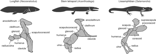
Disparity of skeletal pectoral appendages to work with from lobe-fins to tetrapods.

Again, it’s a lengthy, detailed study (31 pages) but designed as a review and meta-analysis that introduces readers to the data and ideas and then builds on them in new ways. I feel that this was a synthesis that was badly needed to tie together disparate observations and speculations on what the many, many obvious bumps, squiggles, crests and tuberosities on fossil tetrapods/cousins “mean” in terms of soft tissues. The figures here tell the basic story; Julia, as usual, rocked it with some lovely scientific illustration! Short message: the large number of pectoral limb muscles in living tetrapods probably didn’t evolve until limbs with digits evolved, but that number might go back to the common ancestor of all tetrapods, rather than more recently. BUT there are strong hints that earlier tetrapodomorph “fishapods” had some of those novel muscles already, so it was a more stepwise/gradual pattern of evolution than a simple punctuated event or two.

Colour maps of reconstructed right fin/limb muscles in tetrapodomorph sarcopterygian (~”fishapod”) and tetrapod most recent common ancestors. Some are less ambiguous than others.

That study opens the way to do proper biomechanical studies (like the Mussaurus study) of muscle actions, functions… even locomotor dynamics (like the mouse study)– and ooh, I’ve now tied all three studies together, tidily wrapped up with a scientific bow! There you have it. I’m looking forward to sharing more new science in 2018. We have some big, big plans!

Read Full Post »

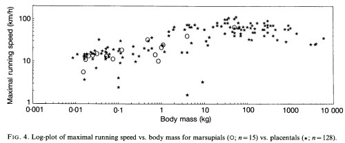
I still have my original photocopy, from my grad school days circa 1996, of the 1983 Ted Garland classic paper “The relation between maximal running speed and body mass in terrestrial mammals”, festooned with my comments and highlighter pen marks and other scribblings. That paper remains the backbone of many research questions I am interested in today, and I often think about its underlying concepts. Here’s the key scatterplot from that paper, which I could almost replot by hand from memory, it is so full of implications (and can be clicked to embiggen it, perhaps even speedily depending on your internet connection):

Garland 1983- max speed

Stomach-Churning Rating: 1/10; data and their ramifications; offal-free.

The major points (IMO there are less exciting ones about which theoretical scaling model the data best fit) of the paper are: (1) the fastest-running mammals are neither the smallest nor largest, but those around ~100 kg body mass; (2) if you fit a linear equation to the data (see above; hashed line), it seems like speed increases with body mass linearly (with no limit to that increase, within the body mass range of the data), but if you analyze individual groups of mammals they either don’t change speed significantly with size or they get slower– refer back to point #1 and the polynomial regression that is shown in the figure above (curved line). That’s the biological-question-driven science at the core of the paper (with some methods-y questions at their foundation; e.g. should we use a linear or polynomial regression to fit the data? The latter fits best, and gives a different answer from the former, so it matters.).

But what also fascinates me is the question of data. As the author, who taught me Evolution as an undergrad at U Wisconsin (this had a big impact on me), fully admits in the paper, the ~3-page table of data “necessarily sacrifices some accuracy for completeness”. This paper is about a big question, how mammal speed changes with size, and so its big question explicitly allows for some slop in the data (I will return to this issue of slop later). But given that very few of the data points have very accurate measurements for speed, or for body mass for that matter, how much can we trust an x-y plot of those data, no matter what method is used? Oh there is so much opportunity here for geeky pedantry and niggling scrutiny of data points, true, but hold on…

Plenty of follow-up papers have mused over that latter question, and spin-off ones. Here are some of their plots, re-analyzing the same or very similar data in different contexts. A look at how these papers examine these data and related questions/methods leads into some avenues of science that fascinate me:

Garland 1988- max perf

Garland and Baudinette (link to pdf here) checked whether placental (i.e. most; including us) mammals could run/hop faster than marsupial (pouched; e.g. kangaroos) mammals. Their results said “not really”, as the plot intimates. Scatter in the data, especially between 0.01-10 kg, confounds the issue- there’s a lot of specialization going on (notably, animals that are very slow for their size, e.g. sloths). But marsupials are not, as had been suggested before, inferior to placentals in some basic way such as running ability.

GarlandJanis1993-Fig5

Above, Garland and Janis 1993 (link to pdf here) examined how the ratio of metatarsal (“sole bones” of the lower end of the leg/foot) vs. femur (thigh bone) length relate to speed, with evolutionary relationships taken into account. The methods (“independent contrasts” and its conceptual kin; I won’t delve into that morass more here!) did not exist for looking at phylogeny’s effects on the results in Garland’s 1983 paper. Yet “cursoriality” (relative elongation of the lower limb) had been thought to relate to running speed for over 80 years at that time, so that was what they tested: how much does limb-elongation correlate in a positive way with maximal running speed? They found that the answer was “sort of”, but that other things like home range size, energetics, ecology, etc. might explain as much/more, so caveat emptor. And by looking at the plot above, it’s evident that there’s a lot of specialization (scatter, along the x and/or y axes– check out the giraffe/Giraffa and cheetah/Acinonyx outliers, for example). While ungulates seemed to have a better relationship of speed and limb dimensions, their predatory carnivoran relatives did not.Christiansen 2002- max speed

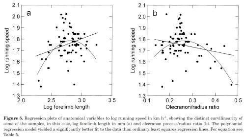
Christiansen was one of two studies in 2002 that looked back on those Garland 1983 data in a new way, and like the 1993 study with Janis considered these data in light of limb lengths too.  The plot above delved into how running speed changes with lengths of forelimb bones, again finding appreciable curvilinearity (indirectly supporting the non-linear scaling idea– even at large sizes, relatively longer-legged mammals aren’t faster). The plot on the right side (b) measured the relative length of the olecranon process; the “funny bone” that acts as a lever for support of the elbow joint against gravity. Again, even mammals that have stouter elbow-supporting processes aren’t faster; there’s a “happy medium” of elbow-osity for optimizing running speed (and huge scatter in the data!). Ultimately, this analysis concluded that it wasn’t speed that animal anatomy seemed to be optimizing overall, especially as size increased, but rather energetic cost, although there was a lot of variation in the data and accounting for phylogeny only muddled things up more (as it tends to do).

diaz2002

Iriarte-Diaz was the other 2002 study to tackle the speed-vs-size issue. It focused primarily on whether mammal speeds showed “differential” (i.e. non-linear) scaling with size, as per the polynomial regression in Garland’s 1983 study. It showed that smaller mammals seemed to either get slightly slower with increasing size or else not change maximal speed (depending on detailed methods/data stuff that don’t matter here), whereas bigger mammals exhibited very strong declines of speed with size past a threshold (optimal) body mass.

So, repeated analyses of Garland’s 1983 data (and modifications of those data) at least uphold the fundamental conclusion that big land mammals cannot move quickly, in an absolute sense (meters/sec or kph or mph) — and much more so in a relative sense (e.g. body lengths/second or other normalized metrics). We might then ask why, and my research scrutinizes this issue in terms of the fundamental mechanisms of movement biomechanics and anatomy that might help to explain why, but for brevity I won’t go there in this post. I want to wander elsewhere.

I want to wander back to those data used in the above (and other) studies. All of the studies discuss the quality of the data and bemoan the lack of quality. I’d agree with them that it’s hard to imagine most of the data being consistently off in a biased way that would fundamentally alter their conclusions. But I still worry. We should worry about the data points for the extreme animals- the fastest, slowest, largest and smallest. We should worry about subjectively removing “outliers” such as hippos or cheetahs, as they do change some of the results.

I worry about elephants, for example: my work has shown that they can “run” about 7 meters/second or ~25 kph; not the 35 kph used as data for African elephants (from speedometer-y anecdotal estimates)– ~1.4 times the speed we’ve been able to measure for both species. See this old “blog post” (sort of) for more information on the tortuous history of characterizing elephant speeds and gaits. And are a white rhino and hippo able to run at this same 25 kph speed as the original data in the 1983 study state, or faster/slower? No one has really nicely measured this so we can’t be sure, but I can imagine it being off by a similar 40% or so. On the other hand, if the bigger animals in the dataset are slower than the original data, that actually strengthens the conclusion that bigger animals are slower, so who cares that much, in the grand scheme of things?

We could worry about plenty of other maximal speed data points, and the “average” adult body masses assumed (although I doubt those would change the results as much as the speed errors). Maybe another question is, in doing such broad-scale analyses should we only include data points that have maximal precision (e.g. elephants, horses, cheetahs, greyhounds, humans and a few others)? We’d maybe be able to do a study of 20 or so species. I doubt it would show much that is different if we did, although I expect that sample size and noise would begin to dampen out the signal. See below.

However, a double standard begins to become evident here. In modern biomechanics (and probably the rest of biology/science), there’s a strong emphasis on data quality and technologically precise measurement. Garland’s 1983 study might be hard to get past peer review today (or maybe not). We agonize over single-species studies trying hard to measure animals’ maximal speeds (a very hard thing to be sure of in terms of motivation, but not intractable unless one takes an almost antiscientific/overly cynical view that animals could always be holding back some critical reserve unless they run for their lives– is that reserve 1%, 10% or 100%? Probably closer to the middle, in good studies). We measure multiple animals and many trials, in field and/or lab conditions, with documented video footage at high resolution and frame rate, with GPS tracking or other tools to maximize precision. We take pride in these high standards today. That’s what makes scientists wriggle uncomfortably when we look back at the data in those older maximal speed papers and ponder how few data points are verified, documented, precise and essentially trustworthy.

So should broad studies be working by the same standards as narrow studies? (I’m far, far, far from the first scientist to think about this but it’s interesting for me at least to think about it in this case and others) There is potential tension here between empiricists who want precise data and theoreticians who want to tackle those Big Questions, and that’s a pattern one can see throughout much of science. I sit on the fence myself, doing both approaches. I can think of plenty of similar examples, in “big data” palaeobiology, morphometrics, genomics, physics and so on. Some of those fields have nice databases with quality control over the data; they’ve maybe solved this problem to a large degree. This tiny area of mammalian maximal speeds hasn’t solved it, but how urgent is the need to?

On the flip side, even if the data points have some error of 10-20% or even 40% that error will probably be largely random, not biased toward assuming that bigger or smaller animals are slower than they truly are, or medium-sized animals faster. We still have the reliable cheetah data point (and racehorses, and greyhounds) showing >100 kph (and 70 kph) speeds for ~100 (and ~40, 400ish) kg animals, so there is evidence for a peak of maximal speed (the cheetah outlier, and one might also throw in pronghorn antelope or others that are pretty damn fast but not yet well measured) at medium body size. I expect there would be incremental overall progress if we did improve the data quality, and that would still be nice (comforting!) but it would be a tough, tough slog. Indeed, my team is doing its share of that, already tackling the data point for giraffes this year (stay tuned!). The potential gains are still there, especially for understanding the unique biology of individual species– that noise in the data (or specialization, if you prefer) is interesting!!! We need that kind of work, partly because the big questions, sexy as they are, still depend on having data quality as a foundation, and old questions still need revisiting from time to time as data quality is improved by those in the trenches of gathering it.

My team’s journal club has gone over the Garland paper lately and we’re hitting the others later this summer, but I wanted to throw these thoughts out there on this blog now to see if they generated any fun discussion, or they might introduce others to the science of maximal speeds and what we do/don’t know. One thing we don’t know much about is what kinds of patterns non-mammalian groups exhibit today. Chris Clemente did some great work on this with lizards, finding a pattern similar to the mammal one. I’ve struggled in my work to move toward trying to address similar questions for extinct groups, but there the data quality presents a challenge I find exciting rather than depressing, although I still have to shrug when I see limb lengths or proportions being used as a proxy for speed. We can do better.

So I’d love to hear your thoughts on any of the points here. Maybe some of the old-timers have stories from ye olden days when Garland’s work was originally published; I’d love to hear those, or other points/questions/favourite papers.

Read Full Post »

Who needs “Ice Road Truckers” when you have the “John’s Freezer” team on the road with fossils, amphibians, felids and 3D phenotype fun? No one, that’s who. We’re rocking the Cheltenham Science Festival for our first time (as a group), and pulling out all the stops by presenting two events! Here’s the skinny on them, with updates as the week proceeds.

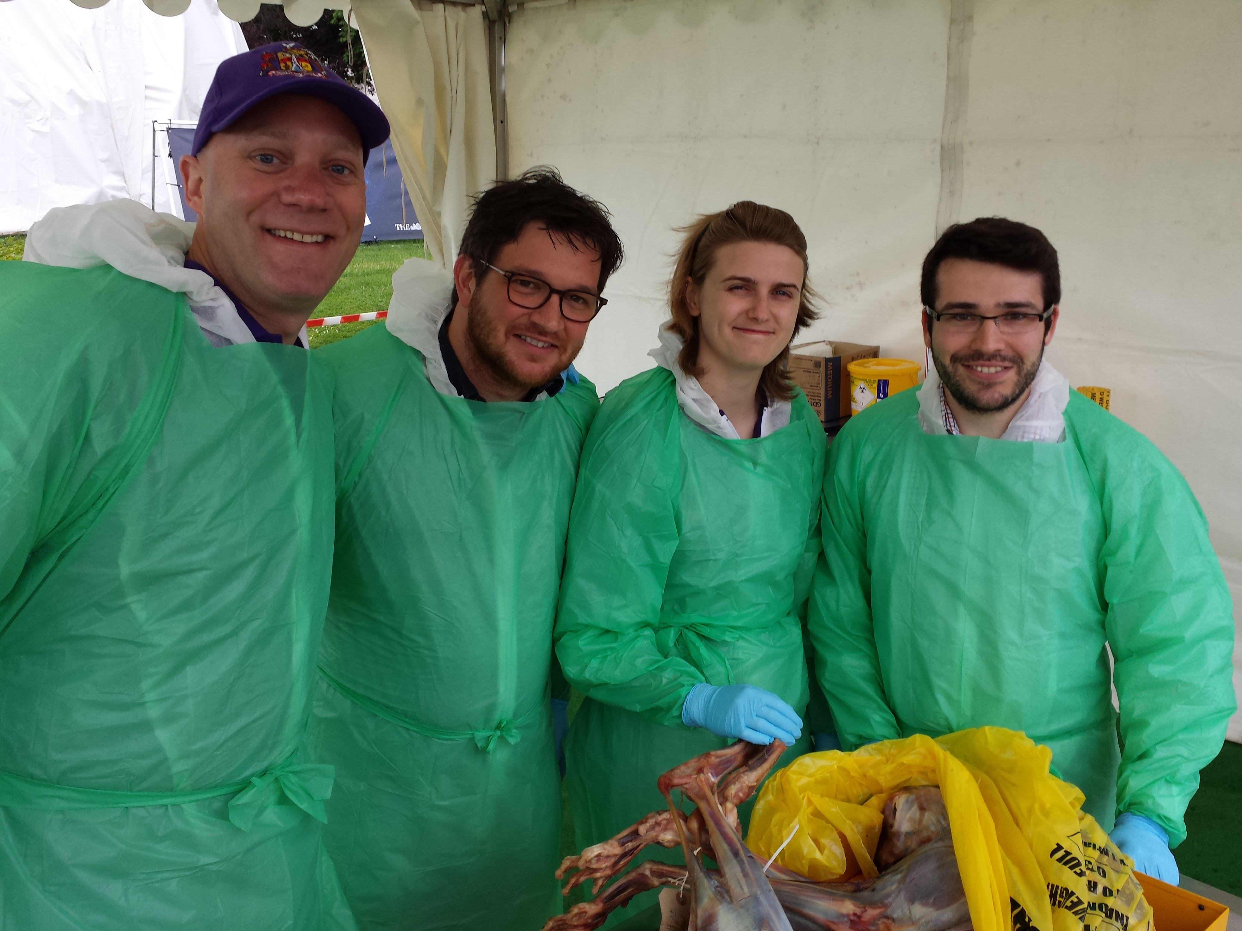
Stomach-Churning Rating: 2/10 for now (just bones), but it could change once the cheetah dissection is under way… 8/10 bloody cheetah bits but only at the end (updated)

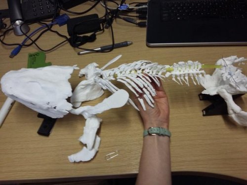
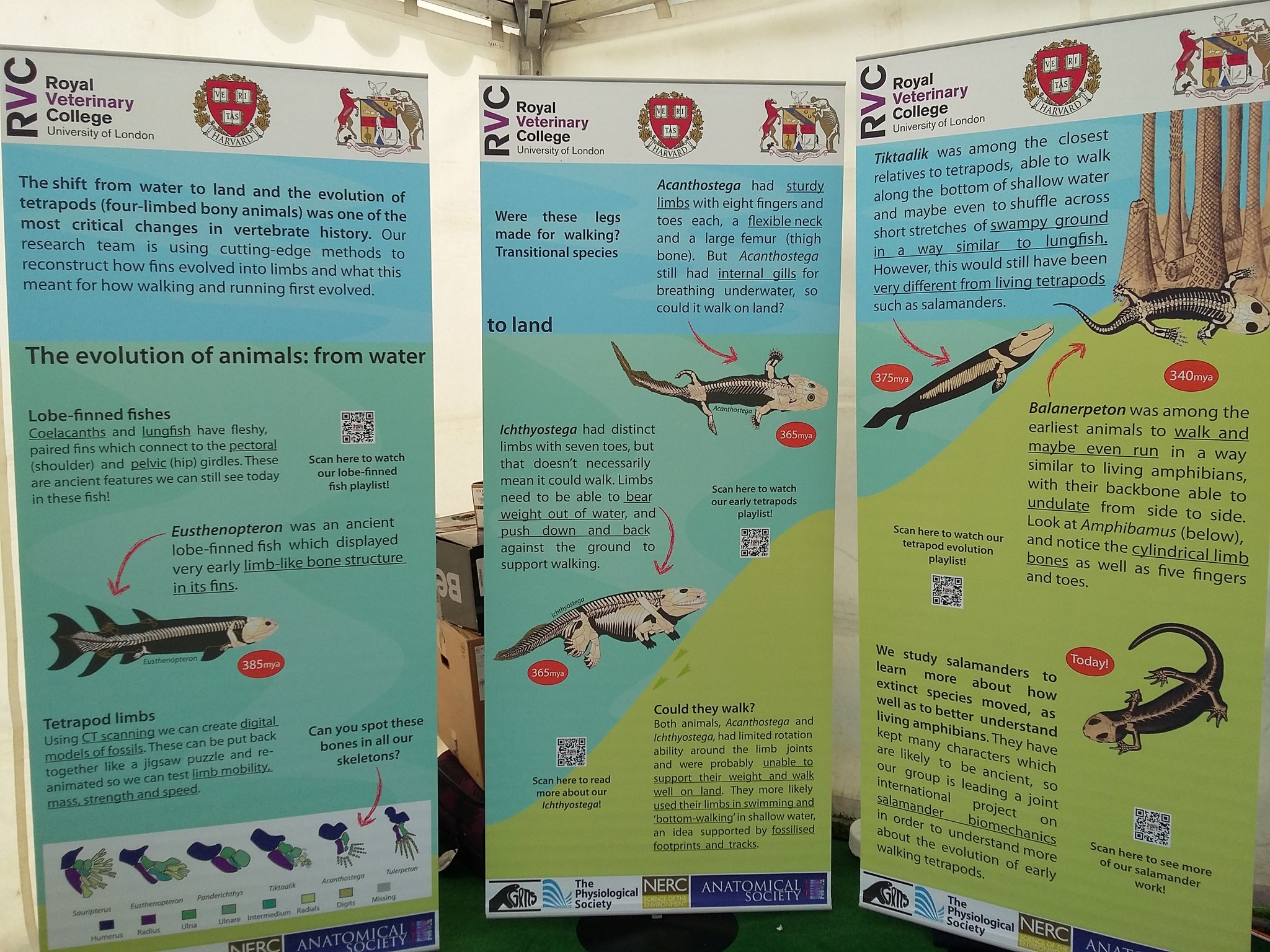
Right now, Lauren Sumner-Rooney (of “Anatomy To You” and other fame) is on-site with a rotating team of others from our lab, in the “Free Activity Tents” area of the Imperial Gardens/Square, inside a marquee where we’ll be showing off our NERC-funded tetrapod research all week. This “First Steps” event features not only our past and present work with Jenny Clack, Stephanie Pierce, Julia Molnar and others on Ichthyostega & its “fishapod” mates, but also our “scampering salamanders” research in Spain, Germany and England. I’ve blogged a lot about all that, and won’t repeat it here, but you can see a 3D-printed Ichthyostega skeleton, view the skeleton in a virtual reality 3D environment, see related specimens and engage in kid-friendly activities, and talk to our team about this and other related research.

Ichthyostega 3D printed backbone is born!

Ichthyostega 3D printed backbone is born!

The central themes of that event are how bone structure relates to function and how we can use such information, along with experimental measurements and computer models of real salamanders, to reconstruct how extinct animals might have behaved as well as how swimming animals became walking ones. How did fins transform into limbs and what did that mean for how vertebrates made the evolutionary transition onto land? If you know my team’s work, that encapsulates our general approach to many other problems in evolutionary biomechanics (e.g. how did avian bipedalism evolve?). Added benefits are that you too can explore this theme in a hands-on way, and you can talk with us about it in person. That continues all week (i.e. until Saturday evening); I’ll be around from Thursday afternoon onwards, too. Kids of all ages are welcome!

Ichthyostega 3D print taking shape!

Ichthyostega 3D print taking shape!

Then, on Saturday for our second free event we join forces with Ben Garrod (master of primate evolution, the secrets of bones, and “Attenborough and the Giant Dinosaur”) and RVC’s forensic pathologist Alexander Stoll as well as Sophie Regnault (“sesamoid street” PhD student w/me). As the “Large Animal Dissection” title hints, it’s not the right kind of gig to bring small kids to. There will be blood and stuff— we’ll be dissecting a cheetah together from 10am-4pm. This will involve walking through all the major organ systems, giving evolutionary anecdotes, and plenty more, with an aim to understand how the magnificent adaptations of cheetahs evolved—but also to investigate what problem(s) this animal faced that led to its sad demise. By the day’s end, there will just be a skeleton left. Get a front row seat early for this event, which serendipitously ties into “Team Cat”’s Leverhulme Trust-funded research project (we wanted a big animal and it just happened to be a cheetah; I had hoped for a giant croc or a shark or something but can’t complain!).

Ichthyostega 3D print is ready!

Ichthyostega 3D print is ready!

If you miss these events, please do cry bitter tears of regret. But don’t despair, there will be another “big cat dissection” in the London area in ~November (watch here for details), and plenty more fossil tetrapod stuff to come, plus a LOT more dinosaurs on the horizon!

Guess the bones! (photo by Zoe Self)

Guess the bones! (photo by Zoe Self)

And please come back to this blog post for more pics and stories as the week carries on… For hashtag afficionados, you can follow the fun on Twitter etc. at #firststepsCSF16. What a world we live in!

Update 1: While you’re here, check out our Youtube playlists of tetrapod-related videos:

Lobe-finned fishes

Ichthyostega‘s awesome anatomy

Tetrapod evolution: Tiktaalik to salamanders!

Update 2: Photos of our main stand (about tetrapod evolution)

csf2016-display

Our poster/banner display looks nice.

20160609_143548

Our tent brings in some punters.

20160609_151508

Our bones excite people young and old, sighted and blind.

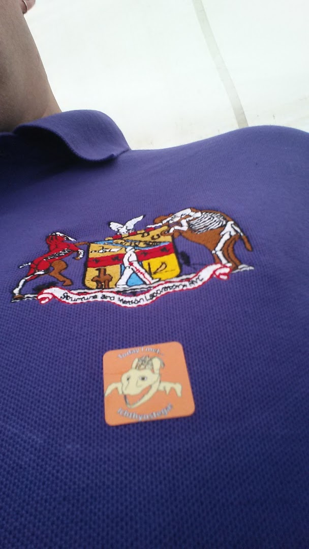
20160610_090009

Fun with stickers and lab t-shirts.

And…

Update 3: Cheetah meat & greet
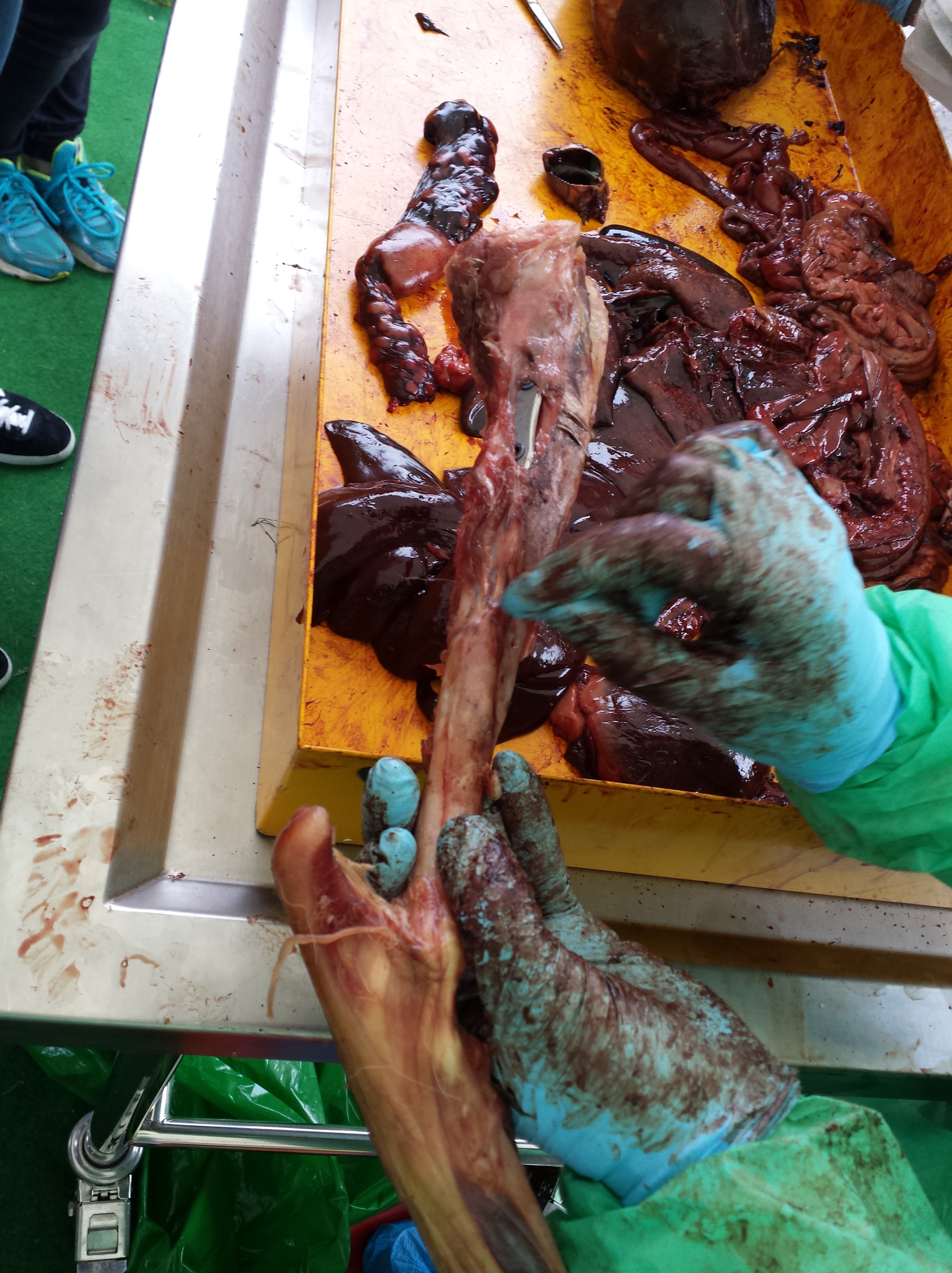
Ben, Alex, Sophie and I tackled the cheetah dissection today and it went GREAT! Much better than I’d optimistically expected. Rain didn’t scare the crowds off and neither did the gore, which there was some of (gelatinous spinal cords, lumpy tumors and at least one flying tiny bit of cheetah flesh that landed on a good-natured audience member!). Photos will tell the tale:

20160611_091345

Peek-a-boo!

20160611_093828

Sophie and Alex help us get set up in our tent.

20160611_094554

Our initial rough schedule- although we ended up improvising more after lunch.

20160611_095500

Dissectors assemble!

20160611_105126

The beast revealed. It was skinned by the museum that loaned it to us.

20160611_110748

Alex showing his talent: removing the viscera in one piece from end to end, starting with the tongue.

20160611_153148

Impressive finding of a surgical fixture (plate and wires) on the tibia, which had been used to hold the shattered bone back together long enough for it to heal. Added to the kidney disease and liver-spleen-lung cancer, this cheetah was in the sorriest shape of any cadaver I’ve seen yet.

20160611_153213

Cheetah coming to pieces: (from bottom) lumbar/pelvic region, hindlimb, thorax, forelimb and other bits.

20160611_155452

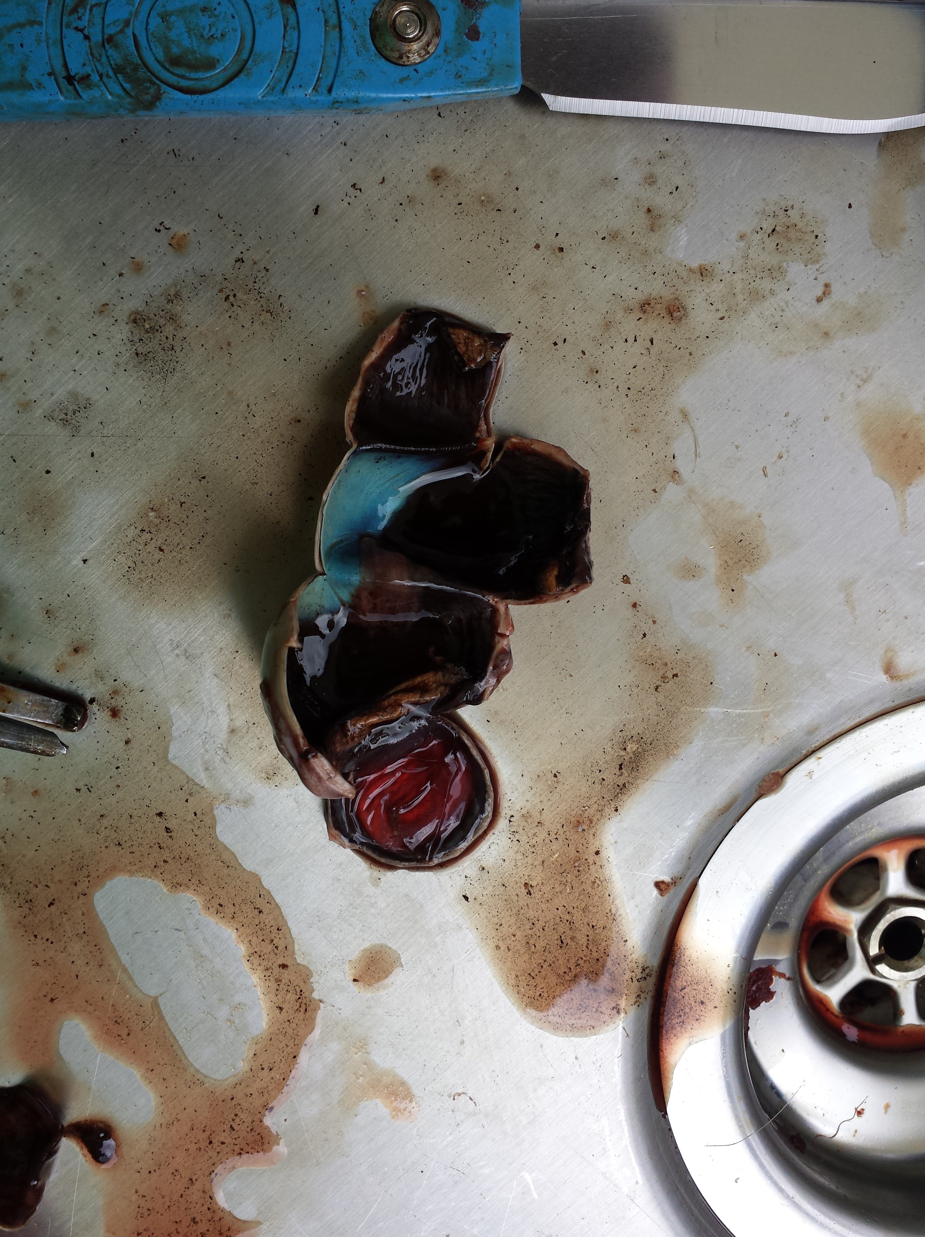
Dr Adam Rutherford, an eye expert, did a nice dissection of the cheetah’s eye, here showing the tapetum lucidum (reflective membrane), which shows up as light blue colour. Its small size befits the not-very-nocturnal habits of cheetahs.

20160611_155459

The lens of the cheetah’s eye. Now cloudy because of dehydration and crystalization, but still fascinating to see.

Want to see more images and the enthusiastic responses from the audience (we got some great feedback)? Check out Twitter’s #cheltscifest feed, or more simply my Storify condensation of the cheetah-related tweets here.

Read Full Post »

Last year we finally, after about 14 years of slow work, released our biomechanical model of an ostrich’s hindleg. We showed how it informed us about the potential leverages (moment arms; contributions to mechanical advantage of the joints) of all of the muscles. It was a satisfying moment, to understate it, to finally publish this work from my postdoc at Stanford. Today, we begin to deliver on that model’s promise. And it only took 4 years or so, roughly? The journal Royal Society Interface has published our study of how we used this musculoskeletal model to simulate walking and running dynamics. Those simulations join an intimidatingly broad and complex literature using similar models to study human (and some other primate) locomotion or other functions at the level of individual muscles (for whole limbs/bodies) in vast detail and growing rigor. I have Dr. Jeffery Rankin, a research fellow finishing up his post with me after ~6 years of hard work on many projects, to thank for driving this work forward, and Dr. Jonas Rubenson (now at PennState) for his patient collaboration that has continued since the early 2000’s.

Stomach-Churning Rating: 2/10; computer models of muscle actions. The underlying anatomical data are goopy, as prior ostrich-dissection-focused posts show!

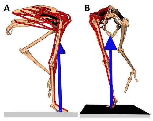
Our model; in right side view (on the left) and frontal view (on the right), with muscles in red and the leg's force as the blue arrow; frozen at the middle of a step.

Our model; in right side view (on the left) and frontal view (on the right), with muscles in red and the leg’s force as the blue arrow; frozen at the middle of a running step.

Simulations like these predict things that we can’t easily measure in living animals, such as how much force muscles and tendons generate, how quickly those tissues change length, how much mechanical energy they thus contribute to the joints, limbs and whole body, how much metabolic energy their actions cost, and much more. There are more ways to use these tools than I have space or time to explain, but simply put we forced our ostrich simulation to match experimental measurements of the motions and forces of a representative walking and running ostrich stride, from contact of one foot until the next time that foot hit the ground. It then used optimization methods (minimizing target criteria like muscle stress) to estimate how the muscles and tendons were used to generate those motions and forces. This is a ways ahead of some prior ostrich simulations such as this one that I recall from classes during my PhD studies.

Any modeller worth their salt knows that their models and simulations are wrong at some level. This is much as most science is “wrong”; i.e. a simplification of reality with some errors/noise introduced by assumptions, variation, methods and such. But generally these kinds of musculoskeletal dynamic simulations hold up pretty well against experimental data. A standard “validation” is to test how closely the simulations’ predictions of muscle activity match the “real” (measured in life, also with some uncertain error) activity of muscles. Science still lacks those data for ostriches, but fortunately measurements from other birds (by Steve Gatesy and colleagues) indicate that muscles tend to follow fairly conservative patterns. Grossly speaking, avian leg muscles tend to either be active mainly when the foot is on the ground (stance phase) or off the ground (swing phase). Some studies acknowledge that this is an oversimplification and other muscles do act across those two phases of a stride, either in multiple pulses or as “transitional” (stance-to-swing or swing-to-stance) switch-hitters in their activations. Our ostrich predictions matched the qualitative patterns for avian muscle activations measured to date, so that’s good. The results also reinforced the notion of transitional or multi-phasic muscle activation as still having some importance, which bears more study.

Yet what did the simulations with our ostrich model tell us that other ostrich experiments or other bird species didn’t? Three main things. First, we could calculate what the primary functions of muscles were; they can act as motors (generating energy), brakes (absorbing energy), springs (bouncing energy back and forth) or struts (just transmitting force). We could then sum up what whole muscle groups were doing overall. The image below shows how these broad functions of groups vary across the stance phase (swing phase is harder to condense here so I’ve left it out).

Positive work can speed things up; negative work can slow things down.

Positive work can speed things up; negative work can slow things down. Solid bars are running; striped bars are walking. (from our Fig. 13) You may need to click to em-broaden this image for the gory biomechanical details.

There’s a lot going on there but a few highlights from that plot are that the hip extensor (antigravity) muscles (biarticular hip/knee “hamstrings”) are acting like motors, the knee extensors (like our quadriceps) are mainly braking as in other animals and the ankle is fairly springy as its tendons (e.g. digital flexors; gastrocnemius) suggest. We often characterize birds as “knee-driven” but it’s more accurate biomechanically to say that their hips drive (power; i.e. act as motors) their motion, whereas their knees still act as brakes — in both cases as in many other land vertebrates. Thus in some ways bird legs don’t work so unusually. Birds like ostriches are, though, a little odd in how much they rely on their hamstring muscles to power locomotion (at the hip) rather than their caudofemoral muscles, which are reduced. Zooming in on some particular muscles such as parts of the hip or knee extensors, the functions sometimes weren’t as predictable as their similar anatomy might suggest. Some muscles had parts that turned on during swing phase and other parts used during stance phase. Neural control and mechanics can produce some unexpected patterns.

Second, we looked at one important methodological issue. Simulations of musculoskeletal dynamics can vary from simple static (assuming each instant of a motion is independent from the others; e.g. ignoring acceleration, inertia, tendon effects, etc.) to more complex grades of fully dynamic flavours (e.g. assuming rigid or flexible tendons). We looked across this spectrum of assumptions, for both walking and running gaits, with the expectation that more static assumptions would work less well (vs. more dynamic ones; by various criteria) for running vs. walking. This also showed us how much tendons influence our simulations’ estimates of muscle mechanics—a fully rigid tendon will make the muscle do all of the work (force times length change) whereas a flexible tendon can literally take up some (or even all) of that slack, allowing muscles to remain closer to their isometric (high force-generating, negligible length change) quasi-optimum.

Nicely, our predicted muscle functions weren’t influenced much by these methodological variations. However, static assumptions  clearly were in some ways less appropriate for running than for walking, as were flexible tendons. Somewhat surprisingly, making the simulations more dynamic didn’t lower the levels of activation (and thus presumably the metabolic costs) of muscles, but actually raised those levels. There are good reasons why this might be realistic but it needs further study. It does muddy the waters for the issue of whether assuming that rapid locomotion can be modelled as static is a “bad” thing such as for estimating maximal speeds—yes, tendons can do more (elastic energy storage, etc.) if more dynamic models are used, but co-contraction of antagonistic muscles against each other also brings in some added costs and might lead to slower speed estimates. We’ll see in future work…

Finally, one often overlooked (sometimes even undiscussed!) aspect of these simulations is that they may silently add in extra forces to help muscles that are struggling to support and move their joints. The justification is typically that these extra “reserve actuators” are passive tissues, bony articular forces and other non-muscular interactions. We found that the hip joint muscles of ostriches were very weak at resisting abduction (drawing the thigh away from the body) and this needed resisting during the stance phase, so our simulations had very high reserve actuators switched on there. That fits the anatomy pretty well and needs more investigation.

Want to know more? Happily, not only is the paper free for anyone to view but so are all of the data including the models (modified slightly from our last paper’s). So, although the software (Opensim) isn’t ideal for 4-year-olds to play with (it is fancy engineering stuff), if you have the interest and dilligence it is there to play with and re-use and all that. But also watch this space for future developments, as there is more to happen with our steadily improving models of ostriches and other beasties. Anyway, while this paper is very technical and challenging to explain I am not too bashful to say it’s one of the finer papers from my career; a big stride forward from what we’ve done before. I have been looking forward for a long time to us getting this paper out.

P.S. Our peer reviewers were splendid- tough but constructive and fair. The paper got a lot better thanks to them.

Read Full Post »

Deck the ‘Nets With PeerJ Papers— please sing along!

♬Deck the ‘nets with PeerJ papers,
Fa la la la la, la la la la.
‘Tis the day to show our labours,
Fa la la la la, la la la la.

Downloads free; CC-BY license,
Fa la la, la la la, la la la.
Read the extant ratite science,
Fa la la la la, la la la la.

See the emu legs before you
Fa la la la la, la la la la.
Muscles allometric’ly grew.
Fa la la la la, la la la la.

Follow the evolvin’ kneecaps
Fa la la la la, la la la la.
While we dish out ratite recaps 
Fa la la la la, la la la la.

Soon ostrich patellar printing
Fa la la la la, la la la la.
Hail anat’my, don’t be squinting
Fa la la la la, la la la la.

Dissections done all together
Fa la la la la, la la la la.
Heedless of the flying feathers,
Fa la la la la, la la la la♪

(alternate rockin’ instrumental version)

Stomach-Churning Rating: 5/10: cheesy songs vs. fatty chunks of tissue; there are no better Crimbo treats!

Today is a special day for palaeognath publications, principally pertaining to the plethora of published PeerJ papers (well, three of them anyway) released today, featuring my team’s research! An early Crimbo comes this year in the form of three related studies of hind limb anatomy, development, evolution and biomechanics in those flightless feathered freaks of evolutionary whimsy, the ratites! And since the papers are all published online in PeerJ (gold open access), they are free for anyone with internet access to download and use with due credit. These papers include some stunning images of morphology and histology, evolutionary diagrams, and a special treat to be revealed below. Here I’ll summarize the papers we have written together (with thanks to Leverhulme Trust funding!):

1) Lamas, L., Main, R.P., Hutchinson, J.R. 2014. Ontogenetic scaling patterns and functional anatomy of the pelvic limb musculature in emus (Dromaius novaehollandiae). PeerJ 2:e716 http://dx.doi.org/10.7717/peerj.716 

My final year PhD student and “emu whisperer” Luis Lamas has published his first paper with co-supervisor Russ Main and I. Our paper beautifully illustrates the gross anatomy of the leg muscles of emus, and then uses exhaustive measurements (about 6524 of them, all done manually!) of muscle architecture (masses, lengths, etc.) to show how each of the 34 muscles and their tendons grew across a more than tenfold range of body mass (from 6 weeks to 18 months of age). We learned that these muscles get relatively, not just absolutely, larger as emus grow, and their force-generating ability increases almost as strongly, whereas their tendons tend to grow less quickly. As a result, baby emus have only about 22% of their body mass as leg muscles, vs. about 30% in adults. However, baby emus still are extremely athletic, more so than adults and perhaps even “overbuilt” in some ways.

This pattern of rapidly growing, enlarged leg muscles seems to be a general, ancestral pattern for living bird species, reflecting the precocial (more independent, less nest-bound), cursorial (long-legged, running-adapted) natural history and anatomy, considering other studies of ostriches, rheas, chickens and other species close to the root of the avian family tree. But because emus, like other ratites, invest more of their body mass into leg muscles, they can carry out this precocial growth strategy to a greater extreme than flying birds, trading flight prowess away for enhanced running ability. This paper adds another important dataset to the oft-neglected area of “ontogenetic scaling” of the musculoskeletal system, or how the locomotor apparatus adapts to size-/age-related functional/developmental demands as it grows. Luis did a huge amount of work for this paper, leading arduous dissections and analysis of a complex dataset.

Superficial layer of leg muscles in an emu, in right side view.

Superficial layer of leg muscles in an emu, in right side view. Click any image here to emu-biggen. The ILPO and IC are like human rectus femoris (“quads”); ILFB like our biceps femoris (“hams”); FL, GM and GL much like our fibularis longus and gastrocnemius (calf) muscles, but much much bigger! Or, perhaps FL stands for fa la la la la?

Data for an extra set of emus studied by coauthor Russ Main in the USA, which grew their muscles similarly to our UK group. The exponents (y-axis) show how much more strongly the muscles grown than isometry (maintaining the same relative size), which is the dotted line at 1.0.

Data for an extra set of emus studied by coauthor Russ Main in the USA, which grew their muscles similarly to our UK group. The exponents (y-axis) show how much more strongly the muscles grew than isometry (maintaining the same relative size), which is the dotted line at 1. The numbers above each data point are the # of individuals measured. Muscle names are partly above; the rest are in the paper. If you want to know them, we might have been separated at birth!

2) Regnault, S., Pitsillides, A.A., Hutchinson, J.R. 2014. Structure, ontogeny and evolution of the patellar tendon in emus (Dromaius novaehollandiae) and other palaeognath birds. PeerJ 2:e711 http://dx.doi.org/10.7717/peerj.711

My second year PhD student Sophie Regnault (guest-blogger here before with her rhino feet post) has released her first PhD paper, on the evolution of kneecaps (patellae) in birds, with a focus on the strangeness of the region that should contain the patella in emus. This is a great new collaboration combining her expertise in all aspects of the research with coauthor Prof. Andy Pitsillides‘s on tissue histology and mine on evolution and morphology. This work stems from my own research fellowship on the evolution of the patella in birds, but Sophie has taken it in a bold new direction. First, we realized that emus don’t have a patella– they just keep that region of the knee extensor (~human quadriceps muscle) tendon as a fatty, fibrous tissue throughout growth, showing no signs of forming a bony patella like other birds do. This still blows my mind! Why they do this, we can only speculate meekly about so far. Then, we surveyed other ratites and related birds to see just how unusual the condition in emus was. We discovered, by mapping the form of the patella across an avian family tree, that this fatty tendon seems to be a thing that some ratites (emus, cassowaries and probably the extinct giant moas) do, whereas ostriches go the opposite direction and develop a giant double-boned kneecap in each knee (see below), whereas some other relatives like tinamous and kiwis develop a more “normal”, simple flake-like bit of bone, which is likely the state that the most recent common ancestor of all living birds had.

There’s a lot in this paper for anatomists, biomechanists, palaeontologists, ornithologists, evo-devo folks and more… plenty of food for thought. The paper hearkens back to my 2002 study of the evolution of leg tendons in tetrapods on the lineage that led to birds. In that study I sort of punted on the question of how a patella evolved in birds, because I didn’t quite understand that wonderful little sesamoid bone. And now, 12 years later, we do understand it, at least within the deepest branches of living birds. What happened further up the tree, in later branches, remains a big open subject. It’s clear there were some remarkable changes, such as enormous patellae in diving birds (which the Cretaceous Hesperornis did to an extreme) or losses in other birds (e.g., by some accounts, puffins… I am skeptical)– but curiously, patellae that are not lost in some other birds that you might expect (e.g., the very non-leggy hummingbirds).

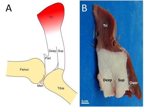
Fatty knee extensor tendon of emus, lacking a patella. The fatty tissue is split into superficial (Sup) and deep regions, with a pad corresponding to the fat pad in other birds continuous with it and the knee joint meniscus (cushioning pad). The triceps femoris (knee extensor) muscle group inserts right into the fatty tendon, continuing over it. A is a schematic; B is a dissection.

Fatty knee extensor tendon of an emu, showing the absence of a patella. The fatty tissue is split into superficial (Sup) and deep regions, with a pad corresponding to the fat pad in other birds continuous with it and the knee joint meniscus (cushioning pad). The triceps femoris (knee extensor) muscle group inserts right into the fatty tendon, continuing on over it. A is a schematic; B is a dissection.

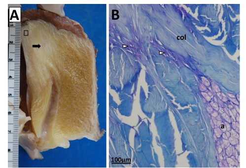
Sectioning of a Southern Cassowary's knee extensor tendon, showing: A Similar section  as in the emu image above. revealing similar regions and fibrous tissue (arrow), with no patella, just fat; and B, with collagen fibre bundles (col), fat cells (a), and cartilage-like tissue (open arrows) labelled.

Sectioning of a Southern Cassowary’s knee extensor tendon, showing: A, Similar section as in the emu image above. revealing similar regions and fibrous tissue (arrow), with no patella, just fat; and B, With collagen fibre bundles (col), fat cells (a), and cartilage-like tissue (open arrows) labelled.

Evolution of patellar form in birds. White branches indicate no patella, blue is a small flake of bone for a patella, green is something bigger, yellow is a double-patella in ostriches, and grey is uncertain. Note the uncertainty and convergent evolution of the patella in ratite birds, which is remarkable but fits well with their likely convergent evolution of flightlessness and running adaptations.

Evolution of patellar form in birds. White branches indicate no patella, blue is a small flake of bone for a patella, green is something bigger, yellow is a double-patella in ostriches, black is a gigantic spar of bone in extinct Hesperornis and relatives, and grey is uncertain. Note the uncertainty and convergent evolution of the patella in ratite birds (Struthio down to Apteryx), which is remarkable but fits well with their likely convergent evolution of flightlessness and running adaptations.

3) Chadwick, K.P., Regnault, S., Allen, V., Hutchinson, J.R. 2014. Three-dimensional anatomy of the ostrich (Struthio camelus) knee joint. PeerJ 2:e706 http://dx.doi.org/10.7717/peerj.706

Finally, Kyle Chadwick came from the USA to do a technician post and also part-time Masters degree with me on our sesamoid grant, and proved himself so apt at research that he published a paper just ~3 months into that work! Vivian Allen (now a postdoc on our sesamoid bone grant) joined us in this work, along with Sophie Regnault. We conceived of this paper as fulfilling a need to explain how the major tissues of the knee joint in ostriches, which surround the double-patella noted above, all relate to each other and especially to the patellae. We CT and MRI scanned several ostrich knees and Kyle made a 3D model of a representative subject’s anatomy, which agrees well with the scattered reports of ostrich knee/patellar morphology in the literature but clarifies the complex relationships of all the key organs for the first time.

This ostrich knee model also takes Kyle on an important first step in his Masters research, which is analyzing how this morphology would interact with the potential loads on the patellae. Sesamoid bones like the patella are famously responsive to mechanical loads, so by studying this interaction in ostrich knees, along with other studies of various species with and without patellae, we hope to use to understand why some species evolved patellae (some birds, mammals and lizards; multiple times) and why some never did (most other species, including amphibians, turtles, crocodiles and dinosaurs). And, excitingly for those of you paying attention, this paper includes links to STL format 3D graphics so you can print your own ostrich knees, and a 3D pdf so you can interactively inspect the anatomy yourself!

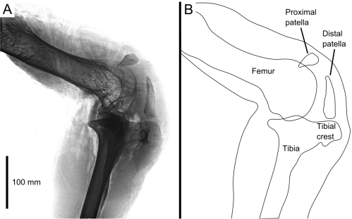
(A) X-ray of an ostrich knee in side view, and (B) labelled schematic of the same.

Ostrich knee in side view: A, X-ray, and (B) labelled schematic.

3D model of an ostrich knee, showing: A, view looking down onto the top of the tibia (shank), with the major collateral ligaments (CL), and B, view looking straight at the front of the knee joint, with major organs of interest near the patella, sans muscles.

3D model of an ostrich knee, showing: A, View looking down onto the top of the tibia (shank), with the major collateral ligaments (CL), and B, View looking straight at the front of the knee joint, with major organs of interest near the patella, sans muscles.

You can view all the peer review history of the papers if you want, and that prompts me to comment that, as usual at PeerJ (full disclosure: I’m an associate editor but that brings me £0 conflict of interest), the peer review quality was as rigorous at a typical specialist journal, and faster reviewing+editing+production than any other journal I’ve experienced. Publishing there truly is fun!

Merry Christmas and Happy Holidays — and good Ratite-tidings to all!

And stay tuned- the New Year will bring at least three more papers from us on this subject of ratite locomotion and musculoskeletal anatomy!

♬Should auld palaeognathans be forgot, 
And never brought for scans? 
Should publications be soon sought, 
For auld ratite fans!♪

Read Full Post »

Title says it all? Sometimes a spade needs to be called a spade.

From time to time the Structure & Motion Lab at the RVC gets cool videos of animals doing different behaviours, be that slow-mo/high-speed videos, x-ray videos. motion capture or whatever. Actually, we get cool videos pretty much every day but some of them (such as a racehorse galloping on a treadmill) seem mundane to us, much as our visitors are impressed.

Here are some examples of the stuff we’ve filmed recently. It all seems to belong on this blog as an example of anatomy in motion, but has no good home here otherwise and no other cohesive threads uniting the disparate videos.

Hence the title. Enjoy!

The above two videos were made by Renate Weller, Emily Sparkes and others. Looping GIF of the last one, via Marko Bosscher:

X-Ray_Hamster

Shin-Ichi Fujiwara, myself and others made that video some years ago; research yet to be finished.

The above two high-speed videos were captured many moons ago with Alexis Wiktorowicz, Karin Jespers and others; more research yet to be finished.

Ashley Heers made this video for the “Fossil Wonderlands” documentary in 2013-14.

Check us out on BBC2 tonight in Cat Watch, with more videos!


Read Full Post »

[This is the original, unedited text of my shorter, tighter (and I think actually better) News & Views piece for Nature, on the paper described below)

Ambitious experimental and morphological studies of a modern fish show how a flexible phenotype may have helped early “fishapods” to make the long transition from finned aquatic animals into tetrapods able to walk on land.

Stomach-Churning Rating: 1/10. Cute fish. Good science. Happy stomachs!

Photo by Antoine Morin, showing Polypterus on land.

Photo by Antoine Morin, showing Polypterus on land.

Napoleon Bonaparte’s military excursions into Egypt in 1798-1799 led a young French naturalist, Ètienne Geoffroy Saint-Hilaire, to cross paths with a strange fish that had paired lungs and could “walk” across land on its stubby, lobelike fins. In 1802, he dubbed this fish “Polyptère bichir”1, today known as the Nile bichir, Polypterus bichir La Cepède 1803. The bichir’s mélange of primitive and advanced traits helped to catapult Geoffroy into scholarly conflict with the reigning naturalist Georges Cuvier back in France and to establish Ètienne as a leading anatomist, embryologist and early evolutionary researcher of repute even today2. Now, on their own excursion under the very “evo-devo” flag that the discoverer of Polypterus helped raise, Canadian scientists Standen et al.3 suggest how the remarkable plasticity of the skeleton of Polypterus (the smaller west African relative of P. bichir, P. senegalus or “Cuvier’s bichir”) reveals a key part of the mechanism that might have facilitated the gradual transition from water to land and thus from “fishapods” to tetrapods (four-limbed vertebrates).

In a bold experiment, the authors raised 149 young bichirs on land and in water for eight months, then studied how they moved on land vs. in water, and also how the ultimate shape of the skeletal elements of the paired front fin bases differed between the land- and water-raised bichirs. Standen et al.3 discovered that both the form and function of the fins’ foundations transformed to better satisfy the constraints of moving on land. Land-acclimated bichirs took faster steps on land, their fins slipped across the substrate less, they held their fins closer to their body, their noses stayed more aloft and their tails undulated less, with less variable motions overall—behaviours that the authors had predicted should appear to enhance walking abilities on land. In turn, the bones of the neck and shoulder region altered their shape to produce a more mobile fin base with greater independence of fin from neck motion, along with improved bracing of the ventral “collarbone” region. These environmentally-induced traits should have fostered the locomotor changes observed in “terrestrialized” fish and aided the animals in resisting gravity, and they represent a common biological phenomenon termed developmental plasticity4,5. Interestingly, the land-reared fish could still swim about as well as the wholly aquatic cohort, so there was not a clear trade-off between being a good swimmer and a good walker, which is surprising.

Considered alone, the developmental plasticity of bichir form and function shows how impressive these amphibious fish are. But Standen et al.’s study3  ventured further, to apply the lessons learned from bichir ontogeny to a phylogenetic context and macroevolutionary question. The phenotypic plasticity during bichir development, they infer, could have been harnessed during the evolutionary transformation of fins for swimming into limbs for walking, in the “fishapod” ancestors of tetrapods. Indeed, bichirs are close to the base of the family tree of fishes6, and other living relatives of tetrapods have reduced or lost their fins (lungfishes) or adapted to strange deep-sea swimming lifestyles, never walking on land (coelacanths). Thus perhaps bichirs and the “fishapod” lineage share what Geoffroy would have called “unity of type”, today termed homology, of their developmental plasticity in response to a land environment. Surveying the fossil record of early “fishapods” and tetrapods, Standen et al.3 found that the macroevolutionary changes of neck and shoulder anatomy in these gradually more land-adapted animals parallel those they observed in terrestrialized Polypterus, providing ancillary support for their hypothesis.

A further test of the application of Polypterus’s plasticity to fossil tetrapods is naturally difficult. However, the “fishapod” lineage has some exceptional examples of fossil preservation. With sufficient sample sizes (e.g. fossil beds that reveal growth series, such as the Late Devonian Miguasha site in Canada7) and palaeoenvironmental gradients in fish or tetrapods, one could imagine performing a rigorous indirect test. Even small samples could be helpful– for example, the early tetrapod Ichthyostega exhibits some developmental changes in its forelimb suggesting that it became more terrestrial as it grew, whereas the related Acanthostega does not evidence such changes8— this hints at some developmental plasticity in the former animal.

During the Devonian period (~360-420 million years ago), were the “fishapod” ancestors of tetrapods floundering about on land now and then, gradually shifting from anatomy and behaviours that were more developmentally plastic (as in bichirs) to ones that were more canalized into the terrestrialized forms and functions that more land-adapted tetrapods retained? An attractive possibility is that the developmental plasticity could have led to fixation (reduction of plasticity), an evolutionary phenomenon called genetic assimilation, which another intellectual descendant of Geoffroy, Conrad Hal Waddington, promoted from the 1950s onwards9, a concept that now enjoys numerous cases of empirical support10 that this one may eventually join.

The nature of the genetic and developmental mechanism that bichirs use to achieve the observed developmental plasticity is still unclear. If it has a high enough degree of heritability, then it could be selected for in cross-generational experiments with bichirs. With sufficient time and luck raising these unusual fish, the hypothesis that their plastic response to a terrestrial environment can become genetically assimilated could be directly tested. This study could thus become an epic exemplar of how genetic assimilation can contribute not only to microevolutionary change but also to major macroevolutionary events, as was presciently suggested in a seminal review of developmental plasticity4.

This genetic assimilation is the Polypterus study’s reasonable speculation, and one that Geoffroy likely would have applauded, all the more for involving his beloved bichirs. Much as Napoleon’s landfall in Egypt was not a lasting success, bichirs never left wholly terrestrial descendants despite their malleable locomotor system. But the same type of plastic developmental mechanism that bichirs use today to make tentative, floppy incursions of the terrestrial realm might have been harnessed by our own “fishapod” forebears, leaving a far more revolutionary dynasty upon the Earth.


References

  1.  Geoffroy, E. (1802). Histoire naturelle et description anatomique d’un nouveau genre de poisson du Nil, nommé polyptère. Annales du Muséum d’Histoire Naturelle 1:57-68.
  2. Le Guyader, H., & Grene, M. (2004) Geoffroy Saint-Hilaire: A Visionary Naturalist. Univ. Chicago Press.
  3. Standen, E. M., Du, T. Y., & Larsson, H. C. E. (2014). Developmental plasticity and the origin of tetrapods. Nature, published online.
  4. West-Eberhard, M. J. (1989). Phenotypic plasticity and the origins of diversity. Annual Review of Ecology and Systematics 20:249-278.
  5. Pigliucci, M., Murren, C. J., & Schlichting, C. D. (2006). Phenotypic plasticity and evolution by genetic assimilation. Journal of Experimental Biology 209(12):2362-2367.
  6. Near, T. J., Dornburg, A., Tokita, M., Suzuki, D., Brandley, M. C., & Friedman, M. (2014). Boom and bust: ancient and recent diversification in bichirs (Polypteridae: Actinopterygii), a relictual lineage of ray‐finned fishes. Evolution 68:1014-1026.
  7. Cloutier, R. (2013). Great Canadian Lagerstätten 4. The Devonian Miguasha Biota (Québec): UNESCO World Heritage Site and a Time Capsule in the Early History of Vertebrates.Geoscience Canada40:149-163.
  8. Callier, V., Clack, J. A., & Ahlberg, P. E. (2009). Contrasting developmental trajectories in the earliest known tetrapod forelimbs.Science324:364-367.
  9. Waddington, C. H. (1953). Genetic assimilation of an acquired character. Evolution 7:118-126.
  10. Crispo, E. (2007). The Baldwin effect and genetic assimilation: revisiting two mechanisms of evolutionary change mediated by phenotypic plasticity. Evolution 61:2469-2479.

Read Full Post »

Older Posts »