Feeds:
Posts
Comments

Posts Tagged ‘inverts’

Our special guest post this week comes from Dr. Liz Clark of Yale University in New Haven, Connecticut, USA. She continues to bring biomechanics-fu to echinoderms– the weird marine critters like seastars and sea urchins. Including fossils, as you’ll see today! You may remember her from blog posts such as “Guest Post: Brittle Star Arms Are Weird“.

Stomach-Churning Rating: 1/10; echinoderms are inoffensive.

Imagine that you’re stuck in a cardboard box on the beach, holding a small stick. Could you use the stick to move yourself forward? What would you do? You could try digging into the sediment ahead of you to pull yourself along. You could try rowing side to side, as if you were in a rowboat. Or maybe it’s not possible and you’d give up, decide to stay put, and wave your stick in the air for help.

Believe it or not, this is a strange-but-important dilemma that some paleobiologists- like me!- have been wrestling with for generations. My research specialty is in the biomechanics of locomotion– how organisms use their bodies to get from one place to the next (through walking or swimming, for instance). We can learn a lot about an animal by studying their locomotion, such as why their body is shaped the way that it is, or what role they occupy in their ecosystem. Animal motion is a major inspiration for robotic design, and I work with engineers to apply the novel insights on animal locomotion from my research to create new kinds of devices.

Studying the biomechanics of motion in living organisms is (relatively) straightforward. We can use high-speed cameras, motion capture software, and 3D imaging tools to visualize and understand how organisms move in real-time, informing our inferences about how they perform certain tasks. Inferring locomotion in fossil organisms, on the other hand, is tricky since we can’t observe the organism’s behavior like we could if the organism were alive. Instead of being able to watch the organism move, we’re left with a snapshot of the animal frozen in place in a rock. We’re also missing a lot of physical information: locomotion in most animals requires soft tissue and hard skeletal structures, but typically with fossils, only the hard structures get preserved.

However, we can often garner some insights from living organisms to determine the locomotion strategies that fossil organisms use. Most organisms in the fossil record look at least somewhat similar to organisms alive today. If our fossil has four legs, for instance, we can study locomotion in living tetrapods (four-legged animals) to help us create a framework for deriving inferences about locomotion in our extinct tetrapod fossil animal. But for some really strange-looking animals- ones without obvious modern analogues- we’re not so lucky. For me, this is where the fun begins.

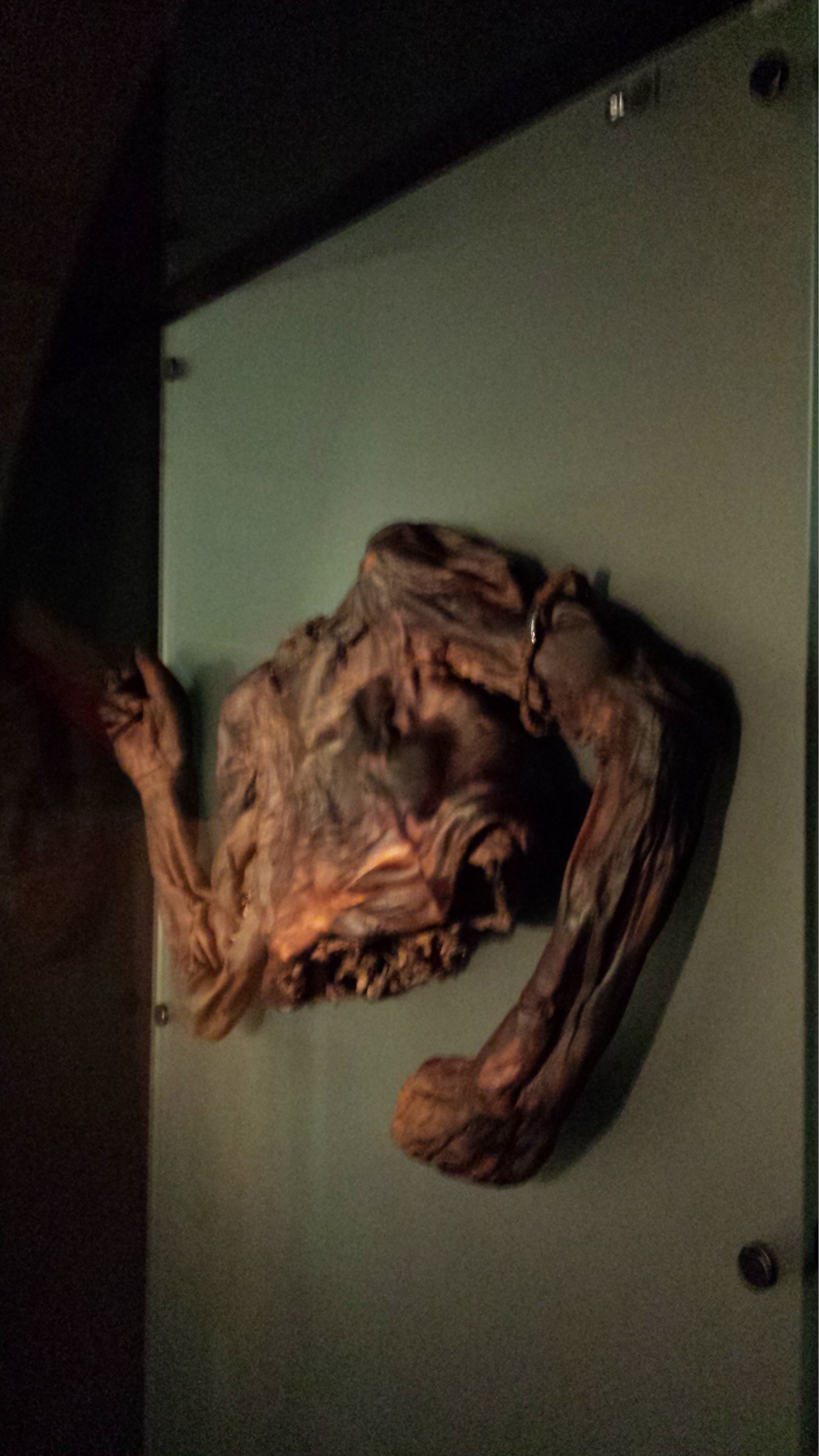
Figure 1: Stylophorans! Here are four fossilized stylophorans from the Helderberg Group of the Early Devonian (YPM 036413)

So getting back to the cardboard box and the stick. These are metaphorical examples of the different locomotion strategies that have been proposed for a group of fossil animals known as stylophorans (Figure 1). Stylophorans are extinct organisms related to sea stars and sea urchins, but with a body structure unlike any organism on the planet today. They have a large, relatively flat body called a theca (i.e., the cardboard box), and a long, thin segmented tail known as the aulacophore (i.e. the stick) (Figure 2). They’re known in the paleontological community as some of “the strangest-looking animals of all time.”

Figure 2: Stylophoran anatomy. The “theca” is the body cavity, and the “aulacophore” comprises of the proximal aulacophore, the stylocone, and the distal aulacophore.

By reconstructing stylophoran locomotion, we can unlock the mechanics of a unique system for motion and its potential applications to engineering. We can also understand more about how this organism lived and functioned in its ancient ecosystem. And, by developing a new approach to understand locomotion in stylophorans, we can apply this strategy to analyze locomotion and movement in other unusual fossil animals as well!

For years, scientists have been documenting the incredible array of stylophoran diversity in the fossil record and making their best predictions about how they would have been able to move (or not!). These predictions are based on their morphology– the structure of an organism’s body. For stylophorans, that means the shape and structure of the theca and aulacophore. There are a variety of stylophoran thecal shapes, ranging from ovoid in Enopleura to trapezoidal in Ceratocystis to almost crescent-shaped in Cortnurnocystis. There’s a similarly wide array of aulacophore morphologies as well.

Figure 3: Left: One half of the concretion within which the stylophoran fossil we analyzed is preserved. Right: The 3D digital image of the stylophoran fossil, created by micro-CT scanning the fossil specimen.

We developed a new approach using 3D imaging (Figure 3) to create a digital model of a stylophoran specimen. We used the model to test if several different locomotion strategies that had been proposed before were physically possible or impossible for a stylophoran to actually perform.

First, we used a micro-CT scanner to image a fossil stylophoran. This outputs a digital 3D picture of the stylophoran fossil that we can look at and analyze on a computer. Next, we developed a program to calculate the joint centers- the point at which one skeletal structure rotates relative to another-within the digitized stylophoran’s aulacophore (Figure 4). This created a digital marionette– a rig of our stylophoran fossil that flexes at the junctures between aulacophore segments as it would have in life. We then rotated each segment at the joint center to calculate the aulacophore’s total range of motion– a reconstruction of how far the aulacophore could flex in each direction (Figure 5).

Figure 4: A look into some of the nuts and bolts of the 3D model we created. Tri-colored axes demarcate where the joint centers are in the proximal aulacophore. 

We used this 3D range of motion model to evaluate several locomotion strategies that had been previously hypothesized for this group of stylophorans. One hypothesis suggested that these stylophorans dug their aulacophores into the substrate– sediment on the ocean floor- to pull themselves forward. Another suggested that they moved the aulacophore side to side in order to push themselves along. We found that the first hypothesis would have been impossible to conduct based on the range of motion we calculated, but the second strategy was theoretically possible! We’ll need to do more work to see how likely it was that stylophorans would have actually used this technique. Nevertheless, through this investigation, our team produced the first objective, data-driven methodology for analyzing locomotion in fossil invertebrates, which is a big step in the right direction for the study of fossil invertebrate biomechanics! Our technique can be applied to study other organisms with rigid skeletons as well, like crabs, insects, or sea stars, for instance, and we’re looking forward to seeing our technique used to uncover more interesting locomotion strategies!

Figure 5: A snapshot of the 3D model where we can observe how dorsal and ventral range of motion compare to the originally preserved orientation of the aulacophore (highlighted in green).

Do you want to know more? You can! We published a paper on this topic here!

Read Full Post »

I had a spare hour in Cambridge this weekend so I dared the crowds in the revamped UMZC’s upper floor. In my prior visit and post I’d experienced and described the lower floor, which is almost exclusively mammals. This “new” floor has everything else that is zoological (animal/Metazoa) and again is organized in an evolutionary context. And here is my photo tour as promised!

Inviting, soft lighting perfuses the exhibits from the entryway onwards.

All images can be clicked to mu-zoom in on them.

Stomach-Churning Rating: 5/10 for spirit animals, by which I mean dissected/ghostly pale whole specimens of animals in preservative fluids.

The exhibits are on a square balcony overlooking the lower floor, so you can get some nice views. It does make the balcony crowded when the museum is busy, so take that in mind if visiting. Strollers on this upper floor could be really difficult. But the ceiling is very tall so it is not cramped in a 3D sense. The lower floor is more spacious.

Like phylogenies? You got em! Tucked away at the beginning of each major group; not occupying huge valuable space or glaringly obvious like AMNH in NYC but still noticeable and useful. To me, it strikes a good balance; gives the necessary evolutionary context for the displayed specimens/taxa.

Introductory panels explain how names are given to specimens, how specimens are preserved and more.

The exhibits give due focus to research that the UMZC is doing or has been famous for. Hey I recognize that 3D tetrapod image in the lower left! 🙂

There is ample coverage of diversity throughout Metazoa but my camera tended to be drawn to the Vertebrata. Except in some instances like these.

Some larger chelicerates.

Some smaller, shadowy sea scorpion (eurypterid) fossils.

Watch here for more about ophiuroids (brittlestars) in not too long!

A BIG fish brain! Interesting!
Before I go through specimens in evolutionary “sequence”, I will feature another thing i really liked: lots of dissected spirit-specimens that show off cool anatomy/evolution/adaptation (and technical skills in anatomical preparation). Mostly heads; mostly fish.

Salps and other tunicates! Our closest non-vertebrate relatives- and some insight into how our head and gut came to be.

Salp-reflection.

Lamprey head: not hard to spot the commonalities with the salps; but now into Vertebrata.

Hagfish head: as a fellow cyclostome/agnathan, much like a lamprey but never forget the slime glands!

Shark head. Big fat jaws; all the better to bite prey with!

Lungfish (Protopterus) head showing the big crushing tooth plates (above).

Sturgeon vertebrae: tweak some agnathan/shark bits and here you are.

Worm (annelid) anatomy model, displaying some differences from/similarities to Vertebrata. (e.g. ventral vs. dorsal nerve cord; segmentation)

Dissected flipper from a small whale/other cetacean. Still five fingers, but other specializations make it work underwater.

Wonderful diversity of tooth and jaw forms in sharks, rays and relatives. I like this display a lot.

More of the above, but disparate fossil forms!

On with the evolutionary context! Woven throughout the displays of modern animals are numerous fossils, like these lovely placoderms (lineage interposed between agnathans, sharks and other jawed fish).

Goblin shark head.

I seem to always forget what ray-finned fish this is (I want to say wolffish? Quick Googling suggests maybe I am right), but see it often and like its impressive bitey-ness.

Bichir and snakefish; early ray-finned fish radiations.

Armoured and similar fish today.

Armoured fish of the past; some convergent evolution within ray-fins.

Convergence- and homology- of amphibious nature in fish is another evolutionary pattern exemplified here.

Gorgeous fossils of ray-finned fish lineages that arose after the Permian extinctions, then went extinct later in the Triassic.

Note the loooooong snout on this cornetfish but the actual jaws are just at the tip.

Flying fish– those ray-fins are versatile.

Diversity of unusual ray-finned fish, including deep-water and bottom-dwelling forms.

Can you find the low-slung jaws of a dory?

Recent and fossil perch lineage fish.

It’s hard to get far into talking about evolution without bringing up the adaptive radiation of east African cichlid fish, and UMZC researchers are keen on this topic too.

Lobe-fins! Everybody dance!

Rhizodonts & kin: reasons to get out of Devonian-Carboniferous waters.

A Cretaceous fossil coelacanth (skull); not extremely different from living ones’.

Let’s admire some fossil and modern lungfish skulls, shall we? Big platey things  (here, mainly looking at the palate) with lots of fusions of tiny bones on the skull roof.

Eusthenopteron fossils aren’t that uncommon but they are still great to see; and very important, because…

OK let’s stop messing around. The UMZC has one of the best displays of fossil stem-tetrapods in the world! And it should.

Another look at the pretty Acanthostega models.

Acanthostega vs. primate forelimb: so like us.

Ichthyostega parts keep Acanthostega company.

A closer look at the “Mr. Magic” Ichthyostega specimen, which takes some unpacking but is incredibly informative and was a mainstay of our 2012 model. Back of skull, left forelimb, and thorax (from left to right here).

Eucritta, another stem-tetrapod.

Closer look at Eucritta‘s skull.

Weird stem-tetrapod Crassigyrinus, which we’re still trying to figure out. It’s a fabulous specimen in terms of completeness, but messy “roadkill” with too many damn bones.

The large skull of Crassigyrinus, in right side view.

Early temnospondyl (true amphibian-line) skulls and neck.

Nectrideans or the boomerangs of the Palaeozoic.

Cool fossil frogs.

Giant Japanese salamander!

Fire salamanders: not as colourful as the real thing, but here revealing their reproductive cycle in beautiful detail.

Closeup of oviduct in above.

Sexual dimorphism in Leptodactylus frogs: the males have bulging upper arms to (I am assuming) help them hold onto females during amplexus (grasping in mating competitions).

Did I forget that Leptodactylus has big flanges on the humerus in males, to support those muscles? Seems so.

An early stem-amniote, Limnoscelis (close to mammals/reptiles divergence); cast.

Grand sea turtle skeleton.

One of my faves on display: a real pareiasaurian reptile skeleton, and you can get a good 3D look around it.

Details on above pareiasaurian.

Mammals are downstairs, but we’re reminded that they fit into tetrapod/amniote evolution nonetheless.

Let there be reptiles! And it was good.

Herps so good.  (slow worm, Gila monster, glass lizard)

A curator is Dr Jason Head so you bet Titanoboa is featured!

Crocodylia: impressive specimens chosen here.

It ain’t a museum without a statuesque ratite skeleton. (There are ~no non-avian dinosaurs here– for those, go to the Sedgwick Museum across the street, which has no shortage!)

Avian diversity takes off.

Glad to see a tinamou make an appearance. They get neglected too often in museums- uncommon and often seemingly unimpressive, but I’m a fan.

I still do not understand hoatzins; the “cuckoo” gone cuckoo.

Dodo parts (and Great Auk) near the entrance.

Wow. What an oilbird taxidermy display! :-O

There we have it. Phew! That’s a lot! And I left out a lot of inverts. This upper floor is stuffed with specimens; easier there because the specimens are smaller on average than on the lower floor. Little text-heavy signage is around. I give a thumbs-up to that– let people revel in the natural glory of what their eyes show them, and give them nuggets of info to leave them wanting more so they go find out.

Now it’s in your hands– go find out yourself how lovely this museum is! I’ve just given a taste.

Read Full Post »

A photo blog post for ya here! I went to Dublin on a ~28 hour tour, for a PhD viva (now-Dr Xia Wang; bird feather/flight evolution thesis) earlier this month. And I made a beeline for the local natural history museum (National Museum of Ireland, Natural History building) when I had free time. So here are the results!

Stomach-Churning Rating: Tame; about a 1/10 for most, but I am going to break my rule about showing human bodies near the end. Just a warning. The bog bodies were too awesome not to share. So that might be 4/10-8/10 depending on your proclivities. They are dry and not juicy or bloody, and don’t look as human as you’d expect.

Simple Natural History museum entrance area.

Simple Natural History museum entrance area.

Adorable frolicking topiaries outside the NHM.

Adorable frolicking topiaries outside the NHM.

Inside, it was a classical Victorian-style, dark wood-panelled museum stuffed with stuffed specimens. It could use major refurbishment, but I do love old-fashioned exhibits. Get on with it and show us the animals; minimize interpretive signage and NO FUCKING INTERACTIVE COMPUTER PANELS! So by those criteria, I liked it. Some shots of the halls: hall2 hall1 hall3 hall4 hall5 hall6 And on to the specimens!

Giant European deer ("Irish elk"). I looked at these and thought, "why don't we see female deer without antlers ever? then noticed one standing next to these; photo was crappy though. :(

Giant European deer (“Irish elk”). I looked at these and thought, “why don’t we see female deer without antlers ever? then noticed one standing next to these (you can barely see it in back); too bad my photo is crappy.

Superb mounted skeleton of giraffe (stuffed skin was standing near it).

Superb mounted skeleton of giraffe (stuffed skin was standing near it).

A sheep or a goat-y thingy; I dunno but it shows off a nice example of the nuchal ligament (supports the head/neck).

A sheep-y or a goat-y beastie; I dunno but it shows off a nice example of the nuchal ligament (supports the head/neck).

Yarr, narwhals be internet gold!

Yarr, narwhals be internet gold!

Giant blown glass models of lice!

Giant blown glass models of lice!

Who doesn't like a good giant foramanifera image/models? Not me.

Who doesn’t like a good giant foramanifera image/model?

"That's one bigass skate," I murmured to myself.

“That’s one bigass skate,” I murmured to myself.

"That's one bigass halibut," I quipped.

“That’s one bigass halibut,” I quipped.

Tatty basking shark in entry hall.

Tatty basking shark in entry hall.

Irish wolfhound, with a glass sculpture of its spine hanging near it, for some reason.

Irish wolfhound, with a glass sculpture of its spine hanging near it, for some reason.

Stand back folks! The beaver has a club!

Stand back everyone! That beaver has a club!

Skull of a pilot whale/dolphin.

Skull of a pilot whale/dolphin.

Nice anteater skeleton and skin.

Nice anteater skeleton and skin.

Nice anteater skeleton and skin.

Nice wombat skeleton and skin.

Sad display of a stuffed rhino with the horn removed, and signage explaining the problem of thefts of those horns from museum specimens of rhinos worldwide.

Sad display of a stuffed rhino with the horn removed, and signage explaining the problem of thefts of those horns from museum specimens of rhinos worldwide.

But then the stuffed animals started to get to me. Or maybe it was the hangover. Anyway, I saw this…
creepy proboscis (1) creepy proboscis (2)

A proboscis monkey mother who seemed to be saying “Hey kid, you want this yummy fruit? Tough shit. I’m going to hold it over here, out of reach.” with a disturbing grimace. That got me thinking about facial expressions in stuffed museum specimens of mammals more, and I couldn’t help but anthropomorphize as I toured the rest of the collection, journeying deeper into surreality as I progressed. What follows could thus be employed as a study of the Tim-Burton-eseque grimaces of stuffed sloths. Click to emslothen.

sloths (1) sloths (5)sloths (4) sloths (3) sloths (2)

Tree anteater has a go at the awkward expression game.

Tree anteater has a go at the awkward expression game.


This completed my tour of the museum; there were 2 more floors of specimens but they were closed for, sigh, say it with me… health and safety reasons. Balconies from which toddlers or pensioners or drunken undergrads could accidentally catapult themselves to their messy demise upon the throngs of zoological specimens below. But the National Museum’s Archaeology collection was just around the block, so off I went, following whispered tales of bog bodies. There will be a nice, calm, pretty photo, then the bodies, so if peaty ~300 BCE cadavers are not your cup of boggy tea, you can depart this tour now and lose no respect.

Impressive entrance to the National Museum's Archaeology building.

Impressive entrance to the National Museum’s Archaeology building.

The bog bodies exhibit is called “Kingship and Sacrifice“. It is packed with cylindrical chambers that conceal, and present in a tomb-like enclosed setting, the partial bodies of people that were killed and then tossed in peat bogs as honoraria for the ascension of a new king. The peaty chemistry has preserved them for ~2300 years, but in a dessicated, contorted state. The preservation has imparted a mottled colouration and wrinkled texture not far off from a Twix chocolate bar’s. Researchers have studied the bejesus out of these bodies (including 3D medical imaging techniques) and found remarkable details including not just wounds and likely causes of death (axes, strangling, slit throats etc) but also clothing, diet, health and more.

Here they are; click to (wait for it)… emboggen:

BogBodies (1) BogBodies (2) BogBodies (3) BogBodies (4) BogBodies (5) BogBodies (6)

Did you find the Celtic armband on one of them?

Finally (actually this happened first; my post is going back in time), I visited UCD’s zoology building for the PhD viva and saw a few cool specimens there, as follows:

Giant deer in UCD zoology building foyer.

Giant deer in UCD zoology building foyer, with a lovely Pleistocene landscape painted on the wall behind it.

Sika deer in awkward posture in Univ Coll Dublin zoology building's foyer.

Sika deer in an awkward posture (what is it supposed to be doing?) in Univ Coll Dublin zoology building’s foyer.

The pose of this ?baboon? struck me as very peculiar, and menacing- reminiscent of a vampire bat's pose, to me.

The pose of this ?baboon?mandrill struck me as very peculiar and menacing- reminiscent of a vampire bat’s pose.

A whole lotta chicken skeletons in a UCD teaching lab.

A whole lotta chicken skeletons in a UCD teaching lab.

After the viva we went out for some nice Chinese food and passed some Dublin landmarks like this:

Trinity College entrance, I think.

Trinity College entrance, I think.Former Irish Parliament; now the Bank of Ireland.

And we wandered into a very posh Irish pub called the Bank (on College Green), which displayed this interesting specimen, as well as some other features shown below:

Replica of illuminated old Gaelic manuscript.

Replica of illuminated 9th Century gospel manuscript “The Book of Kells”, with gorgeous Celtic art.

Vaults near toilets in the Bank pub.

Vaults near toilets in the Bank pub. Almost as cool as having giant freezers down there.

Nice glass ceiling of the Bank pub.

Nice glass ceiling of the Bank pub.

And Irish pub means one big, delicious thing to me, which I will finish with here– much as I finished that night off:

Ahhh...

Ahhh… ice cold.

Read Full Post »