Feeds:
Posts
Comments

Posts Tagged ‘rhinos’

One of my favourite museums in the world, and certainly one of the best natural history museums in the UK, is Cambridge’s Museum of Zoology, AKA “University Museum of Zoology at Cambridge” (UMZC). It is now nearing a lengthy completion of renovations; the old museum exhibits and collections were excellent but needed some big changes along with the re-fabbed “David Attenborough Building” that houses them. As a longtime fan of the exhibits and user of the collection (and microCT scanner), I hurried to see the new museum once it officially opened.

And that makes a great excuse to present a photo-shoot from my visit. This focuses on the “mammal floor” below the entrance- the upper floor(s?) are still being completed and will have the birds, non-avian tetrapods, fish, etc. But the UMZC is strong in mammals and so it is natural for them to feature them in this chock-full-o-specimens display. Less talk, more images. Here we go!

All images can be clicked to mu-zoom in on them.

Stomach-Churning Rating: 3/10; bones and taxidermy and innocuous jars.

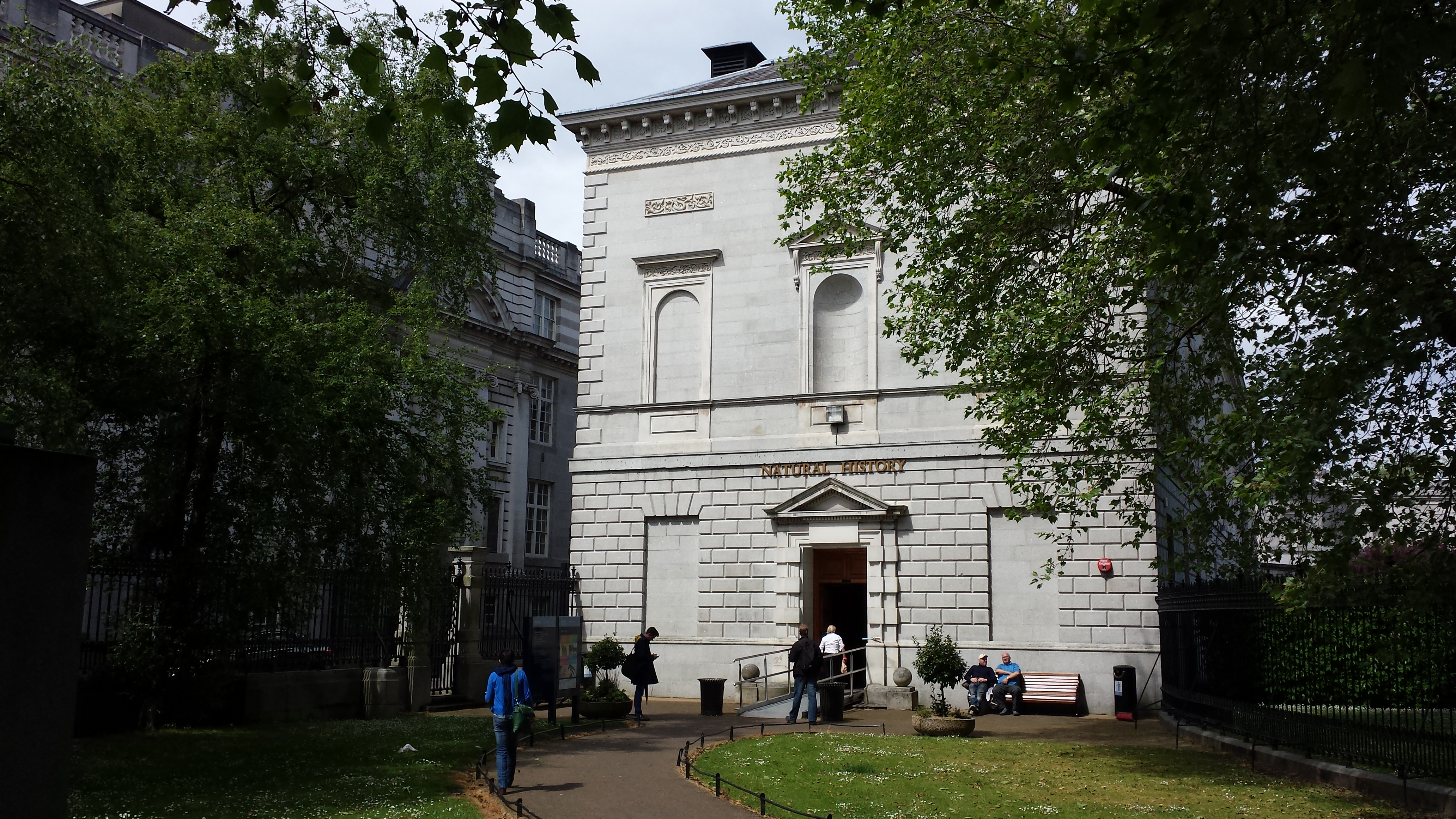
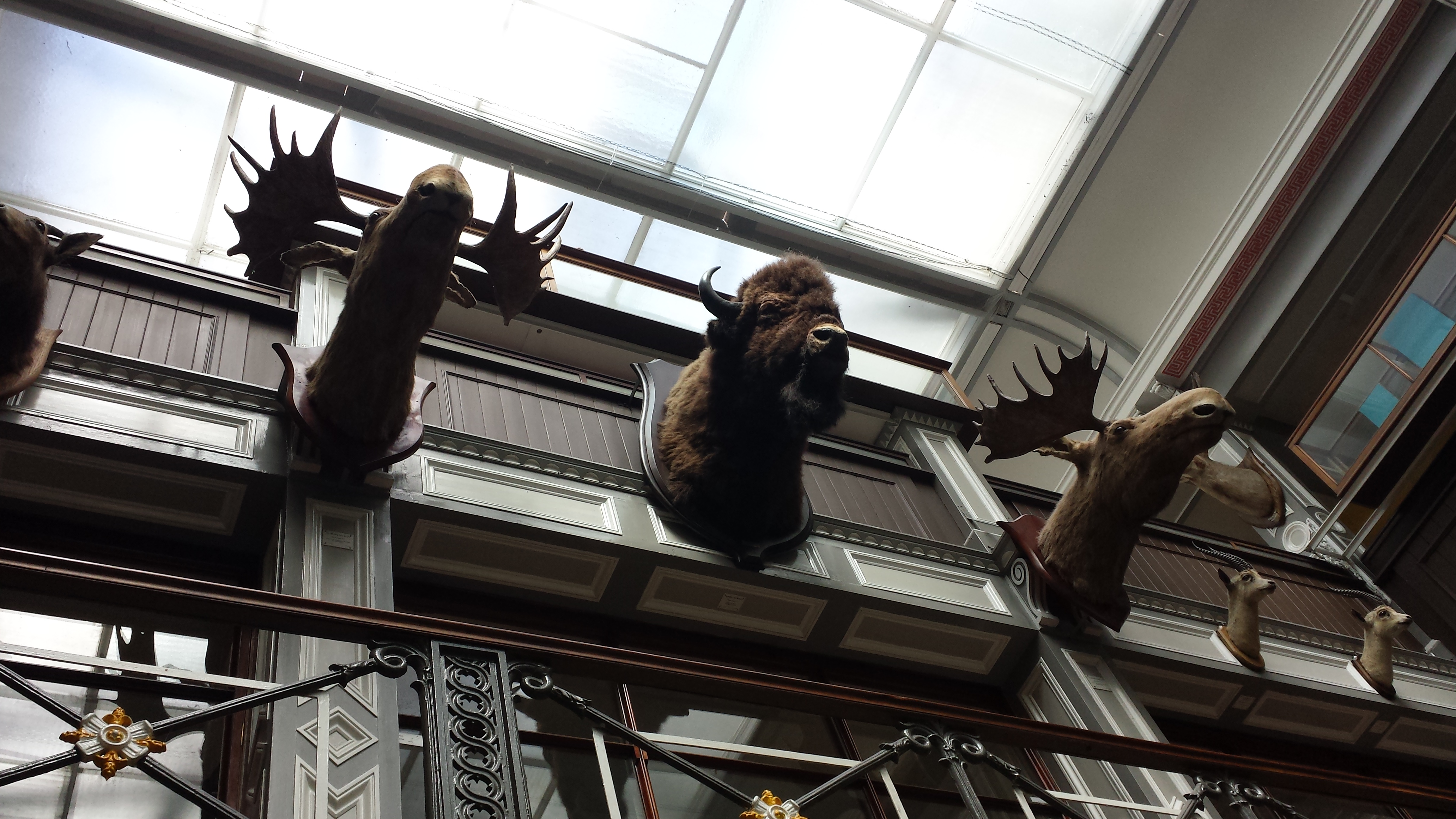
The building. The whale skeleton that hung outside for years is now cleaned up and housed right inside; you walk under it as you enter.
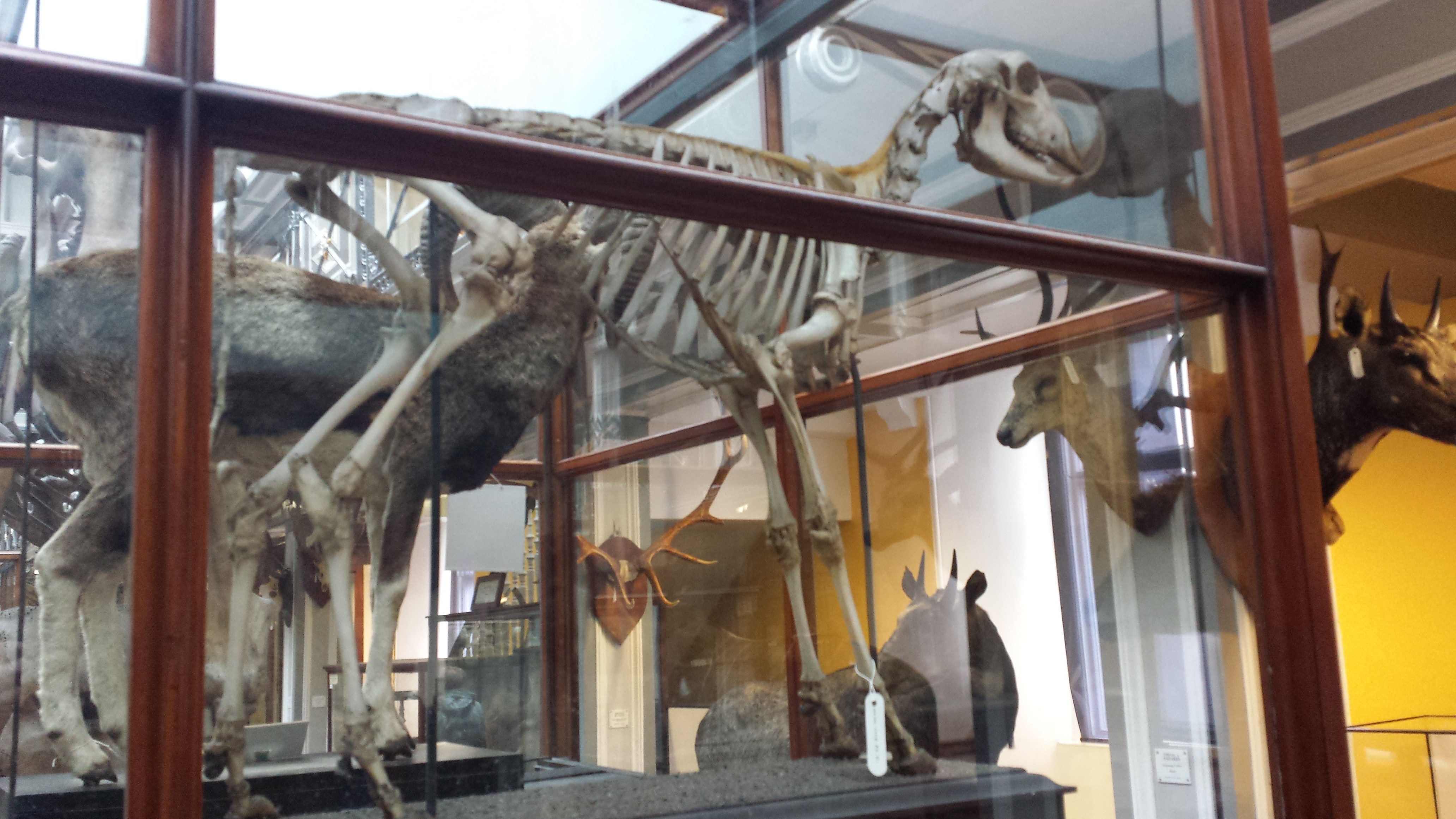
Entrance.

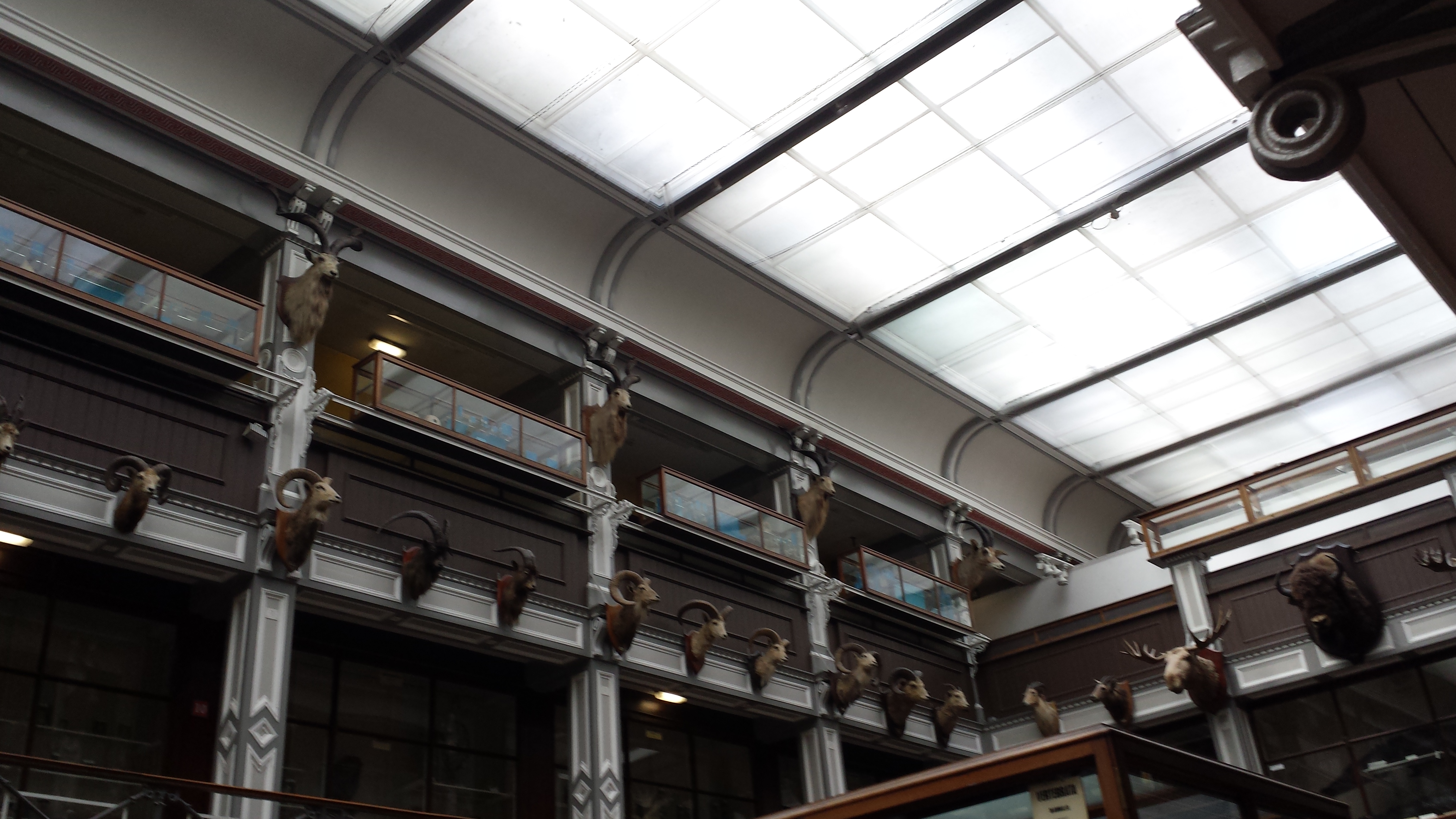
First view past the entryway: lots of cool specimens.

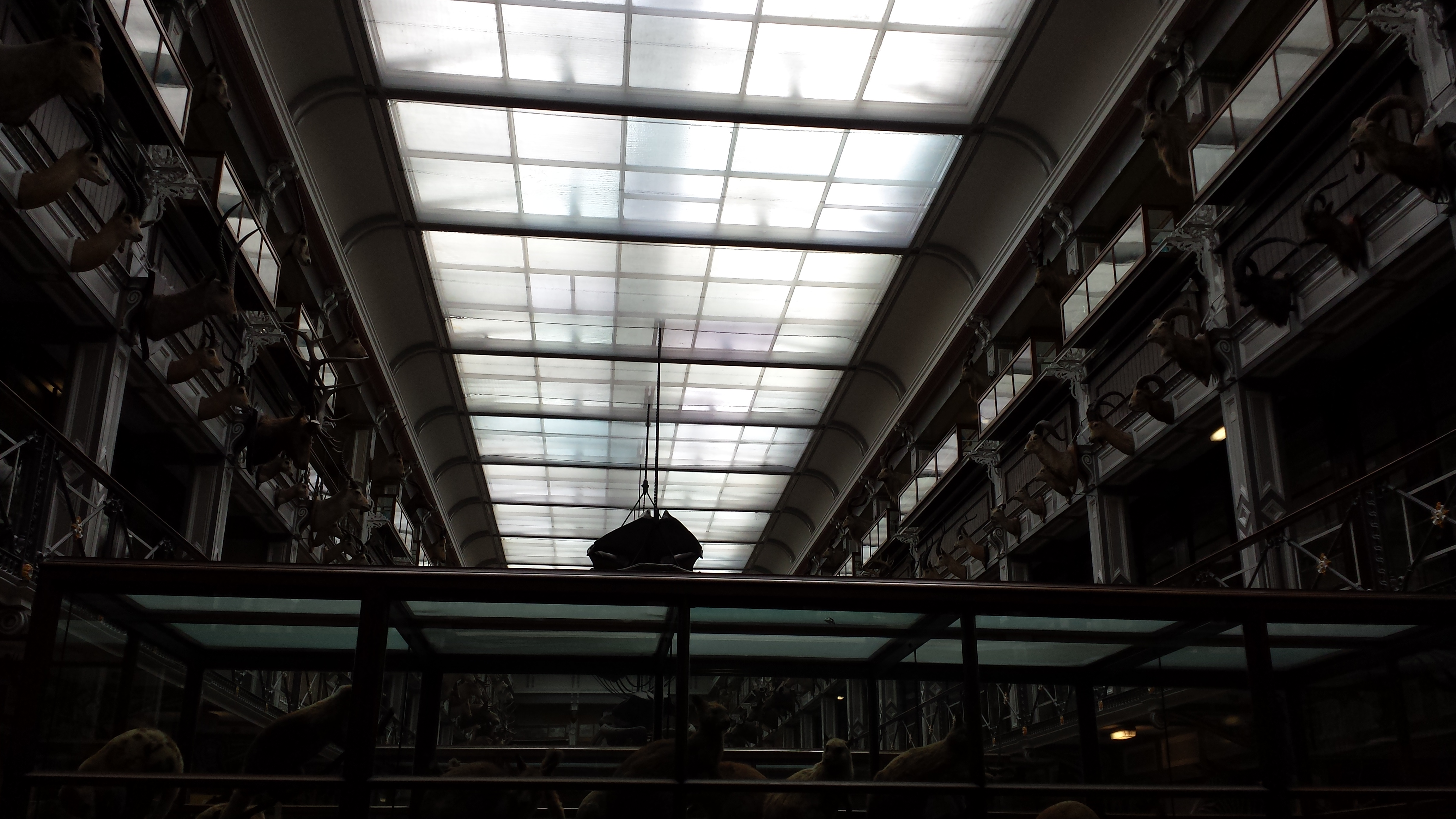
View from the walkway down into the ground/basement level from the entry. As specimens-per-unit-volume goes, the UMZC still scores highly and that is GOOD!

Explanation of frog dissection image below.

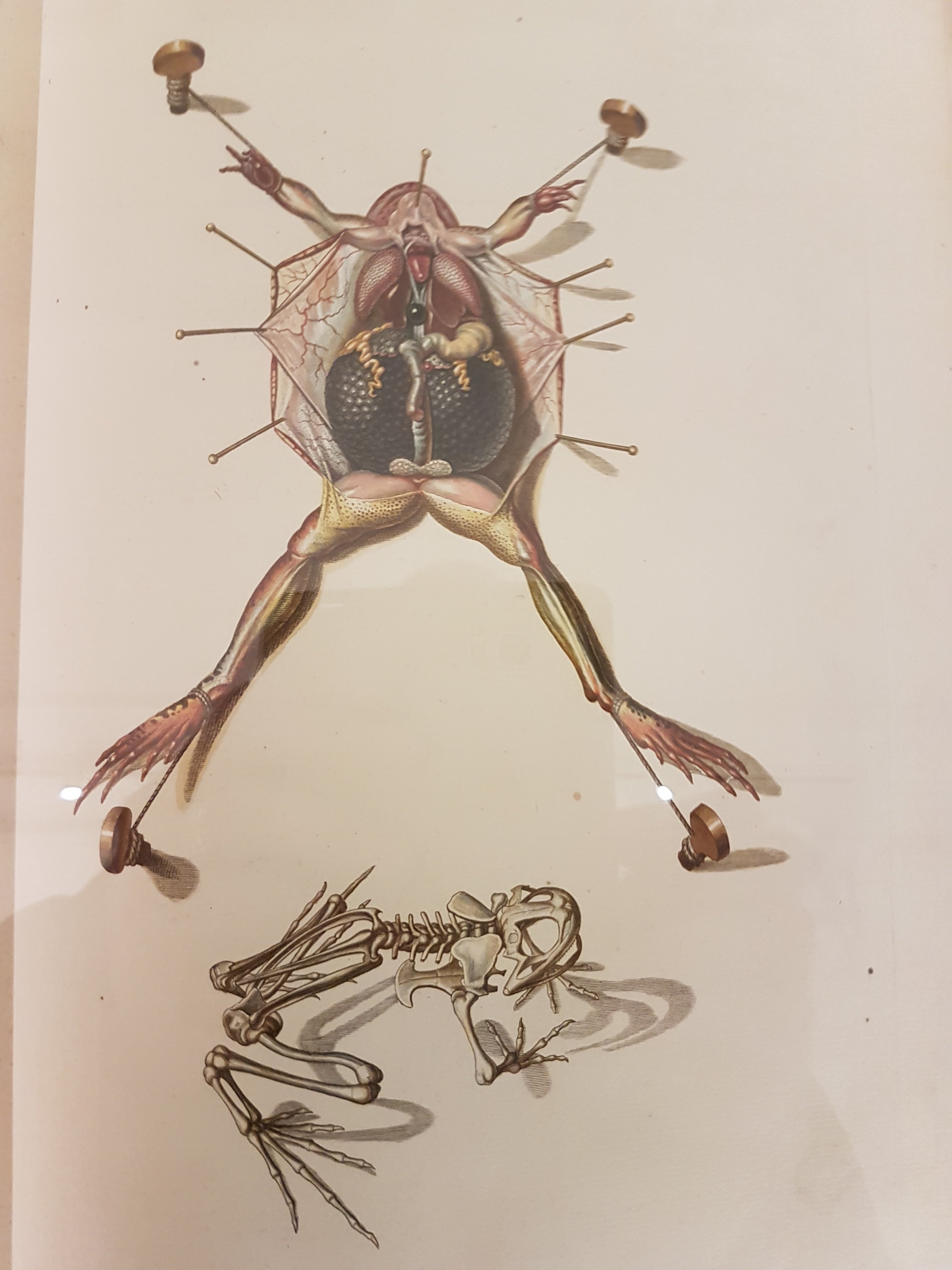
Gorgeous old frog dissection illustration; such care taken here.

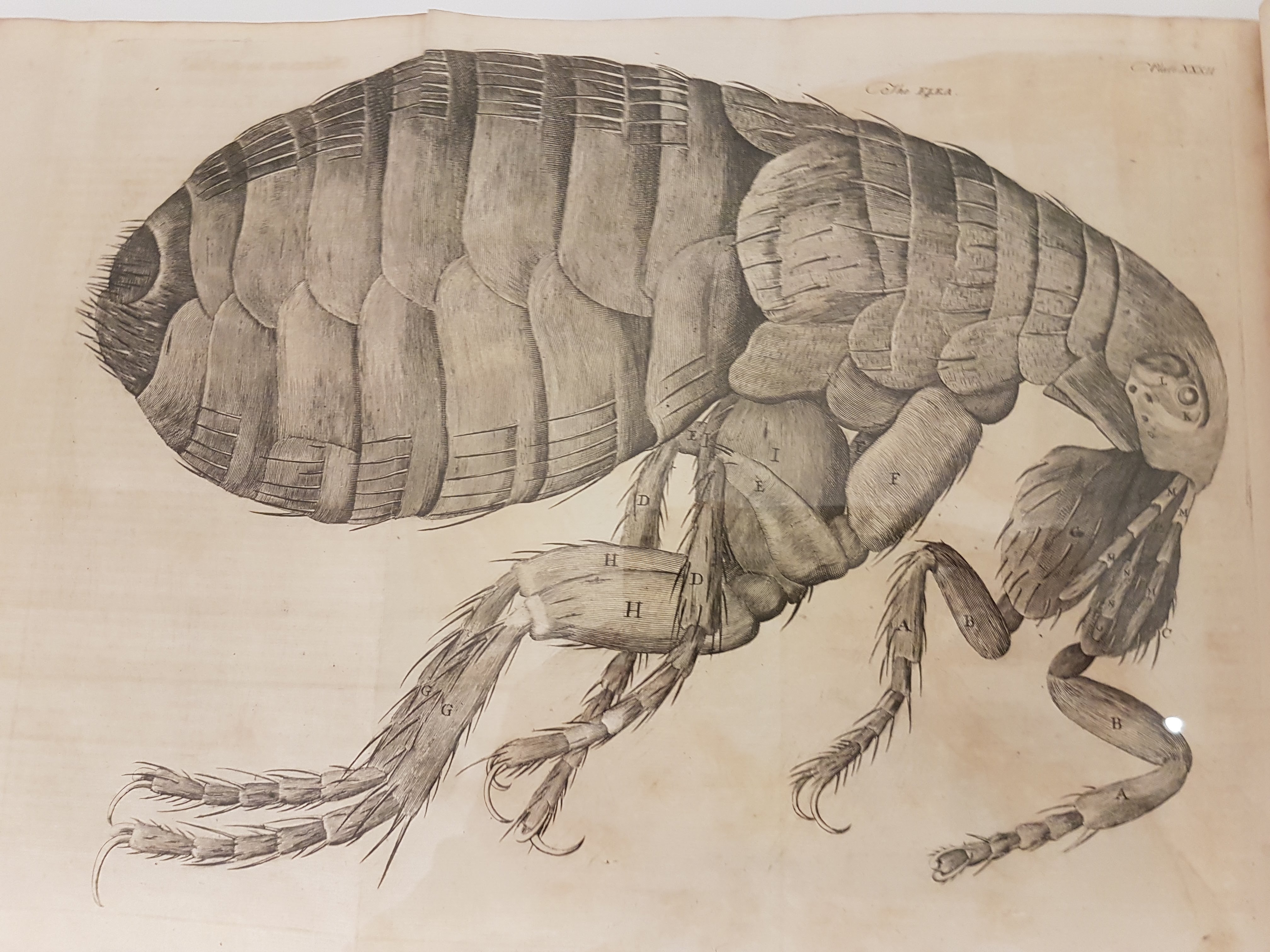
Leeuwenhoek’s flea woodcut; I think from Arcana Naturae Detecta (1695). There is an impressive display of classic natural history books near the entryway.

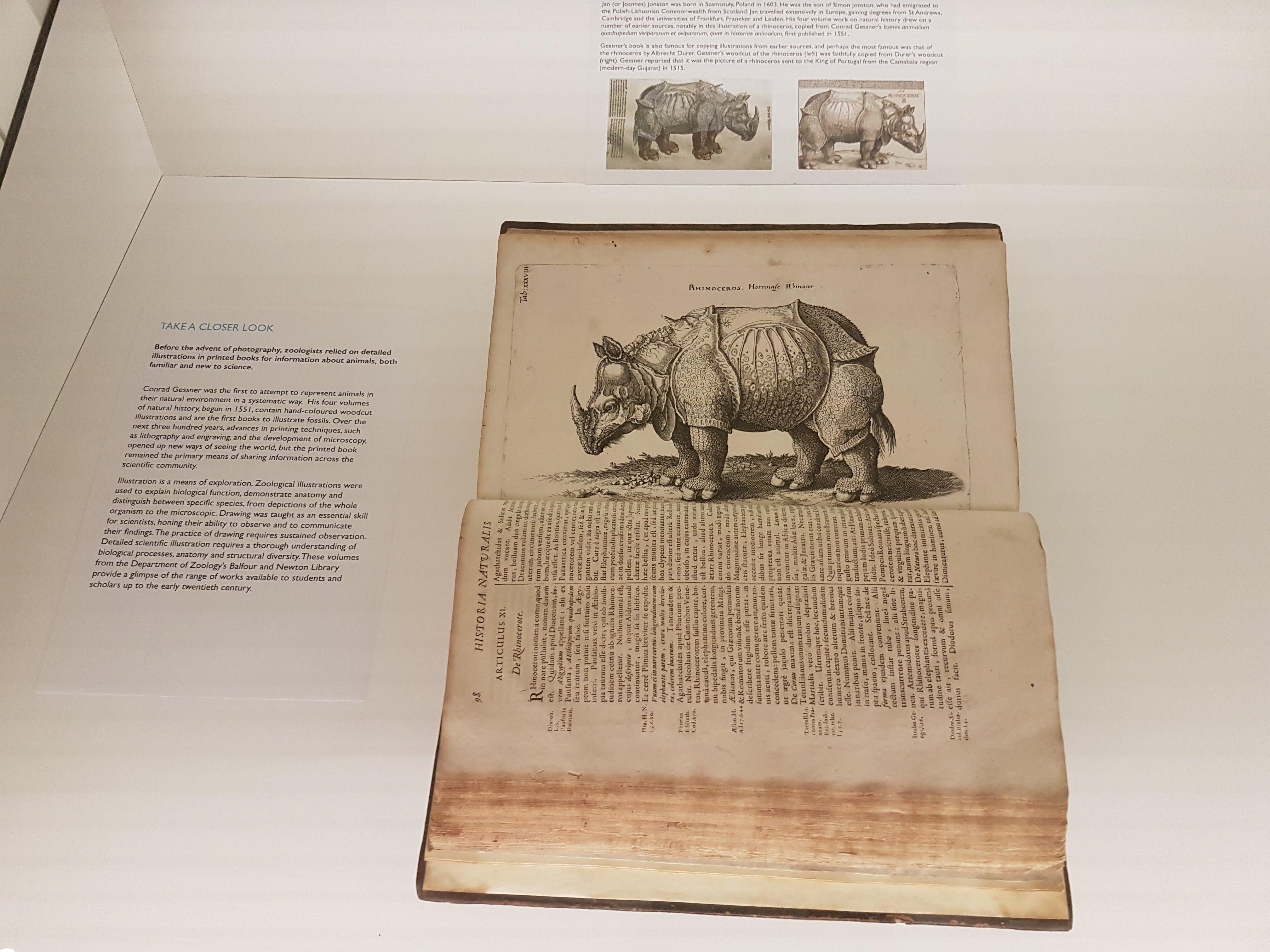
Dürer/other rhino art image and info.

Darwin was famed for collecting beetles when he should have been studying theology at Cambridge as a youth, and here is some of his collection. Dang.

Darwin’s finches!

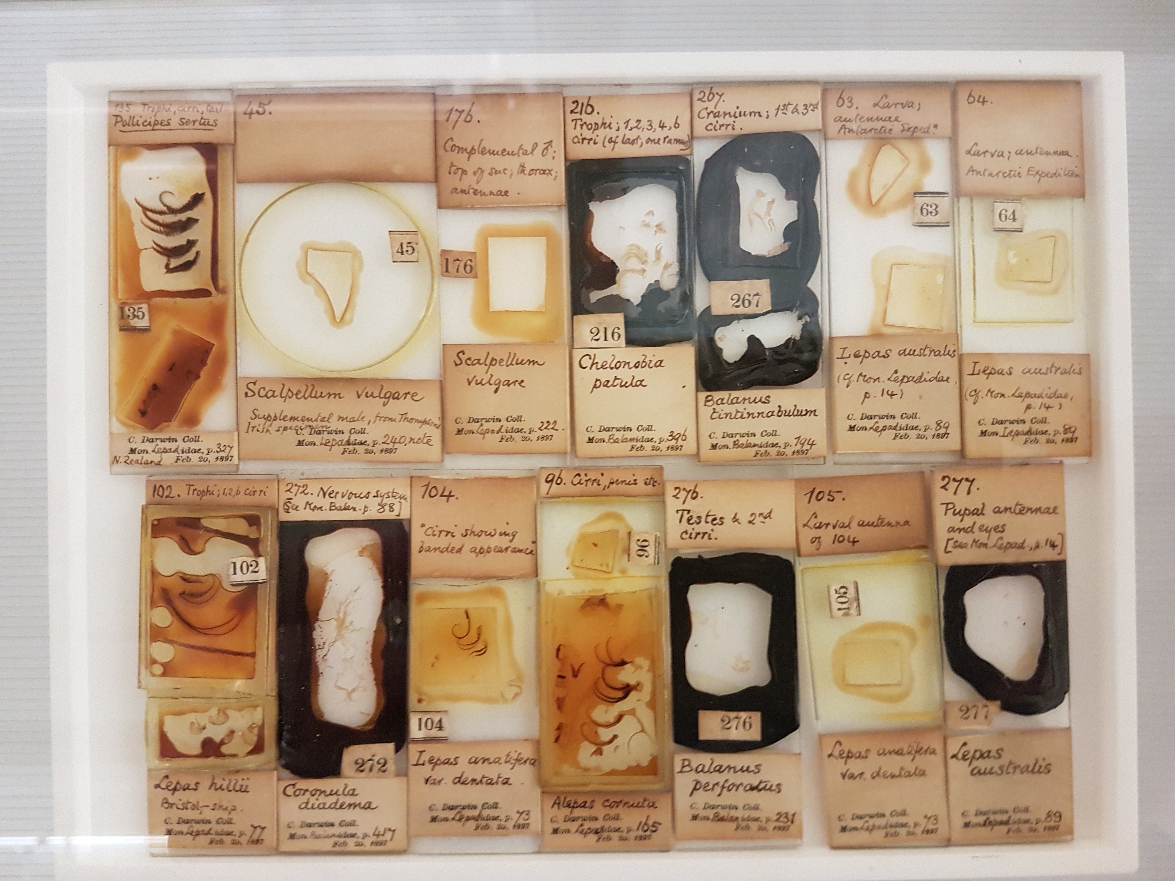
Darwin kicked off some of his meticulous work with volumes on barnacles; specimens included here; which helped fuel insights into evolution (e.g. they are “retrograde” crustaceans, not mollusks).

Darwin’s voyage: fish & other preserved specimens.

I think this is a solitaire weka (flightless island bird; see Comment below). I’ve never seen them displayed w/skeleton + taxidermy; it’s effective here.

Eryops cast. More early tetrapods will surely be featured on the upper floor; this one was on the timeline-of-life-on-Earth display.

I LOVE dioramas and this seabird nesting ground display is very evocative, especially now that I’ve visited quite a few such islands.

Mammal introduction; phylogenetic context.

Monotreme glory.

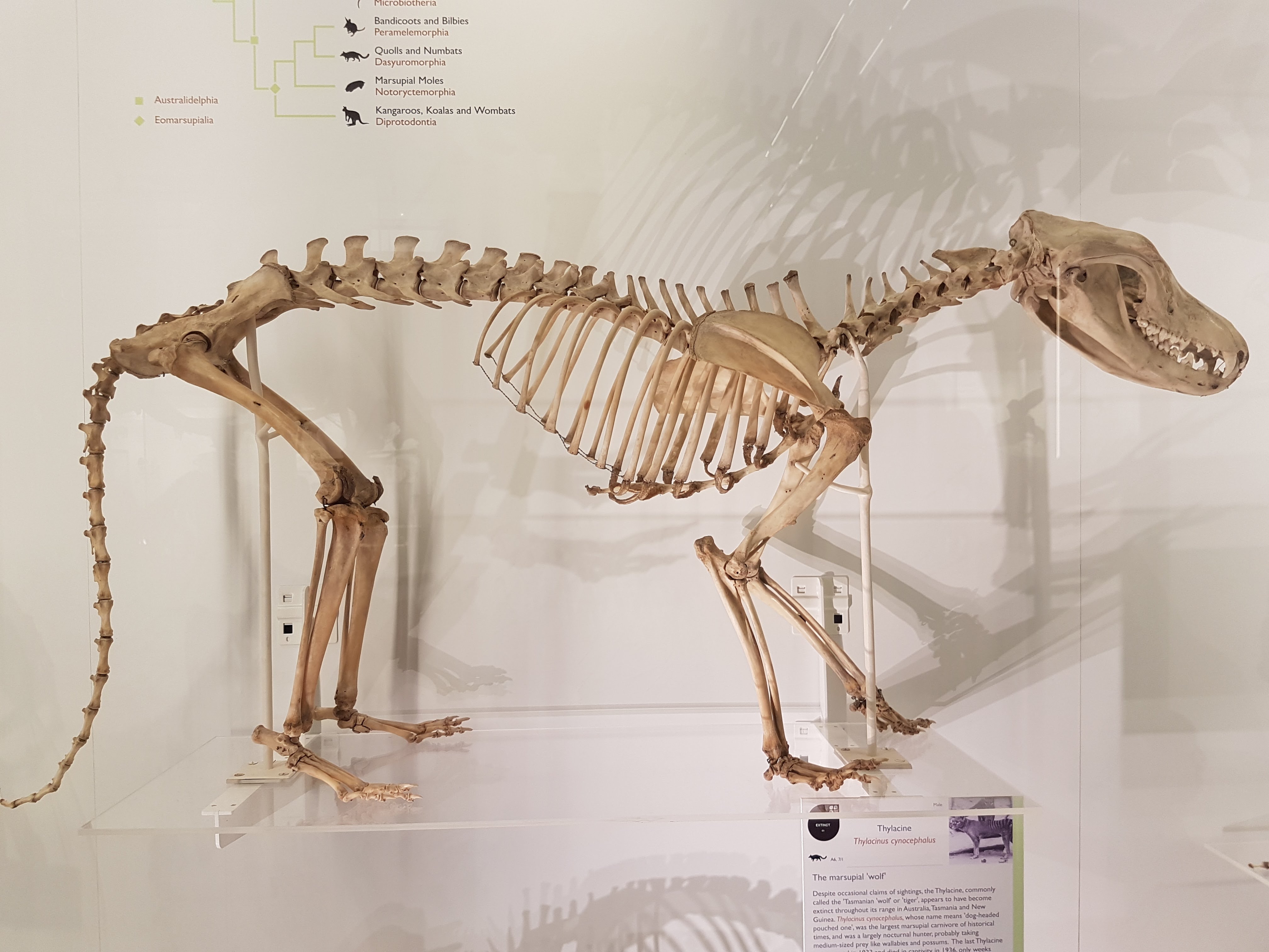
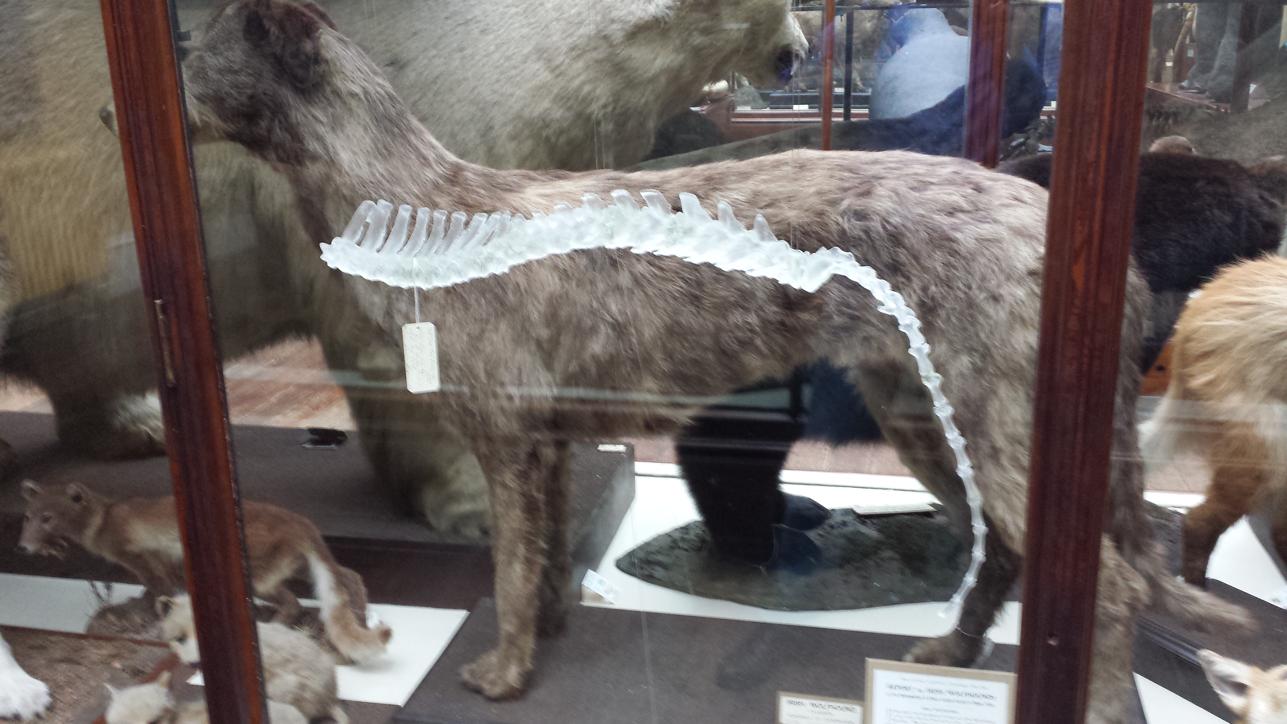
UMZC is well endowed with thylacines and this one is lovely.
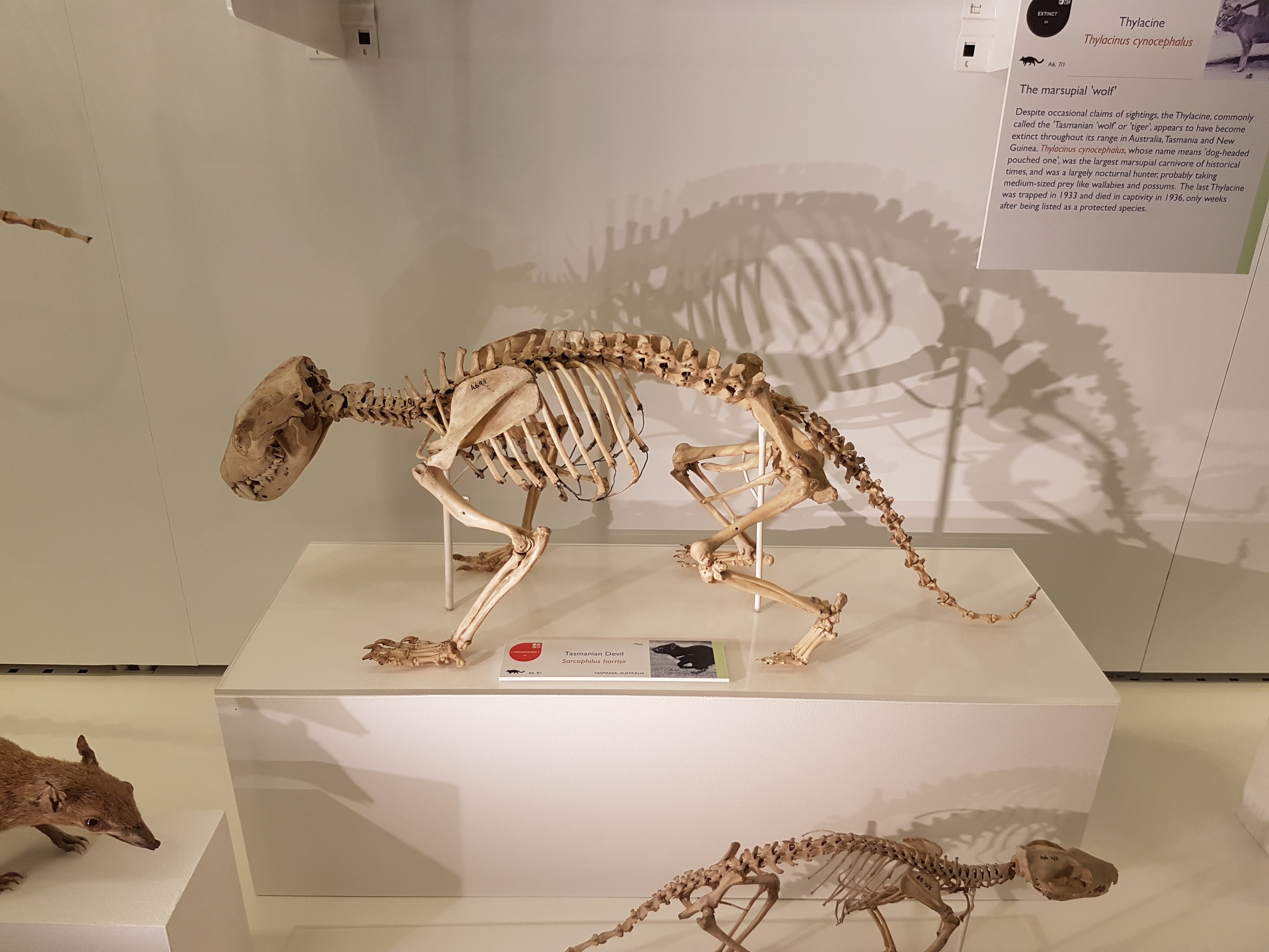
“TAZ FEEL NAKED!”

Narwhal above!

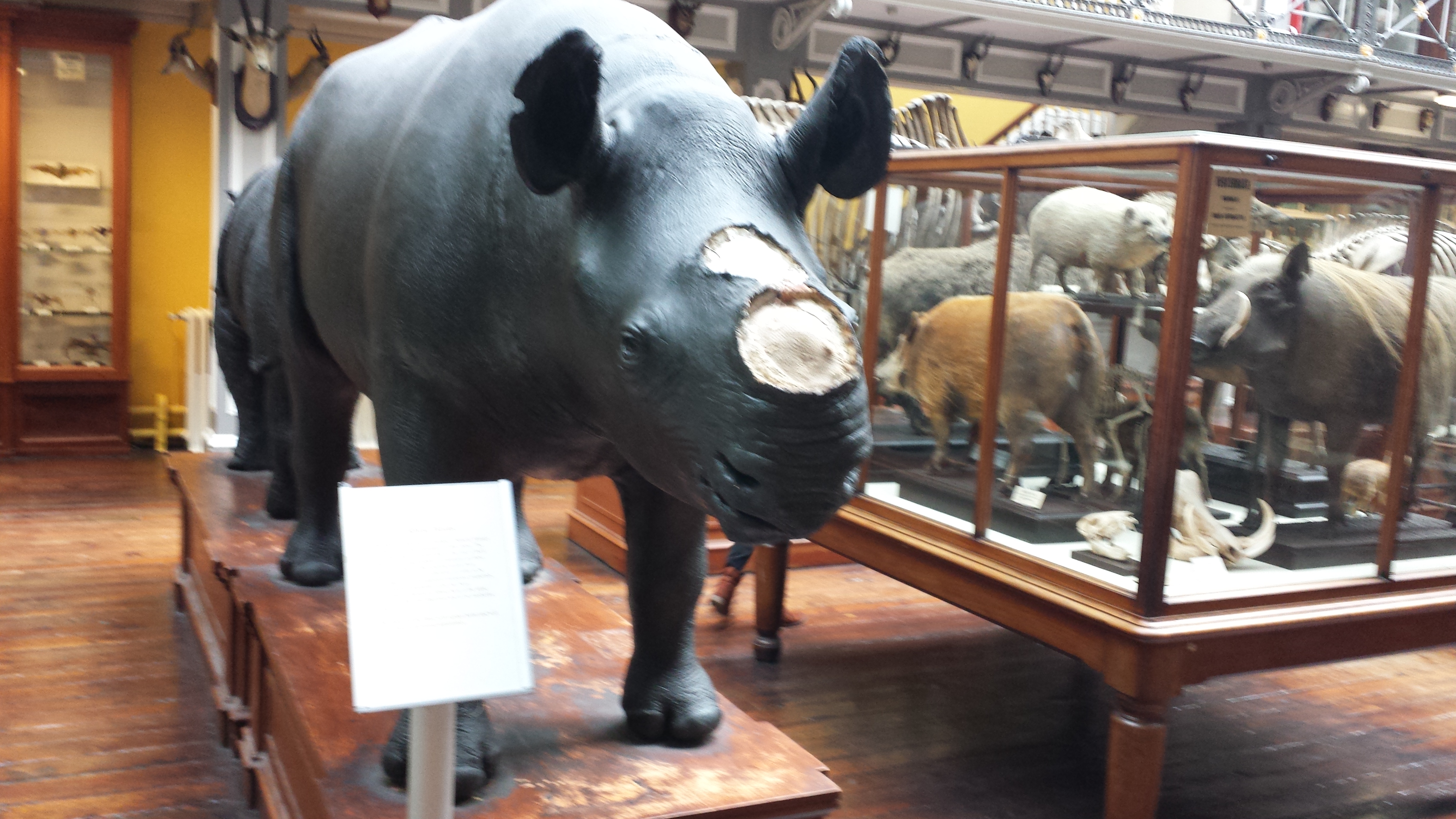
Rhinocerotoidea past, present, and fading glory. 😦

Ceratotherium white rhino. The horn is not real; sadly museums (and even zoos) across the world have to worry about theft of such things, given that some people think these horns are magic.

Ceratotherium staring match. You lose.

Ceratotherium stance.

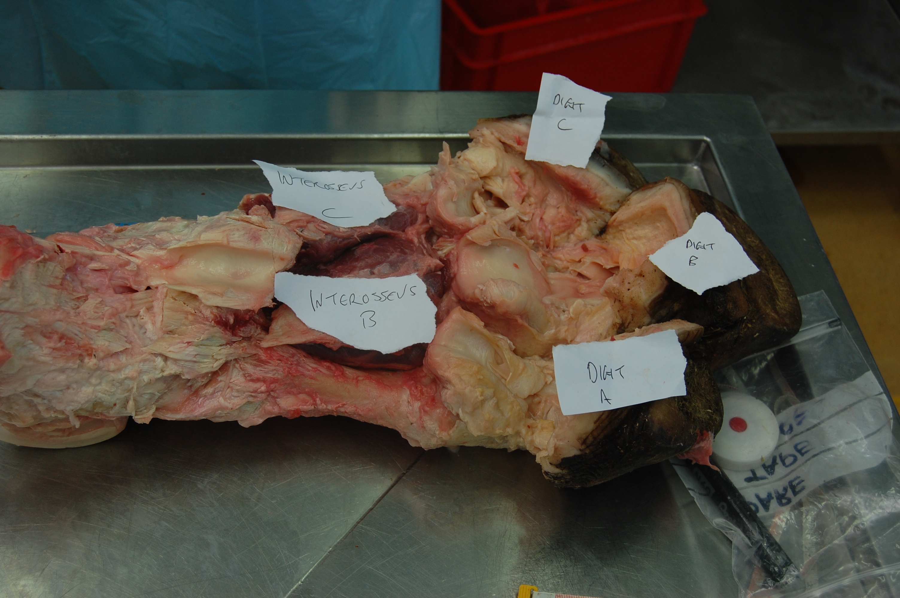
Foot of a Sumatran rhino juxtaposed with a horse’s for Perissodactyla didaction.

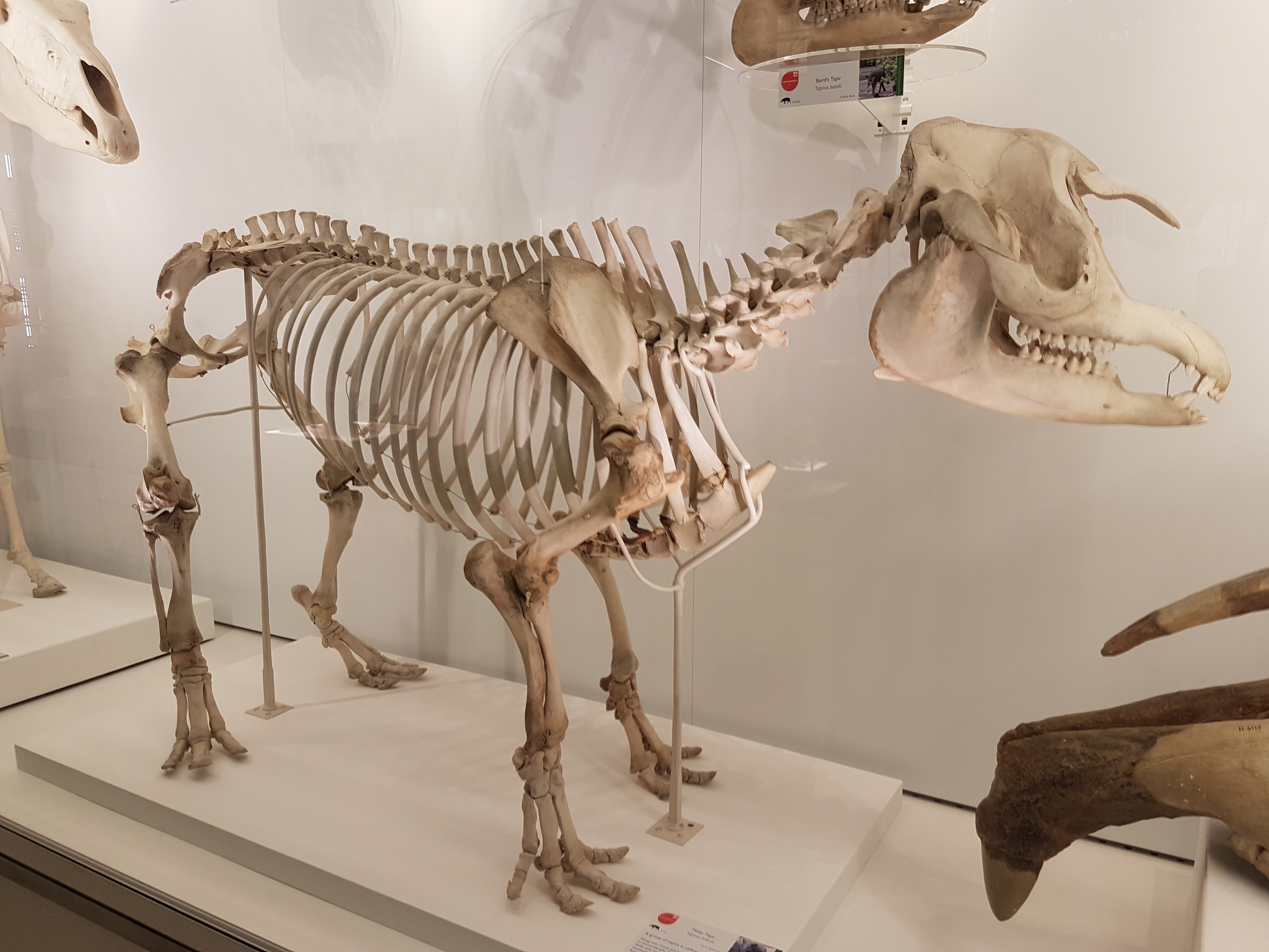
A tapir. As a kid, I used to wander around the house pretending to be a tapir but I did not know what noise they’d make so I’d say “tape tape tape!”.

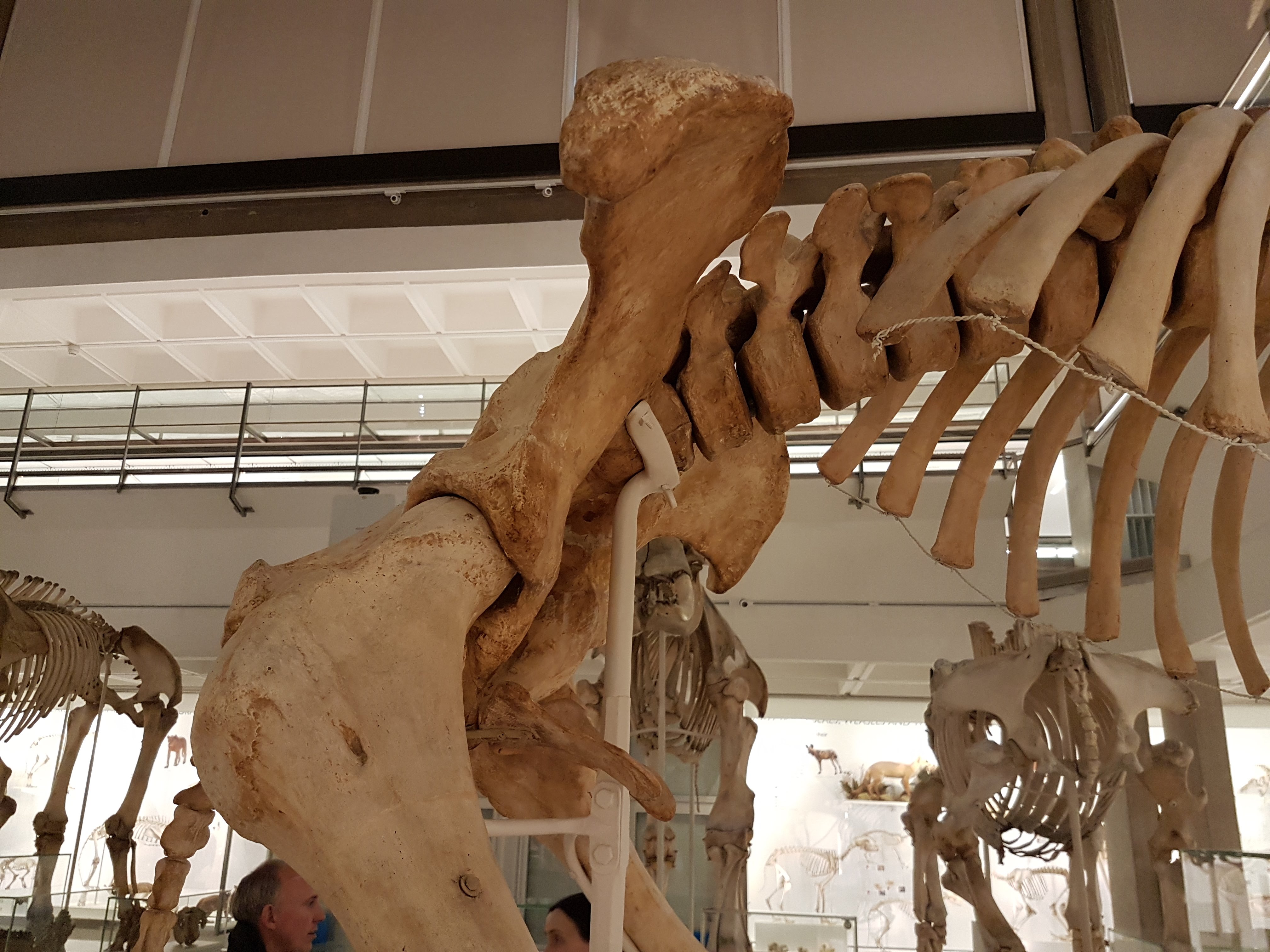
Big Southern Elephant Seal.

Squat little fur seal.

Hippopotamus for the lot of us. (baby included)

Hippo facedown.

Skull of a dwarf Madagascar hippo.

Cave bear and sabretooth cat make an impressive Ice Age demo.
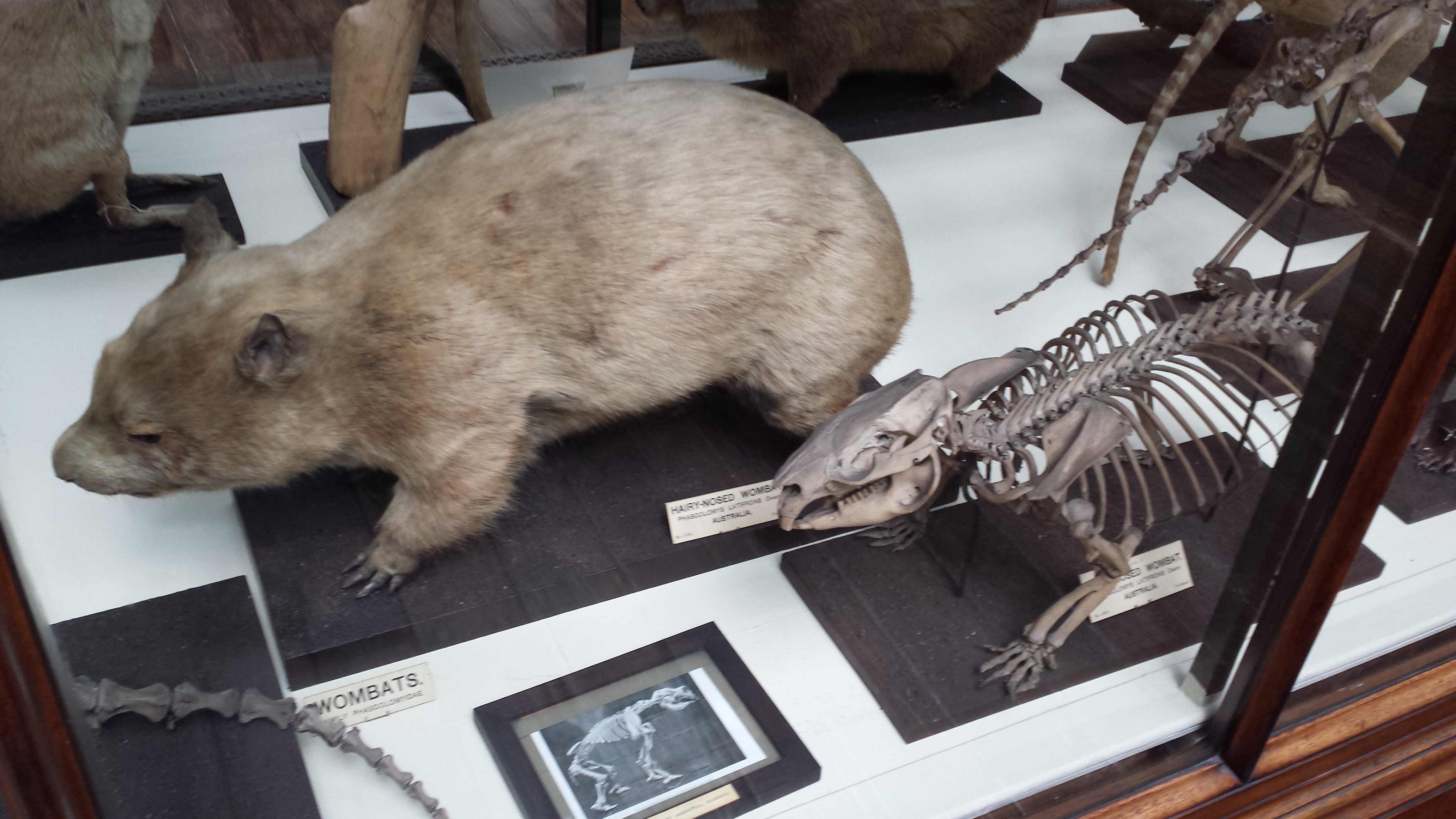
It’s a wombat.

Ain’t no don like a Diprotodon! (also note its modern miniature cousin the wombat, below)

Diprotodon facial.

Diprotodon shoulder: big clavicles bracing that joint region.

Diprotodon knee: even in big marsupials, the “parafibula”/lateral sesamoid of the knee is still generally present. And why it is there/what it does deserves much more study.

Diprotodon hip. I just find this animal’s anatomy fascinating head-to-tail.

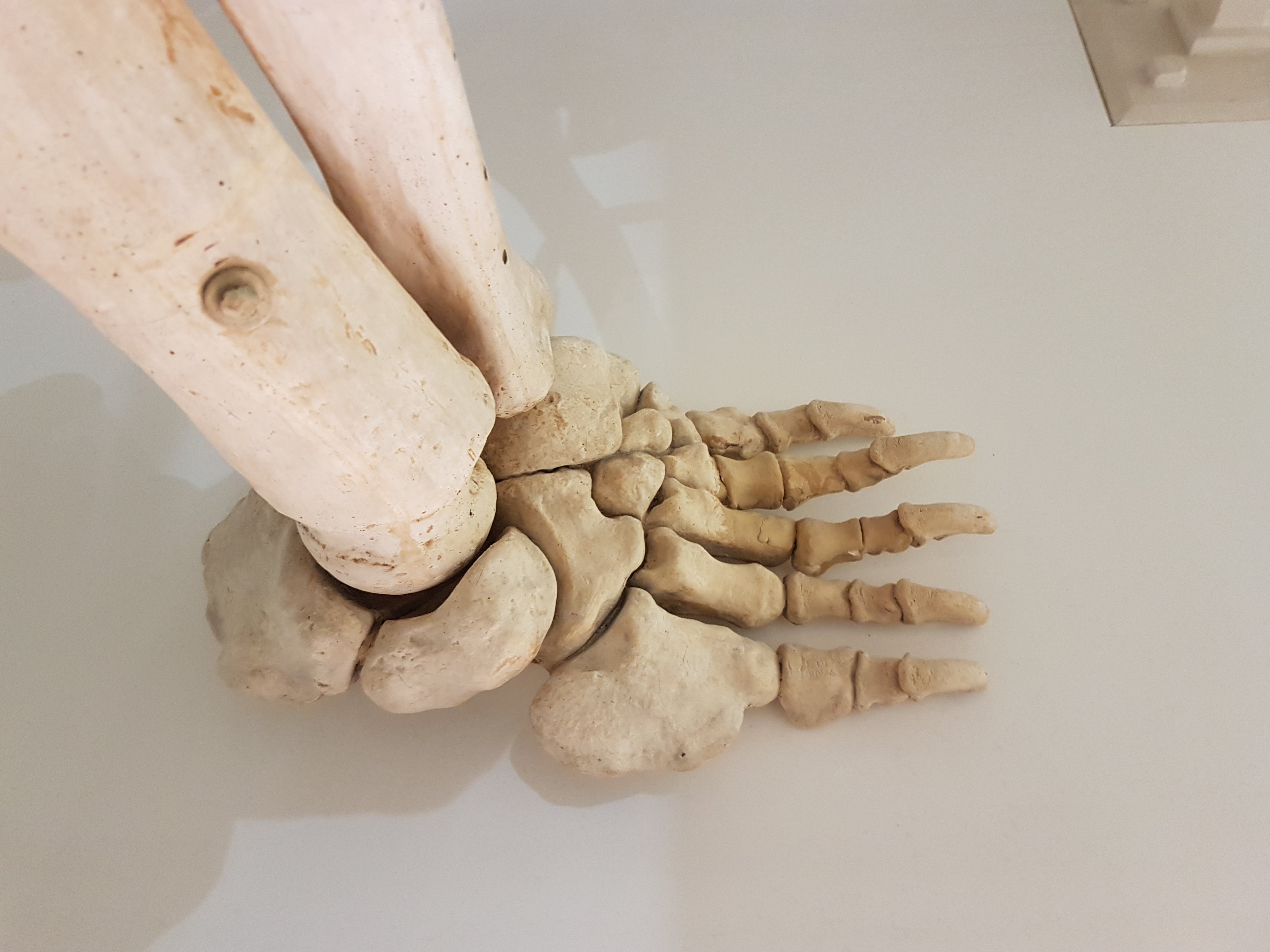
Diprotodon front foot. Absolutely freakish.

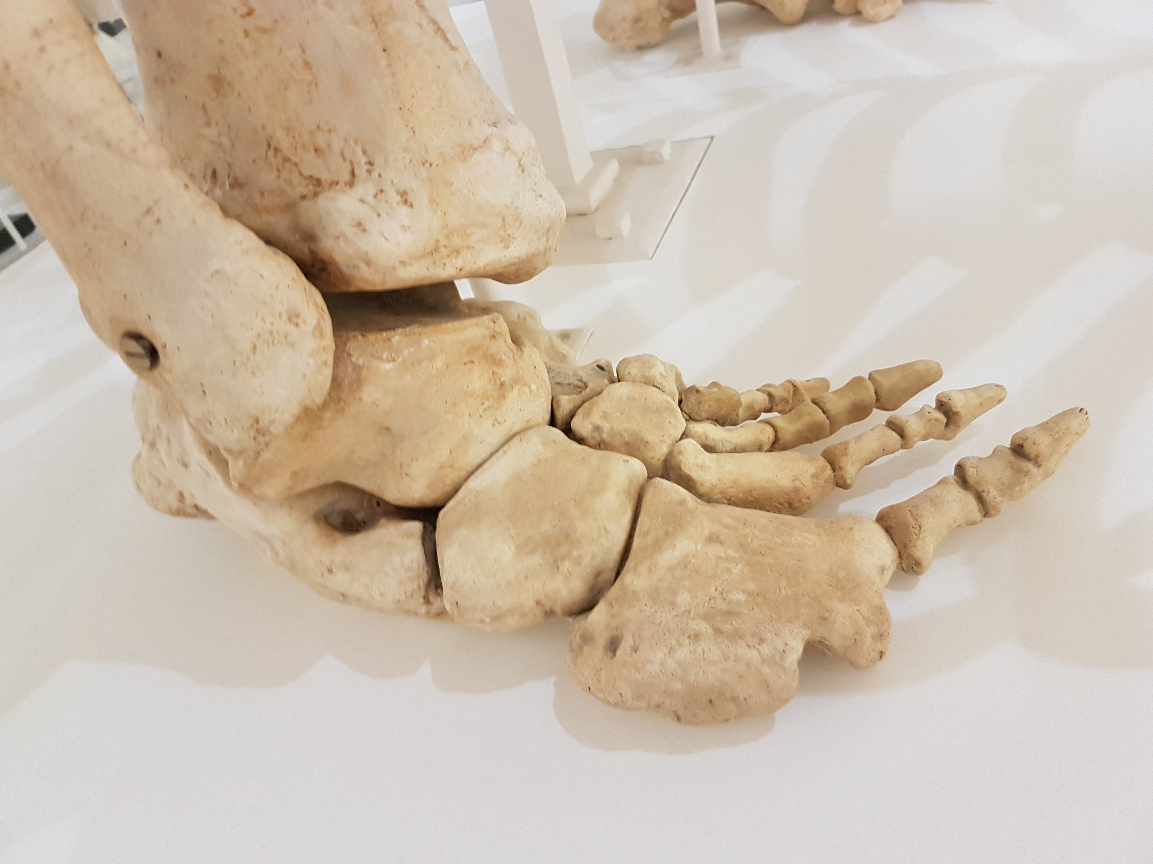
Diprotodon hind foot. Even weirder.

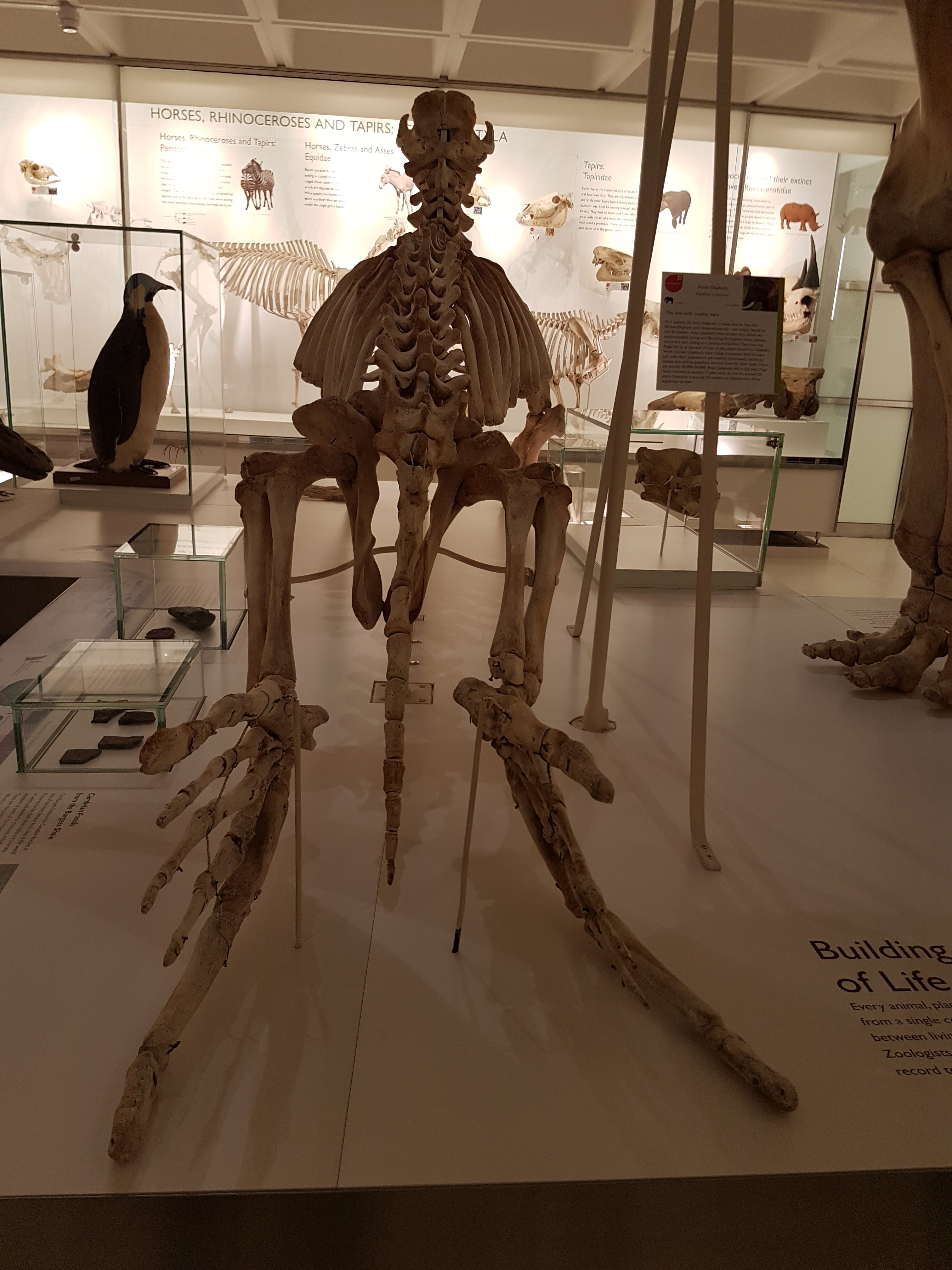
Your view after having been trampled in a supine position by a Diprotodon. Not a good way to go.

Diprotodon got back.

Elephant seal’s butt continues my series of photos of big animals’ bottoms.

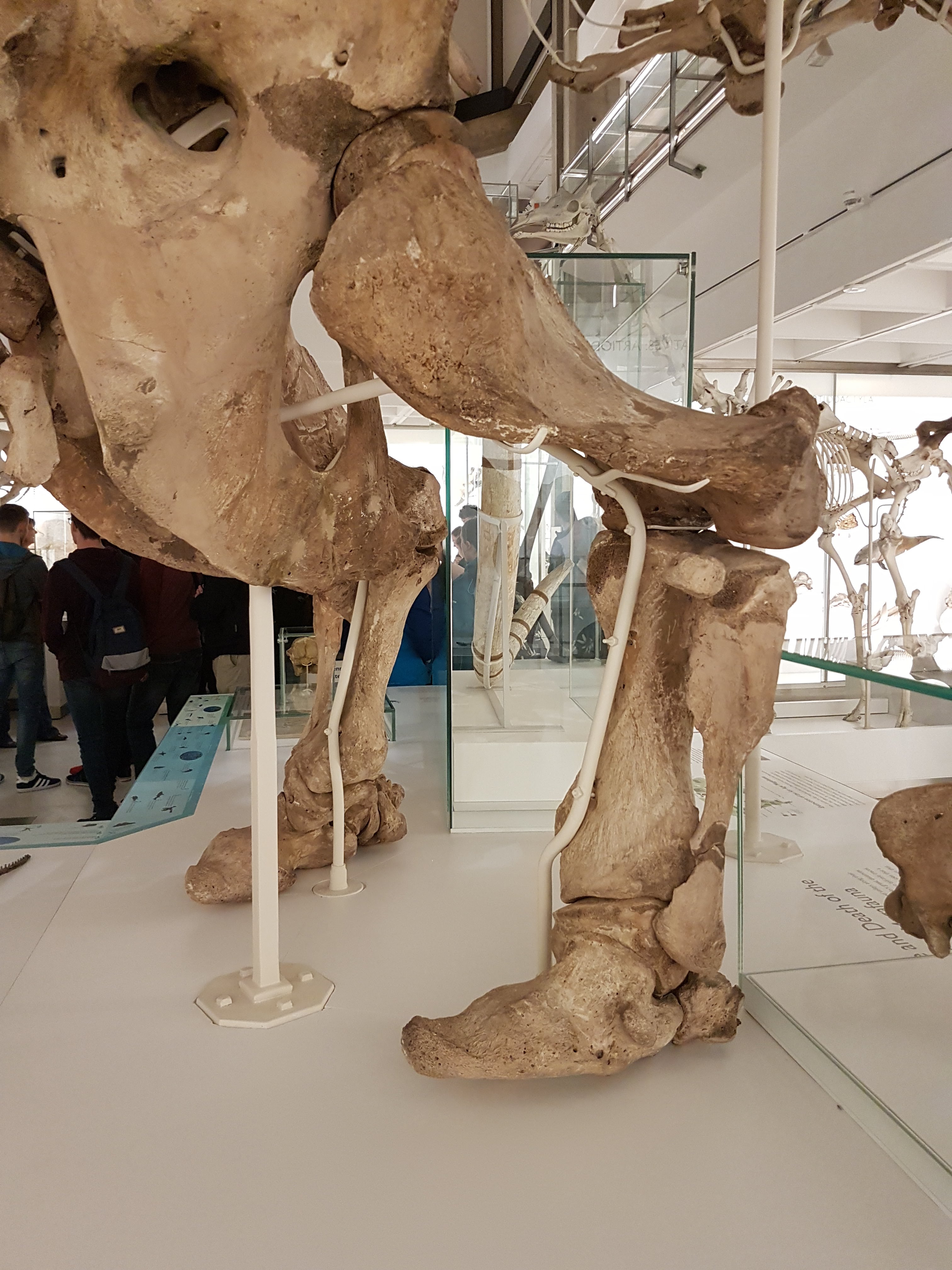
Asian elephant’s butt view.

African elephant butt.

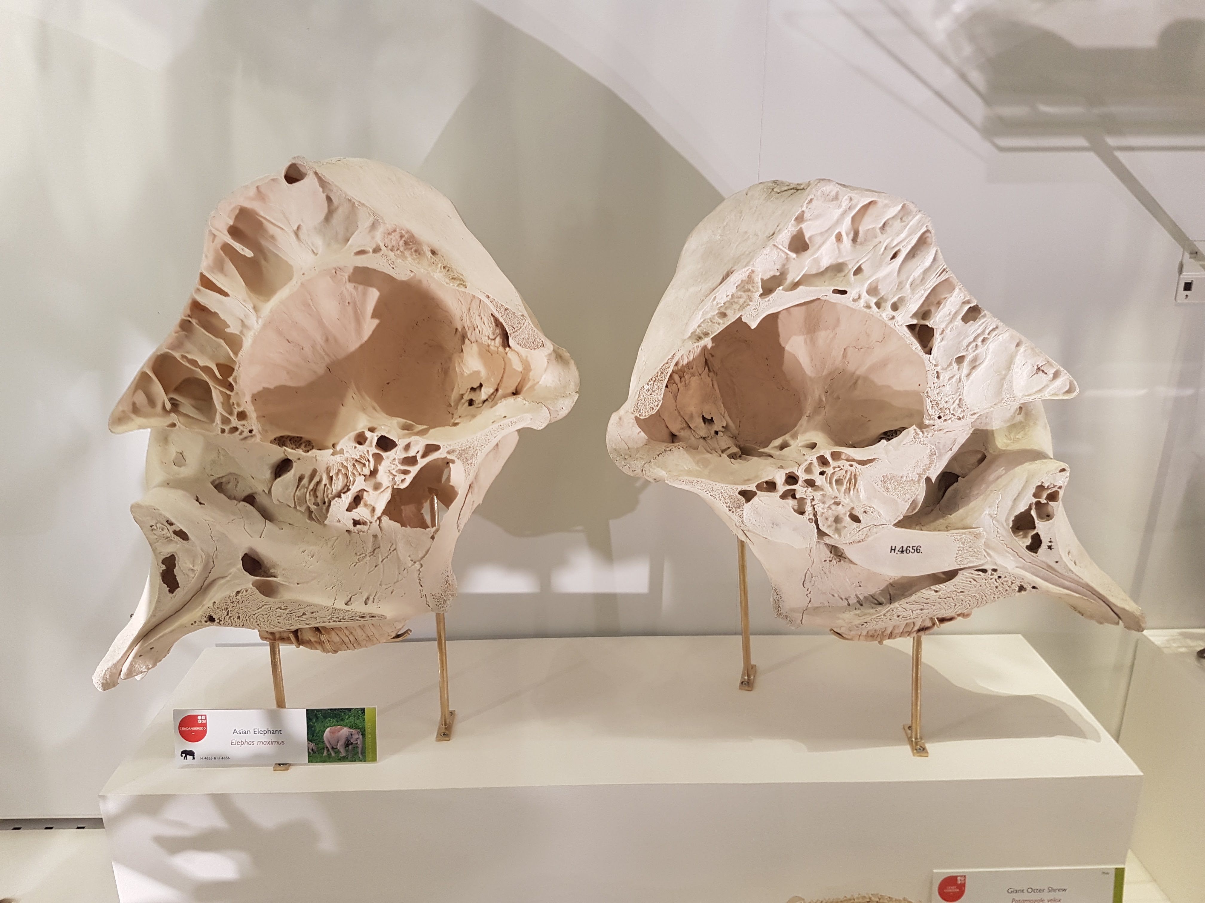
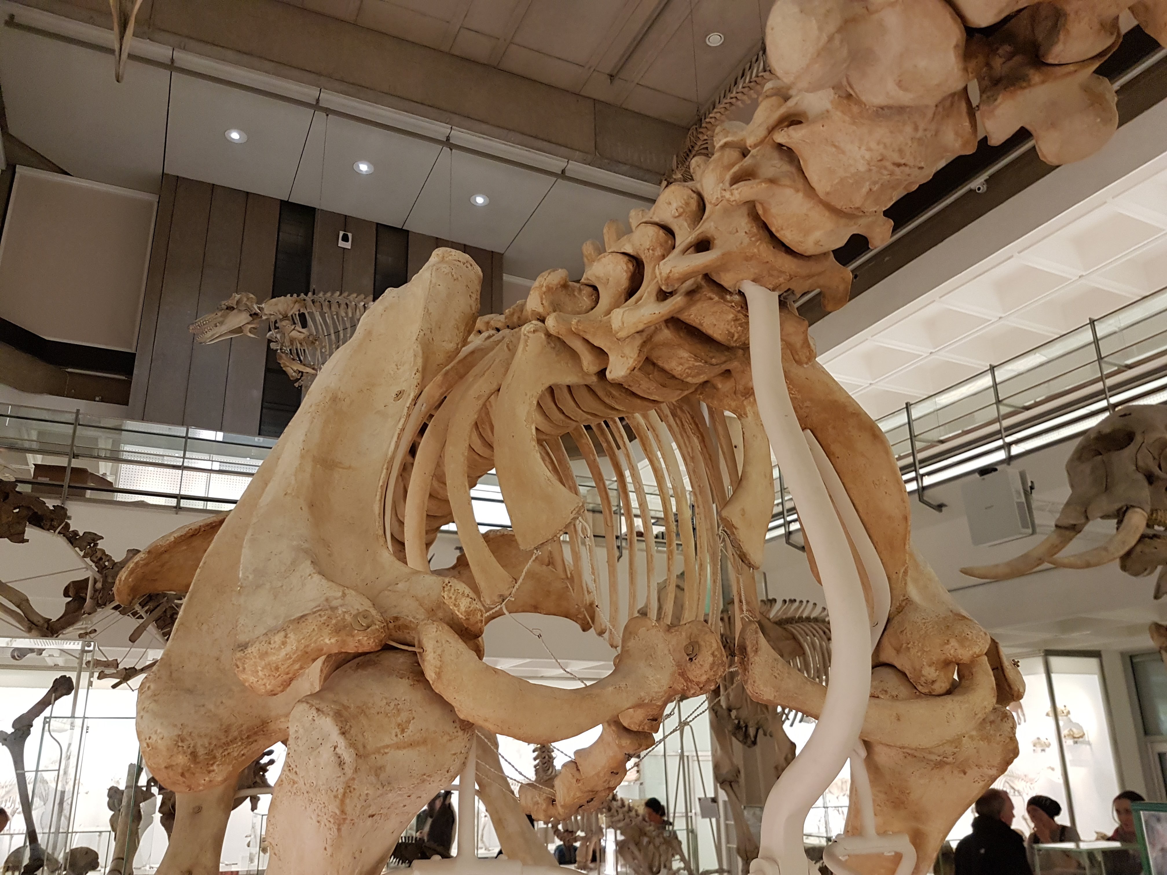
Sectioned elephant skull to show pneumatic resonating chambers.

Paenungulates: hyraxes, Sirenia, elephants & kin (evolutionary demo).

AND MY HYRAX!
Sorry. Had to.

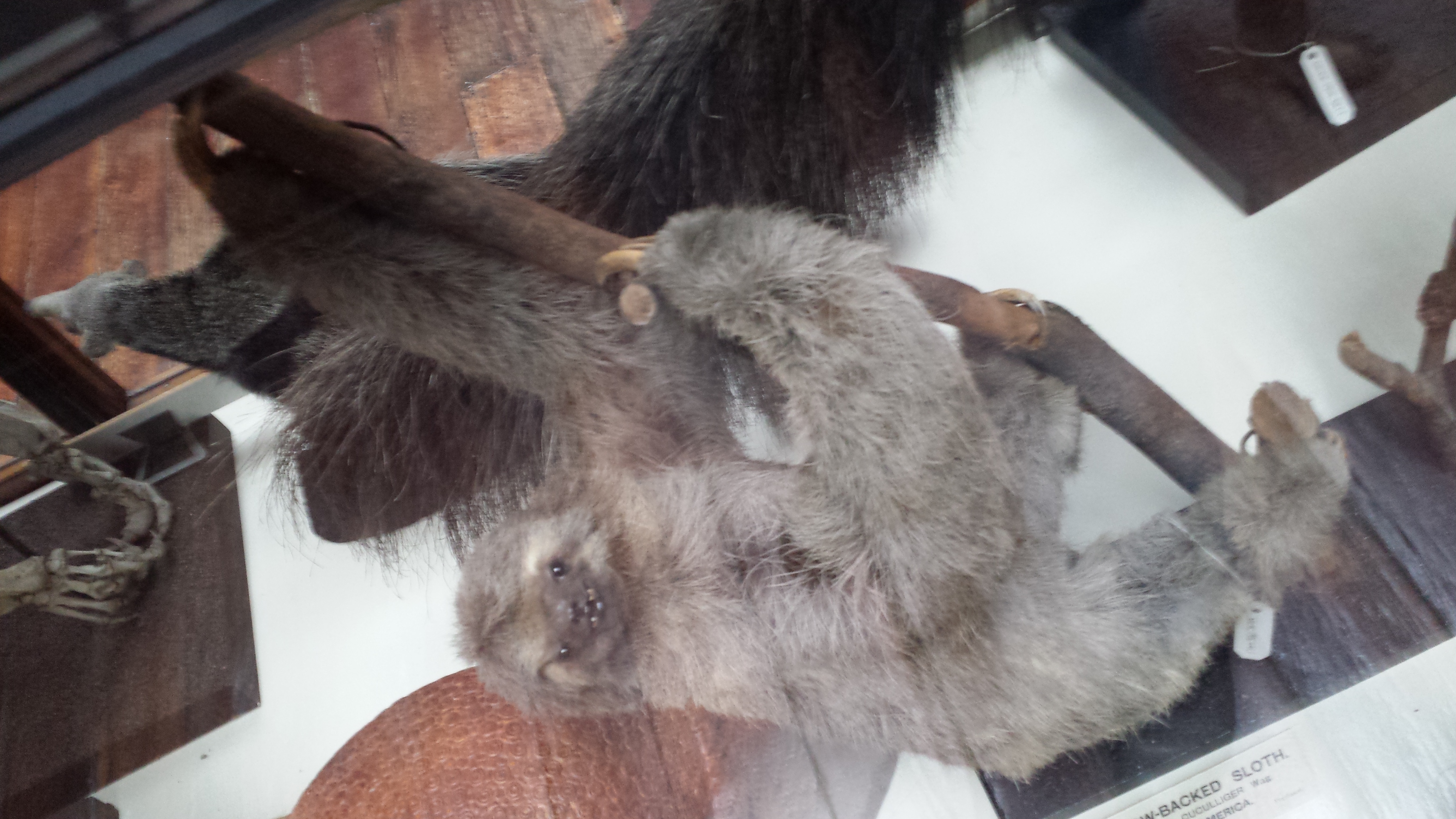
Megatherium side view.

Megatherium. Yeah!

Megatherium hindlegs fascinate me. Well-heeled.
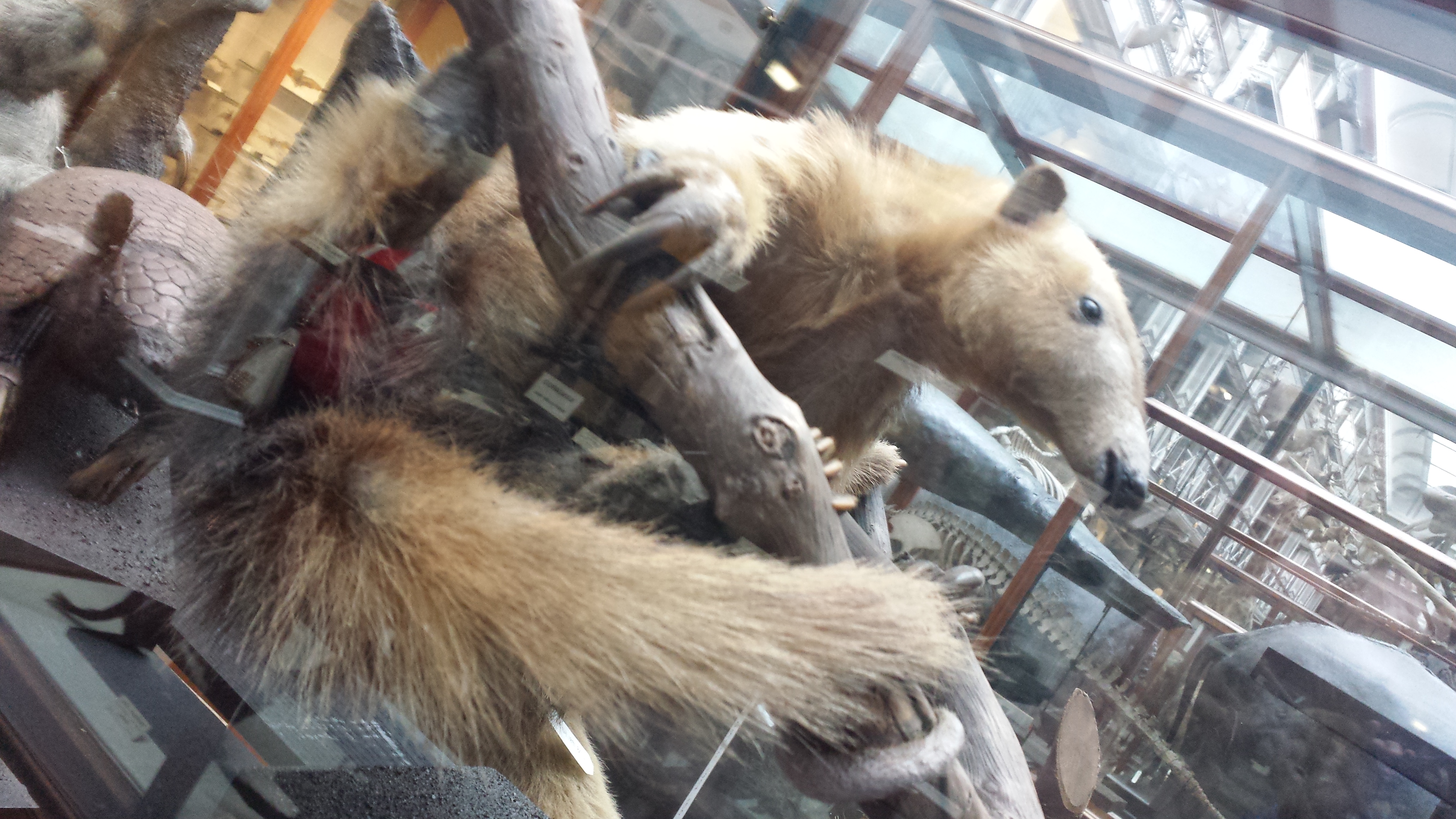
Tamandua duo.

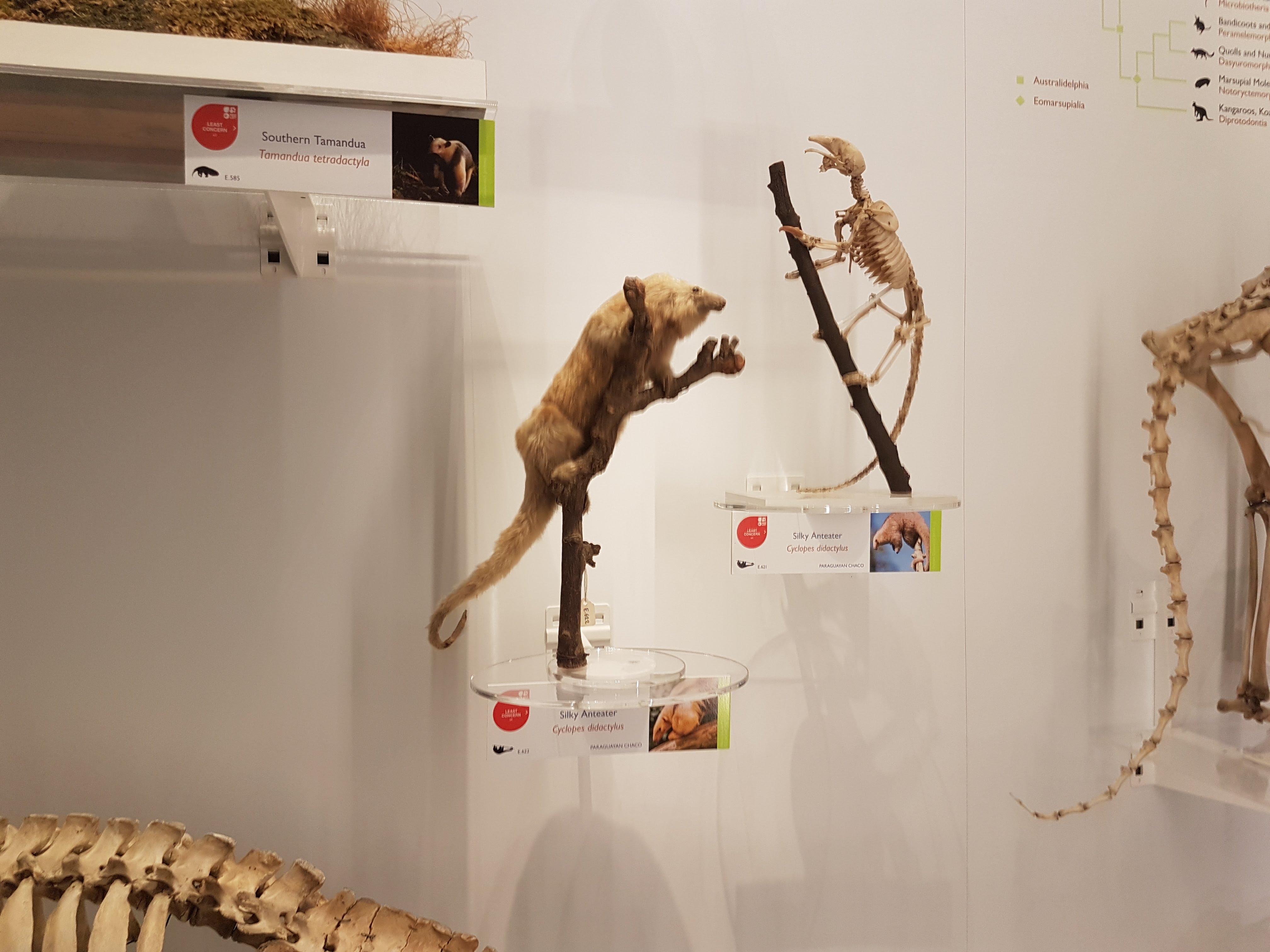
Silky anteater; wonderful.
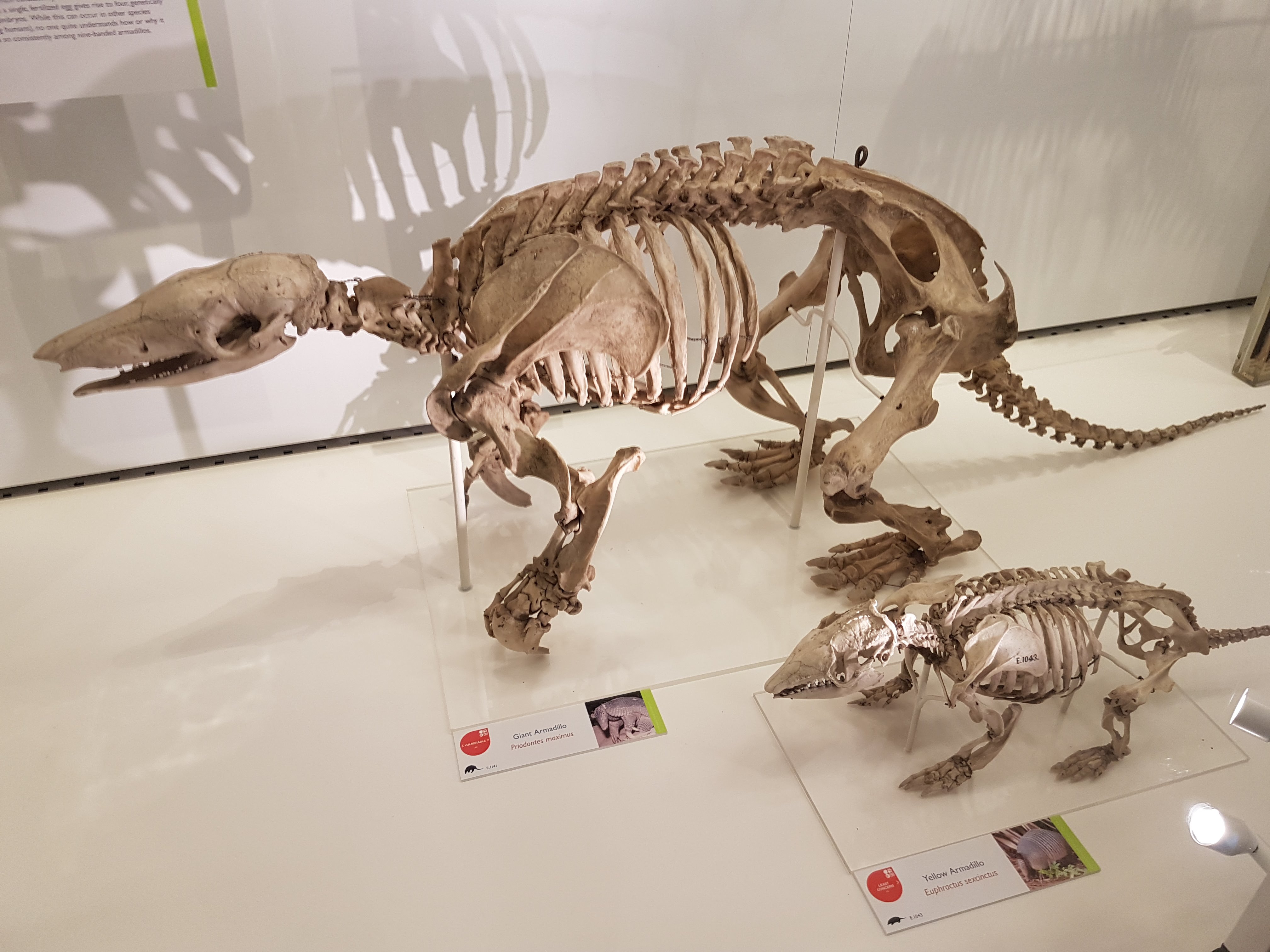
Armadillos.

Anteaters round out a fab display on Xenarthra.

The UMZC has everything from aardvarks to zebus. Here, conceptualized with other Afrotheria.

Golden moles: the more I read about them, the more they fascinate me.

We can all use some more solenodons in our lives!

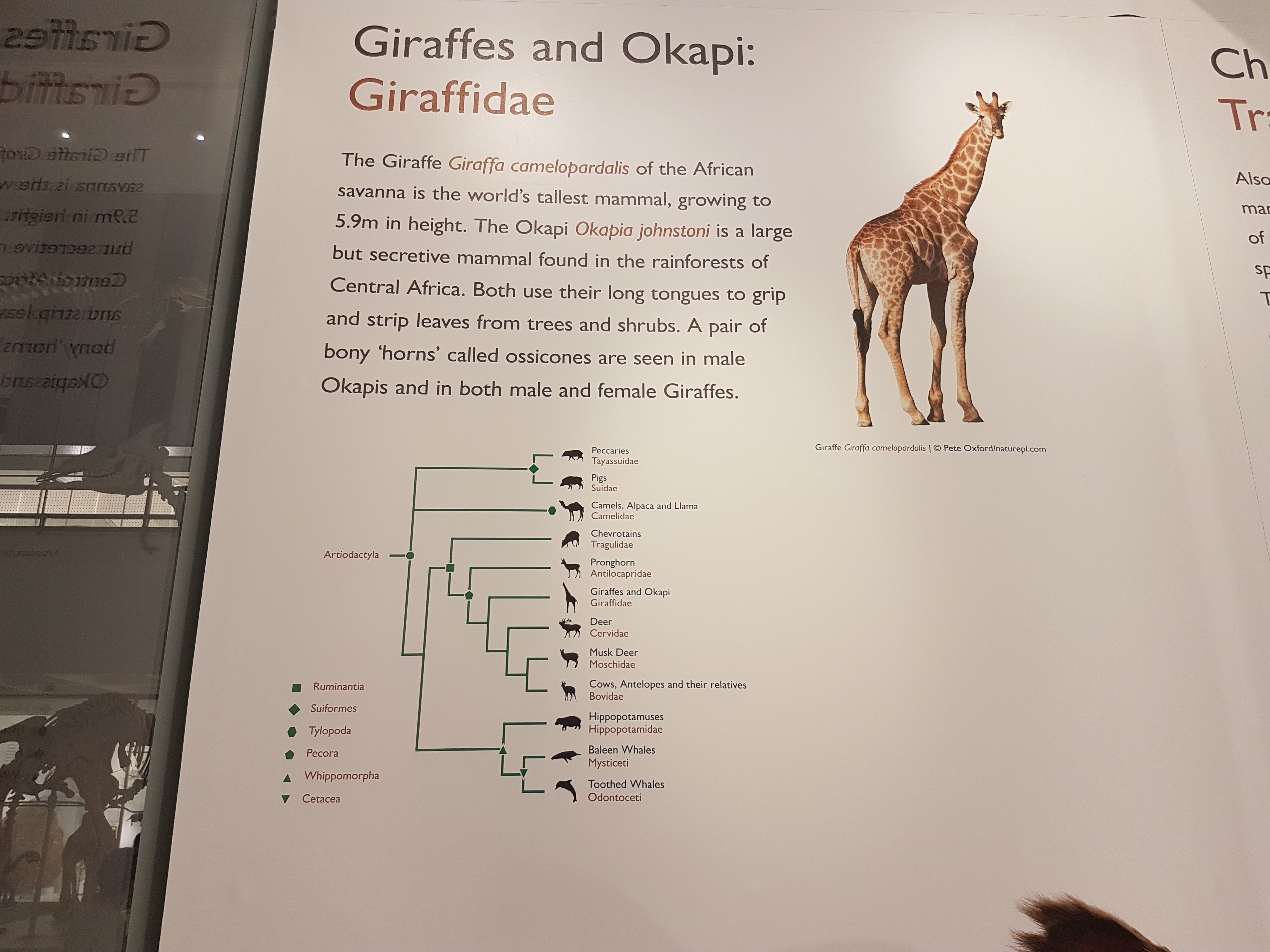
Example of the phylogenetic context used throughout exhibits.

If you’ve got a good Okapi taxidermy, you’d better use it.

It’s a giraffe. Did you guess right?

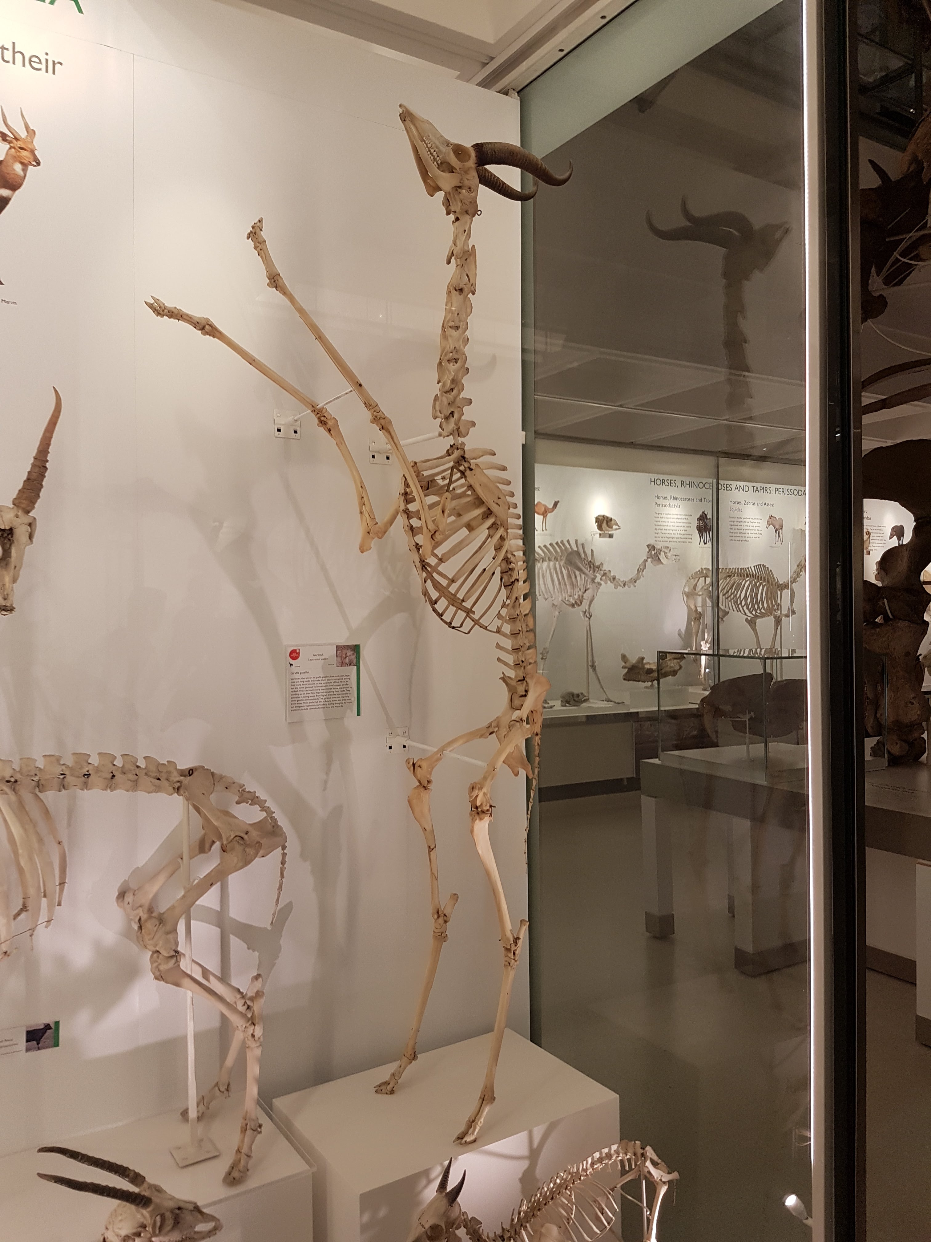
Gerenuk showing off its bipedal capacity.

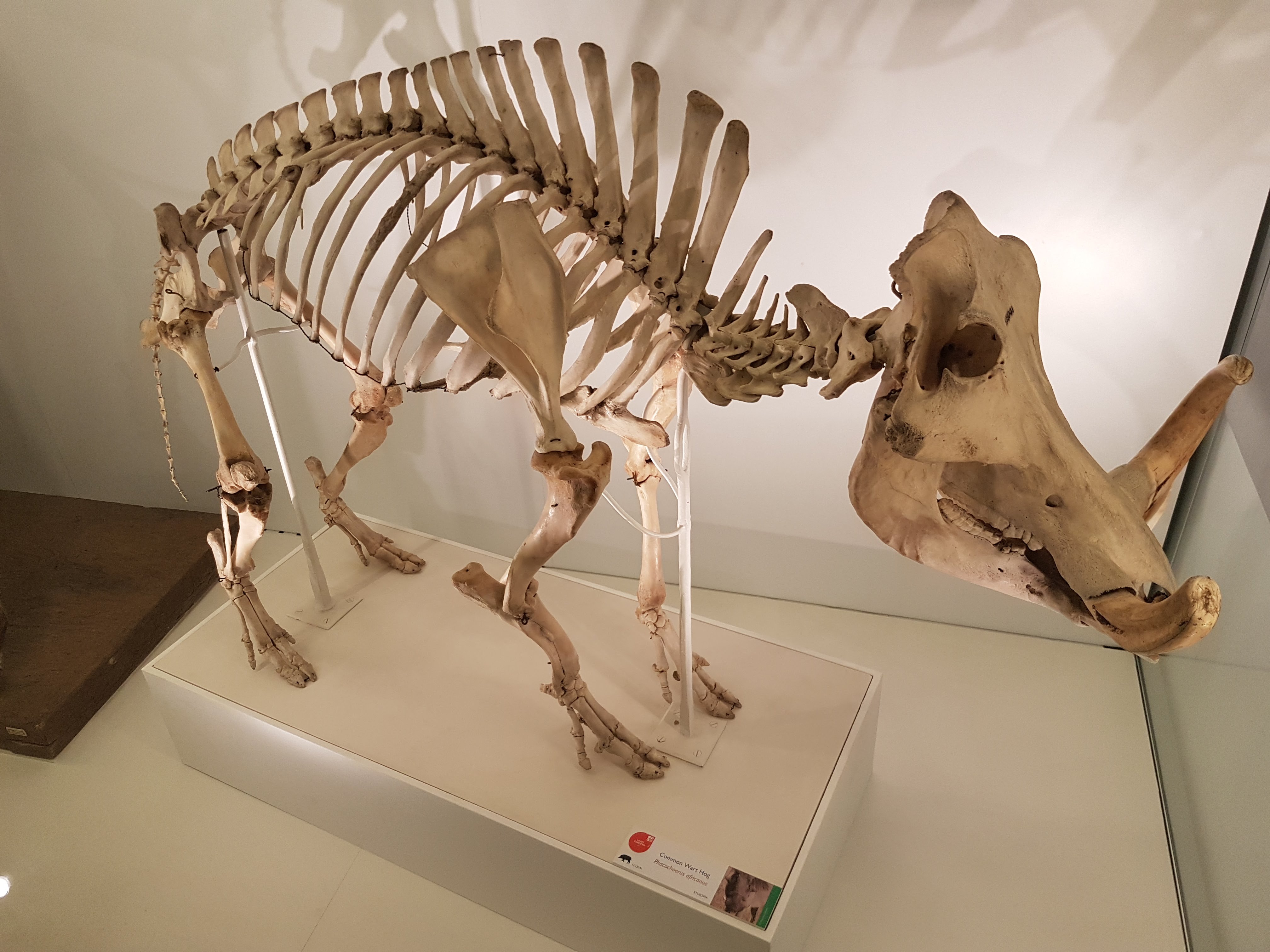
Warthogs have an inner beauty.

Pangolin. Glad to see it back on exhibit.

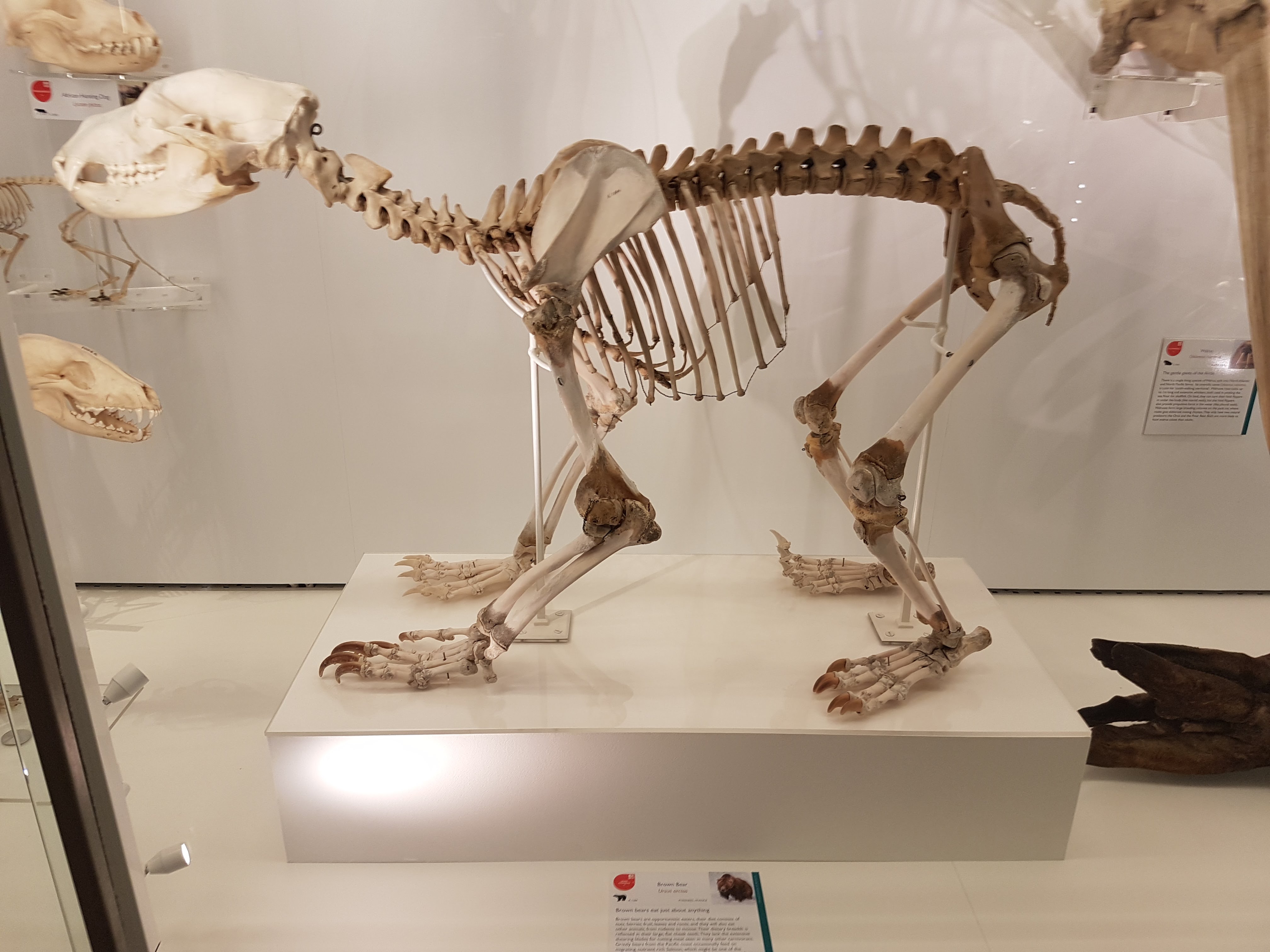
Nice little brown bear.

Double-barrelled shot of hyenas.
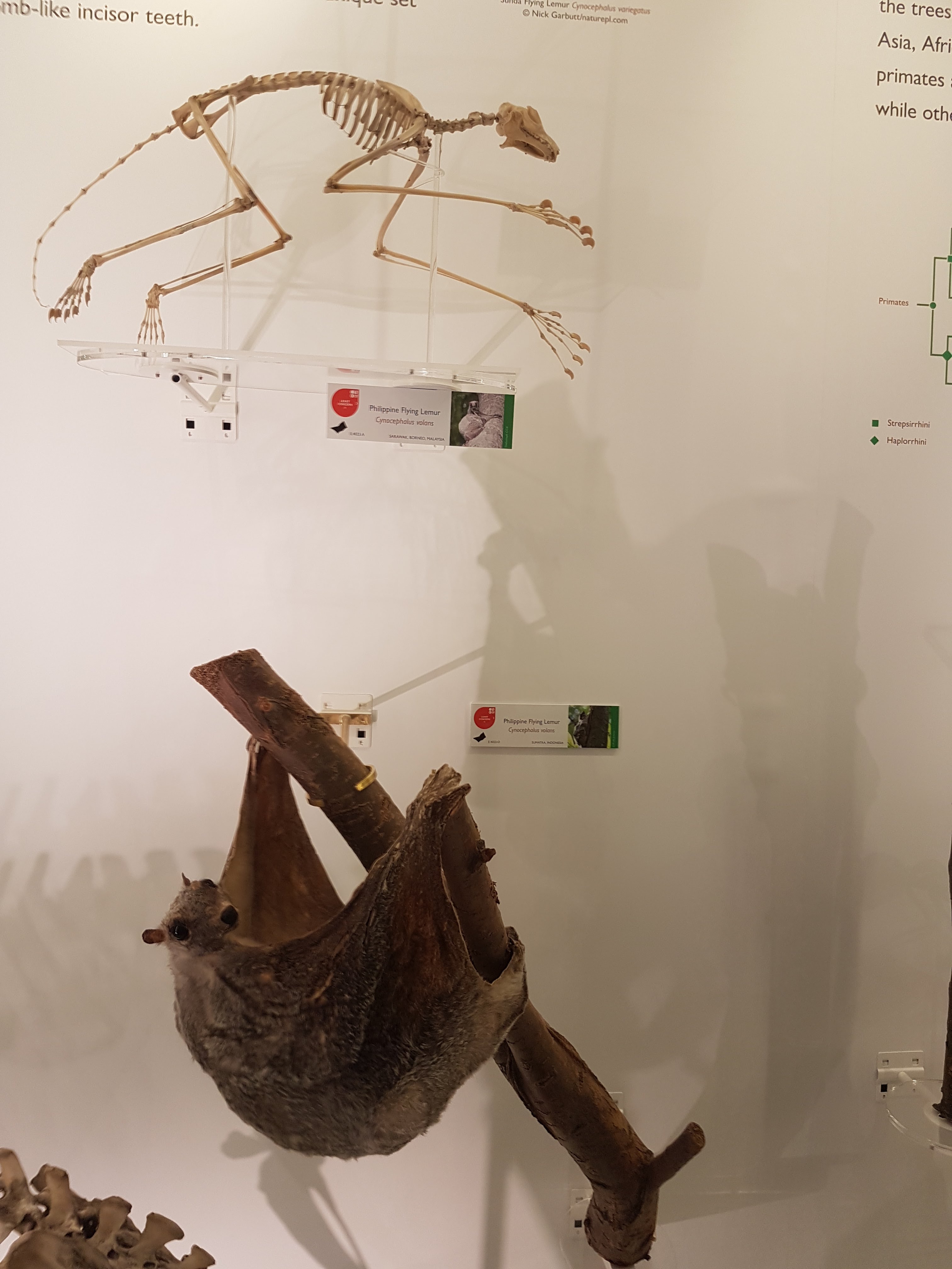
Colugo!

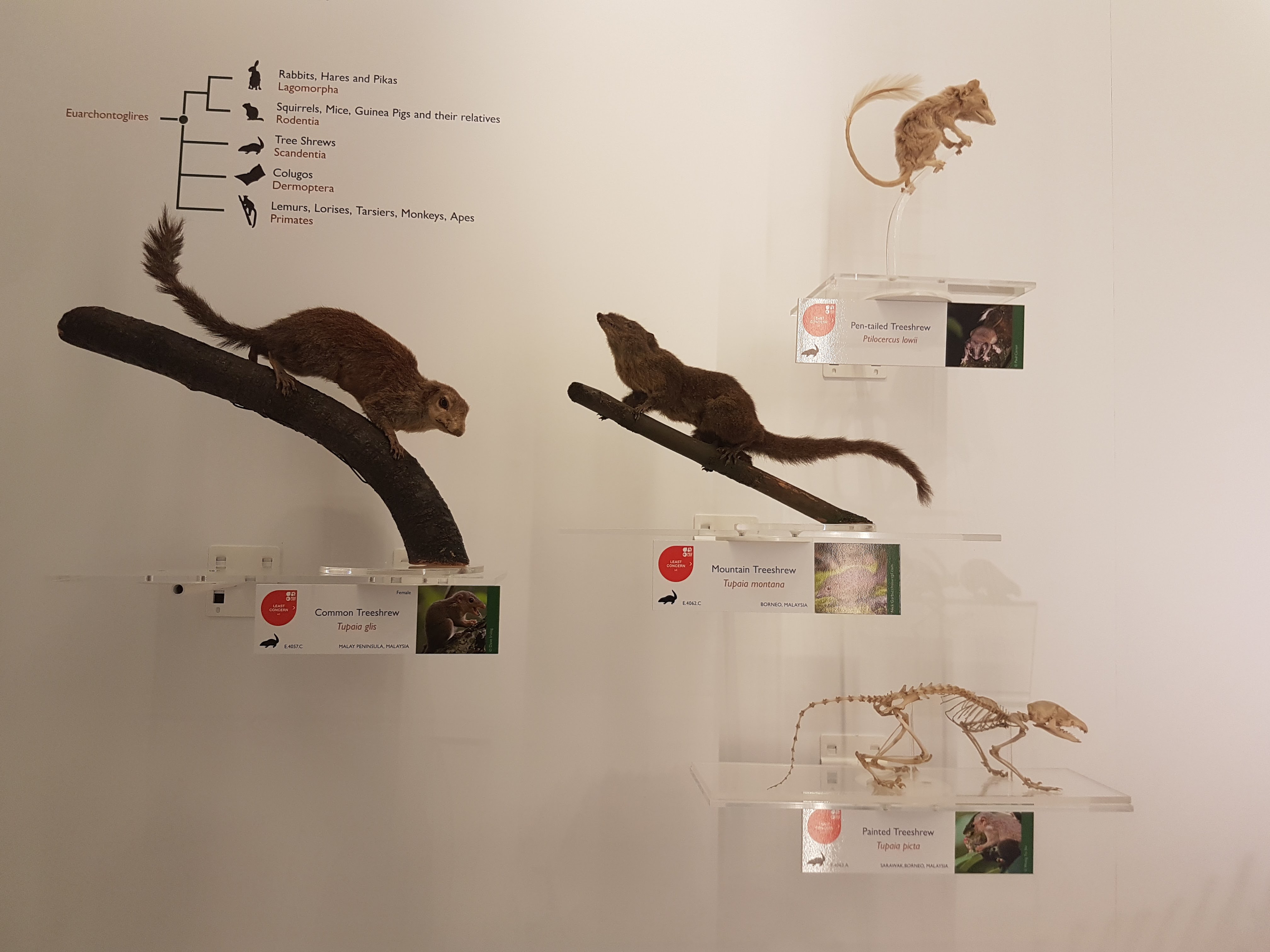
Nice to see some Scandentia featured.

My brain says this is a springhare (Pedetes) so I am going with what my brain says and anyway I really like this display.

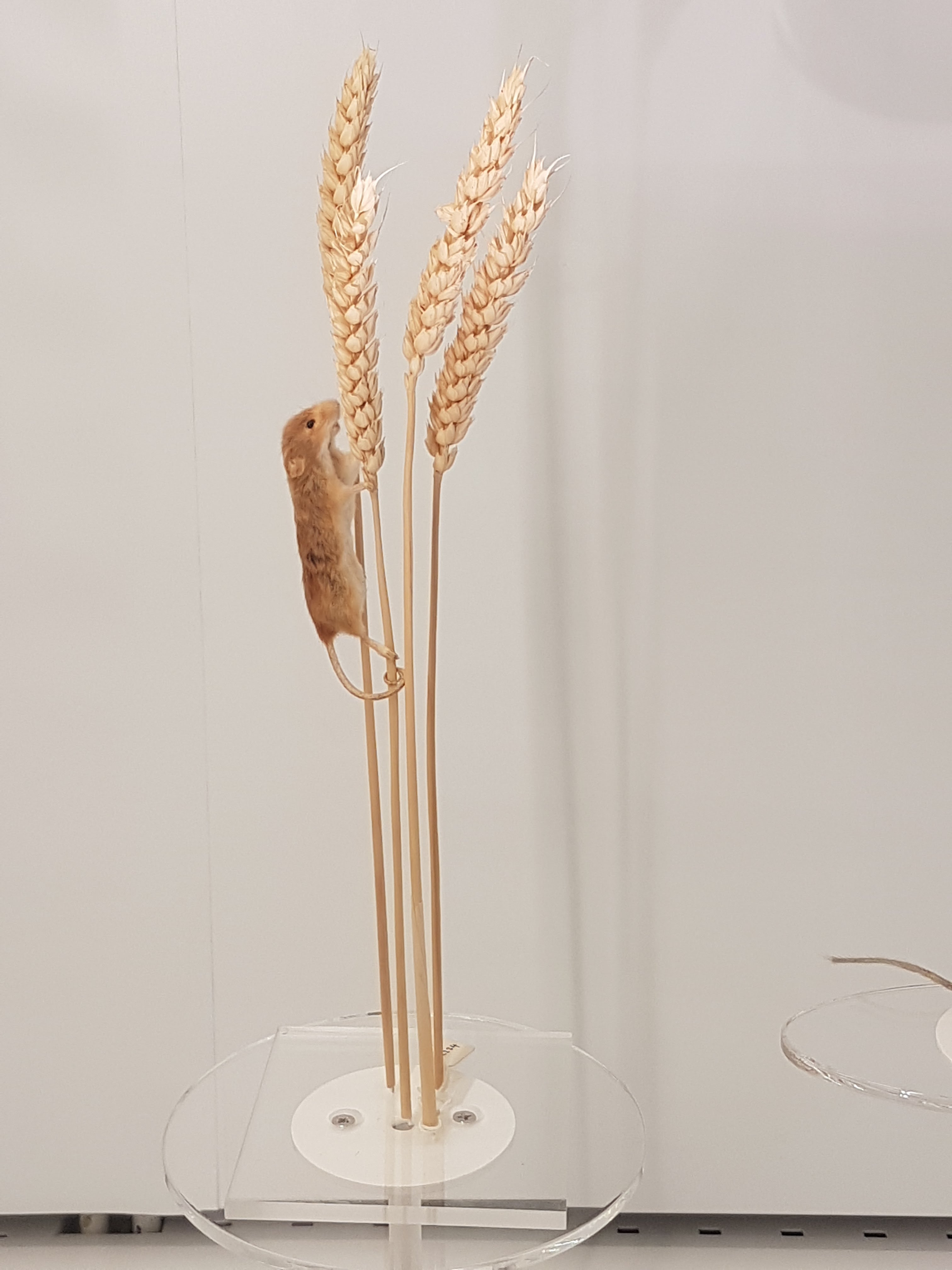
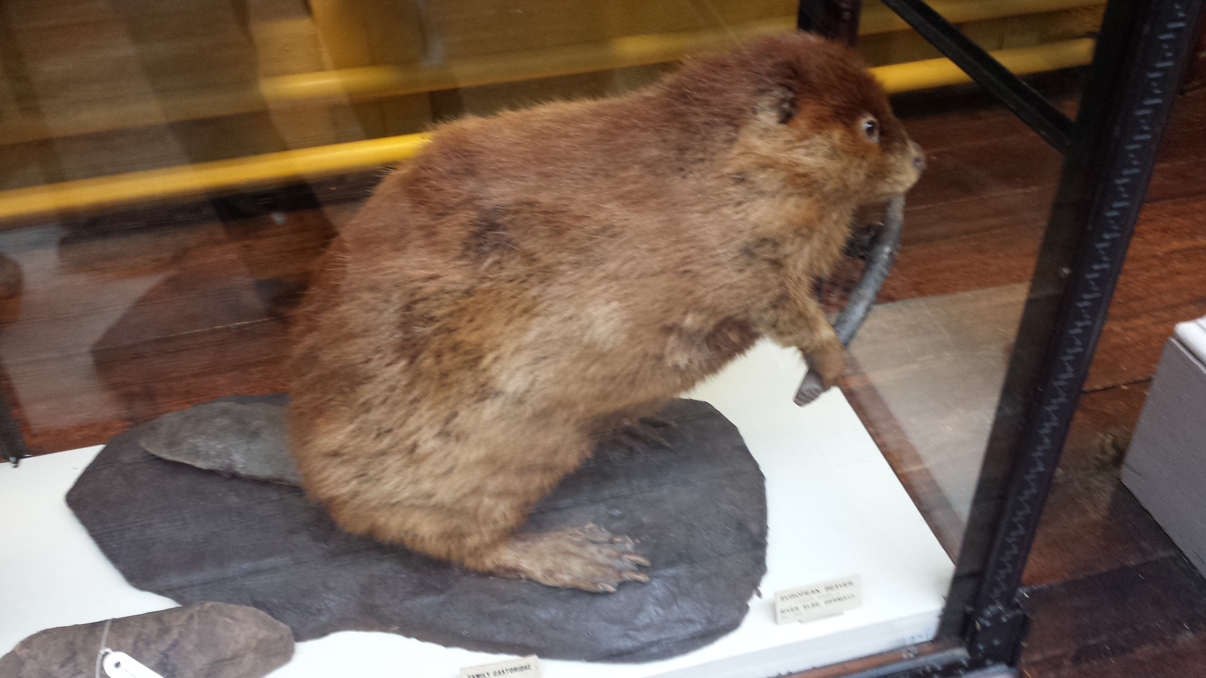
When I saw this I thought, “That’s a nice… rodent thingy.” And so “rodent thing” it shall be labelled here. Enjoy the rodent thingy. Some serious taxidermy-fu in action.

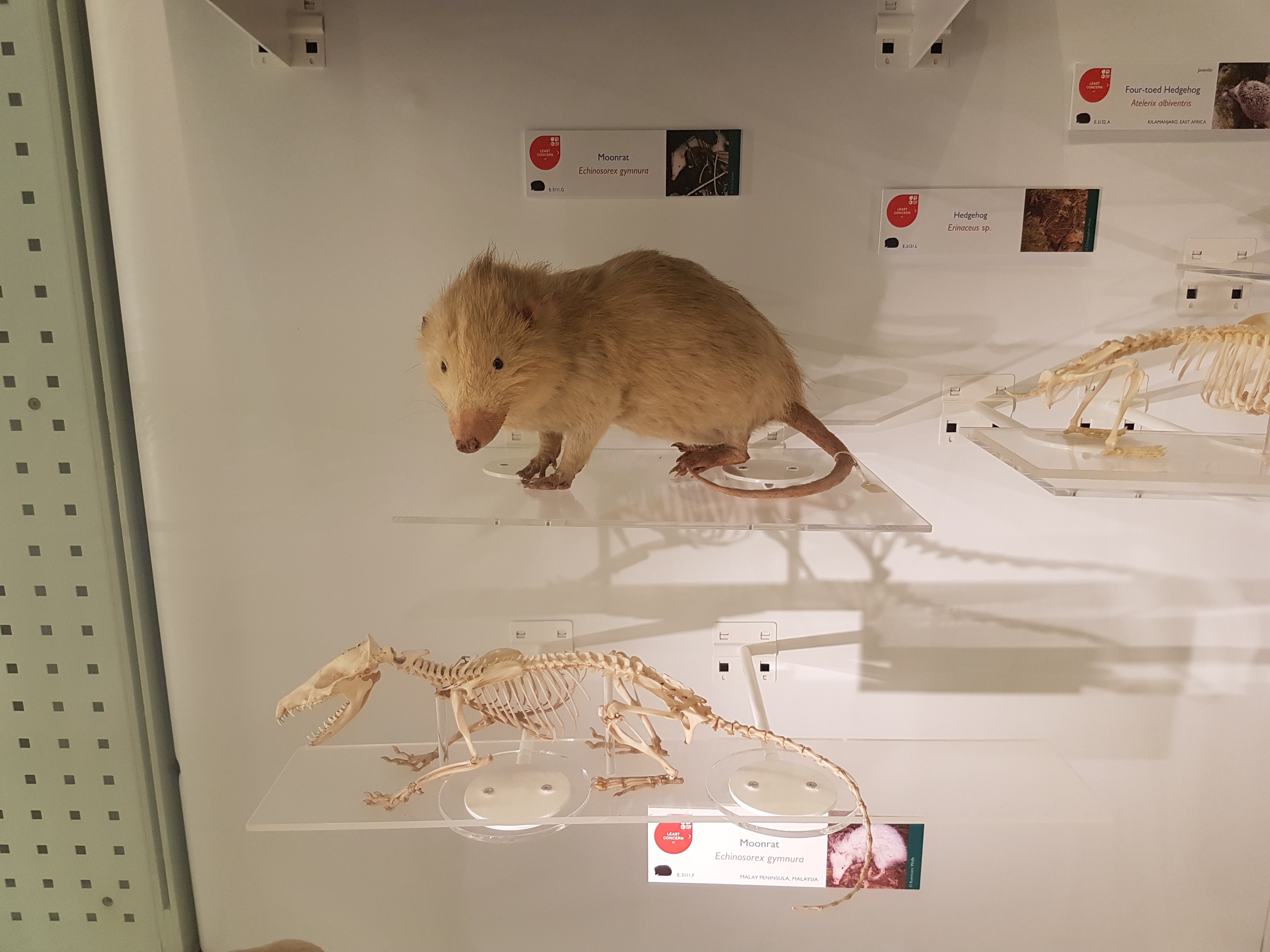
Moonrats– now there’s something you seldom see a full display of. Well done!

That’s part I of this sneak peek at the evolving exhibits- I will put up a part II once the upper floor exhibits open. I highly encourage a visit!

For Mike: gimlet

Read Full Post »

It has been almost three months since my last post here, and things have fallen quiet on our sister blog Anatomy to You, too. I thought it was time for an update, which is mostly a summary of stuff we’ve been doing on my team, but also featuring some interesting images if you stick around. The relative silence here has partly been due to me giving myself some nice holiday time w/family in L.A., then having surgery to fix my right shoulder, then recovering from that and some complications (still underway, but the fact that I am doing this post is itself evidence of recovery).

Stomach-Churning Rating: 4/10; semi-gruesome x-rays of me and hippo bits at the end, but just bones really.

X-ray of my right shoulder from frontal view, unlabelled

X-ray of my right shoulder from frontal view, unlabelled

Labelled x-ray

Labelled x-ray

So my priorities shifted to those things and to what work priorities most badly needed my limited energy and time. I’ve also felt that, especially since my health has had its two-year rough patch, this blog has been quieter and less interactive than it used to be, but that is the nature of things and maybe part of a broader trend in blogs, too. My creative juices in terms of social media just haven’t been at their ~2011-2014 levels but much is out of my control, and I am hopeful that time will reverse that trend. Enough about all this. I want to talk about science for the rest of this post.

My team, and collaborators as well, have published six recent studies that are very relevant to this blog’s theme- how about we run through them quickly? OK then.

  1. Panagiotopoulou, O., Pataky, T.C., Day, M., Hensman, M.C., Hensman, S., Hutchinson, J.R., Clemente, C.J. 2016. Foot pressure distributions during walking in African elephants (Loxodonta africana). Royal Society Open Science 3: 160203.

Our Australian collaborators got five African elephants together in Limpopo, South Africa and walked them over pressure-measuring mats, mimicking our 2012 study of Asian elephants. While sample sizes were too limited to say much statistically, in qualitatively descriptive terms we didn’t find striking differences between the two species’ foot pressure patterns. I particularly like how the centre of pressure of each foot (i.e. abstracting all regional pressures down to one mean point over time) followed essentially the same pattern in our African and Asian elephants, with a variable heelstrike concentration that then moved forward throughout the step, and finally moved toward the outer (3rd-5th; especially 3rd) toes as the foot pushed off the ground, as below.

African elephant foot COP traces vs. time in red; Asian elephant in orange. Left and right forefeet above; hindfeet below.

African elephant foot COP traces vs. time in red; Asian elephant in orange-yellow. Left and right forefeet above; hindfeet below.

Gradually, this work is moving the field toward better ability to use similar techniques to compare elephant foot mechanics among species, individuals, or over time– especially with the potential of using this method (popular in human clinical gait labs) to monitor foot (and broader musculoskeletal) health in elephants. I am hopeful that a difference can be made, and the basic science we’ve done to date will be a foundation for that.

  1. Panagiotopoulou, O., Rankin, J.W., Gatesy, S.M., Hutchinson, J.R. 2016. A preliminary case study of the effect of shoe-wearing on the biomechanics of a horse’s foot. PeerJ 4: e2164.

Finally, about six years after we collected some very challenging experimental data in our lab, we’ve published our first study on them. It’s a methodological study of one horse, not something one can hang any hats on statistically, but we threw the “kitchen sink” of biomechanics at that horse (harmlessly!) by combining standard in vivo forceplate analysis with “XROMM” (scientific rotoscopy with biplanar fluoroscopy or “x-ray video”) to conduct dynamic analysis of forefoot joint motions and forces (with and without horseshoes on the horse), and then to use these data as input values for finite element analysis (FEA) of estimated skeletal stresses and strains. This method sets the stage for some even more ambitious comparative studies that we’re finishing up now. And it is not in short supply of cool biomechanical, anatomical images so here ya go:

fig5-vonmises

Above: The toe bones (phalanges) of our horse’s forefoot in dorsal (cranial/front) view, from our FEA results, with hot colours showing higher relative stresses- in this case, hinting (but not demonstrating statistically) that wearing horseshoes might increase stresses in some regions on the feet. But more convincingly, showing that we have a scientific workflow set up to do these kinds of biomechanical calculations from experiments to computer models and simulations, which was not trivial.

And a cool XROMM video of our horse’s foot motions:

  1. Bates, K.T., Mannion, P.D., Falkingham, P.L., Brusatte, S.L., Hutchinson, J.R., Otero, A., Sellers, W.I., Sullivan, C., Stevens, K.A., Allen, V. 2016. Temporal and phylogenetic evolution of the sauropod dinosaur body plan. Royal Society Open Science 3: 150636.

I had the good fortune of joining a big international team of sauropod experts to look at how the shapes and sizes of body segments in sauropods evolved and how those influenced the position of the body’s centre of mass, similar to what we did earlier with theropod dinosaurs. My role was minor but I enjoyed the study (despite a rough ride with some early reviews) and the final product is one cool paper in my opinion. Here’s an example:

fig6a-bates-sauropod-com-evol

The (embiggenable-by-clicking) plot shows that early dinosaurs shifted their centre of mass (COM) backwards (maybe related to becoming bipedal?) and then sauropods shifted the COM forwards again (i.e. toward their forelimbs and heads) throughout much of their evolution. This was related to quadrupedalism and giant size as well as to evolving a longer neck; which makes sense (and I’m glad the data broadly supported it). But it is also a reminder that not all sauropods moved in the same ways- the change of COM would have required changes in how they moved. There was also plenty of methodological nuance here to cover all the uncertainties but for that, see the 17 page paper and 86 pages of supplementary material…

  1. Randau, M., Goswami, A., Hutchinson, J.R., Cuff, A.R., Pierce, S.E. 2016. Cryptic complexity in felid vertebral evolution: shape differentiation and allometry of the axial skeleton. Zoological Journal of the Linnean Society 178:183-202.

Back in 2011, Stephanie Pierce, Jenny Clack and I tried some simple linear morphometrics (shape analysis) to see how pinniped (seal, walrus, etc) mammals changed their vertebral morphology with size and regionally across their backbones. Now in this new study, with “Team Cat” assembled, PhD student Marcela Randau collected her own big dataset for felid (cat) backbones and applied some even fancier techniques to see how cat spines change their shape and size. We found that overall the vertebrae tended to get relatively more robust in larger cats, helping to resist gravity and other forces, and that cats with different ecologies across the arboreal-to-terrestrial spectrum also changed their (lumbar) vertebral shape differently. Now Marcela’s work is diving even deeper into these issues; stay tuned…

fig2-randau-measurements

Example measurements taken on felid vertebrae, from the neck (A-F) to the lumbar region (G-J), using a cheetah skeleton.

  1. Charles, J.P., Cappellari, O., Spence, A.J., Hutchinson, J.R., Wells, D.J. 2016. Musculoskeletal geometry, muscle architecture and functional specialisations of the mouse hindlimb. PLOS One 11(4): e0147669.

RVC PhD student James Charles measured the heck out of some normal mice, dissecting their hindlimb muscle anatomy, and using microCT scans produced some gorgeous images of that anatomy too. In the process, he also quantified how each muscle is differently specialized for the ability to produce large forces, rapid contractions or fine control. Those data were essential for the next study, where we got more computational!

mouse-mimics

  1. Charles, J.P., Cappellari, O., Spence, A.J., Wells, D.J., Hutchinson, J.R. 2016. Muscle moment arms and sensitivity analysis of a mouse hindlimb musculoskeletal model. Journal of Anatomy 229:514–535.

James wrangled together a lovely musculoskeletal model of our representative mouse subject’s hindlimb in the SIMM software that my team uses for these kinds of biomechanical analyses. As we normally do as a first step, we used the model to estimate things that are hard to measure directly, such as the leverages (moment arms) of each individual muscle and how those change with limb posture (which can produce variable gearing of muscles around joints). James has his PhD viva (defense) next week so good luck James!

mouse-simm

The horse and mouse papers are exemplars of what my team now does routinely. For about 15 years now, I’ve been building my team toward doing these kinds of fusion of data from anatomy, experimental biomechanics, musculoskeletal and other models, and simulation (i.e. estimating unmeasurable parameters by telling a model to execute a behaviour with a given set of criteria to try to perform well). Big thanks go to collaborator Jeff Rankin for helping us move that along lately. Our ostrich study from earlier this year shows the best example we’ve done yet with this, but there’s plenty more to come.

I am incredibly excited that, now that my team has the tools and expertise built up to do what I’ve long wanted to do, we can finally deliver the goods on the aspirations I had back when I was a postdoc, and which we have put enormous effort into pushing forward since then. In addition to new analyses of horses and mice and other animals, we’ll be trying to push the envelope more with how well we can apply similar methods to extinct animals, which brings new challenges– and evolutionary questions that get me very, very fired up.

Here we are, then; time has brought some changes to my life and work and it will continue to as we pass this juncture. I suspect I’ll look back on 2016 and see it as transformative, but it hasn’t been an easy year either, to say the least. “Draining” is the word that leaps to mind right now—but also “Focused” applies, because I had to try to be that, and sometimes succeeded. I’ve certainly benefited a lot at work from having some talented staff, students and other collaborators cranking out cool papers with me.

I still have time to do other things, too. Once in a while, a cool critter manifests in The Freezers. Check out a hippo foot from a CT scan! It’s not my best scan ever (noisy data) but it shows the anatomy fairly well, and some odd pathologies such as tiny floating lumps of mineralized soft tissue here and there. Lots to puzzle over.

Read Full Post »

A photo blog post for ya here! I went to Dublin on a ~28 hour tour, for a PhD viva (now-Dr Xia Wang; bird feather/flight evolution thesis) earlier this month. And I made a beeline for the local natural history museum (National Museum of Ireland, Natural History building) when I had free time. So here are the results!

Stomach-Churning Rating: Tame; about a 1/10 for most, but I am going to break my rule about showing human bodies near the end. Just a warning. The bog bodies were too awesome not to share. So that might be 4/10-8/10 depending on your proclivities. They are dry and not juicy or bloody, and don’t look as human as you’d expect.

Simple Natural History museum entrance area.

Simple Natural History museum entrance area.

Adorable frolicking topiaries outside the NHM.

Adorable frolicking topiaries outside the NHM.

Inside, it was a classical Victorian-style, dark wood-panelled museum stuffed with stuffed specimens. It could use major refurbishment, but I do love old-fashioned exhibits. Get on with it and show us the animals; minimize interpretive signage and NO FUCKING INTERACTIVE COMPUTER PANELS! So by those criteria, I liked it. Some shots of the halls: hall2 hall1 hall3 hall4 hall5 hall6 And on to the specimens!

Giant European deer ("Irish elk"). I looked at these and thought, "why don't we see female deer without antlers ever? then noticed one standing next to these; photo was crappy though. :(

Giant European deer (“Irish elk”). I looked at these and thought, “why don’t we see female deer without antlers ever? then noticed one standing next to these (you can barely see it in back); too bad my photo is crappy.

Superb mounted skeleton of giraffe (stuffed skin was standing near it).

Superb mounted skeleton of giraffe (stuffed skin was standing near it).

A sheep or a goat-y thingy; I dunno but it shows off a nice example of the nuchal ligament (supports the head/neck).

A sheep-y or a goat-y beastie; I dunno but it shows off a nice example of the nuchal ligament (supports the head/neck).

Yarr, narwhals be internet gold!

Yarr, narwhals be internet gold!

Giant blown glass models of lice!

Giant blown glass models of lice!

Who doesn't like a good giant foramanifera image/models? Not me.

Who doesn’t like a good giant foramanifera image/model?

"That's one bigass skate," I murmured to myself.

“That’s one bigass skate,” I murmured to myself.

"That's one bigass halibut," I quipped.

“That’s one bigass halibut,” I quipped.

Tatty basking shark in entry hall.

Tatty basking shark in entry hall.

Irish wolfhound, with a glass sculpture of its spine hanging near it, for some reason.

Irish wolfhound, with a glass sculpture of its spine hanging near it, for some reason.

Stand back folks! The beaver has a club!

Stand back everyone! That beaver has a club!

Skull of a pilot whale/dolphin.

Skull of a pilot whale/dolphin.

Nice anteater skeleton and skin.

Nice anteater skeleton and skin.

Nice anteater skeleton and skin.

Nice wombat skeleton and skin.

Sad display of a stuffed rhino with the horn removed, and signage explaining the problem of thefts of those horns from museum specimens of rhinos worldwide.

Sad display of a stuffed rhino with the horn removed, and signage explaining the problem of thefts of those horns from museum specimens of rhinos worldwide.

But then the stuffed animals started to get to me. Or maybe it was the hangover. Anyway, I saw this…
creepy proboscis (1) creepy proboscis (2)

A proboscis monkey mother who seemed to be saying “Hey kid, you want this yummy fruit? Tough shit. I’m going to hold it over here, out of reach.” with a disturbing grimace. That got me thinking about facial expressions in stuffed museum specimens of mammals more, and I couldn’t help but anthropomorphize as I toured the rest of the collection, journeying deeper into surreality as I progressed. What follows could thus be employed as a study of the Tim-Burton-eseque grimaces of stuffed sloths. Click to emslothen.

sloths (1) sloths (5)sloths (4) sloths (3) sloths (2)

Tree anteater has a go at the awkward expression game.

Tree anteater has a go at the awkward expression game.


This completed my tour of the museum; there were 2 more floors of specimens but they were closed for, sigh, say it with me… health and safety reasons. Balconies from which toddlers or pensioners or drunken undergrads could accidentally catapult themselves to their messy demise upon the throngs of zoological specimens below. But the National Museum’s Archaeology collection was just around the block, so off I went, following whispered tales of bog bodies. There will be a nice, calm, pretty photo, then the bodies, so if peaty ~300 BCE cadavers are not your cup of boggy tea, you can depart this tour now and lose no respect.

Impressive entrance to the National Museum's Archaeology building.

Impressive entrance to the National Museum’s Archaeology building.

The bog bodies exhibit is called “Kingship and Sacrifice“. It is packed with cylindrical chambers that conceal, and present in a tomb-like enclosed setting, the partial bodies of people that were killed and then tossed in peat bogs as honoraria for the ascension of a new king. The peaty chemistry has preserved them for ~2300 years, but in a dessicated, contorted state. The preservation has imparted a mottled colouration and wrinkled texture not far off from a Twix chocolate bar’s. Researchers have studied the bejesus out of these bodies (including 3D medical imaging techniques) and found remarkable details including not just wounds and likely causes of death (axes, strangling, slit throats etc) but also clothing, diet, health and more.

Here they are; click to (wait for it)… emboggen:

BogBodies (1) BogBodies (2) BogBodies (3) BogBodies (4) BogBodies (5) BogBodies (6)

Did you find the Celtic armband on one of them?

Finally (actually this happened first; my post is going back in time), I visited UCD’s zoology building for the PhD viva and saw a few cool specimens there, as follows:

Giant deer in UCD zoology building foyer.

Giant deer in UCD zoology building foyer, with a lovely Pleistocene landscape painted on the wall behind it.

Sika deer in awkward posture in Univ Coll Dublin zoology building's foyer.

Sika deer in an awkward posture (what is it supposed to be doing?) in Univ Coll Dublin zoology building’s foyer.

The pose of this ?baboon? struck me as very peculiar, and menacing- reminiscent of a vampire bat's pose, to me.

The pose of this ?baboon?mandrill struck me as very peculiar and menacing- reminiscent of a vampire bat’s pose.

A whole lotta chicken skeletons in a UCD teaching lab.

A whole lotta chicken skeletons in a UCD teaching lab.

After the viva we went out for some nice Chinese food and passed some Dublin landmarks like this:

Trinity College entrance, I think.

Trinity College entrance, I think.Former Irish Parliament; now the Bank of Ireland.

And we wandered into a very posh Irish pub called the Bank (on College Green), which displayed this interesting specimen, as well as some other features shown below:

Replica of illuminated old Gaelic manuscript.

Replica of illuminated 9th Century gospel manuscript “The Book of Kells”, with gorgeous Celtic art.

Vaults near toilets in the Bank pub.

Vaults near toilets in the Bank pub. Almost as cool as having giant freezers down there.

Nice glass ceiling of the Bank pub.

Nice glass ceiling of the Bank pub.

And Irish pub means one big, delicious thing to me, which I will finish with here– much as I finished that night off:

Ahhh...

Ahhh… ice cold.

Read Full Post »

A quick plug here for BBC Radio 4’s fourth episode of “Just So Science”, playing at 13:45 GMT today (this is the link). I was interviewed a few weeks ago for the show “How the Rhinoceros Got His Skin,” a la the classic Kipling tale. This series is revisiting Kipling’s tales in light of modern evolutionary science and evidence, whereas Kipling only had crude, Lamarckian or early Darwinian insight. Check out their earlier episodes on whales, leopards and armadillos– good stuff, and with real scientists.  Richard Dawkins may appear again (EDIT: yep! Dawkins manifested) in this episode to provide some gravitas and evolution street cred, too.

And Freezersaurus gets a big plug! From the website: ” Rhinos and horses have much in common. John Hutchinson studies both, but just don’t ask to look inside his freezer.” 🙂  NOTE: I am not a vet (I am a biologist), and definitely not a horse specialist like others in our lab, but I do study horses a little, in a comparative context.

While the original Kipling story focuses on rhino skin, and the producers were interested because of my popular post here on rhino skin, we discussed other issues such as gait, fossil record, feet, and more. I owe thanks to rhino skin expert Dr Tobin Hieronymus for helping me bone up on the unusual skin of rhinos, which has a surprising amount in common with the tough hide of walruses, boars, some water deer, and a few other species. It’s not just normal thickened skin, as Tobin and others have shown. Anyway, I don’t want to give away what’s on the radio programme; afterwards I might embellish this post more with some rhino anatomy and mechanics facts.

Coincidentally, I’m receiving four white rhinoceros feet today from a zoo mortality. So it’s rhino-fest here!

I hope you like the show— please let me know what you think in the comments below! I really enjoyed listening to it, but I’d like to know what you thought.

White rhinoceros forelimb (left), ready for dissection.

White rhinoceros forelimb (left side), ready for dissection.

How did the rhinoceros get her foot tendons?

How did the rhinoceros get her foot muscles?

Read Full Post »

A vignette from research I’m engaged in with a couple of different projects follows. Below is a photo I took of two humeri (upper arm bones; humerus is singular).

One is from a Black Rhinoceros; Diceros bicornis (modern; specimen #H.6481 from the University Museum of Zoology, Cambridge), which was collected in 1873 in Bogos, Abyssinia by zoologist ?Edward? Gerrard.

The other, larger one is from a giant long-necked and (presumably) hornless rhinocerotoid; Paraceratherium [AKA Indricotherium, Baluchitherium] (extinct of course; specimen #NHMUK PV M 12251 from The Natural History Museum, London); which was collected in 1911 in the Siwalik Hills of India by palaeontologist Forster Cooper. My photo is shown with kind permission of the Natural History Museum, London.

For an idea of scale, the smaller one is 39 cm (just over a foot) long, so about the same length as your humerus, give or take a bit. It comes from an animal that probably weighed around one tonne (1000 kg; 2200 lbs) or so. Look back at the picture, and pause to reflect on the scale. This is one of the largest living land animals right here, and despite that size it is quite an athlete (watch the classic John Wayne chasing-animals-around-Africa film Hatari! if you want elegant proof, or browse Youtube videos of boisterous rhinos).

But any living rhino pales in comparison to the giant Oligocene form, whose humerus is twice the length (~80 cm; almost as long as your entire leg, probably) and quite a bit more robust. The best estimates of mass for such an animal are up to 15-20 tonnes, on a par with the largest mammoths and other elephant relatives. That’s like a ten-rhino rhino! Sure, they all pale somewhat in scale against the largest sauropods (or whales, which cheat by living in water). Yet for my money (warning: subjective value judgement ahead!) a rhinoceros is cooler than any sauropod at the same size, and sauropods are extinct so we have less left to study. (I’m being deliberately provocative for my sauropod researcher friends, but in a loving way)

The scale, and often cramped conditions, make it hard getting a good photo of a Paraceratherium skeleton or reconstruction, but here’s one I took at Tokyo’s Museum of Nature and Science.

Now, of course if you know me, you know I am thinking about how such giant land animals moved. Authors such as Gregory Paul and Per Christiansen have made arguments based on real data, both qualitative anatomy and quantitative bone dimension measurements, that even giant rhinos like Paraceratherium could trot and gallop much like living rhinos do, despite their giant size. They have inferred from the limb joint structure that these giant rhinos were more crouched, were less columnar (vertical-limbed) than living elephants are (although I’ve shown with my team that this characterization of elephants is quite misleading; they get quite un-columnar, rather crouched, as they attain faster speeds). If Paul and Christiansen were correct, it would be remarkable. I can’t definitively show either way, just yet. But I want to see how well this argument holds up with other data and methods, so I’ve been planning to test this idea for a long time. We’ll see how it goes.

Anyway, that was my brief tale of two scales. On one hand we have living “giants” in the form of the five currently remaining species of rhinoceroses, which are quite extraordinary in many ways, albeit in big trouble. On the other hand we have amazing, mysterious uber-giants like Paraceratherium, two or more times the size in linear dimensions and an order of magnitude greater in weight. Both are certainly giants by any measure of size in land animals.

But was the bigger rhino living in a rather different world, even more dominated by gravity than its smaller relative is today? (No, gravity was no different! It was only 30 or so million years ago; relatively recent!) Or did they live in relatively similar worlds of just being “bloody huge and devastatingly powerful, thank you very much”? I find that question really exciting and wondrous to ponder. What do you think?

Read Full Post »

In case you haven’t heard, Saturday, September 22nd, 2012 (today, at this writing) is World Rhino Day! The main websites include here and here.  Ivan Kwan has also posted a fantastic blog entry “Rhinos are not prehistoric survivors” for WRD2012- check it out! And if you haven’t seen the WitmerLab’s AWESOME Visible Interactive Rhino site, you really really need to (in fact, quit reading this and go there first; it is soooooo good!).

I’ve written about the global rhino crisis before, and about rhino foot pathologies. The title of today’s post may be “cute”, or at least goofy, but the real situation is as grim as the images I’ll share. I won’t repeat the explanation, but all five living species of rhinoceroses are in serious trouble. There’s a good chance that most or all of them will go extinct quite soon– see the previous links for more information on this. Javan and Sumatran rhinos are dangling the most precariously over the precipice of extinction. My goal in this post is to share the beautiful, complex and exotic anatomy of rhinoceros anatomy and movement, and the joy of contributing new scientific information about poorly understood species.

Stomach-Churning Rating: 7/10— dissections, and there are a couple of pics where the specimens are not so fresh, and there’s big skin, and a huge heart.

Baby white rhinoceros. Will frozen specimens like this be all we have of rhinos someday?

The purpose of today’s rhino post is to share a bit more; especially images; of the work my team has done on rhinoceros gait and limb anatomy; all of it unpublished but hopefully coming soon. We’ve steadily been collecting data since ~2005. Because my previous post went through some of this, I’ll keep it brief and image-focused.

First, a video of one of our amusing encounters with a white rhinoceros, at Woburn Safari Park. In this study, we wanted to measure, for the first time really, the gaits (footfall patterns) that a white rhinoceros uses at different speeds, and how often it uses those different gaits. We attached a GPS unit on a horse surcingle around the rhino’s torso, which measured the animal’s speed once a second. We then observed 5 individuals (1 at a time over various days), following them in my station wagon (estate car) across the safari park. We filmed them with a conventional camcorder to document their gaits, and concentrated on the two periods of the day that they’d normally be active: when released from their overnight barn, and when coming in for the night back to that barn. They got rather excited and frisky some of those times. The GPS belt then kept recording speeds for the rest of the day; unsurprisingly, the rhinos generally did not do much. I have to thank Nick Whiting, rhino handler, for his help making this research happen. I’ve been meaning  for too long to finish the final paper… soon, I hope! Enjoy this tense scene of a rhino investigating my car (driven by me and with an undergraduate student filming) then having a nice canter/gallop across the field (accompanied by my jubilant narration).

Like our foot pressure research, we aim that this work provides baseline data useful to caretakers of rhinos; for example, to test if a particular animal is lame. This follows what we’ve successfully done with elephant gaits and feet, translating basic research into more clinical application. But my major scientific interest is in understanding more about what makes any rhinoceros, even a 2-tonne White rhino, so much more athletic than any elephant (even a baby or 2-tonne small adult Asian elephant). As the video shows, they can use a variety of gaits including cantering and galloping, and trotting at slower running speeds. No elephant ever does that, and no one knows precisely why. The leg bones are more robust, but the muscles aren’t that dramatically larger in rhinos.

An Indian rhinoceros forelimb- note the characteristic knobbly hide, unlike the smoother, more elephant-like hide of a White rhinoceros.

Similarly, the anatomical work we do with rhinos is intended to not only be useful science for comparative biologists like me, showing how rhino limbs work and how they differ from those of other animals, but also to aid clinicians in comparing normal vs. pathological anatomy. For conveying that anatomical work, I’m lucky to have been granted permission to use a professional photographer’s pictures of some of my freezers’ rhino specimens– big thanks to James King-Holmes and the Science Photo Library. The watermarked images below belong to them. I ask that you do not use them elsewhere, honouring their license to me for personal usage on this website (and I will only use them here). I’m in all the images, which makes me feel weird putting them up here, but it’s about the rhinos (and freezers), not me. First: the infamous “rhino foot freezer”, featuring some of its denizens:

Second, a re-introduction to multifarious contents of Freezersaurus, but this time featuring rhino feet (here, a skinned white rhino foot that we had already studied):

…and inside we go (and I begin to get frosty and numb-fingered from holding a foot; my smile soon fades):

Taking a rest with the skinned white rhinoceros foot:

And now warming up at the “digital freezer”, our CT scanner, and preparing to scan another rhinoceros foot, which segues nicely out of this image sequence:

Now over to some 3D anatomy– segmented reconstructions of rhinoceros fore (top) and hind (bottom) feet, from CT scans; if you’ve frequented this blog you know the drill. Here, the longest bones are the metacarpals/metatarsals and the upper bones are the carpals/tarsals, then the bones near the botttom are the phalanges, which connect to the hooves (visible in the bottom image):

I’ll wrap up with a series of images of basic limb muscle anatomy from dissections we’ve done of baby and adult Indian and White rhinoceroses. First, here’s what a rhino looks like underneath the skin:

But ahh that skin, that fabled “pachyderm” skin! A rhino’s greatest defense is also a real chore to get through in a dissection.  Here, we enlist the help of a crane and hook, hurrying to get down to the muscles of this forelimb before rotting takes over too much (as with other big animals, this is a tough race against time even in chilly England!):

Here is a closer look at that amazing armoured skin; sometimes 10cm or so thick:

Back to the forelimb muscles– stocky and well-defined for this athletic animal:

(late addition) Here are the massive shoulder muscles, such as the serratus and latissimus dorsi (this is a left limb in side view; head is toward the left):

And now a close look at the forearm muscles:

And then over to the hindlimb, here from an adult Indian rhino, whose thigh bone (femur) shows the characteristic giant “third trochanter” (toward the bottom centre of the image), which is an expanded bony attachment for the giant “gluteobiceps” muscle complex that retracts the femur for the power stroke in locomotion. Also, this specimen showed fascinating anatomy that I’d never seen before: the third trochanter has a thin bar of bone that extends up (toward the bottom left in the image) to fuse with the greater trochanter, opposite the head of the femur (upper left corner):

Damn my photography skills, cutting off the edge of that image and instead giving a view of my boots! Anyway, another interesting feature of that femur: the medial (inner) condyle of the femur (knee joint surface) has a pink stripe of worn cartilage. This is indicative of at least a moderate stage of arthritis, shown here (look for the pinkness amidst the shiny, healthy white cartilage on the upper right side). It is an exemplar of serious welfare problems that some captive, and probably some wild as well, rhinos face:

(late addition) Back up the limb, this baby White rhino shows the massive thigh muscles, especially that “gluteobiceps” that attaches to the third trochanter, noted above, and also showing the hamstrings:

Moving down the limb, we encounter the glorious three-toed perissodactyl foot of rhinos, and the robust hooves/nails, which are reasonably healthy in this animal– unlike others I’ve seen:

And the sole of that foot, showing a fairly healthy pad, below. Toward the rear (away from the nails), it culminates in a modest-sized fat pad, or digital cushion, akin to that in elephants but far less well developed and lacking the false “sixth toe” (predigit) (see also CT scan movie of the hindfoot above):

Here’s a view inside that marvelous foot, showing the HUGE digital flexor tendons. These help support the toes against gravity and, in theory, can act to curl them up– although in a rhino’s foot, as in an elephant’s, the toes are more like a single functional hoof, with reduced independence compared to a carnivore or primate:

And that ends our tour of rhinoceros limb anatomy and function. Help spread the word of how precious and threated rhinos are; educate yourself and others! And if you overhear someone talking about using rhino horn for medicine, try to politely educate them on the utter fallacy of this tradition. It is this cruel, greedy, ignorant practice that needs to die; not rhinos. I don’t enjoy receiving dead rhinos, on a personal level, even though the science excites me. I’d rather have many more alive and living good, healthy lives. And my team is trying to do what we can to help others on the “front lines” of rhino conservation make that happen.

For example, Will Fowlds, vet and co-owner of Amakhala Game Reserve, South Africa, recently sent us some images of a white rhino that had been caught in a poacher’s foot snare some years ago. The poor rhino still was having problems healing– we inspected x-ray images and external photos and helped to make an initial diagnosis of osteomyelitis, a nasty infectious, inflammatory foot bone/joint disease. We are following this case to hope that the rhino recovers and contribute help where we can, but the tough job belongs to the keepers/vets on the ground, not to mention the rhinos…

Furthermore, we’ve done foot pressure research covered here, and here is an example of the data we’ve collected (image credit: Dr Olga Panagiotopoulou), showing high pressures on the toes and low pressures on the foot pads:

Big thanks to people on my team that have helped with this and related research: Dr Olga Panagiotopoulou (and Dr Todd Pataky at Shinshu University, Japan), Dr Renate Weller in the VCS Dept at the RVC, Liz Ferrer at Berkeley, and former undergraduate student researchers Sophie Regnault, Richard Harvey, Hinnah Rehman, Richard Sheehan, Kate Jones, Bryony Armson and Suzannah Williams.

A White rhino’s heart, with more images below, all courtesy of William Perez’s Veterinary Anatomy Facebook pages. A mass of around 10kg (22 lbs weight) is not unusual! (Compare with even larger elephant heart)

White rhino closeup: coronary arteries

White rhino: branches of left coronary artery

White rhino heart: right atrium

Read Full Post »

Well, it’s time for the Grand Reveal of what the picture in the previous post is (reproduced below). The guesses ranged from bird to wallaby/kangaroo to the stuff of nightmares. And indeed Nick Gardner got it right first, it is a wallaby. Specifically, it is a Bennett’s Wallaby (Macropus rufogriseus rufogriseus), which is the Tasmanian island form related to the Red-necked Wallaby, and an animal that has gone feral (along with mara and other cool critters) in Whipsnade Zoo near the RVC. You can tell it is a wallaby and not a bird, because there is an “Achilles tendon” attaching to a calcaneal tuber (“heel bone”) on the back side of the limb (shown with asterisk below) that birds lack, and if you look closely the toes are hairy, lack bird-like claws, and a few other details like the profile of the musculature are very different; more mammalian than avian. The stump of the muscular tail (cut off) is also a clue. Although the avian similarity in the case of wallabies is still striking, which is one reason I chose this image. Well done, Nick!

I found this picture in my archives and remembered when it was taken back in ~2005- some lab members received some frozen wallaby legs and thawed them out to use in experiments. They tried to compress the legs in an Instron machine (mechanical testing system; partly visible at the top of the pic) to see what the passive, springlike properties of the legs are like in a wallaby, vs. the properties they could measure in a living animal. (The shiny white reflective areas in the pic are for tracking joint motions) And I thought it was a freaky cool pic, so I shared it.

I also posted that pic because my team has done some in vivo analysis of the leg properties in such animals (previous news story here; paper in preparation), and because we use this technique of loading cadaveric legs in such machines quite routinely. We did this for elephant feet to study how the “sixth toe” of elephants works, and we’re analyzing data (as I write) for how elephant feet and rhino feet deform or move when loaded similarly. This method has a long history; we didn’t invent it; perhaps most famously used for studying horse limb mechanics [pdf example], which have a lot of passive properties (almost everything below the elbow/knee is non-muscular). Many animals’ limbs are tendinous/elastic toward their distal end (toes), so the limbs tend to become less actively controlled by the nervous system and become more of a mechanical control system (sometimes involving a non-neural “preflex“) in that region; although it’s all a matter of relative degree of passive:active control in different situations, species, and limbs.

The picture below shows an x-ray of an elephant’s whole hind foot, in which you should be able to see the bones (brighter white) of the foot surrounded by a lot of soft tissue, mostly more passive kinds like fat, skin, fascial sheets, ligaments and tendon.

Here (further below) is a preliminary image from our elephant foot studies in progress, intended to reveal the passive properties/motions of the feet so we can figure out how those properties are combined with more active control, and how actively elephants control their feet vs. other, possibly more ‘passive-footed’ animals like horses. This is interesting from anatomical and evolutionary perspectives, and for helping with foot health problems that are serious concerns for such animals– more about that later. The arrow in the picture below shows where a lot of the motion is: at the knuckle (metatarsophalangeal) joints of the toes; the rest of the foot tends to rotate around these mobile joints. We can’t peer inside living elephant feet to see if they actually do this, but we can compare the external motions, pressure patterns, and other data from living and dead elephant feet to see how they match up, which is what we’re doing now — and we’re doing the same thing with rhinos, which have cool 3-toed, more “hoof-like” feet, as opposed to the 5-toed, fatter feet of elephants. To get this image, we’ve had to put the foot inside a custom-made device using a car jack to apply a constant load, and a wooden framework to hold the specimen still, and then run it through a CT machine in unloaded and then loaded states to see how the bones move. Here is what the crazy apparatus looks like, with enthusiastic undergrad for scale:

This, below, is a right hind foot (pes) of an Asian elephant, shown from the inside of the foot (toes are numbered 1-4 from the big toe/hallux toward the outside of the foot; 5th toe is not visible). The yellow image is the relaxed, unloaded foot; the green is after applying a large load equivalent to the animal standing on one foot (or running quickly). Notice how the third metatarsal (the long bone that the arrowhead is touching) for the unloaded state is in front of that for the loaded state, whereas yellow and green images of the bones toward the tip of the toes are overlapping more, indicating they did not move much/at all. That tells us that the motion is occuring at the joint indicated (“knuckle”), which makes sense anatomically, because that joint looks like it has a lot of mobility.

Image

Read Full Post »

On a serious note, in case you aren’t aware of it, there is a huge crisis afoot with the world’s rhino populations, because of demonstrably false claims about the health benefits of rhino horn (e.g. curing cancer) leading to a surge in its value worldwide, and thereby a massive upswing in poaching- as well as theft of museum specimens(!?). It may just be a matter of time before zoos’ and safari parks’ rhinos outside of Africa/Asia get hit, too. If sustained, this poaching could wipe out multiple rhino species in a matter of years; it is that severe and seems to already be worse this year than it was last year- and that was a Very Bad Year for rhinos.

However, a lot of people are uniting against the cruel greed and gross ignorance that has fueled the decline of rhinos, and public support seems to be growing. You can contribute, too; one example is this cause, or this one, to name but two. I’ve tried to make this my #1 cause, both on personal/ethical and scientific grounds (e.g. our work on rhino foot mechanics, health and care, to be detailed here later), and have been educating myself about it.

Anatomist extraordinaire Professor Larry Witmer of Ohio University has been contributing scientifically to helping rhinos, in an unexpected (but very sensible) way. This is a perfect example of how important basic science is; if we didn’t know rhino horn/nose anatomy we’d be less able to treat problems when they arise, and such problems can come from unexpected directions. His team’s contributions in the understanding of rhino anatomy are helping in one horrible case (see video below), in which three rhinos had their horns slashed off (along with part of the top of their skulls!) and two have survived so far. The vets in South Africa are trying to treat these two mutilated animals, and Larry’s group has been providing anatomical advice along with superb pictures on their Facebook page, which I want to publicize here because it is such GREAT freezer-based anatomical science and stunning imagery, and a seriously urgent cause. Please take a look. And while you’re at it, check out the Kariega Game Reserve’s Facebook page with more info on the plight of poor Themba and Thandi.

Edit: also check out this great story on our research, by Ann and Steve Toon, rhino conservationists/photographers/journalists.

Read Full Post »