Today, to help thaw you poor Americans out of that Arctic Vortex, we have a guest post bringing the heat, by my PhD student Sophie Regnault! This relates to some old posts about rhinos, which are a mainstay here at the WIJF blog- I’ve posted a lot about the rhino extinction crisis, feet, skin, big and bigger bones, and more, but this is our first rhinoceros-focused, actual published scientific paper! Take it away, Sophie! (We’re planning a few more “guest” blog posts from my team, so enjoy it, folks!)
Almost a year ago to the day, I submitted my first paper written with John Hutchinson and Renate Weller at the RVC and it has (finally!) just been published. To celebrate, I have been allowed to temporarily hijack ‘What’s in John’s Freezer?’ for my first foray into the world of blogging. I started the paper back as an undergraduate veterinary student. It was my first experience of proper research, and so enjoyable that I’m now doing a PhD, studying sesamoid bones like the patella!
We wanted to discover more about the types of bony disease rhinos get in their feet, of which there isn’t much known. Rhinos, of course, are big, potentially dangerous animals – difficult enough to examine and doubly difficult to x-ray clearly because of their thick skin. Unlike diseases which are fairly easy to spot (like abscesses or splitting of the nails and footpad), there is hardly anything out there in the scientific literature on bony diseases in rhino feet. It’s no small issue, either. When your feet each need to support over 900kg (typical for a large white rhino), even a relatively minor problem can be a major pain. Progressing unseen under their tough hide, lesions in the bone can eventually become so serious than the only solution is euthanasia, but even mild conditions can have negative consequences. For example, foot problems in other animals are known to have knock-on effects on fertility, which would be a big deal for programs trying to breed these species in captivity.
Hidden treasures abound! (Photos can be clicked to embiggen)
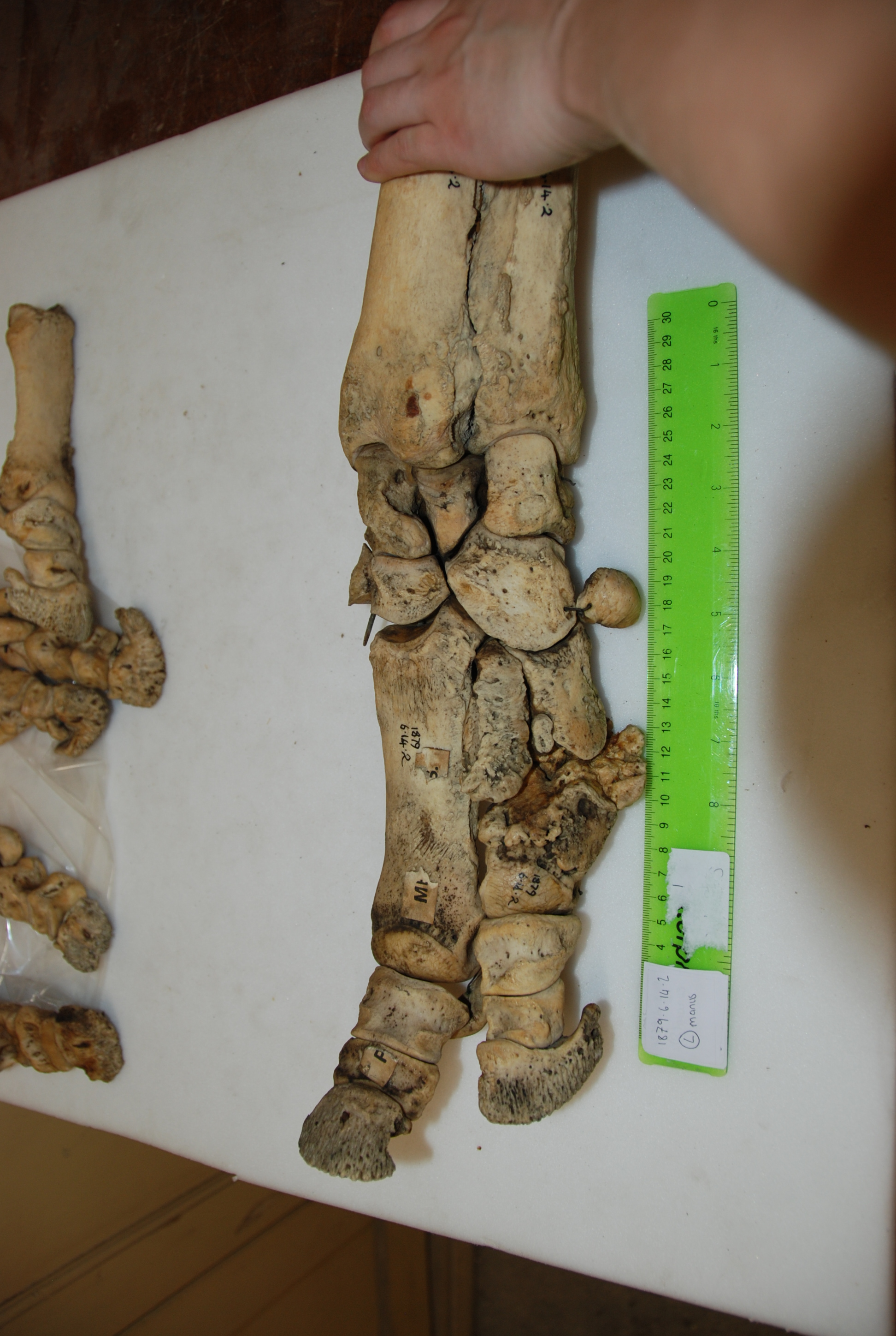
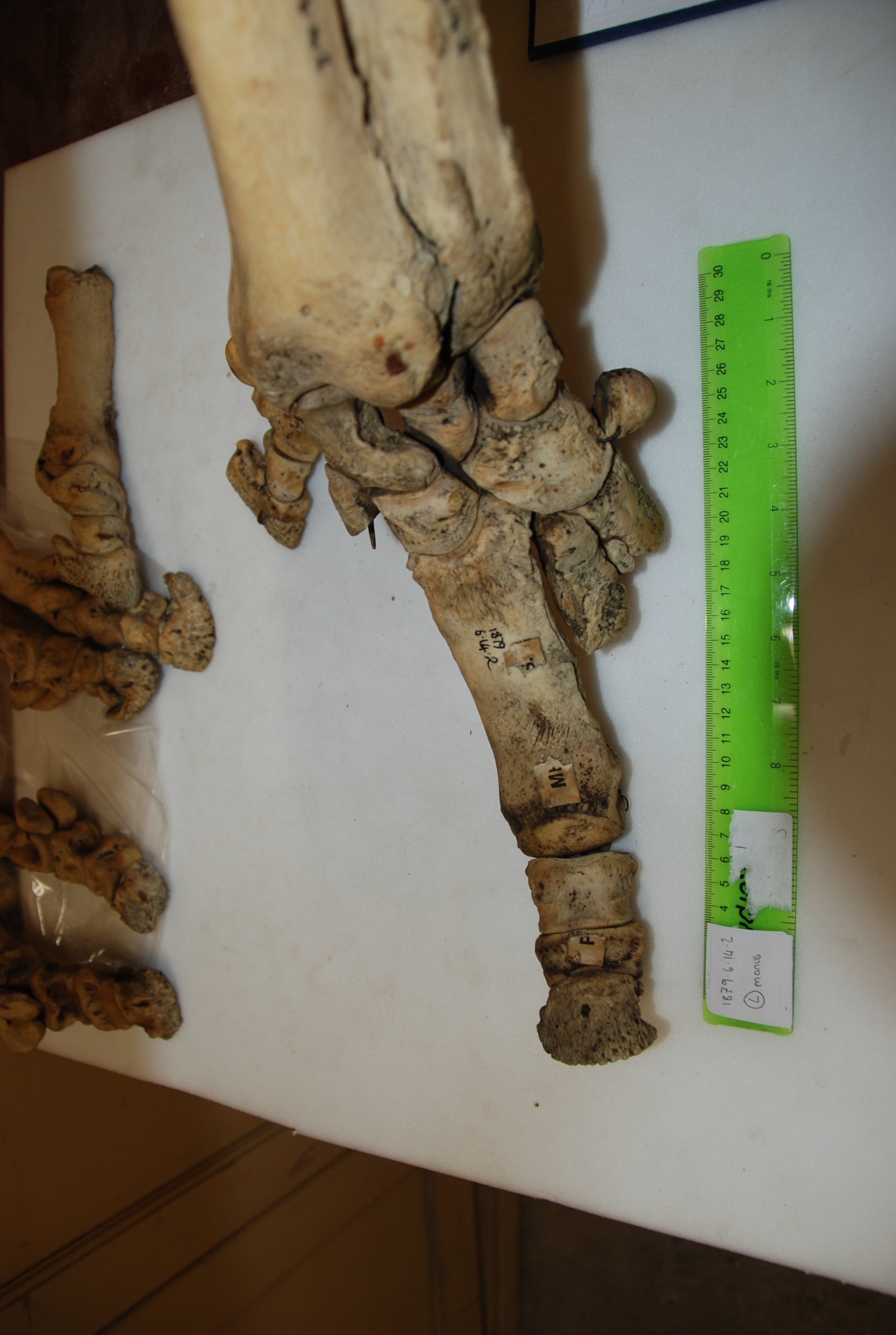
Data gathering was a blast. I got to travel to Cambridge, Oxford, and London during one of England’s better summers, and these beautiful old museums were letting me snoop around their skeleton collections. I’d been there often as a visitor, but it was anatomy-nerd-heaven to go behind the scenes at the Natural History Museum, and to be left alone with drawers and drawers of fantastic old bones. Some of the specimens hadn’t been touched for decades – at Cambridge University Museum of Zoology, we opened an old biscuit tin filled with the smallest rhinoceros foot bones, only to realise they were wrapped in perfectly preserved 1940’s wartime Britain newspaper.
Osteomyelitis… (3 clickable pics above) the toe’s probably not meant to come off like that!
In addition to my museum studies, I had another fun opportunity to do hands-on research. John (of course!) had freezers full of rhino legs (looking disconcertingly like doner kebabs, but maybe that’s just me!), which we CT scanned to see the bones. Although it is a pretty standard imaging technique, at this point I had only just started my clinical studies at the vet hospital, and being able to flick through CT scans felt super badass. Most vet students just get to see some horse feet or dog/cat scans, at best.
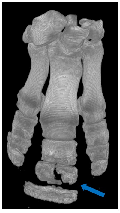
Another osteomyelitis fracture, visible in a CT scan reconstruction.
We expected to find diseases like osteoarthritis (a degenerative joint disease) and osteomyelitis (bone infection and inflammation). Both had previously been reported in rhinoceroses, although it was interesting that we saw three cases of osteomyelitis in only 27 rhinos, perhaps making it a fairly common complication. It’s an ugly-looking disease, and in two of the cases led to the fat, fluffy bones fracturing apart.
We also had several unexpected findings, like flakes of fractured bone, mild dislocations, tons of enthesiophytes (bone depositions at tendon/ligament attachments) and lots of holes in the bones (usually small, occasionally massive). For me, writing up some of these findings was cool and freaky paranoid in equal measures. They hadn’t been much described before, and we were unsure of their significance. Was it normal, or pathological? Were we interpreting it correctly? Discussions with John and Renate (often involving cake) were reassuring, as was the realisation that in science (unlike vet school at the time, where every question seemed to have a concrete answer) you can never be 100% sure of things. Our study has a few important limitations, but has addressed a gap in the field and found some neat new things. Six months into my PhD, I’m enjoying research more than ever, and hoping that this paper will be the first of many (though I promise I won’t keep nicking John’s blog for my own shameless self-promotion if that happens! EDIT BY JOHN: Please do!).
Nasty osteoarthritis wearing away the bone at the joint surface. Most cases occurred in the most distal joint.
Deep holes in some of the bones: infection, injury?
The paper:
Sophie Regnault, Robert Hermes, Thomas Hildebrandt, John Hutchinson, and Renate Weller (2013) OSTEOPATHOLOGY IN THE FEET OF RHINOCEROSES: LESION TYPE AND DISTRIBUTION. Journal of Zoo and Wildlife Medicine: December 2013, Vol. 44, No. 4, pp. 918-927.
A quick plug here for BBC Radio 4’s fourth episode of “Just So Science”, playing at 13:45 GMT today (this is the link). I was interviewed a few weeks ago for the show “How the Rhinoceros Got His Skin,” a la the classic Kipling tale. This series is revisiting Kipling’s tales in light of modern evolutionary science and evidence, whereas Kipling only had crude, Lamarckian or early Darwinian insight. Check out their earlier episodes on whales, leopards and armadillos– good stuff, and with real scientists. Richard Dawkins may appear again (EDIT: yep! Dawkins manifested) in this episode to provide some gravitas and evolution street cred, too.
And Freezersaurus gets a big plug! From the website: ” Rhinos and horses have much in common. John Hutchinson studies both, but just don’t ask to look inside his freezer.” 🙂 NOTE: I am not a vet (I am a biologist), and definitely not a horse specialist like others in our lab, but I do study horses a little, in a comparative context.
While the original Kipling story focuses on rhino skin, and the producers were interested because of my popular post here on rhino skin, we discussed other issues such as gait, fossil record, feet, and more. I owe thanks to rhino skin expert Dr Tobin Hieronymus for helping me bone up on the unusual skin of rhinos, which has a surprising amount in common with the tough hide of walruses, boars, some water deer, and a few other species. It’s not just normal thickened skin, as Tobin and others have shown. Anyway, I don’t want to give away what’s on the radio programme; afterwards I might embellish this post more with some rhino anatomy and mechanics facts.
Coincidentally, I’m receiving four white rhinoceros feet today from a zoo mortality. So it’s rhino-fest here!
I hope you like the show— please let me know what you think in the comments below! I really enjoyed listening to it, but I’d like to know what you thought.
White rhinoceros forelimb (left side), ready for dissection.
A vignette from research I’m engaged in with a couple of different projects follows. Below is a photo I took of two humeri (upper arm bones; humerus is singular).
One is from a Black Rhinoceros; Diceros bicornis (modern; specimen #H.6481 from the University Museum of Zoology, Cambridge), which was collected in 1873 in Bogos, Abyssinia by zoologist ?Edward? Gerrard.
The other, larger one is from a giant long-necked and (presumably) hornless rhinocerotoid; Paraceratherium [AKA Indricotherium, Baluchitherium] (extinct of course; specimen #NHMUK PV M 12251 from The Natural History Museum, London); which was collected in 1911 in the Siwalik Hills of India by palaeontologist Forster Cooper. My photo is shown with kind permission of the Natural History Museum, London.
For an idea of scale, the smaller one is 39 cm (just over a foot) long, so about the same length as your humerus, give or take a bit. It comes from an animal that probably weighed around one tonne (1000 kg; 2200 lbs) or so. Look back at the picture, and pause to reflect on the scale. This is one of the largest living land animals right here, and despite that size it is quite an athlete (watch the classic John Wayne chasing-animals-around-Africa film Hatari! if you want elegant proof, or browse Youtube videos of boisterous rhinos).
But any living rhino pales in comparison to the giant Oligocene form, whose humerus is twice the length (~80 cm; almost as long as your entire leg, probably) and quite a bit more robust. The best estimates of mass for such an animal are up to 15-20 tonnes, on a par with the largest mammoths and other elephant relatives. That’s like a ten-rhino rhino!Sure, they all pale somewhat in scale against the largest sauropods (or whales, which cheat by living in water). Yet for my money (warning: subjective value judgement ahead!) a rhinoceros is cooler than any sauropod at the same size, and sauropods are extinct so we have less left to study. (I’m being deliberately provocative for my sauropod researcher friends, but in a loving way)
The scale, and often cramped conditions, make it hard getting a good photo of a Paraceratherium skeleton or reconstruction, but here’s one I took at Tokyo’s Museum of Nature and Science.
Now, of course if you know me, you know I am thinking about how such giant land animals moved. Authors such as Gregory Paul and Per Christiansen have made arguments based on real data, both qualitative anatomy and quantitative bone dimension measurements, that even giant rhinos like Paraceratherium could trot and gallop much like living rhinos do, despite their giant size. They have inferred from the limb joint structure that these giant rhinos were more crouched, were less columnar (vertical-limbed) than living elephants are (although I’ve shown with my team that this characterization of elephants is quite misleading; they get quite un-columnar, rather crouched, as they attain faster speeds). If Paul and Christiansen were correct, it would be remarkable. I can’t definitively show either way, just yet. But I want to see how well this argument holds up with other data and methods, so I’ve been planning to test this idea for a long time. We’ll see how it goes.
Anyway, that was my brief tale of two scales. On one hand we have living “giants” in the form of the five currently remaining species of rhinoceroses, which are quite extraordinary in many ways, albeit in big trouble. On the other hand we have amazing, mysterious uber-giants like Paraceratherium, two or more times the size in linear dimensions and an order of magnitude greater in weight. Both are certainly giants by any measure of size in land animals.
But was the bigger rhino living in a rather different world, even more dominated by gravity than its smaller relative is today? (No, gravity was no different! It was only 30 or so million years ago; relatively recent!) Or did they live in relatively similar worlds of just being “bloody huge and devastatingly powerful, thank you very much”? I find that question really exciting and wondrous to ponder. What do you think?
In case you haven’t heard, Saturday, September 22nd, 2012 (today, at this writing) is World Rhino Day! The main websites include here and here. Ivan Kwan has also posted a fantastic blog entry “Rhinos are not prehistoric survivors” for WRD2012- check it out! And if you haven’t seen the WitmerLab’s AWESOME Visible Interactive Rhino site, you really really need to (in fact, quit reading this and go there first; it is soooooo good!).
I’ve written about the global rhino crisis before, and about rhino foot pathologies. The title of today’s post may be “cute”, or at least goofy, but the real situation is as grim as the images I’ll share. I won’t repeat the explanation, but all five living species of rhinoceroses are in serious trouble. There’s a good chance that most or all of them will go extinct quite soon– see the previous links for more information on this. Javan and Sumatran rhinos are dangling the most precariously over the precipice of extinction. My goal in this post is to share the beautiful, complex and exotic anatomy of rhinoceros anatomy and movement, and the joy of contributing new scientific information about poorly understood species.
Stomach-Churning Rating: 7/10— dissections, and there are a couple of pics where the specimens are not so fresh, and there’s big skin, and a huge heart.
Baby white rhinoceros. Will frozen specimens like this be all we have of rhinos someday?
The purpose of today’s rhino post is to share a bit more; especially images; of the work my team has done on rhinoceros gait and limb anatomy; all of it unpublished but hopefully coming soon. We’ve steadily been collecting data since ~2005. Because my previous post went through some of this, I’ll keep it brief and image-focused.
First, a video of one of our amusing encounters with a white rhinoceros, at Woburn Safari Park. In this study, we wanted to measure, for the first time really, the gaits (footfall patterns) that a white rhinoceros uses at different speeds, and how often it uses those different gaits. We attached a GPS unit on a horse surcingle around the rhino’s torso, which measured the animal’s speed once a second. We then observed 5 individuals (1 at a time over various days), following them in my station wagon (estate car) across the safari park. We filmed them with a conventional camcorder to document their gaits, and concentrated on the two periods of the day that they’d normally be active: when released from their overnight barn, and when coming in for the night back to that barn. They got rather excited and frisky some of those times. The GPS belt then kept recording speeds for the rest of the day; unsurprisingly, the rhinos generally did not do much. I have to thank Nick Whiting, rhino handler, for his help making this research happen. I’ve been meaning for too long to finish the final paper… soon, I hope! Enjoy this tense scene of a rhino investigating my car (driven by me and with an undergraduate student filming) then having a nice canter/gallop across the field (accompanied by my jubilant narration).
Like our foot pressure research, we aim that this work provides baseline data useful to caretakers of rhinos; for example, to test if a particular animal is lame. This follows what we’ve successfully done with elephant gaits and feet, translating basic research into more clinical application. But my major scientific interest is in understanding more about what makes any rhinoceros, even a 2-tonne White rhino, so much more athletic than any elephant (even a baby or 2-tonne small adult Asian elephant). As the video shows, they can use a variety of gaits including cantering and galloping, and trotting at slower running speeds. No elephant ever does that, and no one knows precisely why. The leg bones are more robust, but the muscles aren’t that dramatically larger in rhinos.
An Indian rhinoceros forelimb- note the characteristic knobbly hide, unlike the smoother, more elephant-like hide of a White rhinoceros.
Similarly, the anatomical work we do with rhinos is intended to not only be useful science for comparative biologists like me, showing how rhino limbs work and how they differ from those of other animals, but also to aid clinicians in comparing normal vs. pathological anatomy. For conveying that anatomical work, I’m lucky to have been granted permission to use a professional photographer’s pictures of some of my freezers’ rhino specimens– big thanks to James King-Holmes and the Science Photo Library. The watermarked images below belong to them. I ask that you do not use them elsewhere, honouring their license to me for personal usage on this website (and I will only use them here). I’m in all the images, which makes me feel weird putting them up here, but it’s about the rhinos (and freezers), not me. First: the infamous “rhino foot freezer”, featuring some of its denizens:
…and inside we go (and I begin to get frosty and numb-fingered from holding a foot; my smile soon fades):
Taking a rest with the skinned white rhinoceros foot:
And now warming up at the “digital freezer”, our CT scanner, and preparing to scan another rhinoceros foot, which segues nicely out of this image sequence:
Now over to some 3D anatomy– segmented reconstructions of rhinoceros fore (top) and hind (bottom) feet, from CT scans; if you’ve frequented this blog you know the drill. Here, the longest bones are the metacarpals/metatarsals and the upper bones are the carpals/tarsals, then the bones near the botttom are the phalanges, which connect to the hooves (visible in the bottom image):
I’ll wrap up with a series of images of basic limb muscle anatomy from dissections we’ve done of baby and adult Indian and White rhinoceroses. First, here’s what a rhino looks like underneath the skin:
But ahh that skin, that fabled “pachyderm” skin! A rhino’s greatest defense is also a real chore to get through in a dissection. Here, we enlist the help of a crane and hook, hurrying to get down to the muscles of this forelimb before rotting takes over too much (as with other big animals, this is a tough race against time even in chilly England!):
Here is a closer look at that amazing armoured skin; sometimes 10cm or so thick:
Back to the forelimb muscles– stocky and well-defined for this athletic animal:
(late addition) Here are the massive shoulder muscles, such as the serratus and latissimus dorsi (this is a left limb in side view; head is toward the left):
And now a close look at the forearm muscles:
And then over to the hindlimb, here from an adult Indian rhino, whose thigh bone (femur) shows the characteristic giant “third trochanter” (toward the bottom centre of the image), which is an expanded bony attachment for the giant “gluteobiceps” muscle complex that retracts the femur for the power stroke in locomotion. Also, this specimen showed fascinating anatomy that I’d never seen before: the third trochanter has a thin bar of bone that extends up (toward the bottom left in the image) to fuse with the greater trochanter, opposite the head of the femur (upper left corner):
Damn my photography skills, cutting off the edge of that image and instead giving a view of my boots! Anyway, another interesting feature of that femur: the medial (inner) condyle of the femur (knee joint surface) has a pink stripe of worn cartilage. This is indicative of at least a moderate stage of arthritis, shown here (look for the pinkness amidst the shiny, healthy white cartilage on the upper right side). It is an exemplar of serious welfare problems that some captive, and probably some wild as well, rhinos face:
(late addition) Back up the limb, this baby White rhino shows the massive thigh muscles, especially that “gluteobiceps” that attaches to the third trochanter, noted above, and also showing the hamstrings:
Moving down the limb, we encounter the glorious three-toed perissodactyl foot of rhinos, and the robust hooves/nails, which are reasonably healthy in this animal– unlike others I’ve seen:
And the sole of that foot, showing a fairly healthy pad, below. Toward the rear (away from the nails), it culminates in a modest-sized fat pad, or digital cushion, akin to that in elephants but far less well developed and lacking the false “sixth toe” (predigit) (see also CT scan movie of the hindfoot above):
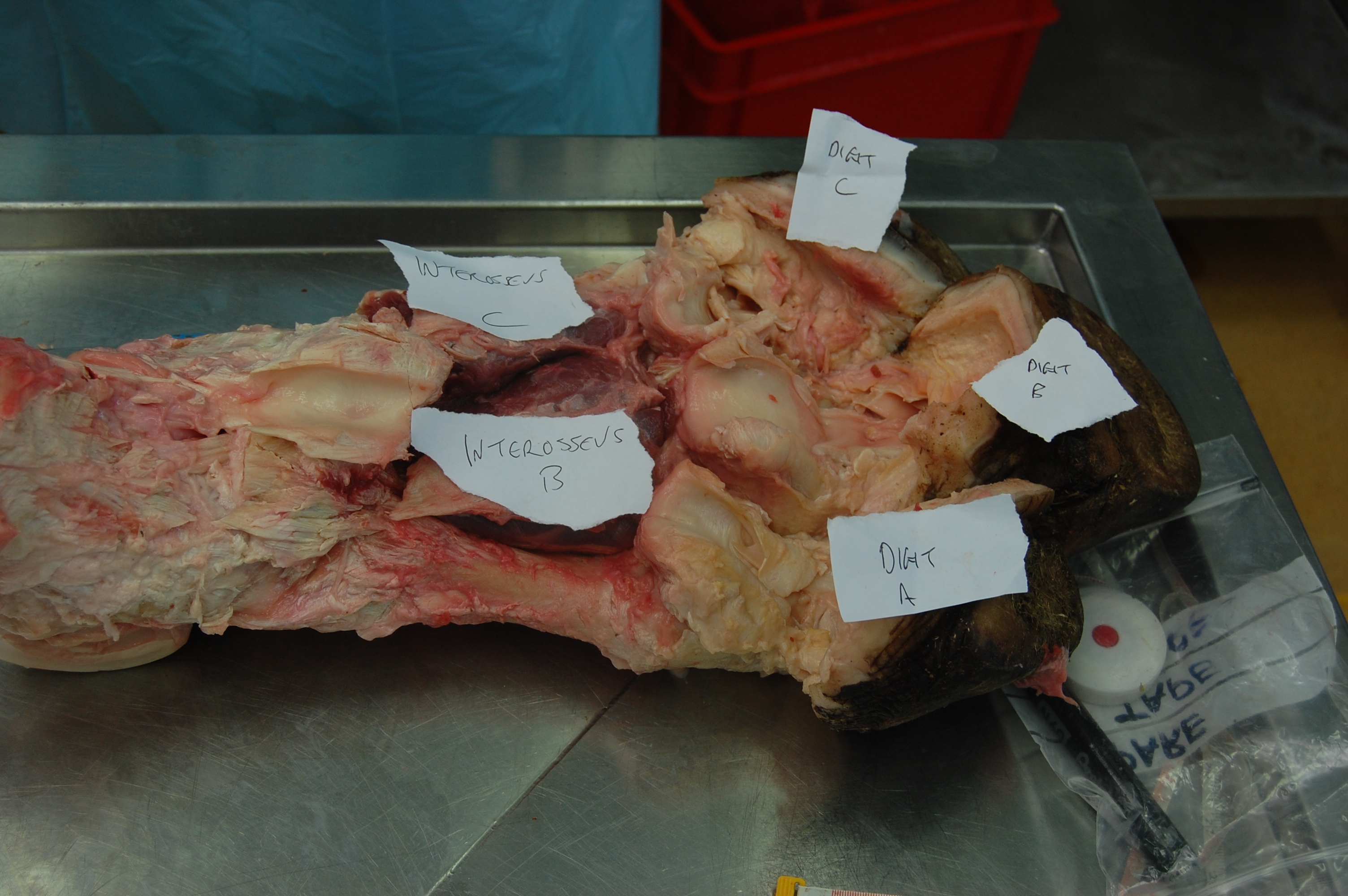
Here’s a view inside that marvelous foot, showing the HUGE digital flexor tendons. These help support the toes against gravity and, in theory, can act to curl them up– although in a rhino’s foot, as in an elephant’s, the toes are more like a single functional hoof, with reduced independence compared to a carnivore or primate:
And that ends our tour of rhinoceros limb anatomy and function. Help spread the word of how precious and threated rhinos are; educate yourself and others! And if you overhear someone talking about using rhino horn for medicine, try to politely educate them on the utter fallacy of this tradition. It is this cruel, greedy, ignorant practice that needs to die; not rhinos. I don’t enjoy receiving dead rhinos, on a personal level, even though the science excites me. I’d rather have many more alive and living good, healthy lives. And my team is trying to do what we can to help others on the “front lines” of rhino conservation make that happen.
For example, Will Fowlds, vet and co-owner of Amakhala Game Reserve, South Africa, recently sent us some images of a white rhino that had been caught in a poacher’s foot snare some years ago. The poor rhino still was having problems healing– we inspected x-ray images and external photos and helped to make an initial diagnosis of osteomyelitis, a nasty infectious, inflammatory foot bone/joint disease. We are following this case to hope that the rhino recovers and contribute help where we can, but the tough job belongs to the keepers/vets on the ground, not to mention the rhinos…
Furthermore, we’ve done foot pressure research covered here, and here is an example of the data we’ve collected (image credit: Dr Olga Panagiotopoulou), showing high pressures on the toes and low pressures on the foot pads:
Big thanks to people on my team that have helped with this and related research: Dr Olga Panagiotopoulou (and Dr Todd Pataky at Shinshu University, Japan), Dr Renate Weller in the VCS Dept at the RVC, Liz Ferrer at Berkeley, and former undergraduate student researchers Sophie Regnault, Richard Harvey, Hinnah Rehman, Richard Sheehan, Kate Jones, Bryony Armson and Suzannah Williams.
A White rhino’s heart, with more images below, all courtesy of William Perez’s Veterinary Anatomy Facebook pages. A mass of around 10kg (22 lbs weight) is not unusual! (Compare with even larger elephant heart)
On a serious note, in case you aren’t aware of it, there is a huge crisis afoot with the world’s rhino populations, because of demonstrably false claims about the health benefits of rhino horn (e.g. curing cancer) leading to a surge in its value worldwide, and thereby a massive upswing in poaching- as well as theft of museum specimens(!?). It may just be a matter of time before zoos’ and safari parks’ rhinos outside of Africa/Asia get hit, too. If sustained, this poaching could wipe out multiple rhino species in a matter of years; it is that severe and seems to already be worse this year than it was last year- and that was a Very Bad Year for rhinos.
However, a lot of people are uniting against the cruel greed and gross ignorance that has fueled the decline of rhinos, and public support seems to be growing. You can contribute, too; one example is this cause, or this one, to name but two. I’ve tried to make this my #1 cause, both on personal/ethical and scientific grounds (e.g. our work on rhino foot mechanics, health and care, to be detailed here later), and have been educating myself about it.
Anatomist extraordinaire Professor Larry Witmer of Ohio University has been contributing scientifically to helping rhinos, in an unexpected (but very sensible) way. This is a perfect example of how important basic science is; if we didn’t know rhino horn/nose anatomy we’d be less able to treat problems when they arise, and such problems can come from unexpected directions. His team’s contributions in the understanding of rhino anatomy are helping in one horrible case (see video below), in which three rhinos had their horns slashed off (along with part of the top of their skulls!) and two have survived so far. The vets in South Africa are trying to treat these two mutilated animals, and Larry’s group has been providing anatomical advice along with superb pictures on their Facebook page, which I want to publicize here because it is such GREAT freezer-based anatomical science and stunning imagery, and a seriously urgent cause. Please take a look. And while you’re at it, check out the Kariega Game Reserve’s Facebook page with more info on the plight of poor Themba and Thandi.
Edit: also check out this great story on our research, by Ann and Steve Toon, rhino conservationists/photographers/journalists.


")


