Feeds:
Posts
Comments

Posts Tagged ‘funky feet’

Back in November 2016 I got an exciting email from colleague Dr. Richard Thomas, who was building a team of experts for a proposed documentary on Jumbo the elephant; the famed proboscidean of the Victorian era (and arguably most famous elephant of all time, first international celebrity animal, etc.). I knew him from social media and from our mutual interests in chicken anatomy and evolution. And that exciting email, for once, worked out! Over coming months I chatted with the film producers and they could see a place for me in the programme, contributing my expertise in elephant (postcranial) anatomy, locomotion, health/welfare etc. Lo and behold, in May 2017 I met Sir David Attenborough at Heathrow and we flew out to New York City to film with the skeleton at the American Museum of Natural History. And to cap it off, I got to meet another childhood science communication hero: Professor David Suzuki of CBC’s “The Nature of Things“– my adrenaline levels were sky high!

Brooklyn neighborhood by our hotel. Lots to do!

The show has aired in the UK and is coming very soon to Canada and the world (details below). Here’s my part of the story.

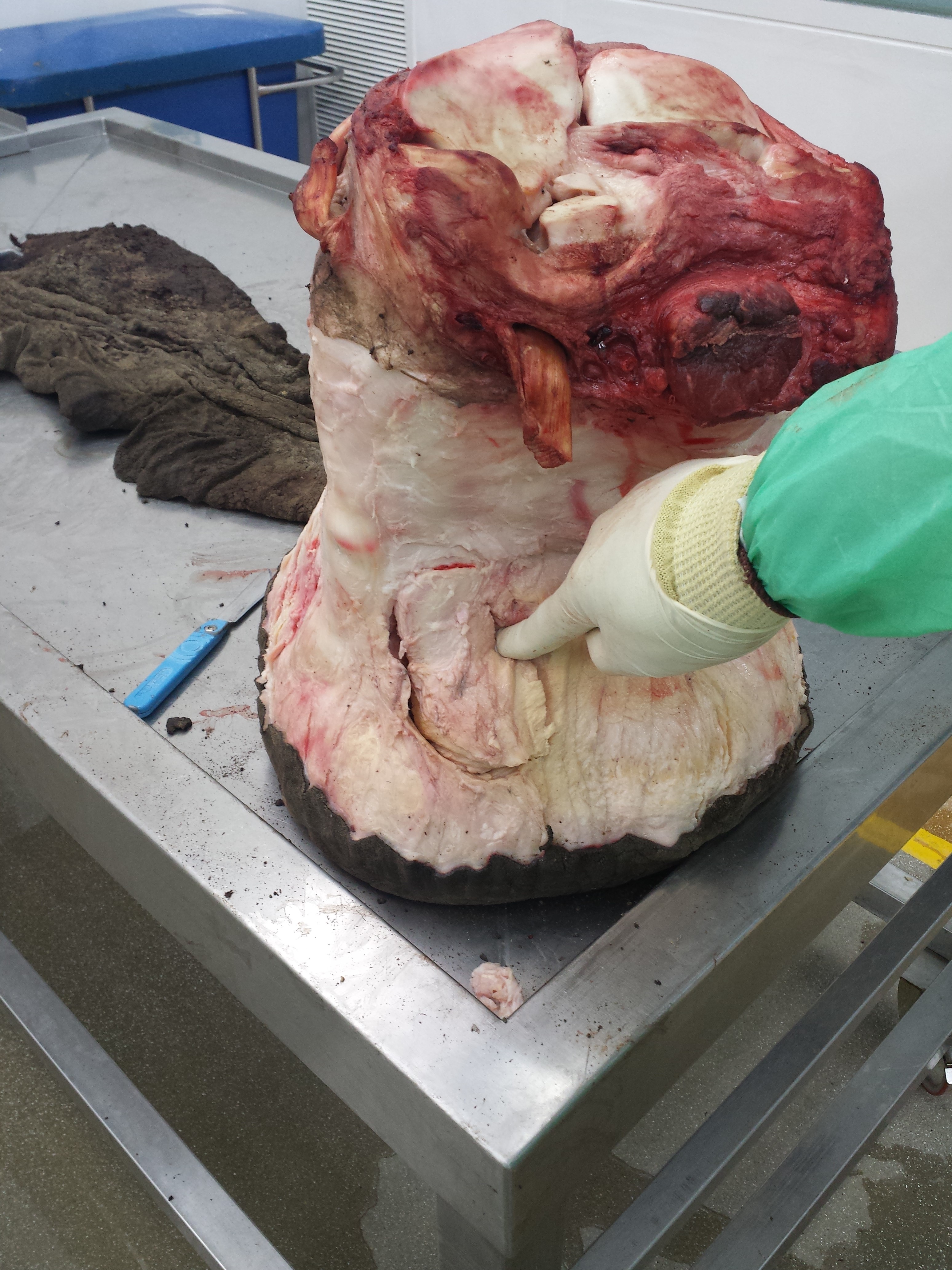
Stomach-Churning Rating: 3/10– bad bones but no blood.

We filmed from 15-19 May 2017 at the AMNH’s warehouse of mammalian skeletal remains, which is housed deep in the Brooklyn Army Terminal; a picturesque site in and of itself. And it is a site with a lot of history— WWI and II, Elvis and more.

It was a hectic week of the usual documentary stuff: repeat the same lines and motions again and again from different angles and with different paces and intonations (I cannot help in these cases but think about the Simpsons “Fallout Boy” episode), from ~9am-5pm, with plenty of downtime watching setup or other bits being filmed. I’m used to all that. But having the time to peer around the collection and chat to Richard and colleague Dr. Holly Miller (handling the tissue isotopes side of the story) about Jumbo’s skeleton was a lot of fun during downtime and filming itself. Not to mention the utter joy of studying one of the most famous museum specimens ever, and an animal widely held to be one of the largest of its kind, with much mystery surrounding its history despite its fame. (Wikipedia does a fair job of summarizing some of this)

Here are some photos to tell the story:

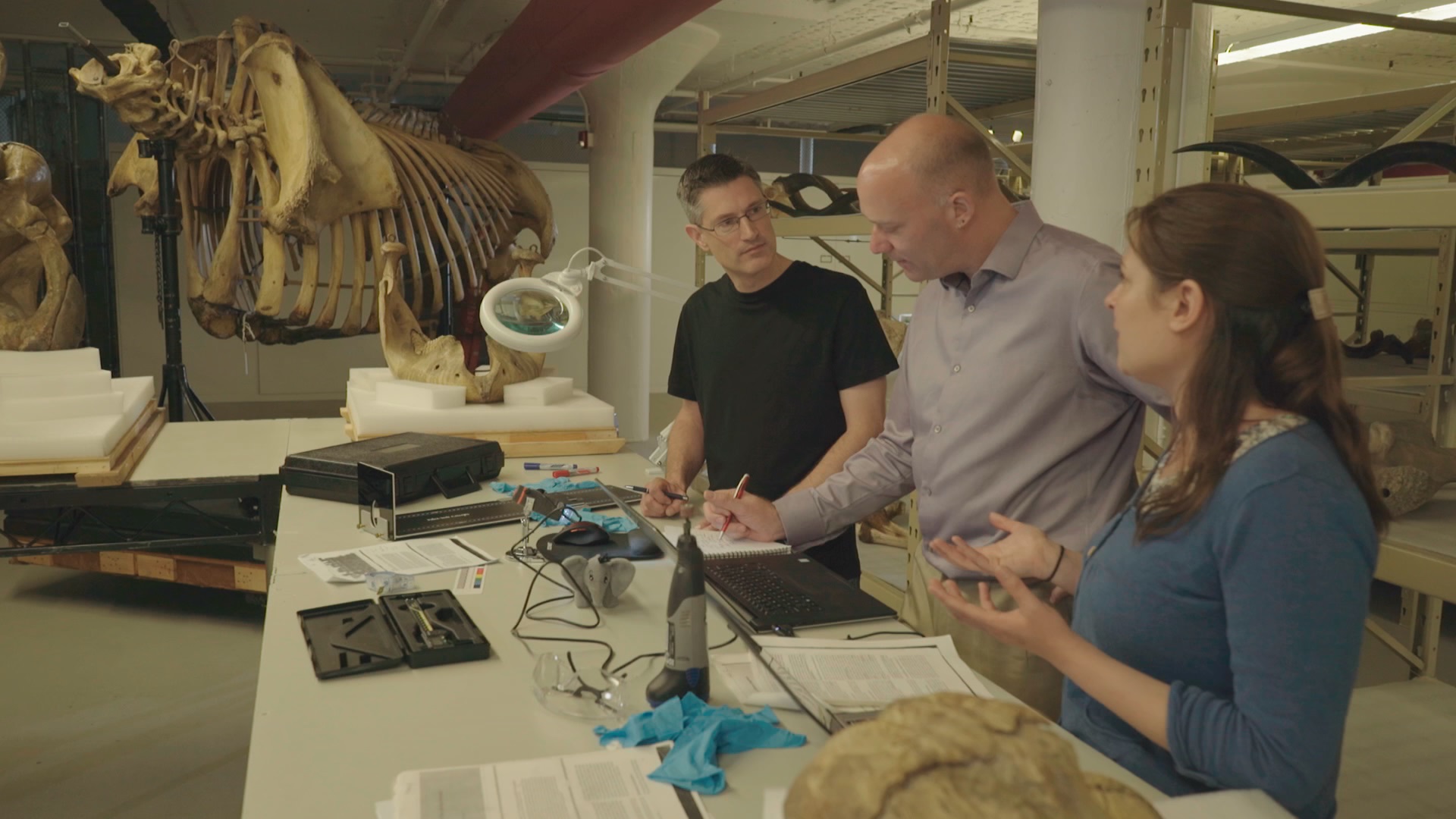
Photo of the team, courtesy of Infield Fly Productions (CBC production, “Jumbo: The Life of An Elephant Superstar”.

The Brooklyn Army Terminal, with a view of the harbour beyond.

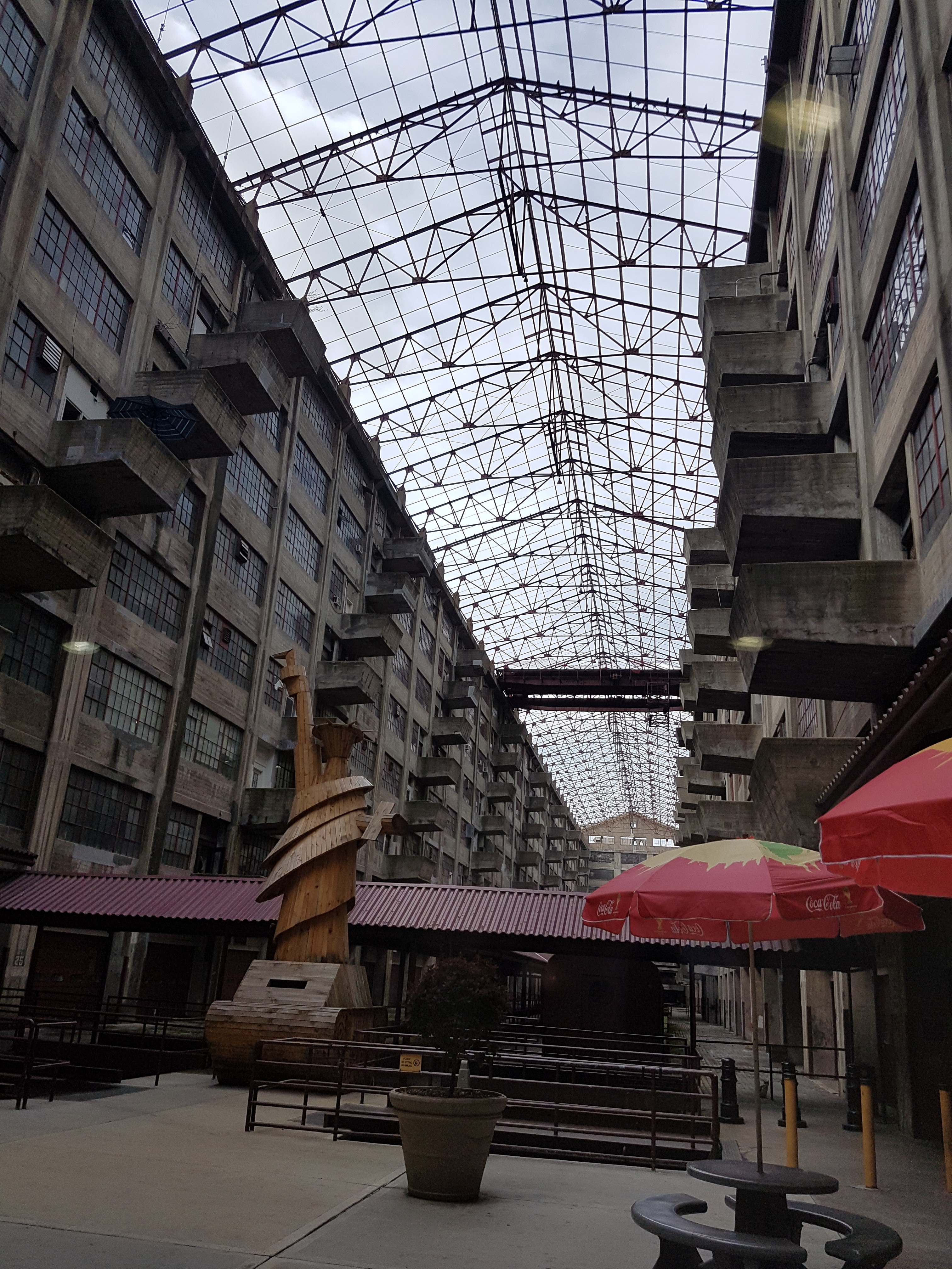
Inside the terminal: old army staging area and an evocative wooden Liberty/tank artwork.

Army terminal cat. Shipping still comes through the terminal so I guess there are plenty of rats and handouts from cat-lovers to keep it going. I miss our cats when I travel so this moment was appreciated.

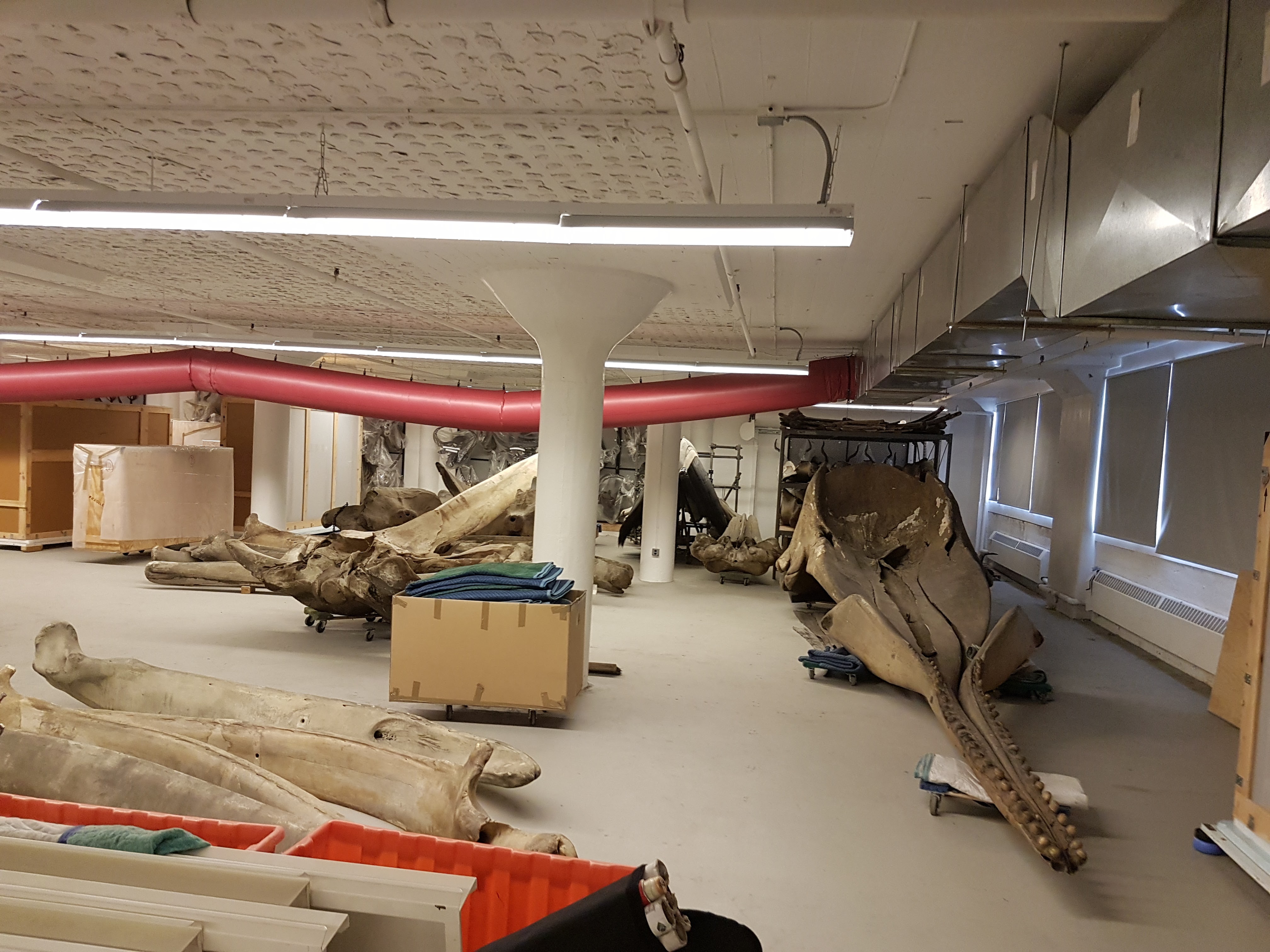
Whale skulls and other specimens inside the AMNH warehouse.

First view of Jumbo’s remains.

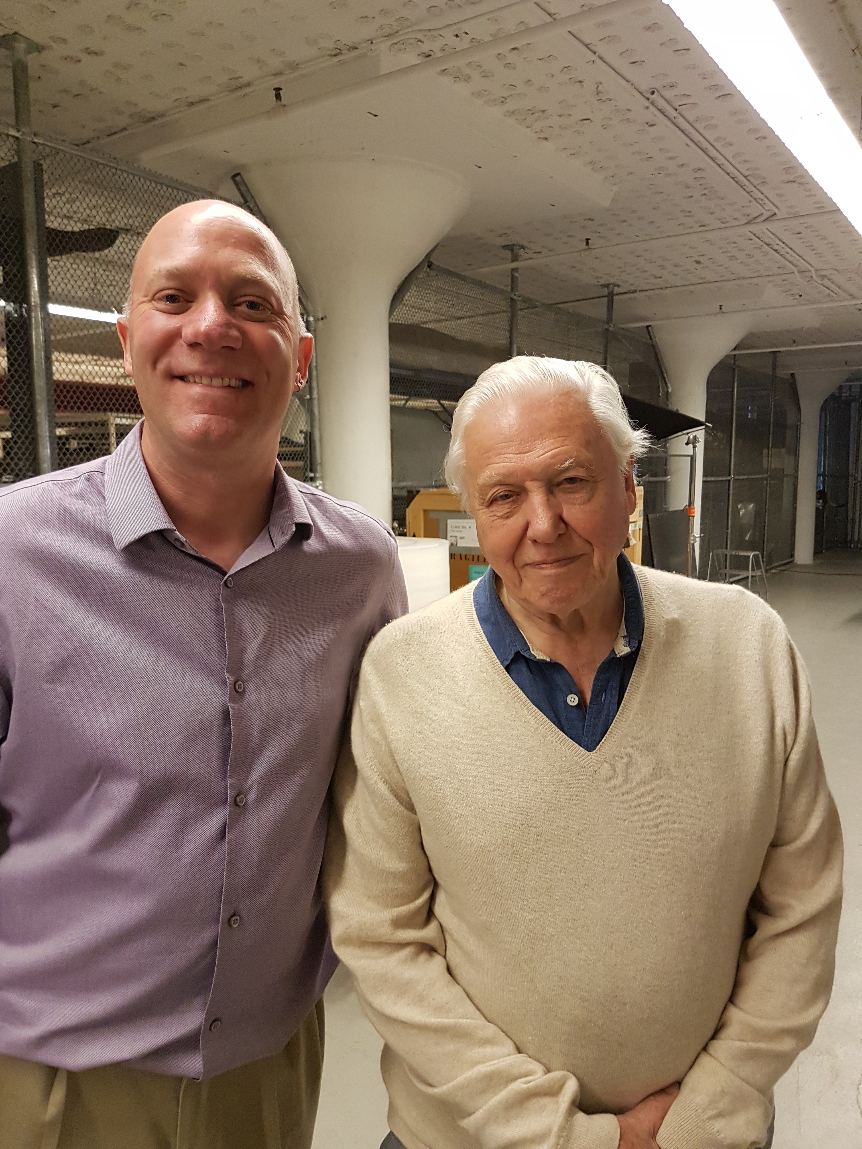
Photo opp with Sir David.

Photo opp with Prof Suzuki.

That’s the setup. I’ve done ~15 other documentary episodes/shows but this was like nothing else– simply an awesome experience.

Now the delivery: we set to studying those bones. We’d seen photos before, and Henry Fairfield Osborn had illustrated the specimen as his type of “Elephas africanus rothschildi” (Sudanese elephant; no longer valid but those were different times– it’s now just a nicely preserved Loxodonta africana africana), so we knew some of what to expect.

Looking at Osborn’s classic monograph. Oddly he didn’t address the GLARING MASSIVE PROBLEMS WITH THE TEETH!

Skull with terrible tooth pathologies– and let’s play spot Mumbo, my daughter’s toy elephant! He might even appear in some TV footage!

We had noted some serious issues with some bones (pathologies). I won’t spoil the message here but will show some images. I know some experts have voiced issues with how the tooth pathologies/growth were explained in some footage but I can’t address that here; it’s not my expertise. The important point to me is that the teeth are incredibly messed up and that can easily be linked to bad diet and other management/health issues, as the documentary explains.

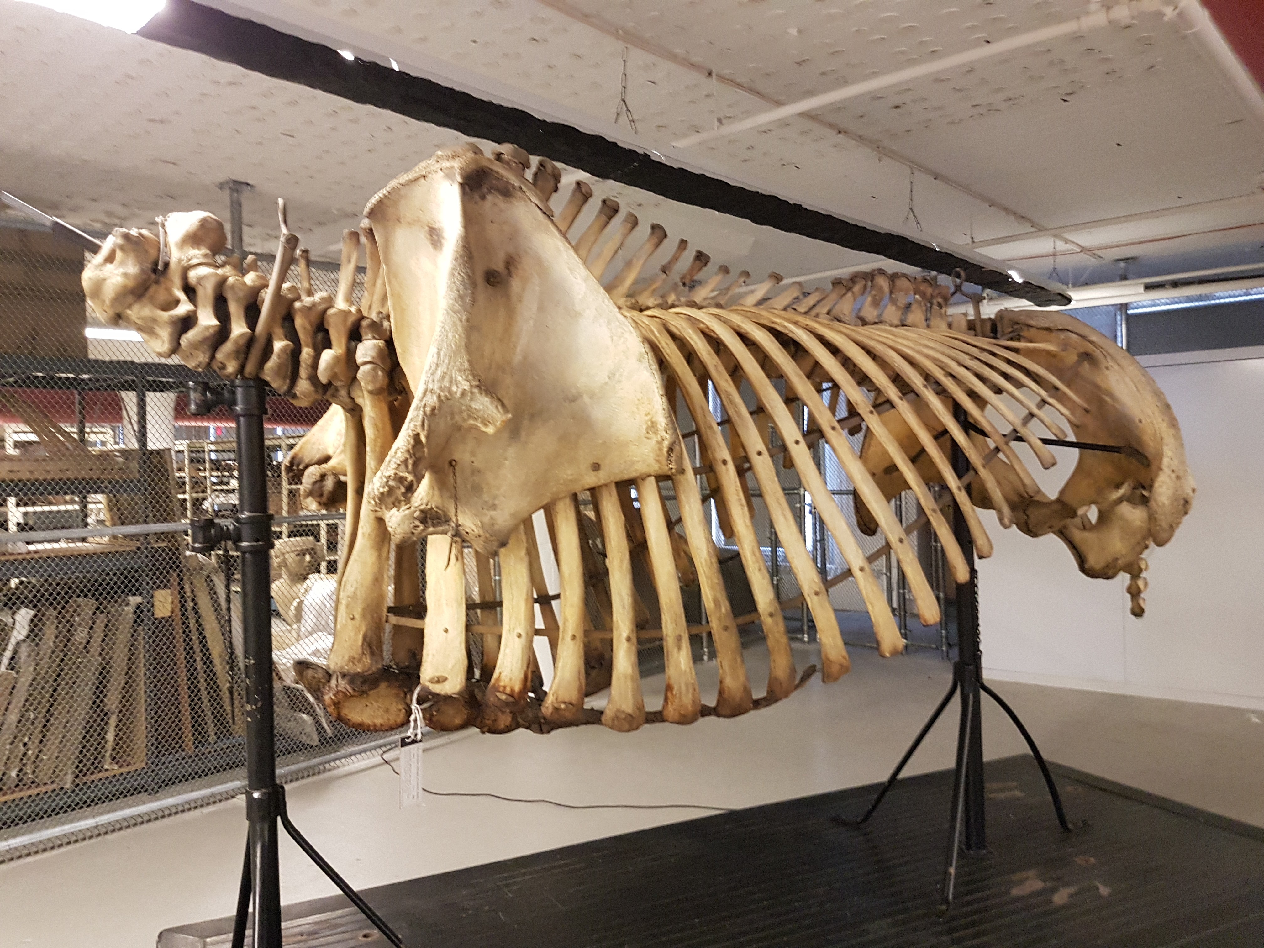
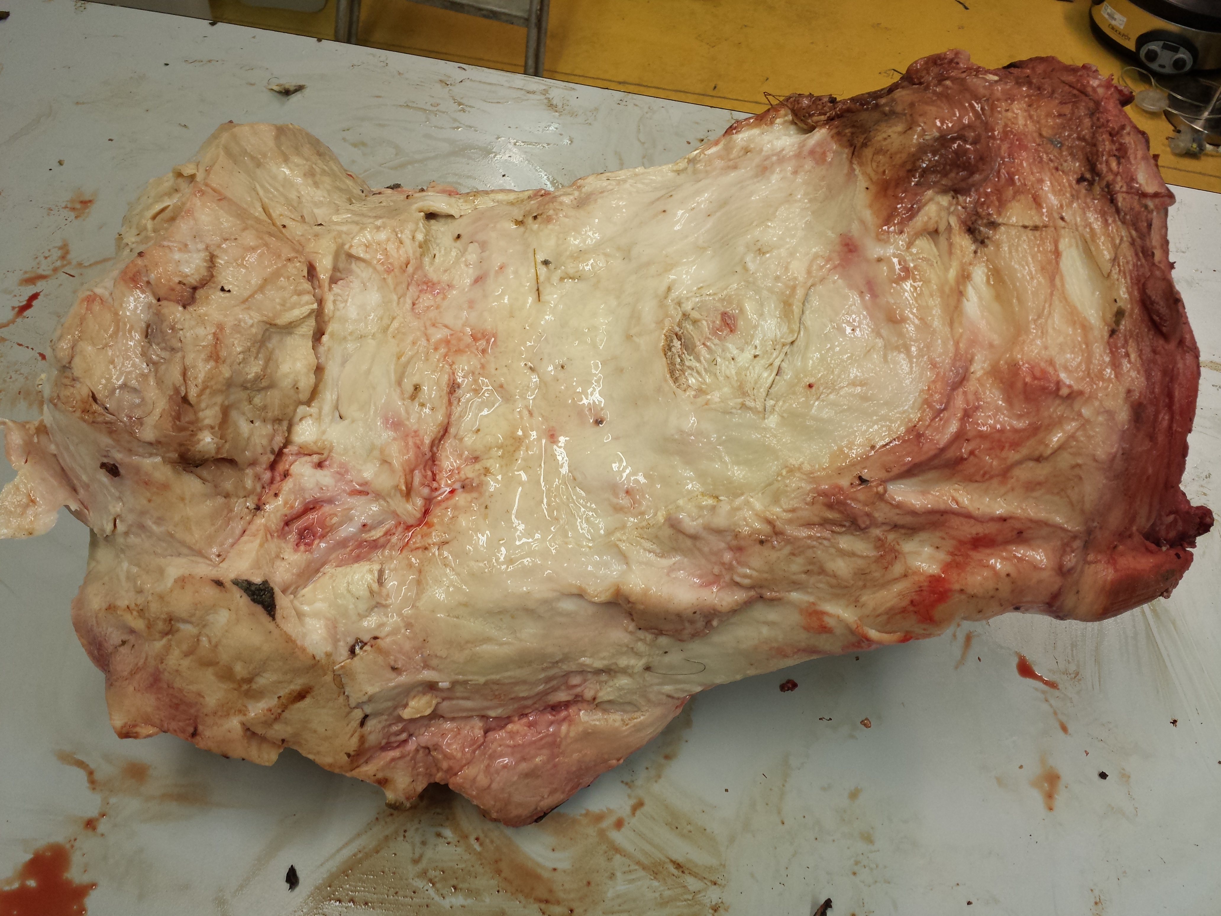
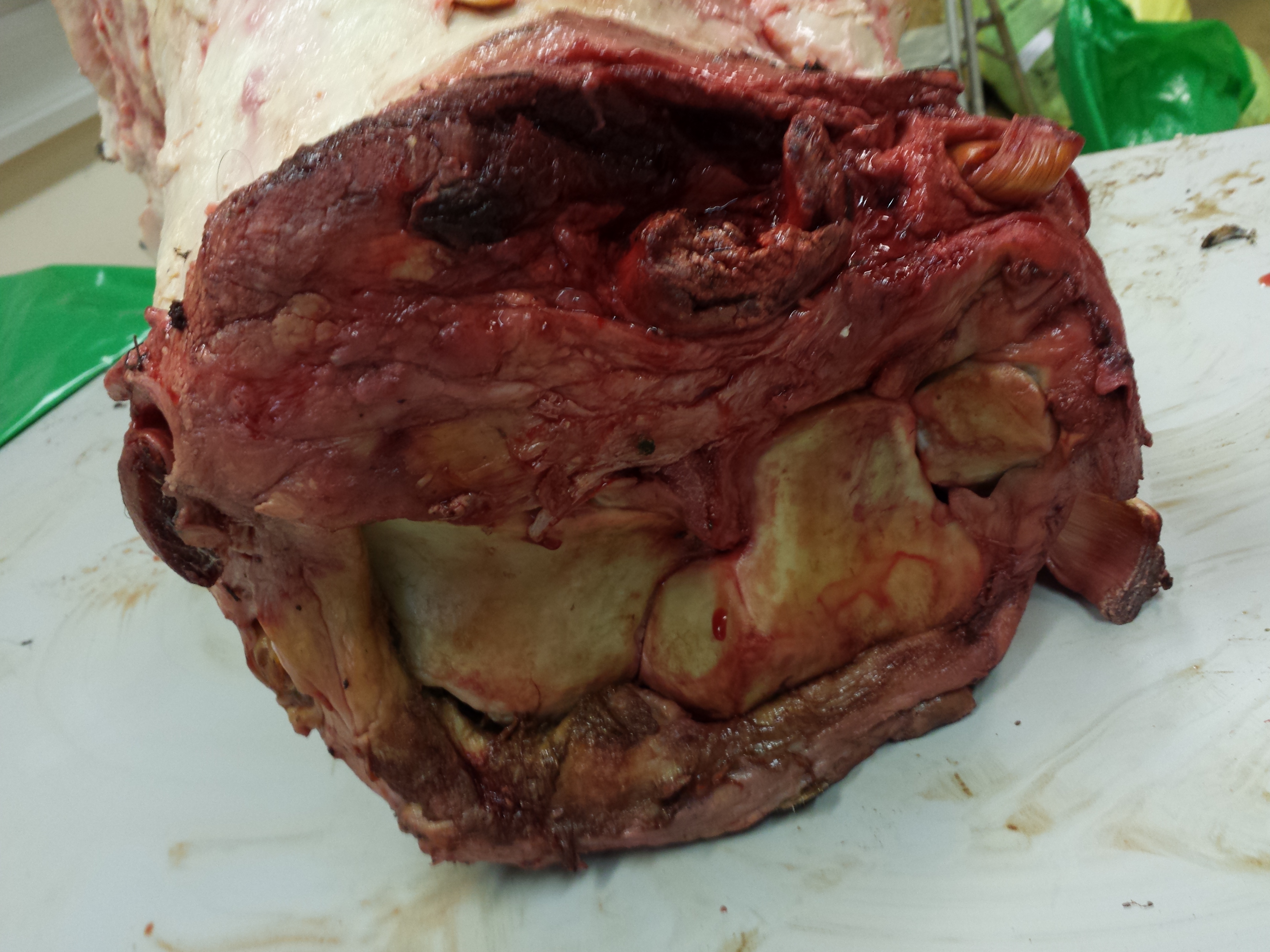
Jumbo’s torso in left side view. Glorious preservation.

Right forelimb, showing that the “growth plates” (epiphyses”) were not all fused, consistent with Jumbo still growing– as expected for an African male elephant in his 20’s.

Right elbow with some pathologies consistent with degenerative joint disease.

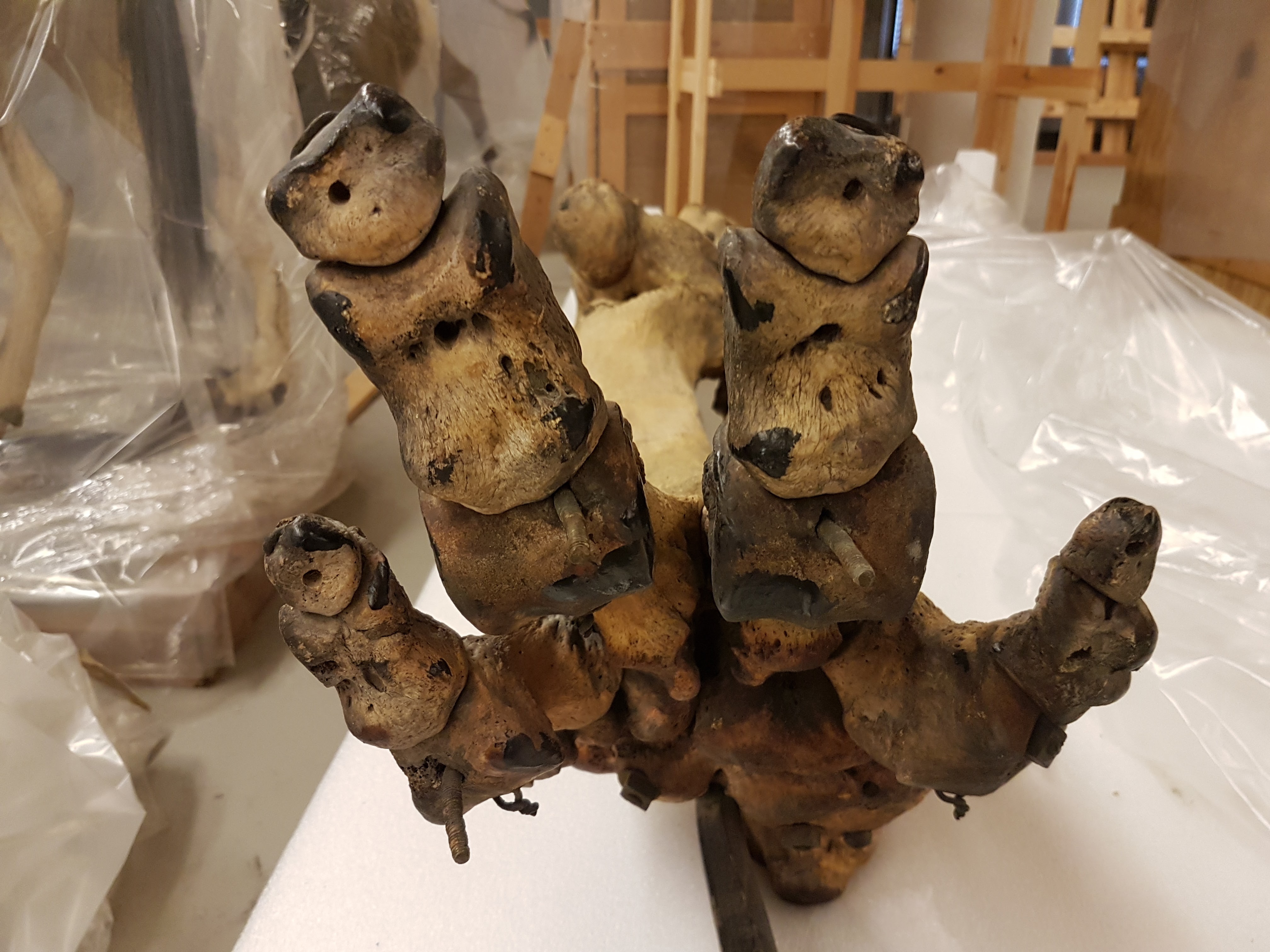
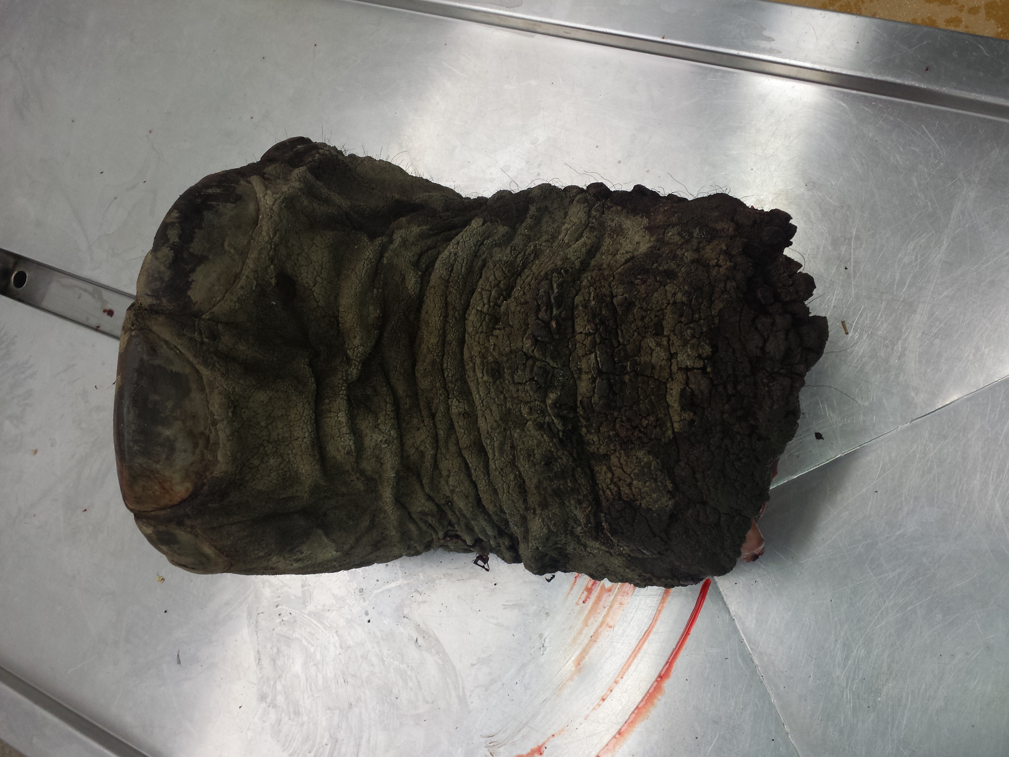
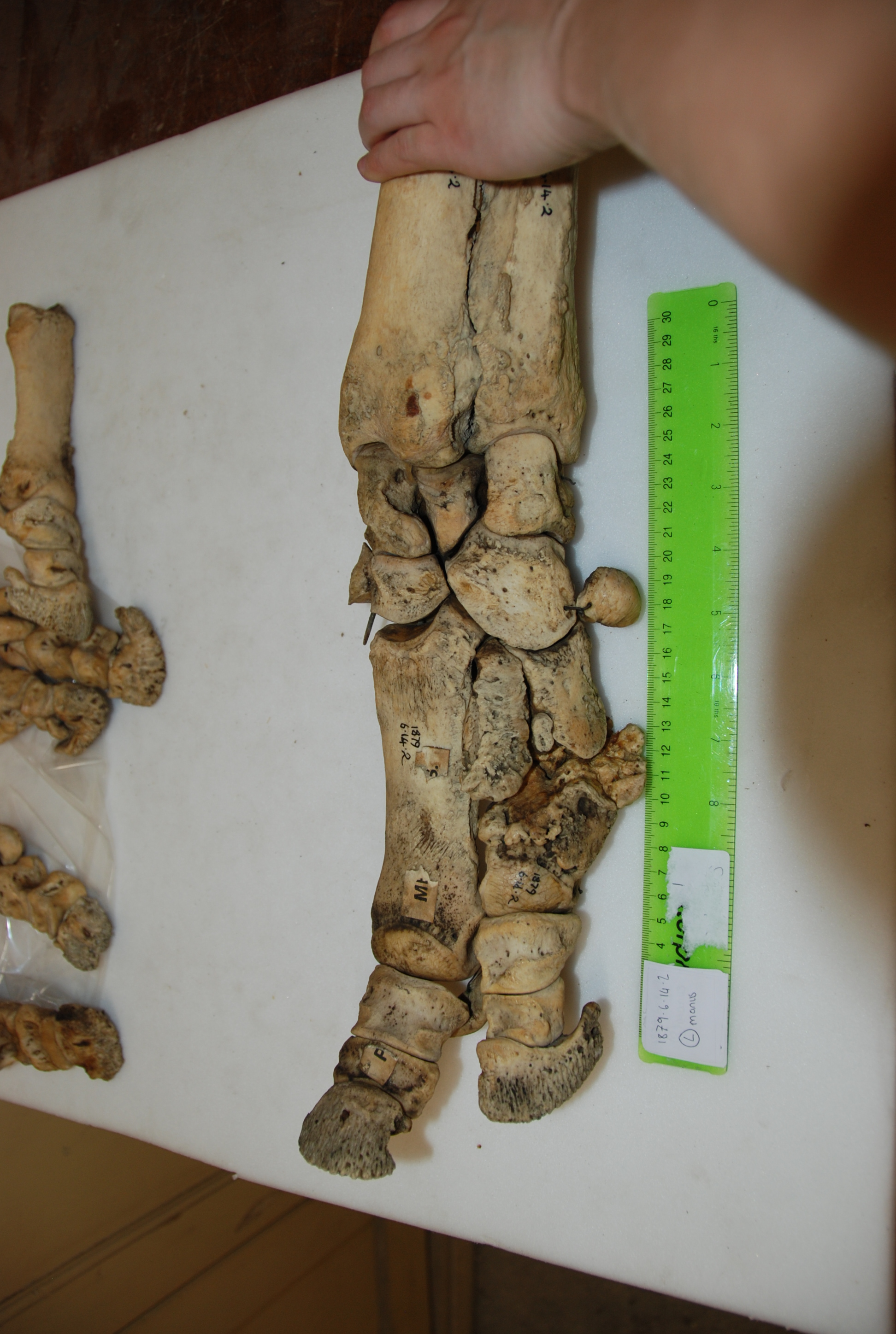
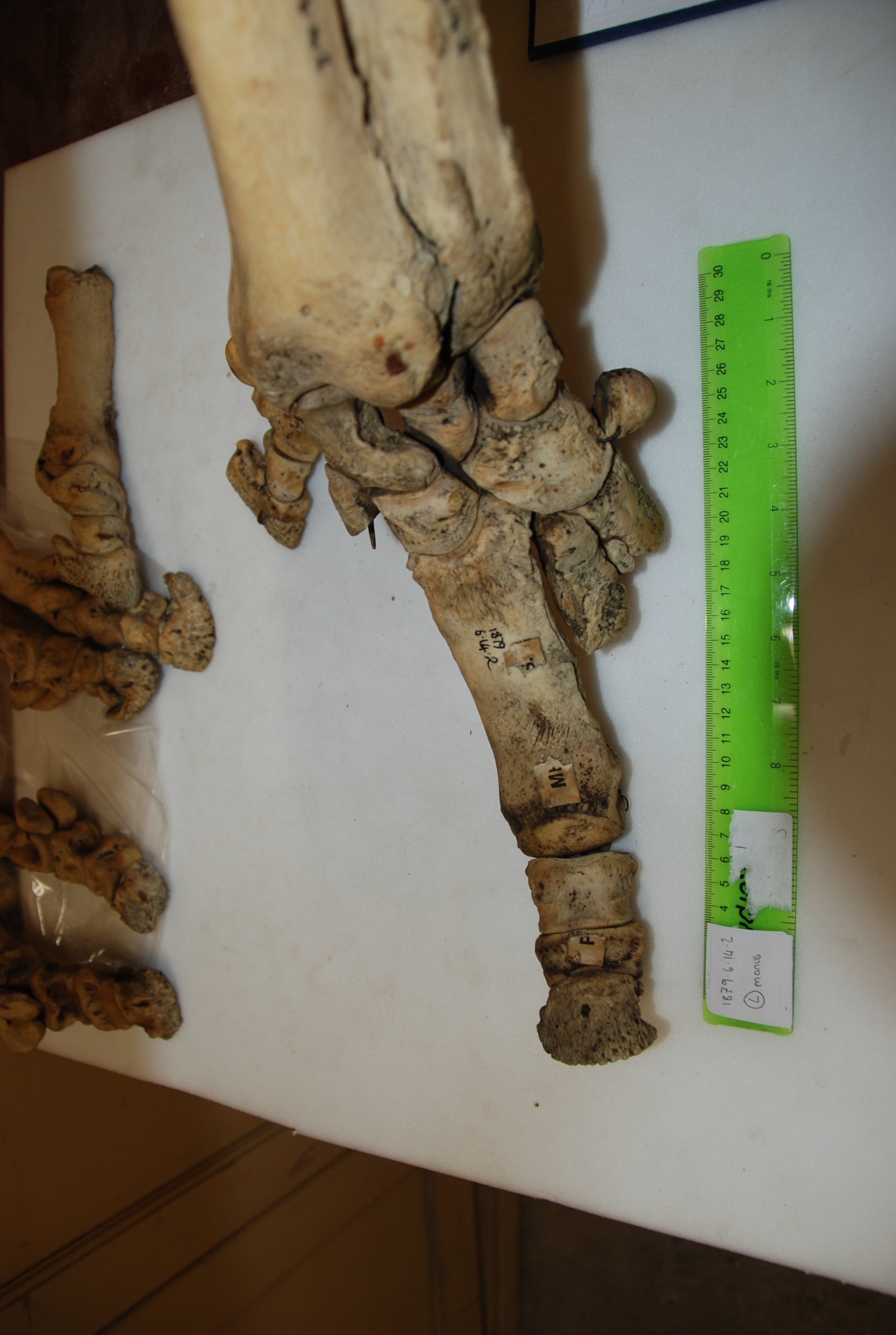
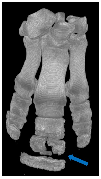
Surprisingly, Jumbo’s feet were not in nasty condition in terms of pathologies. I’d expected to see that. They’d been painted and drilled for mounting, but were not riddled with arthritic changes that I could see.

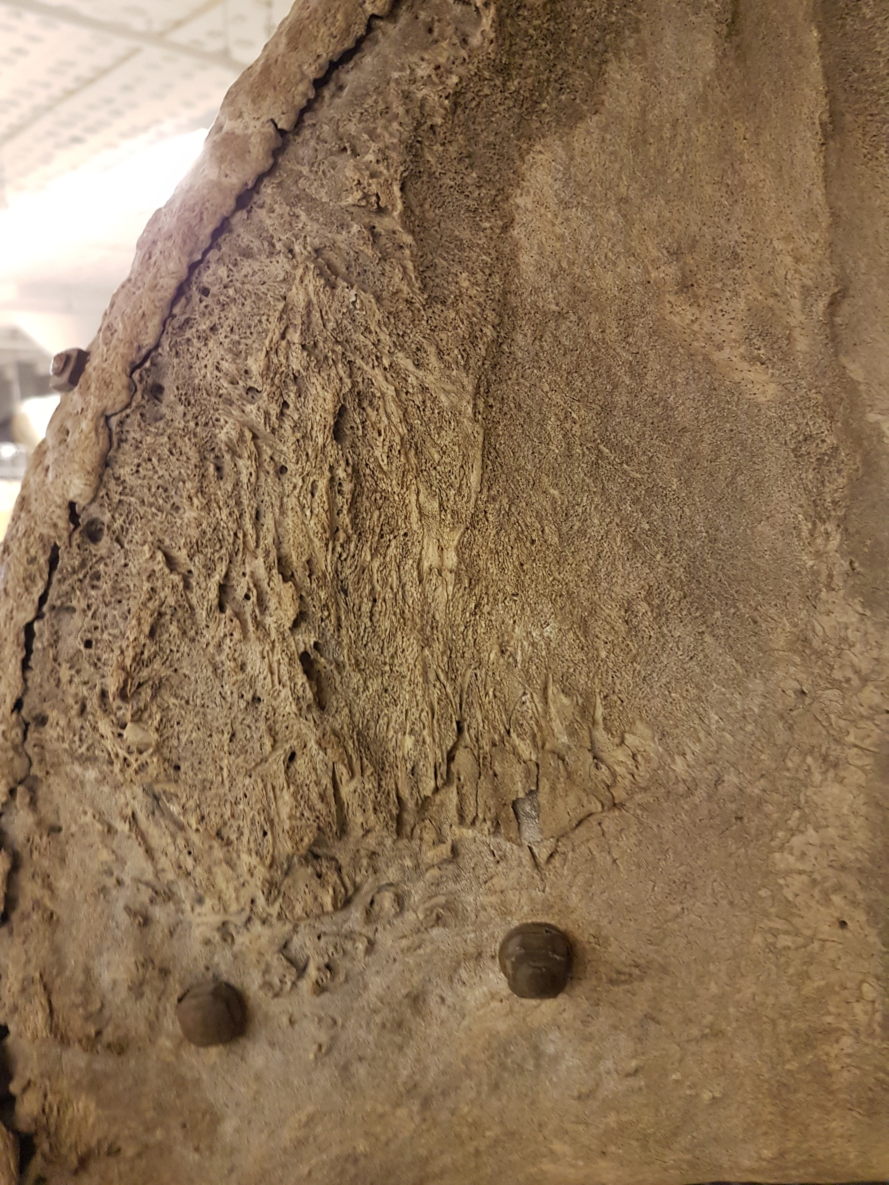
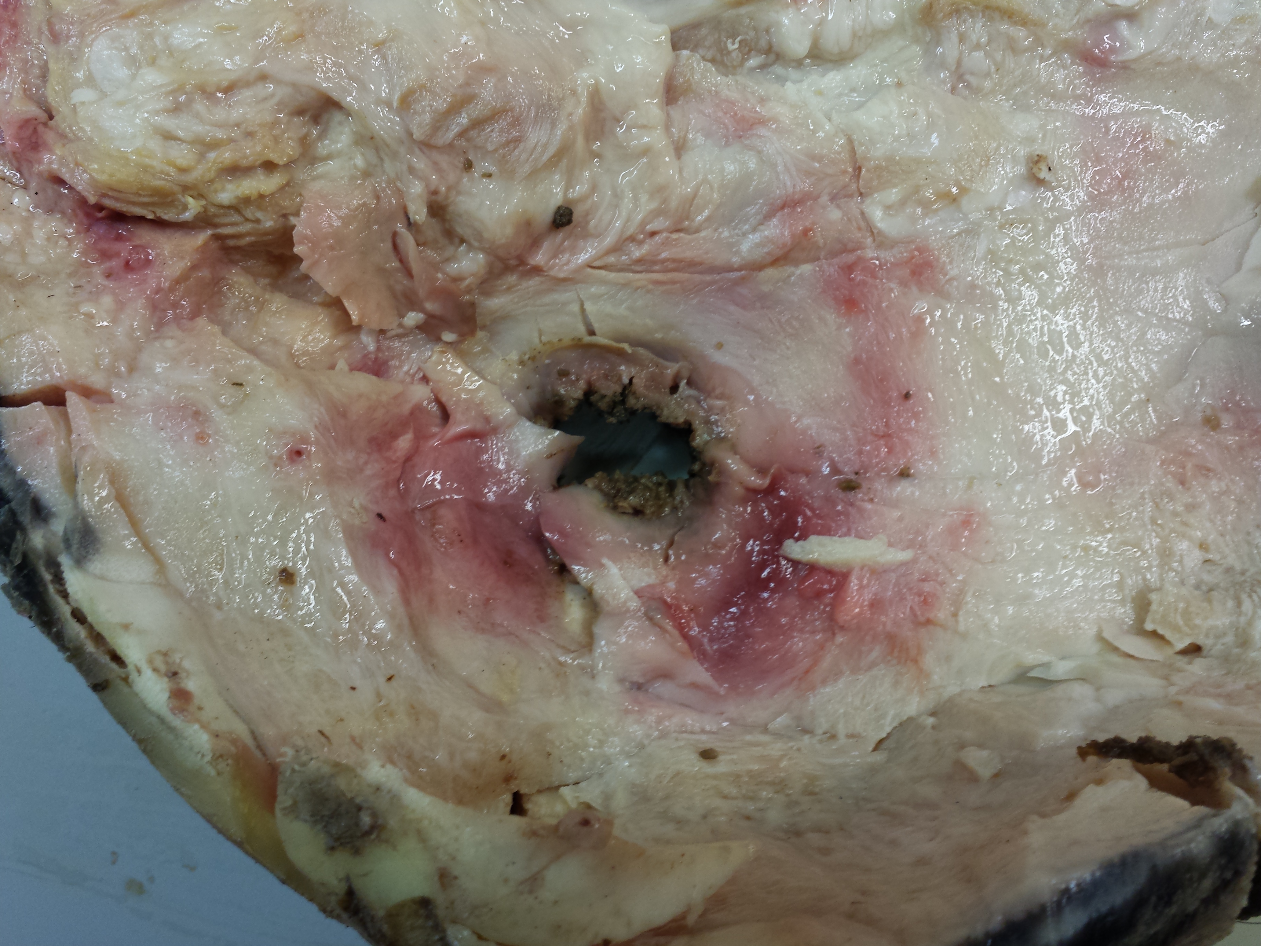
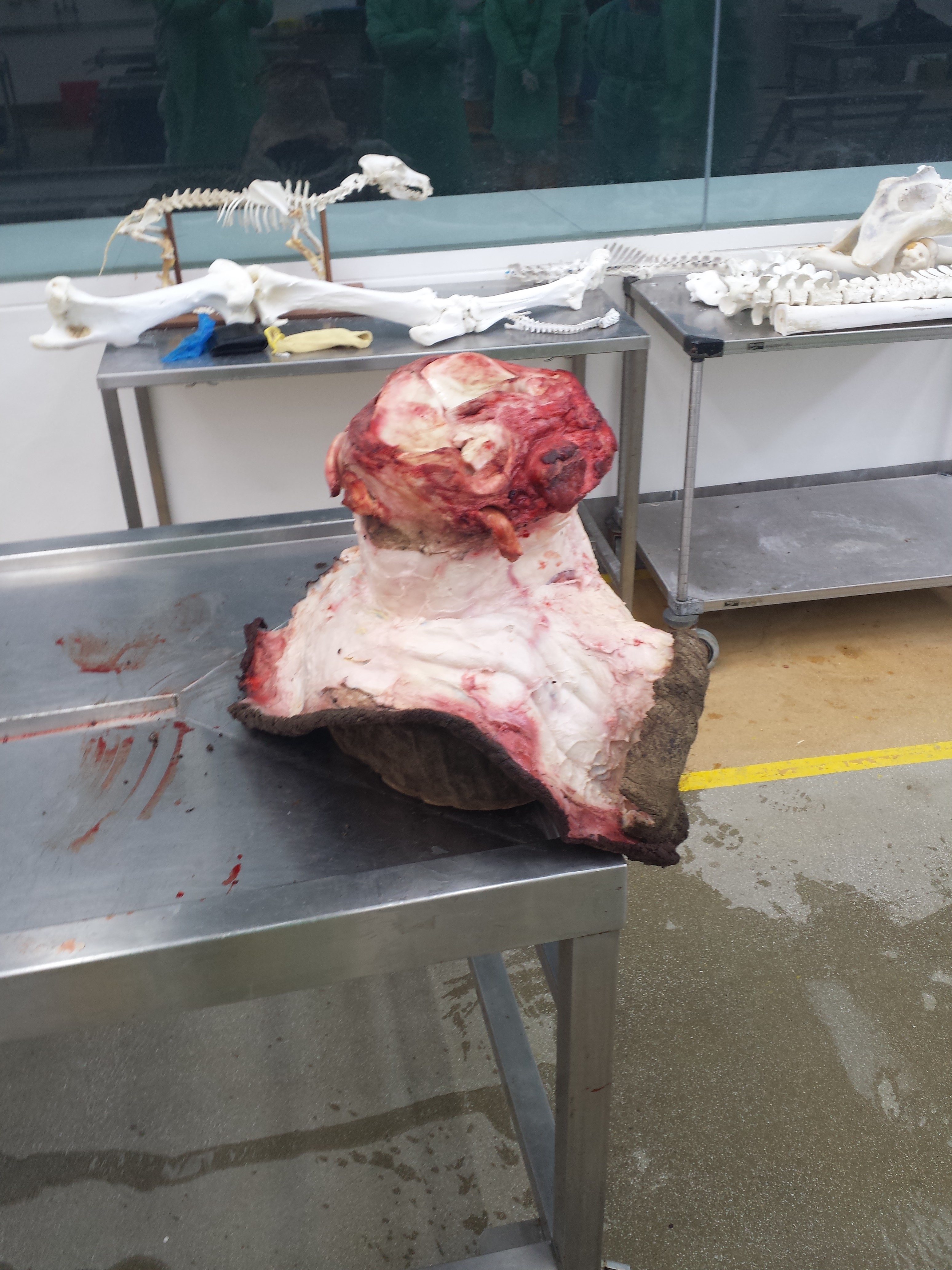
Strange bony plaque on the left pelvis (hip) region; something I’d never seen before in any elephant (and I’ve seen many). Why? The programme offers a reasonable explanation.

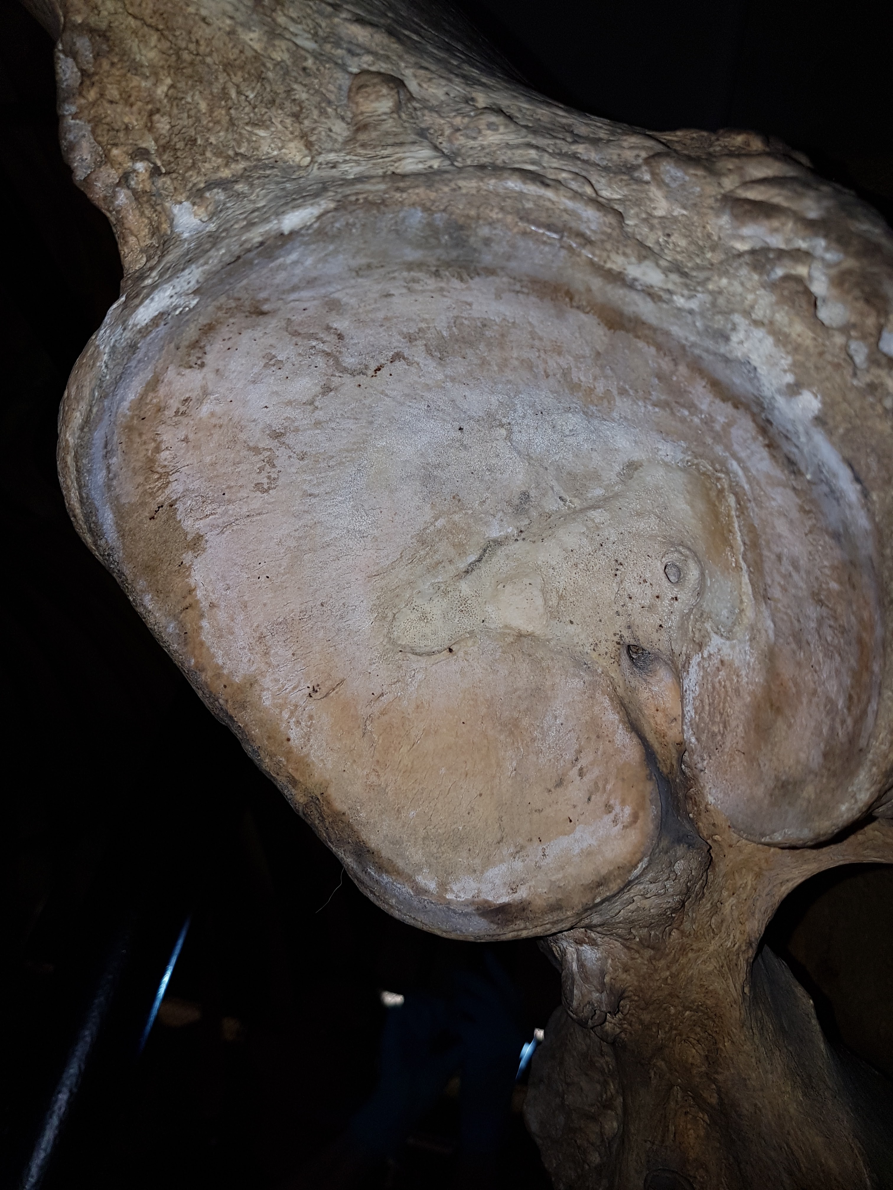
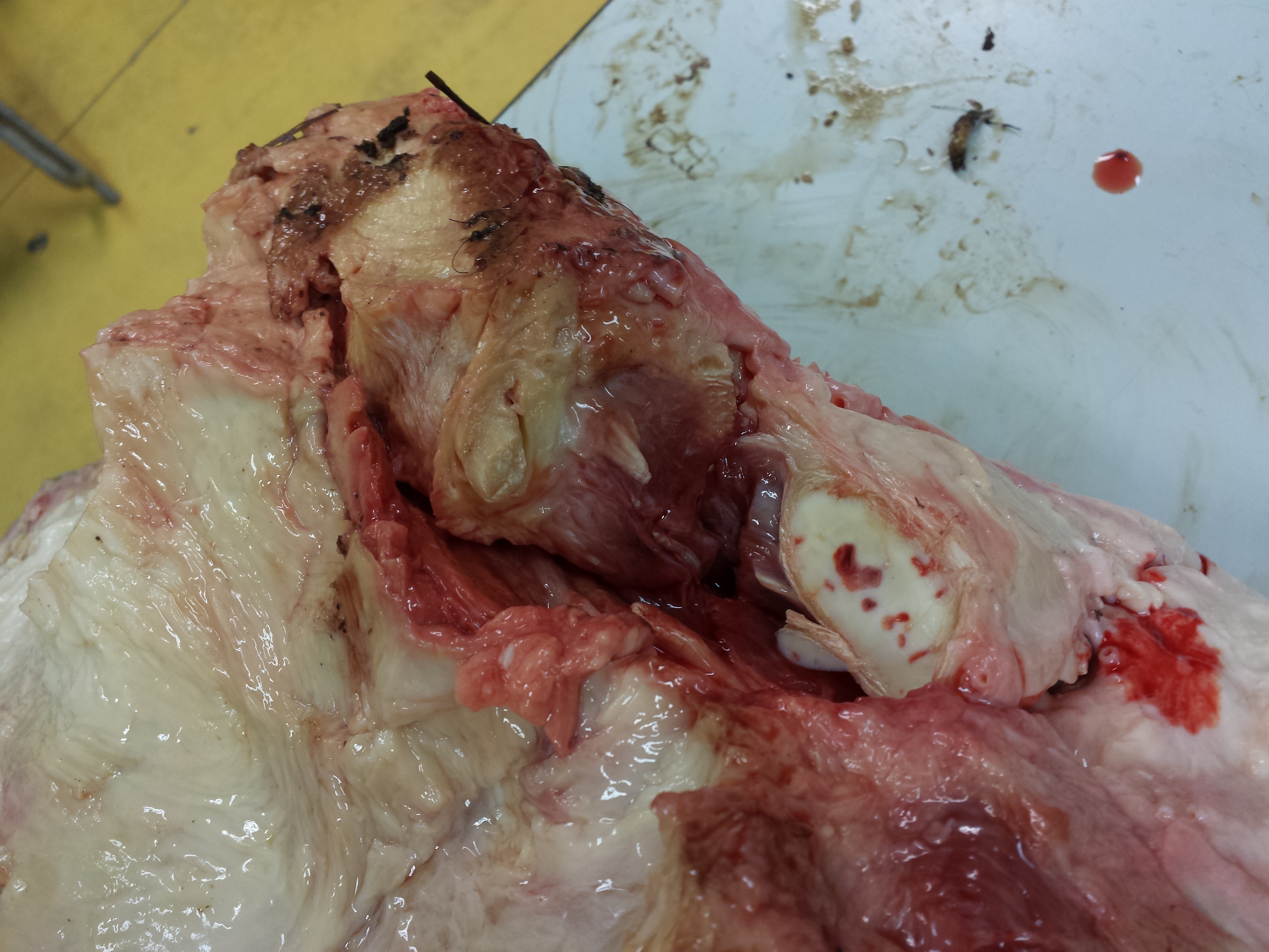
Jumbo’s right hip, with bad erosion of the bone and thus presumably the overlying cartilage. Ouch!

Strange extra prong on one right rib in Jumbo- we didn’t figure that out. It could conceivably be natural variation.

So, poor Jumbo suffered some jumbo-sized problems, and in complex ways. That’s just scratching the surface of what his skeleton tells us, and there’s plenty more in the show plus plenty more we can say later– there’s real science that came out of this programme! I was surprised to find how little had been stated anywhere in the scientific literature about Jumbo’s pathologies.

Sad as Jumbo’s skeletal story is, the broader story of his life and death is sadder still. For purposes of time I don’t think any of the three versions of the show will get to delve into how Jumbo’s mother may have been slashed to death by a broadsword, as the story below describes was the ancient practice:

I’d hate to be “so pestered by a popinjay”, too.

Adding insult to injury, we can reflect on how Jumbo was taken from the Sudan to the east (across the Suez), then on boat to Italy and then overground to Paris, where he lived for a little while until the zoological garden sold him to London. Luckily Jumbo avoided becoming a meal to starving Parisians during the Prussian siege of 1870-1. So he did not become elephant consommé like some of his co-captives did. The more one learns about Jumbo’s life and the life of elephants in captivity in the 1800s, the more harrowing the tale becomes.

Jumbo is THE celebrity elephant. His name has come to mean ‘big’ and ‘bombastic’, from applications to jumbo jets to hot dogs and other (darkly ironic) forms of consumption and extravagance. He has had a jumbo effect on Western culture, but also symbolizes the complex human-elephant relationship, such as the inspiration for “Dumbo’s” own sad story. We love elephants but our fascination with them can also be their undoing, such as poaching for the ivory trade or mistreatment in captivity. Jumbo’s story writ large is also the story of elephants, and our story to learn from. If anything comes out of my participation in the Jumbo documentary for the public’s benefit, I hope it is increased empathy for how we interact with elephants. They are like us in many ways (maybe over-emphasized with anthropomorphism in many accounts), but also unlike us (maybe even unfathomable) in not only their size and anatomy but also in aspects of their prodigious intellect, emotions and social structure. Elephants aren’t just jumbo spectacles. They are jumbo responsibilities for humans now that we dominate the planet so much.

Want to catch a version of the Jumbo show? I’ll try to keep this list up to date:

BBC iplayer now: https://www.bbc.co.uk/iplayer/episode/b09jcxrj/attenborough-and-the-giant-elephant

BBC One: 5:05pm on January 31st

CBC: 8pm on January 7th– trailer is here:

http://www.cbc.ca/player/play/1115035715562

And the international version is coming soon, plus the above versions surely will circulate globally in some ways.

Have a jumbo time (in a good way) in the rest of 2017 and onwards into 2018!

-John

Read Full Post »

It has been almost three months since my last post here, and things have fallen quiet on our sister blog Anatomy to You, too. I thought it was time for an update, which is mostly a summary of stuff we’ve been doing on my team, but also featuring some interesting images if you stick around. The relative silence here has partly been due to me giving myself some nice holiday time w/family in L.A., then having surgery to fix my right shoulder, then recovering from that and some complications (still underway, but the fact that I am doing this post is itself evidence of recovery).

Stomach-Churning Rating: 4/10; semi-gruesome x-rays of me and hippo bits at the end, but just bones really.

X-ray of my right shoulder from frontal view, unlabelled

X-ray of my right shoulder from frontal view, unlabelled

Labelled x-ray

Labelled x-ray

So my priorities shifted to those things and to what work priorities most badly needed my limited energy and time. I’ve also felt that, especially since my health has had its two-year rough patch, this blog has been quieter and less interactive than it used to be, but that is the nature of things and maybe part of a broader trend in blogs, too. My creative juices in terms of social media just haven’t been at their ~2011-2014 levels but much is out of my control, and I am hopeful that time will reverse that trend. Enough about all this. I want to talk about science for the rest of this post.

My team, and collaborators as well, have published six recent studies that are very relevant to this blog’s theme- how about we run through them quickly? OK then.

  1. Panagiotopoulou, O., Pataky, T.C., Day, M., Hensman, M.C., Hensman, S., Hutchinson, J.R., Clemente, C.J. 2016. Foot pressure distributions during walking in African elephants (Loxodonta africana). Royal Society Open Science 3: 160203.

Our Australian collaborators got five African elephants together in Limpopo, South Africa and walked them over pressure-measuring mats, mimicking our 2012 study of Asian elephants. While sample sizes were too limited to say much statistically, in qualitatively descriptive terms we didn’t find striking differences between the two species’ foot pressure patterns. I particularly like how the centre of pressure of each foot (i.e. abstracting all regional pressures down to one mean point over time) followed essentially the same pattern in our African and Asian elephants, with a variable heelstrike concentration that then moved forward throughout the step, and finally moved toward the outer (3rd-5th; especially 3rd) toes as the foot pushed off the ground, as below.

African elephant foot COP traces vs. time in red; Asian elephant in orange. Left and right forefeet above; hindfeet below.

African elephant foot COP traces vs. time in red; Asian elephant in orange-yellow. Left and right forefeet above; hindfeet below.

Gradually, this work is moving the field toward better ability to use similar techniques to compare elephant foot mechanics among species, individuals, or over time– especially with the potential of using this method (popular in human clinical gait labs) to monitor foot (and broader musculoskeletal) health in elephants. I am hopeful that a difference can be made, and the basic science we’ve done to date will be a foundation for that.

  1. Panagiotopoulou, O., Rankin, J.W., Gatesy, S.M., Hutchinson, J.R. 2016. A preliminary case study of the effect of shoe-wearing on the biomechanics of a horse’s foot. PeerJ 4: e2164.

Finally, about six years after we collected some very challenging experimental data in our lab, we’ve published our first study on them. It’s a methodological study of one horse, not something one can hang any hats on statistically, but we threw the “kitchen sink” of biomechanics at that horse (harmlessly!) by combining standard in vivo forceplate analysis with “XROMM” (scientific rotoscopy with biplanar fluoroscopy or “x-ray video”) to conduct dynamic analysis of forefoot joint motions and forces (with and without horseshoes on the horse), and then to use these data as input values for finite element analysis (FEA) of estimated skeletal stresses and strains. This method sets the stage for some even more ambitious comparative studies that we’re finishing up now. And it is not in short supply of cool biomechanical, anatomical images so here ya go:

fig5-vonmises

Above: The toe bones (phalanges) of our horse’s forefoot in dorsal (cranial/front) view, from our FEA results, with hot colours showing higher relative stresses- in this case, hinting (but not demonstrating statistically) that wearing horseshoes might increase stresses in some regions on the feet. But more convincingly, showing that we have a scientific workflow set up to do these kinds of biomechanical calculations from experiments to computer models and simulations, which was not trivial.

And a cool XROMM video of our horse’s foot motions:

  1. Bates, K.T., Mannion, P.D., Falkingham, P.L., Brusatte, S.L., Hutchinson, J.R., Otero, A., Sellers, W.I., Sullivan, C., Stevens, K.A., Allen, V. 2016. Temporal and phylogenetic evolution of the sauropod dinosaur body plan. Royal Society Open Science 3: 150636.

I had the good fortune of joining a big international team of sauropod experts to look at how the shapes and sizes of body segments in sauropods evolved and how those influenced the position of the body’s centre of mass, similar to what we did earlier with theropod dinosaurs. My role was minor but I enjoyed the study (despite a rough ride with some early reviews) and the final product is one cool paper in my opinion. Here’s an example:

fig6a-bates-sauropod-com-evol

The (embiggenable-by-clicking) plot shows that early dinosaurs shifted their centre of mass (COM) backwards (maybe related to becoming bipedal?) and then sauropods shifted the COM forwards again (i.e. toward their forelimbs and heads) throughout much of their evolution. This was related to quadrupedalism and giant size as well as to evolving a longer neck; which makes sense (and I’m glad the data broadly supported it). But it is also a reminder that not all sauropods moved in the same ways- the change of COM would have required changes in how they moved. There was also plenty of methodological nuance here to cover all the uncertainties but for that, see the 17 page paper and 86 pages of supplementary material…

  1. Randau, M., Goswami, A., Hutchinson, J.R., Cuff, A.R., Pierce, S.E. 2016. Cryptic complexity in felid vertebral evolution: shape differentiation and allometry of the axial skeleton. Zoological Journal of the Linnean Society 178:183-202.

Back in 2011, Stephanie Pierce, Jenny Clack and I tried some simple linear morphometrics (shape analysis) to see how pinniped (seal, walrus, etc) mammals changed their vertebral morphology with size and regionally across their backbones. Now in this new study, with “Team Cat” assembled, PhD student Marcela Randau collected her own big dataset for felid (cat) backbones and applied some even fancier techniques to see how cat spines change their shape and size. We found that overall the vertebrae tended to get relatively more robust in larger cats, helping to resist gravity and other forces, and that cats with different ecologies across the arboreal-to-terrestrial spectrum also changed their (lumbar) vertebral shape differently. Now Marcela’s work is diving even deeper into these issues; stay tuned…

fig2-randau-measurements

Example measurements taken on felid vertebrae, from the neck (A-F) to the lumbar region (G-J), using a cheetah skeleton.

  1. Charles, J.P., Cappellari, O., Spence, A.J., Hutchinson, J.R., Wells, D.J. 2016. Musculoskeletal geometry, muscle architecture and functional specialisations of the mouse hindlimb. PLOS One 11(4): e0147669.

RVC PhD student James Charles measured the heck out of some normal mice, dissecting their hindlimb muscle anatomy, and using microCT scans produced some gorgeous images of that anatomy too. In the process, he also quantified how each muscle is differently specialized for the ability to produce large forces, rapid contractions or fine control. Those data were essential for the next study, where we got more computational!

mouse-mimics

  1. Charles, J.P., Cappellari, O., Spence, A.J., Wells, D.J., Hutchinson, J.R. 2016. Muscle moment arms and sensitivity analysis of a mouse hindlimb musculoskeletal model. Journal of Anatomy 229:514–535.

James wrangled together a lovely musculoskeletal model of our representative mouse subject’s hindlimb in the SIMM software that my team uses for these kinds of biomechanical analyses. As we normally do as a first step, we used the model to estimate things that are hard to measure directly, such as the leverages (moment arms) of each individual muscle and how those change with limb posture (which can produce variable gearing of muscles around joints). James has his PhD viva (defense) next week so good luck James!

mouse-simm

The horse and mouse papers are exemplars of what my team now does routinely. For about 15 years now, I’ve been building my team toward doing these kinds of fusion of data from anatomy, experimental biomechanics, musculoskeletal and other models, and simulation (i.e. estimating unmeasurable parameters by telling a model to execute a behaviour with a given set of criteria to try to perform well). Big thanks go to collaborator Jeff Rankin for helping us move that along lately. Our ostrich study from earlier this year shows the best example we’ve done yet with this, but there’s plenty more to come.

I am incredibly excited that, now that my team has the tools and expertise built up to do what I’ve long wanted to do, we can finally deliver the goods on the aspirations I had back when I was a postdoc, and which we have put enormous effort into pushing forward since then. In addition to new analyses of horses and mice and other animals, we’ll be trying to push the envelope more with how well we can apply similar methods to extinct animals, which brings new challenges– and evolutionary questions that get me very, very fired up.

Here we are, then; time has brought some changes to my life and work and it will continue to as we pass this juncture. I suspect I’ll look back on 2016 and see it as transformative, but it hasn’t been an easy year either, to say the least. “Draining” is the word that leaps to mind right now—but also “Focused” applies, because I had to try to be that, and sometimes succeeded. I’ve certainly benefited a lot at work from having some talented staff, students and other collaborators cranking out cool papers with me.

I still have time to do other things, too. Once in a while, a cool critter manifests in The Freezers. Check out a hippo foot from a CT scan! It’s not my best scan ever (noisy data) but it shows the anatomy fairly well, and some odd pathologies such as tiny floating lumps of mineralized soft tissue here and there. Lots to puzzle over.

Read Full Post »

I was recently featured on Daily Planet, a great Canadian science show on TV that lamentably is not broadcast more globally. It is always high quality science communication, aided by the superb hosts Ziya Tong and Dan Riskin (and a talented crew!). What were we doing? Dissecting an elephant’s foot, of course!

Stomach-Churning Rating: 9/10; no-holds-barred dismantling of elephant feet, from the video onwards, and this post is heavy on moist, goopy photos afterwards, with some nasty pathologies. Not nice at all. I’ll give you a chance to turn around while contemplating the cart that we use to carry elephant feet around campus (each foot is 20-30kg; up to 70lbs; so we need the help!), before the video.

no_poo

Here is a snippet of the full segment from Daily Planet:

And here is more of some of my recent dissections. I’ll walk you through two dissections, via photos. This goes back to the roots of this blog: unflinching, gritty examinations of real anatomy! Of course, no elephants were harmed for this work. They died at EU zoos/parks and were sent to me for postmortem examination and research, so we hope that this benefits the future care of elephants. We’re currently finishing up a grand overview paper that describes all of the odd pathologies we’ve observed in elephant feet, for the benefit of zoo keepers and vets who are trying to detect, diagnose and monitor any foot problems.

As the post’s title alludes, elephant feet (and more proximal parts of the limbs) are no stranger to this blog. If you’ve forgotten or are unfamiliar, here are some of my past proboscidean-posts: on elephant foot pathologies (a close sister post to this one), our “six-toed” elephants paper, how to make a computer simulation of an elephant’s limb (umm, paper yet to come!), how we boil and bleach bones to clean them up, and a few others. Last but not least, there was the post that went viral in the early #JohnsFreezer/WIJF days: dissecting an elephant with the “Inside Nature’s Giants” show.

There are two feet in this post, both front right feet (manus is the technical term; singular and plural). The first one is the messier (unhealthy and bloodier, less fresh and clean) one, from the show/video. It is an Asian elephant (Elephas maximus). I kick off with photos I took after the filming, so the foot is already deconstructed:

Skinned foot, oblique front/inside view.

Skinned foot, oblique front/inside view. The wrist is on the right side of the photo; the toes on the left.

Sole ("slipper"), with a hole on the fourth toe showing where the abscess is that let infection in/pus drain out.

Sole (“slipper”), with a hole on the fourth toe showing where the abscess is that let infection in/pus drain out. The slipper here is upside-down.

Top-down view of the sole of the foot, once the slipper is removed.

Top-down view of the sole of the foot, once the slipper is removed; flipped over and rotated 90 degrees clockwise from the above photo. Some of the fat pad of the foot is on the right side of the image; it’s very hard to separate from the keratinous sole of the foot.

Looking down into the fourth toe's abscess on the other side of the above view.

Looking down into the fourth toe’s (ring finger) abscess on the other side of the above view.

Looking down into the third (middle) toe, same view as above. Some redness and greyness where this toe had some of its own pathological issues.

Looking down into the second toe (index finger), same view as above. Some redness and greyness where this toe had some of its own pathological issues like infection and a smaller abscess.

Looking up from the slipper at the fat pad and toes of the foot, where they interface with the sole/slipper. The fat pad is toward the bottom and left side; the five toes are on the upper/right side (knobby subcircular regions on the perimeter of the foot).

Looking up from the slipper (removed) at the fat pad and toes of the foot, where they interface with the sole/slipper. The fat pad is toward the bottom and left side; the five toes are on the upper/right side (knobby subcircular regions on the perimeter of the foot). The very bad infection on the fourth toe is visible on the bottom right.

The sproingy fat pad is worth a video!

And one good wiggle deserves another!

A view down onto the wrist joint. The carpal (wrist) bones are visible at the bottom of the image, whereas the flexor (palmar) tendons and muscles on the back of the "hand" are at the top. There is a LOT of musculotendinous tissue on the back side of an elephant's foot.

A view down onto the wrist joint. The carpal (wrist) bones are visible at the bottom of the image, whereas the flexor (palmar) tendons and muscles on the back of the “hand” are at the top. There is a LOT of musculotendinous tissue on the back side of an elephant’s foot. As you will see in my dissection of the second foot, further below!

Looking down onto the medial (inner/"thumb") border of the foot, where I've exposed the prepollex, or false "sixth finger" by removing the first metacarpal (knuckle) bone.

Looking down onto the medial (inner/”thumb”) border of the foot, where I’ve exposed the prepollex, or false “sixth finger”, by removing the first metacarpal (knuckle) bone.

Removed the prepollex from the foot. The white oval structure is the top of the prepollex; white is cartilage, whereas the red "islands" are blood vessels that have invaded the cartilage and are starting to turn it into patches of bone. So this prepollex is at a very early stage of bone formation, still almost entirely cartilaginous, whereas some older elephants have the prepollex largely formed of bone.

I’ve removed the prepollex from the foot. The white oval structure (bottom right) is the top of the conical prepollex, where it connected to the rest of the foot. White is cartilage, whereas the red “islands” are blood vessels that have invaded the cartilage and are starting to turn it into patches of bone. So this prepollex is at a very early stage of bone formation, still almost entirely cartilaginous, whereas some older elephants have the prepollex largely formed of bone. The fleshy pink tissue adhering to the surface of the prepollex here is a remnant of “abductor” muscle that connects it to the thumb and thus could allow some active control of the prepollex’s mobility.

Well, that was one very pathological elephant’s foot; one of the worst I have ever seen. Every foot I dissect is different and tells me a unique story about that animal’s development, history and health. This one told a very sad tale. What does a somewhat normal elephant’s foot look like? I thawed one out for comparison, and to thin out my overstuffed freezer stock. This one starts off from an intact (if severed) foot so you can witness the stages of dissection:

Whole foot. African elephant (Loxodonta africana).

Whole foot. African elephant (Loxodonta africana). You may spot in later photos that the second and fourth toes’ nails are cracked longitudinally. This happens sometimes in elephants without any obvious health problems such as infection, but if it lasts long enough and conditions are bad enough (e.g. unsanitary conditions getting bacteria into the crack; spreading the crack to let them into the foot tissue), it could worsen.

Nice clean sole.

Nice clean sole. No abscesses or other problems. You can faintly see the cracked toenails here.

Gorgeous white cartilage surfaces of the wrist joints. Nice and healthy-looking. A young animal, in this case.

Gorgeous white cartilage surfaces of the wrist joints. Nice and healthy-looking. A young animal, in this case.

Removing the skin; nice soft whitish connective tissue underneath.

Removing the skin; nice soft whitish connective tissue underneath.

Skinned foot; rear view. The yellowish fat pad is wonderfully visible through the connective tissue sheath.

Skinned foot; rear view. The yellowish fat pad is wonderfully visible through the connective tissue sheath.

Skinned foot; front view. The thin, broad extensor tendons that would draw the fingers forward in life are visible here as longitudinal lines along the foot's surface, running to the toes.

Skinned foot; front view. The thin, broad extensor tendons that would draw the fingers forward in life are visible here as longitudinal lines along the foot’s surface, running to the toes.

Ahh, my favourite thing! I've cut around the prepollex and am pointing at it. It's almost impossible otherwise to see through all the fatty tissue of the fat pad that surrounds it.

Ahh, my favourite thing! I’ve cut around the prepollex and am pointing at it. It’s almost impossible otherwise to see through all the fatty tissue of the fat pad that surrounds it.

Removing the prepollex. It's tiny and enmeshed in connective tissue; harder to see than in the first elephant (photos above).

Removing the prepollex. It’s tiny and enmeshed in connective tissue; harder to see than in the first elephant (photos above).

There is the prepollex! Maybe 12cm long. A little bit of cartilage (white) visible where it connected to the foot. These "sesamoid bones" vary tremendously in elephants I've inspected. I am still getting my head around that, after >10 years of staring at them in >75 feet!

There is the prepollex! Maybe 12cm long. A little bit of cartilage (white) visible where it connected to the foot. These “sesamoid bones” vary tremendously in elephants I’ve inspected. I am still getting my head around that, after >10 years of staring at them in >75 feet!

Gap left by removal of the prepollex, on the median border of the foot; thumb region. Imagine having a little extra thumb growing off the base of your thumb and sticking toward your palm. That's what elephants have.

Gap left by removal of the prepollex, on the median border of the foot; thumb region. Imagine having a little extra thumb growing off the base of your thumb and sticking toward your palm. That’s what elephants have.

Here, removing the slipper/sole of the foot, from the back side forwards. Hard work!

Here, removing the slipper/sole of the foot, from the back side forwards. Hard work!

The slipper. Compare with the image above (same orientation). Nothing wrong here that I could see.

The slipper. Compare with the image above (same orientation). Nothing wrong here that I could see.

Front view of the toes, where they connect to the toenails. This specimen was so fresh that they were surprisingly easy to cut through and remove the foot from the sole.

Front view of the toes, where they connect to the toenails. This specimen was so fresh that they were surprisingly easy to cut through and remove the foot from the sole.

Looking up at the palm. You can see the bulbous fat pad (yellower tissue) bulging out in the centre of the palm, and segments of it extending between each finger, separated by fibrous tracts. I love this anatomy. I can stare at it for hours and still be fascinated after all these years. So complex!

Looking up at the palm. You can see the bulbous fat pad (yellower tissue) bulging out in the centre of the palm, and segments of it extending between each finger, separated by fibrous tracts. I love this anatomy. I can stare at it for hours and still be fascinated after all these years. So complex!

Looking down onto the inside of the toenails, toes 3 and 4. Healthy, relatively intact tissue; no swelling or bleeding or other pathology.

Looking down onto the inside of the toenails, toes 3 and 4. Healthy, relatively intact tissue; no swelling or bleeding or other pathology.

Skinned foot, oblique front/inside view again, as above.

Skinned foot, oblique front/inside view again, as above.

Fat pad removed, looking up through where it was at the palm of the "hands", where the tendons and ligaments connect to the five toes. Each arc-like structure is a toe; the "thumb" (first toe) is on the upper left.

Fat pad removed, looking up through where it was at the palm of the “hands”, where the tendons and ligaments connect to the five toes. Each arc-like structure is a toe; the “thumb” (first toe) is on the upper left.

Elephant's-eye-view looking down onto the fat pad, where the palm of the foot in the image below would be placed in life.

Elephant’s-eye-view looking down onto the fat pad, where the palm of the foot in the image below would be placed in life (i.e. the limb would be coming down vertically, perpendicular to the plane of the image). The fat pad of the foot is visibly thicker toward the back of the foot (bottom of the image), as you’d expect, because the toes occupy most of the front parts.

Palmar tendons and muscles; the common digital extensor muscle group. Clenches the toes. Not a small muscle, either!

Palmar tendons and muscles; the common digital extensor muscle group, which clenches the toes. Not a small muscle, either!

Tendons of the digital flexor muscle exposed.

Tendons of the digital flexor muscle exposed.

Removed the digital flexor muscle so the three major tendons can be seen (the two short side branches to the first and fifth toes have been cut off).

I removed the digital flexor muscle so the three major tendons can be seen (the two short side branches to the first and fifth toes have been cut off).

Forefoot with flexor tendons removed, revealing the channels that they coursed through.

Forefoot with flexor tendons removed, revealing the channels that they coursed through.

Closeup of the glistening channels for the flexor tendons. They are lined with lubricative tissue to help the tendons glide through them. And the tendons do need to be able to glide- although elephant feet look very solid from the outside, and are to an extent, but we've done studies showing that they do move if you apply even a moderate load to them in a cadaver, and thus would move in life, too.

Closeup of the glistening channels for the flexor tendons. They are lined with lubricative tissue to help the tendons glide through them. And the tendons do need to be able to glide- although elephant feet look very solid from the outside, and are to an extent, but we’ve done studies showing that they do move if you apply even a moderate load to them in a cadaver, and thus would move in life, too.

Let’s finish off with some osteology, shall we? First the unhealthy Asian elephant, then the healthy African elephant; same front right feet, just the bones (from my CT scans):

Ouch, indeed!

Much better. And that’s the end!

Wow, that was an elephantine post! I wanted to take yet another opportunity to share the amazing anatomy of elephant feet with you. You’re all now qualified experts if you made it this far!

Any questions?

Read Full Post »

Sick feet, pig feet, boo hoo, in pain you are
Not well heeled; fate sealed, oh no, inflamed they are
And when your trotter’s on the floor
You’re nearly a good boar
Almost a porker

(corrupted from Pink Floyd’s “Pigs (Three Different Ones)” track #3 from the Animals concept album (1977). A song with quite a history- check out some more about it.)

It could happen...

It could happen…

Concept albums often weave back and forth between themes in a non-linear story, returning to refrains and leitmotifs to create their narrative weft and warp. This Freezermas, I’ve already woven in two legs and four legs, cats and other beasts, x-rays and more. Today, I tie in another thread, which extends throughout the blog, but especially into yesterday’s post. This post is about feet and health again. But it is also solely about pigs, which are cool animals whose biomechanics are surprisingly little studied.

It’s a shorter post (in contrast to the 11-17 minute Pink Floyd cousin song); a drum solo if you will; with just three images representing three big pigs and their funky feats of footedness, and the three days left in Freezermas. One image is about ongoing research; the other two about bizarre cases that kinda freak me out (enough to want to know more about them).

Stomach-Churning Rating: 4/10, not for gore but for surreality; things that should not be. Especially the 2nd picture.

pig gif

Above: X-ray GIF (may take a while to load) from our 3D XROMM analyses of foot biomechanics, here showing a pig studied by Dr. Olga Panagiotopoulou (also RVC Fellow Jeff Rankin; and Prof. Steve Gatesy at Brown University). With data like these, we not only can measure how the tiny bones move, but also get better estimates of the loads on the soft tissues within those feet. Those loads should relate to the risks of musculoskeletal injury or disease. This GIF is just a teaser for some fantastic 3D images we’re producing. The pig’s feet were normal. The odd little spheres on them are skin-adhered markers that let us compare how external estimates of skeletal motion compare to actual motion; normally this is a big source of error.

I know little about this case, posted on Reddit (link here), except that the overgrown, grossly deformed toes/hooves of this pig are like nothing I've seen before! This almost gave me nightmares. Poor chicken-footed pig!

I know little about this case (seems to trace back to an original Brazilian news story), posted on Reddit (link here), except that the overgrown, grossly deformed toes/hooves of this pig are like nothing I’ve seen before! This almost gave me nightmares. Poor chicken-footed pig. Foot deformities of this kind in pigs don’t seem to be as much of a problem as in cattle or horses; from the limited literature I’ve seen on this, they seem to have more problems with the soft tissues of their feet, such as  abscesses or inflammation of the digital cushion (padding) of the trotter.

Another crazy case; but this one I was able to track down more about after reading the Reddit post here. The Getty images page says: This photo dated November 24, 2011 shows a Chinese farmer showing off his prize swine, which he named 'Strong Pig', as the disabled animal keeps its 30kgs of body suspended in midair, in Mengcheng, east China's Anhui province. The pig has become an internet sensation around China due to its ability to walk around balancing on its two front legs. TOPSHOTS CHINA OUT AFP PHOTO (Photo credit should read STR/AFP/Getty Images)

Another crazy case; but this one I was able to track down more about after reading the Reddit post here. This news image page says:
“This photo dated November 24, 2011 shows a Chinese farmer showing off his prize swine, which he named ‘Strong Pig’, as the disabled animal keeps its 30kgs of body suspended in midair, in Mengcheng, east China’s Anhui province. The pig has become an internet sensation around China due to its ability to walk around balancing on its two front legs. TOPSHOTS CHINA OUT AFP PHOTO (Photo credit should read STR/AFP/Getty Images)”

Bipedal pigs– two legs good again? I guess so. Well done, Strong Pig. Well done.

Bipedal ability in injured/deformed/spooked quadrupeds is not so unusual- in addition to trained macaques and rats that have been scientifically studied, there are plenty of examples out there on the internet of videos/GIFs of bipedal cats, dogs, and so on… Post your favourites below. Hooray for the marvelous plasticity of the locomotor system! As Pink Floyd famously wrote, “Any fool knows a dog needs a home, a shelter from bipedal pigs.” (or something like that)

Read Full Post »

Why should you care
If you have to trim my hooves?
I’ve got to move with good feet
Or be put down fast.
I know I should trot
But my old vet she cares a lot.
And I’m still living on stone
Even though these feet won’t last.

(mutated from The Who, “Cut My Hair“, Quadrophenia… from the heyday of concept albums and grandiose rock!)

Talkin' bout my osteitis?

Talkin’ bout my osteitis

Day Four of Freezermas. Four posts to go. I can see through time… Hence the silly title for today’s concept album track. Quadrupedophilia did not have a good ring to it, anyway.

Stomach-Churning Rating: 4/10. Reasonably tame; bones and hooves. Some pathologies of those, but not gory.

If Quadrophenia was the story of a man with four personalities (metaphor for the four band members), then quadrupedopheniaphilia is the story of how diverse forms of four-legged animals have lots of problems because of our exploitation of them, which leaves a crisis to resolve: Who are we? Are we caring enough to fix a bad situation we’ve created for our four-legged ungulate comrades?

Four legs good, two legs bad? Not really. I featured ostriches earlier this week and two legs are indeed pretty good. Four-legged cats are great, too. But four-footed big beasties with deformed hooves: those are bad all around. That leads to today’s topic…

But hey, happy 205th funkin’ birthday Charles freakin’ Robert Darwin!

Charles Darwin on his horse “Tommy” in 1868- from the Darwin Correspondence Project, https://www.darwinproject.ac.uk/darwins-photographic-portraits

Today’s post concerns a phenomenon that (Western) civilization has wrought with large hoofed mammals, and evolution is a big part of it (as well as biomechanics and anatomy) . Cynical perspective, with some truth to it: We’ve evolved larger and heavier animals to either do harder and harder work on tough surfaces like concrete floors and tarmac roads, or to stand around while we gawk at them or wait for them to get fat and tasty. Either way, the outcome should come as no surprise: their feet, the interface of that hard ground and their body, eventually start falling apart.

I’ve posted about this several times with respect to rhinos and elephants (here and here and here and here and here), but this post hits closer to home: what goes wrong with the humble hoof of our friend the horse, cow, sheep or other ungulate. It’s where the rubberkeratin hits the road. Ungulates have not evolved to live on dirty, wet concrete floors; to be obese and inactive; or to have hooves that don’t get worn down. So they suffer when they do encounter those modern conditions.

“No foot no horse,” they say, and it’s so true- once the feet start to go (due to hoof overgrowth or cracks, abscesses or other trouble), it’s hard to reverse the pathologies that ensue (arthritis, osteomyelitis, infections, fractures, etc.) and the animals start going lame, then other limbs (supporting greater loads than the affected limb) start to go, too, sometimes.

Jerry the obese, untrimmed-hoof-bearing horse.

Jerry the obese, untrimmed-hoof-bearing horse. “Turkish slippers” is an apt description. DM has more here.

We can do plenty about these problems, and the title track above explains one of them: trimming hooves. Hooves often get overgrown, and if animals are tame enough (requires training!) or are sedated (risky!), hoof care experts (farriers) can rasp/file/saw them down to a more acceptable conformation. If we don’t, and the animals don’t do the trimming themselves by digging or walking around or living on varied surfaces, then the feet can suffer. But there’s still not much evidence for most common species kept in captivity by humans that indicates what the best methods are for avoiding or fixing foot problems.

What we’ve been trying to do at the RVC is use our expertise in evolution, anatomy and biomechanics to find new ways to prevent, detect, monitor or reverse these foot problems. We had BBSRC grant funding from 2009-2012 to do this, and the work continues, as it behooves us to do… Past posts have described some of this research, which spun off into other benefits like re-discovering/illuminating the false sixth toes of elephants. We’re working with several zoos in the UK to apply some of the lessons we’re learning to their animals and management practices.

Above: Thunderous hoof impacts with nasty vibrations, and large forces concentrated on small areas, seem to contribute to foot problems in hoofed mammals. From our recent work published in PLOS ONE.

Foot health check on a white rhino at a UK zoo. Photo by Ann & Steve Toon, http://www.toonphoto.com/

Foot health check on a white rhino at a UK zoo; one of the animals we’ve worked with. Photo by Ann & Steve Toon, http://www.toonphoto.com/

If it works, it’s the most satisfying outcome my research will have ever had, and it will prevent my freezers from filling up with foot-influenced mortality victims.

Again, I’ll tell this tale mainly in photos. First, by showing some cool variations evolved in the feet of hoofed mammals (artiodactyls and perissodactyls; mostly even/odd-toed ungulates of the cow/sheep and horse lineages, respectively). Second, by showing some pretty amazing and shocking images of how “normal” hooves go all wonky.

Two ways to evolve a splayed hoof for crossing soft ground: 2 toes that are flexible and linked to big pads (camel), and 2 main toes that allow some extra support from 2 side toes when needed (elk). At Univ. Mus. Zoology- Cambridge.

Two ways to evolve a splayed hoof for crossing soft ground: 2 toes that are flexible and linked to big pads (camel), and 2 main toes that allow some extra support from 2 side toes when needed (elk). At Univ. Mus. Zoology- Cambridge.

Diversity of camelid foot forms: big clunky, soft Old World camel feet and dainty, sharp highland New World camelids.

Diversity of camelid foot forms: big clunky, soft Old World camel feet and dainty, sharp highland New World camelids. [Image source uncertain]

Moschus, Siberian musk deer with remarkable splayed hooves/claws; aiding it in crossing snowy or swampy ground. At Univ. Mus. Zoology- Cambridge.

Moschus, Siberian musk deer with remarkable splayed hooves/claws; aiding it in crossing snowy or swampy ground. At Univ. Mus. Zoology- Cambridge.

Tragulus, or mouse-deer, with freaky long "splint bones" (evolutionarily reduced sole bones or metatarsals) and dainty hooved feet. At Univ. Mus. Zoology- Cambridge.

Tragulus, or mouse-deer, with freaky long “splint bones” (evolutionarily reduced sole bones or metatarsals) and dainty hooved feet. At Univ. Mus. Zoology- Cambridge.

Overgrown giraffe hooves. An all-too-common problem, and one we're tacking with gusto lately, thanks to PhD student Chris Basu's NERC-funded giraffe project!

Overgrown giraffe hooves. An all-too-common problem, and one we’re tacking with gusto lately, thanks to PhD student Chris Basu’s NERC-funded giraffe project!

Wayyyyyyyyy overgrown hooves of a ?sheep, from the RVC's pathology collection.

Wayyyyyyyyy overgrown hooves of a ?sheep, from the RVC’s pathology collection.

Craaaaaaazy overgrown ?cow hooves, from the RVC's pathology collection.

Craaaaaaazy overgrown ?sheep hooves, from the RVC’s pathology collection.

If we understand how foot form, function and pathology relate in diverse living hoofed mammals, we can start to piece together how extinct ones lived and evolved- like this giant rhinoceros! At IVPP museum in Beijing.

If we understand how foot form, function and pathology relate in diverse living hoofed mammals, we can start to piece together how extinct ones lived and evolved- like this giant rhinoceros! At IVPP museum in Beijing.

So, what do we do now? If we love our diverse hoofed quadrupeds, we need to exert that quadrupedopheniaphilia and take better care of them. Finding out how to do that is where science comes in. I’d call that a bargain. The best hooves ever had?

Read Full Post »

Today, to help thaw you poor Americans out of that Arctic Vortex, we have a guest post bringing the heat, by my PhD student Sophie Regnault! This relates to some old posts about rhinos, which are a mainstay here at the WIJF blog- I’ve posted a lot about the rhino extinction crisisfeet, skin, big and bigger bones, and more, but this is our first rhinoceros-focused, actual published scientific paper! Take it away, Sophie! (We’re planning a few more “guest” blog posts from my team, so enjoy it, folks!)

Almost a year ago to the day, I submitted my first paper written with John Hutchinson and Renate Weller at the RVC and it has (finally!) just been published. To celebrate, I have been allowed to temporarily hijack ‘What’s in John’s Freezer?’ for my first foray into the world of blogging. I started the paper back as an undergraduate veterinary student. It was my first experience of proper research, and so enjoyable that I’m now doing a PhD, studying sesamoid bones like the patella!

We wanted to discover more about the types of bony disease rhinos get in their feet, of which there isn’t much known. Rhinos, of course, are big, potentially dangerous animals – difficult enough to examine and doubly difficult to x-ray clearly because of their thick skin. Unlike diseases which are fairly easy to spot (like abscesses or splitting of the nails and footpad), there is hardly anything out there in the scientific literature on bony diseases in rhino feet. It’s no small issue, either. When your feet each need to support over 900kg (typical for a large white rhino), even a relatively minor problem can be a major pain. Progressing unseen under their tough hide, lesions in the bone can eventually become so serious than the only solution is euthanasia, but even mild conditions can have negative consequences. For example, foot problems in other animals are known to have knock-on effects on fertility, which would be a big deal for programs trying to breed these species in captivity.

Hidden treasures abound!

Hidden treasures abound! (Photos can be clicked to embiggen)

Data gathering was a blast. I got to travel to Cambridge, Oxford, and London during one of England’s better summers, and these beautiful old museums were letting me snoop around their skeleton collections. I’d been there often as a visitor, but it was anatomy-nerd-heaven to go behind the scenes at the Natural History Museum, and to be left alone with drawers and drawers of fantastic old bones. Some of the specimens hadn’t been touched for decades – at Cambridge University Museum of Zoology, we opened an old biscuit tin filled with the smallest rhinoceros foot bones, only to realise they were wrapped in perfectly preserved 1940’s wartime Britain newspaper.

rhino-feet (2)

rhino-feet (4)

rhino-feet (3)

Osteomyelitis… (3 clickable pics above) the toe’s probably not meant to come off like that!

In addition to my museum studies, I had another fun opportunity to do hands-on research.  John (of course!) had freezers full of rhino legs (looking disconcertingly like doner kebabs, but maybe that’s just me!), which we CT scanned to see the bones. Although it is a pretty standard imaging technique, at this point I had only just started my clinical studies at the vet hospital, and being able to flick through CT scans felt super badass. Most vet students just get to see some horse feet or dog/cat scans, at best.

Another osteomyelitis fracture, visible in a CT scan.

Another osteomyelitis fracture, visible in a CT scan reconstruction.

We expected to find diseases like osteoarthritis (a degenerative joint disease) and osteomyelitis (bone infection and inflammation). Both had previously been reported in rhinoceroses, although it was interesting that we saw three cases of osteomyelitis in only 27 rhinos, perhaps making it a fairly common complication. It’s an ugly-looking disease, and in two of the cases led to the fat, fluffy bones fracturing apart.

We also had several unexpected findings, like flakes of fractured bone, mild dislocations, tons of enthesiophytes (bone depositions at tendon/ligament attachments) and lots of holes in the bones (usually small, occasionally massive). For me, writing up some of these findings was cool and freaky paranoid in equal measures. They hadn’t been much described before, and we were unsure of their significance. Was it normal, or pathological? Were we interpreting it correctly? Discussions with John and Renate (often involving cake) were reassuring, as was the realisation that in science (unlike vet school at the time, where every question seemed to have a concrete answer) you can never be 100% sure of things. Our study has a few important limitations, but has addressed a gap in the field and found some neat new things. Six months into my PhD, I’m enjoying research more than ever, and hoping that this paper will be the first of many (though I promise I won’t keep nicking John’s blog for my own shameless self-promotion if that happens!  EDIT BY JOHN: Please do!).

Nasty osteoarthritis wearing away the bone at the joint surface. Most cases occurred in the most distal joint.

Nasty osteoarthritis wearing away the bone at the joint surface. Most cases occurred in the most distal joint.

Deep holes in some of the bones: infection, injury?

Deep holes in some of the bones: infection, injury?

The paper:
Sophie Regnault, Robert Hermes, Thomas Hildebrandt, John Hutchinson, and Renate Weller (2013) OSTEOPATHOLOGY IN THE FEET OF RHINOCEROSES: LESION TYPE AND DISTRIBUTION. Journal of Zoo and Wildlife Medicine: December 2013, Vol. 44, No. 4, pp. 918-927.

Read Full Post »

In case you haven’t heard, Saturday, September 22nd, 2012 (today, at this writing) is World Rhino Day! The main websites include here and here.  Ivan Kwan has also posted a fantastic blog entry “Rhinos are not prehistoric survivors” for WRD2012- check it out! And if you haven’t seen the WitmerLab’s AWESOME Visible Interactive Rhino site, you really really need to (in fact, quit reading this and go there first; it is soooooo good!).

I’ve written about the global rhino crisis before, and about rhino foot pathologies. The title of today’s post may be “cute”, or at least goofy, but the real situation is as grim as the images I’ll share. I won’t repeat the explanation, but all five living species of rhinoceroses are in serious trouble. There’s a good chance that most or all of them will go extinct quite soon– see the previous links for more information on this. Javan and Sumatran rhinos are dangling the most precariously over the precipice of extinction. My goal in this post is to share the beautiful, complex and exotic anatomy of rhinoceros anatomy and movement, and the joy of contributing new scientific information about poorly understood species.

Stomach-Churning Rating: 7/10— dissections, and there are a couple of pics where the specimens are not so fresh, and there’s big skin, and a huge heart.

Baby white rhinoceros. Will frozen specimens like this be all we have of rhinos someday?

The purpose of today’s rhino post is to share a bit more; especially images; of the work my team has done on rhinoceros gait and limb anatomy; all of it unpublished but hopefully coming soon. We’ve steadily been collecting data since ~2005. Because my previous post went through some of this, I’ll keep it brief and image-focused.

First, a video of one of our amusing encounters with a white rhinoceros, at Woburn Safari Park. In this study, we wanted to measure, for the first time really, the gaits (footfall patterns) that a white rhinoceros uses at different speeds, and how often it uses those different gaits. We attached a GPS unit on a horse surcingle around the rhino’s torso, which measured the animal’s speed once a second. We then observed 5 individuals (1 at a time over various days), following them in my station wagon (estate car) across the safari park. We filmed them with a conventional camcorder to document their gaits, and concentrated on the two periods of the day that they’d normally be active: when released from their overnight barn, and when coming in for the night back to that barn. They got rather excited and frisky some of those times. The GPS belt then kept recording speeds for the rest of the day; unsurprisingly, the rhinos generally did not do much. I have to thank Nick Whiting, rhino handler, for his help making this research happen. I’ve been meaning  for too long to finish the final paper… soon, I hope! Enjoy this tense scene of a rhino investigating my car (driven by me and with an undergraduate student filming) then having a nice canter/gallop across the field (accompanied by my jubilant narration).

Like our foot pressure research, we aim that this work provides baseline data useful to caretakers of rhinos; for example, to test if a particular animal is lame. This follows what we’ve successfully done with elephant gaits and feet, translating basic research into more clinical application. But my major scientific interest is in understanding more about what makes any rhinoceros, even a 2-tonne White rhino, so much more athletic than any elephant (even a baby or 2-tonne small adult Asian elephant). As the video shows, they can use a variety of gaits including cantering and galloping, and trotting at slower running speeds. No elephant ever does that, and no one knows precisely why. The leg bones are more robust, but the muscles aren’t that dramatically larger in rhinos.

An Indian rhinoceros forelimb- note the characteristic knobbly hide, unlike the smoother, more elephant-like hide of a White rhinoceros.

Similarly, the anatomical work we do with rhinos is intended to not only be useful science for comparative biologists like me, showing how rhino limbs work and how they differ from those of other animals, but also to aid clinicians in comparing normal vs. pathological anatomy. For conveying that anatomical work, I’m lucky to have been granted permission to use a professional photographer’s pictures of some of my freezers’ rhino specimens– big thanks to James King-Holmes and the Science Photo Library. The watermarked images below belong to them. I ask that you do not use them elsewhere, honouring their license to me for personal usage on this website (and I will only use them here). I’m in all the images, which makes me feel weird putting them up here, but it’s about the rhinos (and freezers), not me. First: the infamous “rhino foot freezer”, featuring some of its denizens:

Second, a re-introduction to multifarious contents of Freezersaurus, but this time featuring rhino feet (here, a skinned white rhino foot that we had already studied):

…and inside we go (and I begin to get frosty and numb-fingered from holding a foot; my smile soon fades):

Taking a rest with the skinned white rhinoceros foot:

And now warming up at the “digital freezer”, our CT scanner, and preparing to scan another rhinoceros foot, which segues nicely out of this image sequence:

Now over to some 3D anatomy– segmented reconstructions of rhinoceros fore (top) and hind (bottom) feet, from CT scans; if you’ve frequented this blog you know the drill. Here, the longest bones are the metacarpals/metatarsals and the upper bones are the carpals/tarsals, then the bones near the botttom are the phalanges, which connect to the hooves (visible in the bottom image):

I’ll wrap up with a series of images of basic limb muscle anatomy from dissections we’ve done of baby and adult Indian and White rhinoceroses. First, here’s what a rhino looks like underneath the skin:

But ahh that skin, that fabled “pachyderm” skin! A rhino’s greatest defense is also a real chore to get through in a dissection.  Here, we enlist the help of a crane and hook, hurrying to get down to the muscles of this forelimb before rotting takes over too much (as with other big animals, this is a tough race against time even in chilly England!):

Here is a closer look at that amazing armoured skin; sometimes 10cm or so thick:

Back to the forelimb muscles– stocky and well-defined for this athletic animal:

(late addition) Here are the massive shoulder muscles, such as the serratus and latissimus dorsi (this is a left limb in side view; head is toward the left):

And now a close look at the forearm muscles:

And then over to the hindlimb, here from an adult Indian rhino, whose thigh bone (femur) shows the characteristic giant “third trochanter” (toward the bottom centre of the image), which is an expanded bony attachment for the giant “gluteobiceps” muscle complex that retracts the femur for the power stroke in locomotion. Also, this specimen showed fascinating anatomy that I’d never seen before: the third trochanter has a thin bar of bone that extends up (toward the bottom left in the image) to fuse with the greater trochanter, opposite the head of the femur (upper left corner):

Damn my photography skills, cutting off the edge of that image and instead giving a view of my boots! Anyway, another interesting feature of that femur: the medial (inner) condyle of the femur (knee joint surface) has a pink stripe of worn cartilage. This is indicative of at least a moderate stage of arthritis, shown here (look for the pinkness amidst the shiny, healthy white cartilage on the upper right side). It is an exemplar of serious welfare problems that some captive, and probably some wild as well, rhinos face:

(late addition) Back up the limb, this baby White rhino shows the massive thigh muscles, especially that “gluteobiceps” that attaches to the third trochanter, noted above, and also showing the hamstrings:

Moving down the limb, we encounter the glorious three-toed perissodactyl foot of rhinos, and the robust hooves/nails, which are reasonably healthy in this animal– unlike others I’ve seen:

And the sole of that foot, showing a fairly healthy pad, below. Toward the rear (away from the nails), it culminates in a modest-sized fat pad, or digital cushion, akin to that in elephants but far less well developed and lacking the false “sixth toe” (predigit) (see also CT scan movie of the hindfoot above):

Here’s a view inside that marvelous foot, showing the HUGE digital flexor tendons. These help support the toes against gravity and, in theory, can act to curl them up– although in a rhino’s foot, as in an elephant’s, the toes are more like a single functional hoof, with reduced independence compared to a carnivore or primate:

And that ends our tour of rhinoceros limb anatomy and function. Help spread the word of how precious and threated rhinos are; educate yourself and others! And if you overhear someone talking about using rhino horn for medicine, try to politely educate them on the utter fallacy of this tradition. It is this cruel, greedy, ignorant practice that needs to die; not rhinos. I don’t enjoy receiving dead rhinos, on a personal level, even though the science excites me. I’d rather have many more alive and living good, healthy lives. And my team is trying to do what we can to help others on the “front lines” of rhino conservation make that happen.

For example, Will Fowlds, vet and co-owner of Amakhala Game Reserve, South Africa, recently sent us some images of a white rhino that had been caught in a poacher’s foot snare some years ago. The poor rhino still was having problems healing– we inspected x-ray images and external photos and helped to make an initial diagnosis of osteomyelitis, a nasty infectious, inflammatory foot bone/joint disease. We are following this case to hope that the rhino recovers and contribute help where we can, but the tough job belongs to the keepers/vets on the ground, not to mention the rhinos…

Furthermore, we’ve done foot pressure research covered here, and here is an example of the data we’ve collected (image credit: Dr Olga Panagiotopoulou), showing high pressures on the toes and low pressures on the foot pads:

Big thanks to people on my team that have helped with this and related research: Dr Olga Panagiotopoulou (and Dr Todd Pataky at Shinshu University, Japan), Dr Renate Weller in the VCS Dept at the RVC, Liz Ferrer at Berkeley, and former undergraduate student researchers Sophie Regnault, Richard Harvey, Hinnah Rehman, Richard Sheehan, Kate Jones, Bryony Armson and Suzannah Williams.

A White rhino’s heart, with more images below, all courtesy of William Perez’s Veterinary Anatomy Facebook pages. A mass of around 10kg (22 lbs weight) is not unusual! (Compare with even larger elephant heart)

White rhino closeup: coronary arteries

White rhino: branches of left coronary artery

White rhino heart: right atrium

Read Full Post »

Here now is the promised blog post, which uses the rhino foot mystery pic as a springboard to address a phenomenon that is a bit better known, partly because it is an even worse situation and involving (arguably) even more charismatic critters: elephants.

A rotating movie of a CT scan reconstruction is a good way to kick this off:

This shows the right hind foot of an Asian elephant that had mild pathology; mostly a roughening of some of the bone surfaces that is called osteitis (proliferative bone growth possibly due to infection or other irritation) and perhaps a mild case of degenerative joint disease such as osteoarthritis. But this is nothing compared to the severe cases we’ve observed in other elephant feet, and indeed may not have anything to do with why this elephant died (I’m not sure; I was given very little medical history for this one).

If you want more elephant anatomy lessons, see the videos from the posting on six-toed elephants. I will proceed assuming some basic familiarity with bones of the feet in animals, although you may be just fine even without that.

About 50% of elephants in captivity die from foot disorders of one kind or another. Elephant keepers spend a huge amount of time and energy taking the best care of elephant feet that they can, but a variety of factors including anatomy, biomechanics, exercise, obesity, ground surface, hygiene, “hoof” care including trimming, nutrition, and much more are part of the very complex causal nexus underlying these disorders. Wild elephants get similar problems, too, but less frequently (e.g. in drought periods, I’m told); there are few solid data on this, however.

Onwards, then! I shall present a cavalcade of horrific examples of the kinds of elephant foot pathology that we have observed in specimens that have come through my freezers at the RVC.

Let’s start with what one of our vets might see on examination of a live elephant at a zoo:

This is an x-ray image of the third (on the left) and fourth (on the right) toes of an elephant’s front foot. The RVC (Dr. Renate Weller and myself) have developed protocols to take such x-rays on live elephants.  The anatomy shown here is pretty normal and non-pathological. So with that in mind, check this out; toes four (on the left) and five (on the right), different animal:

Ouch! Digit 4 (“ring finger”) has a proliferation of bone that is characteristic of an animal with osteomyelitis: a flowering of bone in response to infection and painful swelling, probably caused by an abscess on the toe’s sole/nail. This animal was put down because of its unresolvable misery from this disorder. Oddly, we see toe 4 as well as 3 and 5 as the most commonly pathological; toes 1 and 2 seldom are. We’ll be discussing this in a new paper coming out soon; I’ll get back to that another day.

Assuming such conditions don’t resolve, the next place the foot may end up is in The Freezer at the RVC, and then into our CT scanner before we do postmortem dissections and a report on the pathologies so the zoo knows what went wrong. Here’s an example of what we cut off the end of the fourth toe of such an animal:

Just looks like a glob of tissue, right? The joint between two segments of the toe is visible as a pinkish white structure on the right side, with some bleeding on the cartilage where it wore down to the bone surface. But it gets worse. Here is how that same toe bone looked when we cleaned it up (boiling and bleaching away soft tissues):

Here, that same roughened joint surface is visible at the top of the specimen. Two toe bones have become fused together (the bottom one is not visible), encased in a cocoon of lacy, spiny bone. Again, ouch. The next specimen had a different kind of “ouch”- its fifth toe basically shattered:

That toe is almost unrecognizable, having disintegrated rather than proliferated its bony scaffolding. Other specimens may be in less extreme states of pathology but still likely to have been in pain:

The label here says it all; third toe with a cyst where an infection entered the bone.

This one, the end of a third metatarsal, shows degenerative joint disease with a loss of articular cartilage, and holes where abrasion has worn down into the bone and caused bleeding. In contrast, and to give you a breather from the horrors, here is a healthy, younger elephant’s similar joint surface:

Nice white, fresh, shiny cartilage! Ahhh…

But then we dive back into Grand Guignol-level aberrations:

Here we’re looking at the back side of a right hind foot of an elephant, at the level of the ankle joint. The joint capsule surrounding the ankle joint has been cut open in my dissection to expose the terribly pathological, but still somewhat white and shiny, cartilages (middle of the image) which have been abraded (in some regions) but also extended by new bone formation (in other regions) to creep around the back of the ankle. Here, the bone growth was fulfulling a role to limit joint mobility and thereby restrict painful joint motions- the joint was fusing into an ankylosis (no, not an ankylosaur,  but same Greek root). Here is a closer look, removing the tibia and fibula that were at the top of the screen in the above image, and looking down onto the ankle joint surface:

You should be able to more clearly see how the cartilage and underlying bone are not forming a smooth edge, as they should on the talus (ankle bone), but rather an irregular, jagged contour (area to the right of the label). This animal would have been visibly lame, to say the least; elephant ankles can’t move much even in normal animals but this one was even less mobile. We’ve had some specimens where the ankle was so fused it was totally immobile and took a saw to separate the two sides of the joint. Oddly, I haven’t seen an ankylosis like that in the wrist, which in normal elephants is as flexible a joint as the ankle is inflexible.

Pathologies like these sadly aren’t uncommon in elephant feet but zoo/park keepers are doing their best to turn the trend around. Zoo conditions generally were a lot worse 50 years ago. The pictures below document this, from museum specimens we’ve studied (among many others) at the University Museum of Zoology at Cambridge and the Natural History Museum in London. See what pathologies you can spot! Some are from wild-shot animals, reinforcing that foot pathologies are not just a zoo thing. (click to embiggen)


Zoo/park conditions are improving now— in the UK for example, elephants are being moved into more safari park-like environs and given more varied surfaces to walk on or even dig (e.g. sand at Chester Zoo). But because elephants live long lives, and foot pathologies sometimes cannot be reversible (or even detectable, sometimes), any pathologies existing now may well still be evident, or even worsen despite the best care, for decades to come. The lag time for fixing the global problem of elephant foot pathologies is not a short one. I won’t get into the controversy over whether elephants should be in zoos/parks or not, but at least for the short to medium term they are, and we need to make the best of that. The images in this post help show why, and perhaps point a way toward how.

Read Full Post »

I will go into more detail soon on the broader subject that this involves, but am posting this image as a teaser– what’s up with this foot from my freezer?

Other than the obvious dead-ness and non-attached-to-body-ness…

Read Full Post »

Well, it’s time for the Grand Reveal of what the picture in the previous post is (reproduced below). The guesses ranged from bird to wallaby/kangaroo to the stuff of nightmares. And indeed Nick Gardner got it right first, it is a wallaby. Specifically, it is a Bennett’s Wallaby (Macropus rufogriseus rufogriseus), which is the Tasmanian island form related to the Red-necked Wallaby, and an animal that has gone feral (along with mara and other cool critters) in Whipsnade Zoo near the RVC. You can tell it is a wallaby and not a bird, because there is an “Achilles tendon” attaching to a calcaneal tuber (“heel bone”) on the back side of the limb (shown with asterisk below) that birds lack, and if you look closely the toes are hairy, lack bird-like claws, and a few other details like the profile of the musculature are very different; more mammalian than avian. The stump of the muscular tail (cut off) is also a clue. Although the avian similarity in the case of wallabies is still striking, which is one reason I chose this image. Well done, Nick!

I found this picture in my archives and remembered when it was taken back in ~2005- some lab members received some frozen wallaby legs and thawed them out to use in experiments. They tried to compress the legs in an Instron machine (mechanical testing system; partly visible at the top of the pic) to see what the passive, springlike properties of the legs are like in a wallaby, vs. the properties they could measure in a living animal. (The shiny white reflective areas in the pic are for tracking joint motions) And I thought it was a freaky cool pic, so I shared it.

I also posted that pic because my team has done some in vivo analysis of the leg properties in such animals (previous news story here; paper in preparation), and because we use this technique of loading cadaveric legs in such machines quite routinely. We did this for elephant feet to study how the “sixth toe” of elephants works, and we’re analyzing data (as I write) for how elephant feet and rhino feet deform or move when loaded similarly. This method has a long history; we didn’t invent it; perhaps most famously used for studying horse limb mechanics [pdf example], which have a lot of passive properties (almost everything below the elbow/knee is non-muscular). Many animals’ limbs are tendinous/elastic toward their distal end (toes), so the limbs tend to become less actively controlled by the nervous system and become more of a mechanical control system (sometimes involving a non-neural “preflex“) in that region; although it’s all a matter of relative degree of passive:active control in different situations, species, and limbs.

The picture below shows an x-ray of an elephant’s whole hind foot, in which you should be able to see the bones (brighter white) of the foot surrounded by a lot of soft tissue, mostly more passive kinds like fat, skin, fascial sheets, ligaments and tendon.

Here (further below) is a preliminary image from our elephant foot studies in progress, intended to reveal the passive properties/motions of the feet so we can figure out how those properties are combined with more active control, and how actively elephants control their feet vs. other, possibly more ‘passive-footed’ animals like horses. This is interesting from anatomical and evolutionary perspectives, and for helping with foot health problems that are serious concerns for such animals– more about that later. The arrow in the picture below shows where a lot of the motion is: at the knuckle (metatarsophalangeal) joints of the toes; the rest of the foot tends to rotate around these mobile joints. We can’t peer inside living elephant feet to see if they actually do this, but we can compare the external motions, pressure patterns, and other data from living and dead elephant feet to see how they match up, which is what we’re doing now — and we’re doing the same thing with rhinos, which have cool 3-toed, more “hoof-like” feet, as opposed to the 5-toed, fatter feet of elephants. To get this image, we’ve had to put the foot inside a custom-made device using a car jack to apply a constant load, and a wooden framework to hold the specimen still, and then run it through a CT machine in unloaded and then loaded states to see how the bones move. Here is what the crazy apparatus looks like, with enthusiastic undergrad for scale:

This, below, is a right hind foot (pes) of an Asian elephant, shown from the inside of the foot (toes are numbered 1-4 from the big toe/hallux toward the outside of the foot; 5th toe is not visible). The yellow image is the relaxed, unloaded foot; the green is after applying a large load equivalent to the animal standing on one foot (or running quickly). Notice how the third metatarsal (the long bone that the arrowhead is touching) for the unloaded state is in front of that for the loaded state, whereas yellow and green images of the bones toward the tip of the toes are overlapping more, indicating they did not move much/at all. That tells us that the motion is occuring at the joint indicated (“knuckle”), which makes sense anatomically, because that joint looks like it has a lot of mobility.

Image

Read Full Post »

Older Posts »