I was recently featured on Daily Planet, a great Canadian science show on TV that lamentably is not broadcast more globally. It is always high quality science communication, aided by the superb hosts Ziya Tong and Dan Riskin (and a talented crew!). What were we doing? Dissecting an elephant’s foot, of course!
Stomach-Churning Rating: 9/10; no-holds-barred dismantling of elephant feet, from the video onwards, and this post is heavy on moist, goopy photos afterwards, with some nasty pathologies. Not nice at all. I’ll give you a chance to turn around while contemplating the cart that we use to carry elephant feet around campus (each foot is 20-30kg; up to 70lbs; so we need the help!), before the video.

Here is a snippet of the full segment from Daily Planet:
And here is more of some of my recent dissections. I’ll walk you through two dissections, via photos. This goes back to the roots of this blog: unflinching, gritty examinations of real anatomy! Of course, no elephants were harmed for this work. They died at EU zoos/parks and were sent to me for postmortem examination and research, so we hope that this benefits the future care of elephants. We’re currently finishing up a grand overview paper that describes all of the odd pathologies we’ve observed in elephant feet, for the benefit of zoo keepers and vets who are trying to detect, diagnose and monitor any foot problems.
As the post’s title alludes, elephant feet (and more proximal parts of the limbs) are no stranger to this blog. If you’ve forgotten or are unfamiliar, here are some of my past proboscidean-posts: on elephant foot pathologies (a close sister post to this one), our “six-toed” elephants paper, how to make a computer simulation of an elephant’s limb (umm, paper yet to come!), how we boil and bleach bones to clean them up, and a few others. Last but not least, there was the post that went viral in the early #JohnsFreezer/WIJF days: dissecting an elephant with the “Inside Nature’s Giants” show.
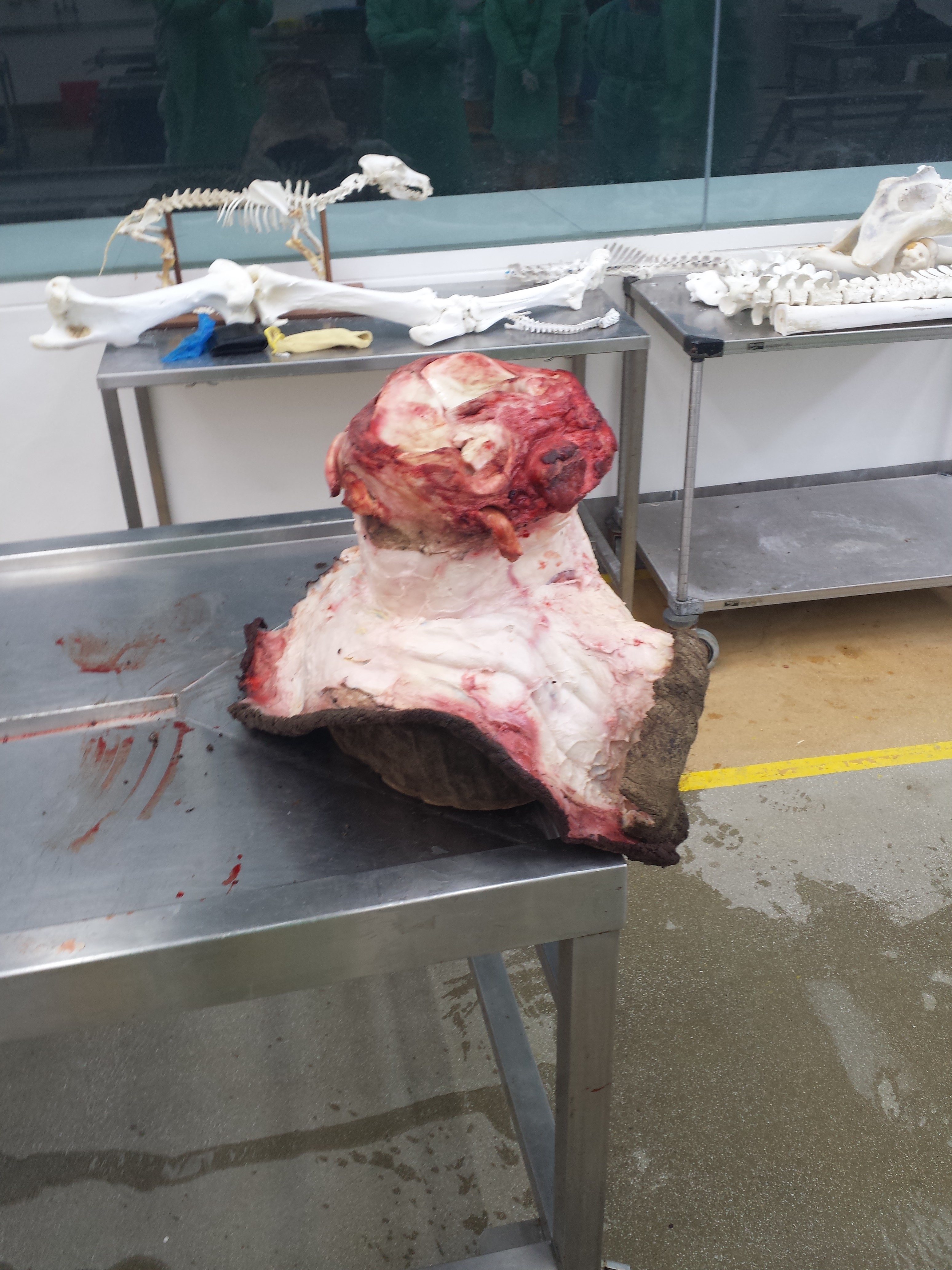
There are two feet in this post, both front right feet (manus is the technical term; singular and plural). The first one is the messier (unhealthy and bloodier, less fresh and clean) one, from the show/video. It is an Asian elephant (Elephas maximus). I kick off with photos I took after the filming, so the foot is already deconstructed:
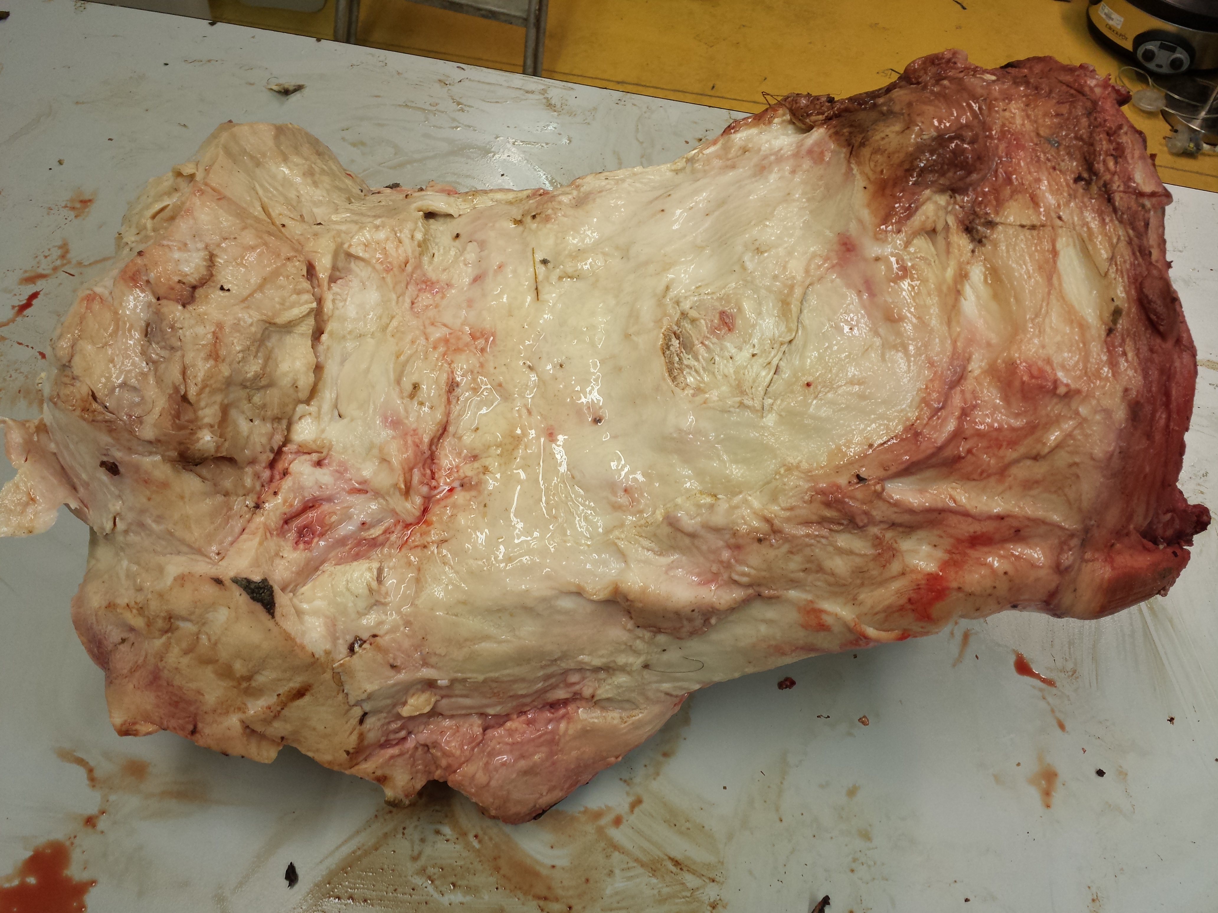
Skinned foot, oblique front/inside view. The wrist is on the right side of the photo; the toes on the left.

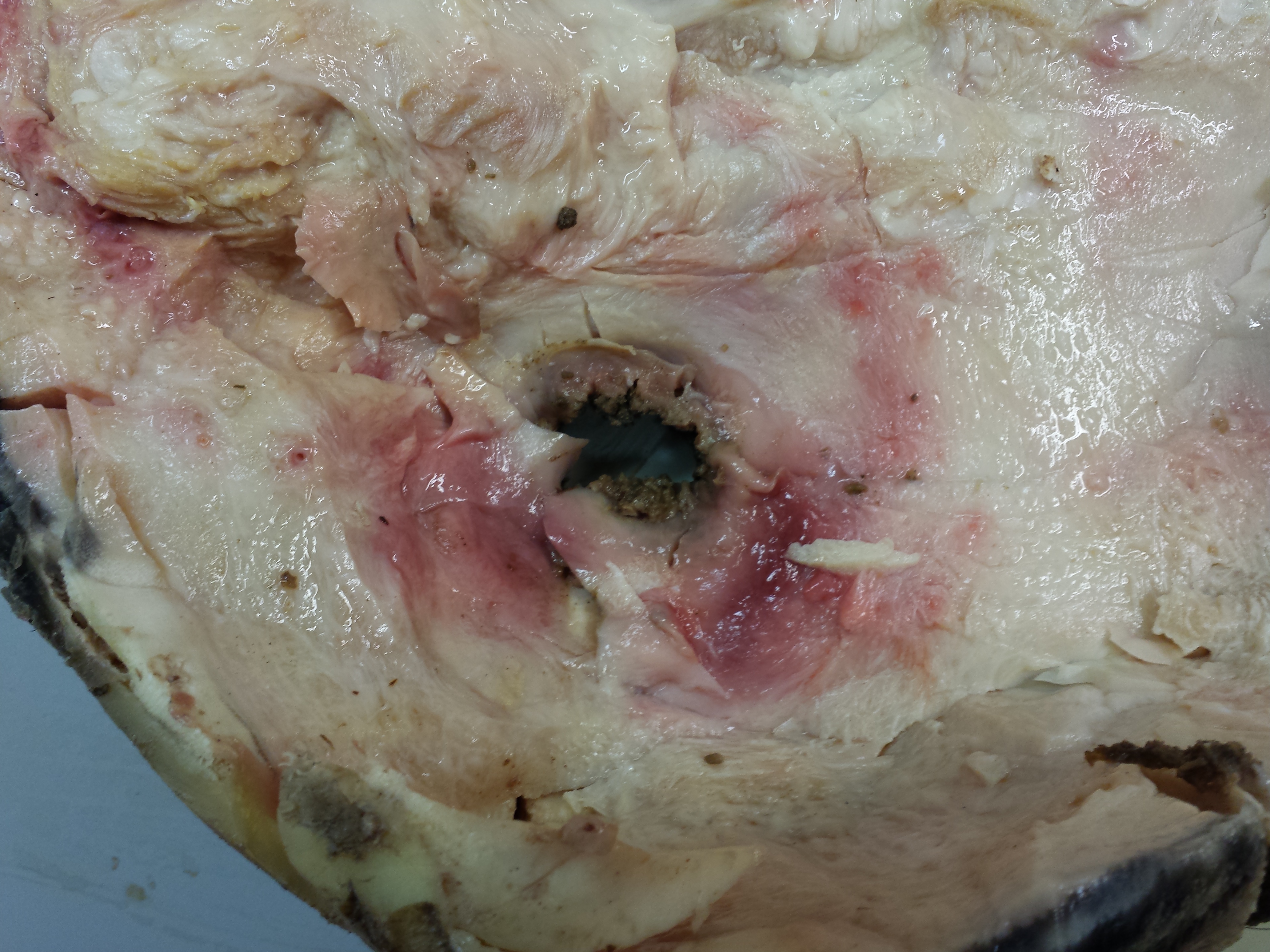
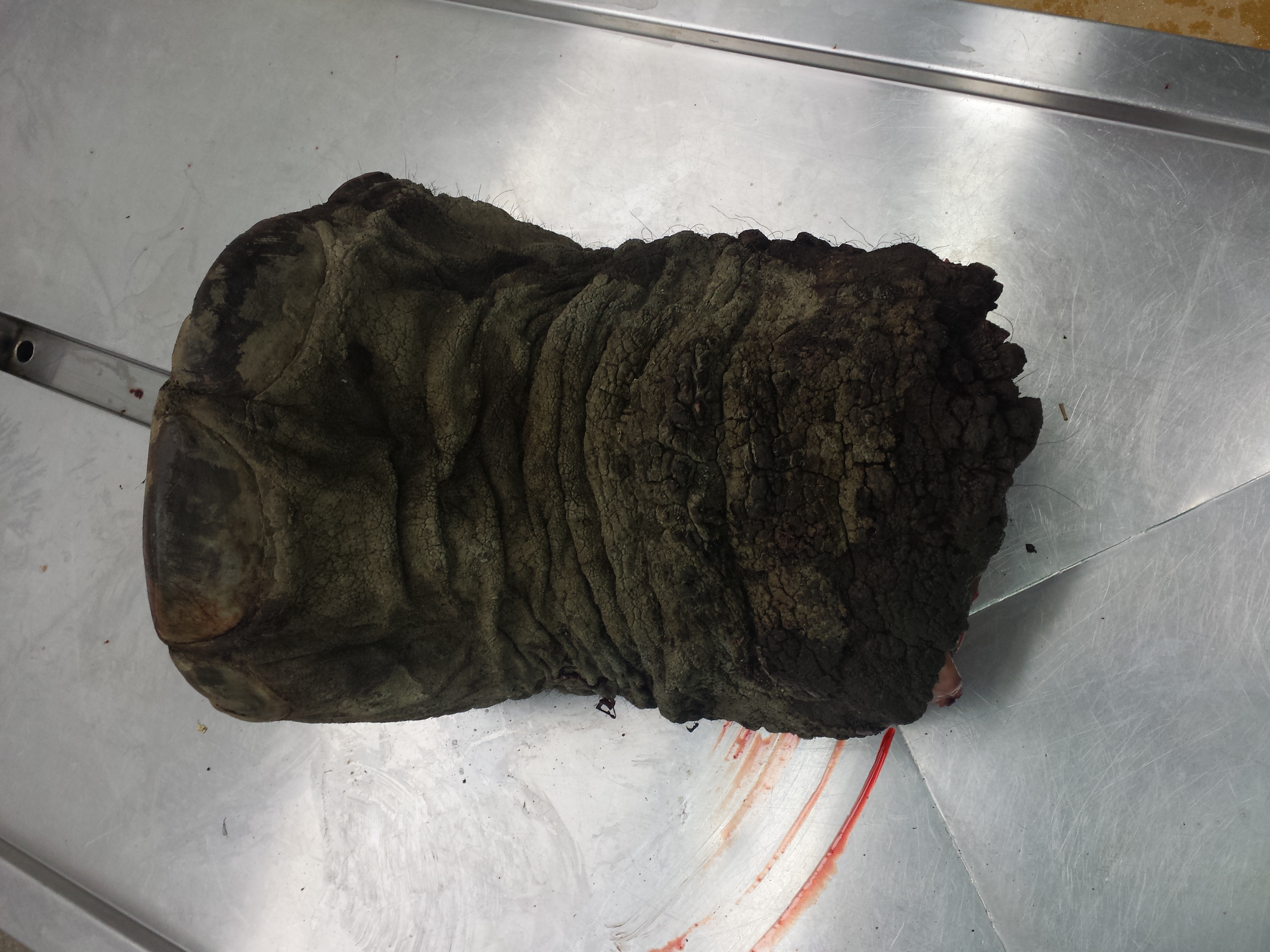
Sole (“slipper”), with a hole on the fourth toe showing where the abscess is that let infection in/pus drain out. The slipper here is upside-down.

Top-down view of the sole of the foot, once the slipper is removed; flipped over and rotated 90 degrees clockwise from the above photo. Some of the fat pad of the foot is on the right side of the image; it’s very hard to separate from the keratinous sole of the foot.
Looking down into the fourth toe’s (ring finger) abscess on the other side of the above view.

Looking down into the second toe (index finger), same view as above. Some redness and greyness where this toe had some of its own pathological issues like infection and a smaller abscess.

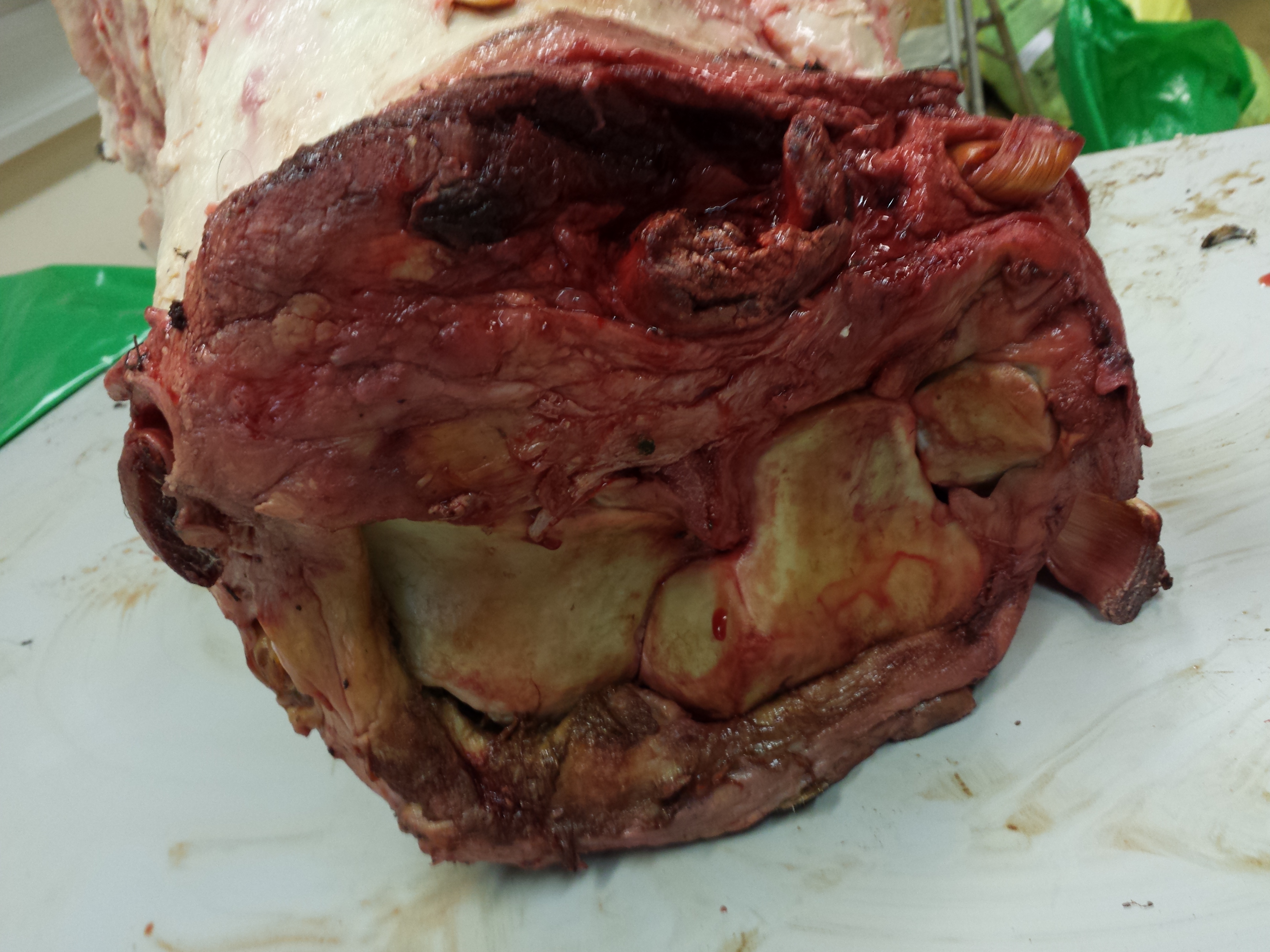
Looking up from the slipper (removed) at the fat pad and toes of the foot, where they interface with the sole/slipper. The fat pad is toward the bottom and left side; the five toes are on the upper/right side (knobby subcircular regions on the perimeter of the foot). The very bad infection on the fourth toe is visible on the bottom right.
The sproingy fat pad is worth a video!
And one good wiggle deserves another!
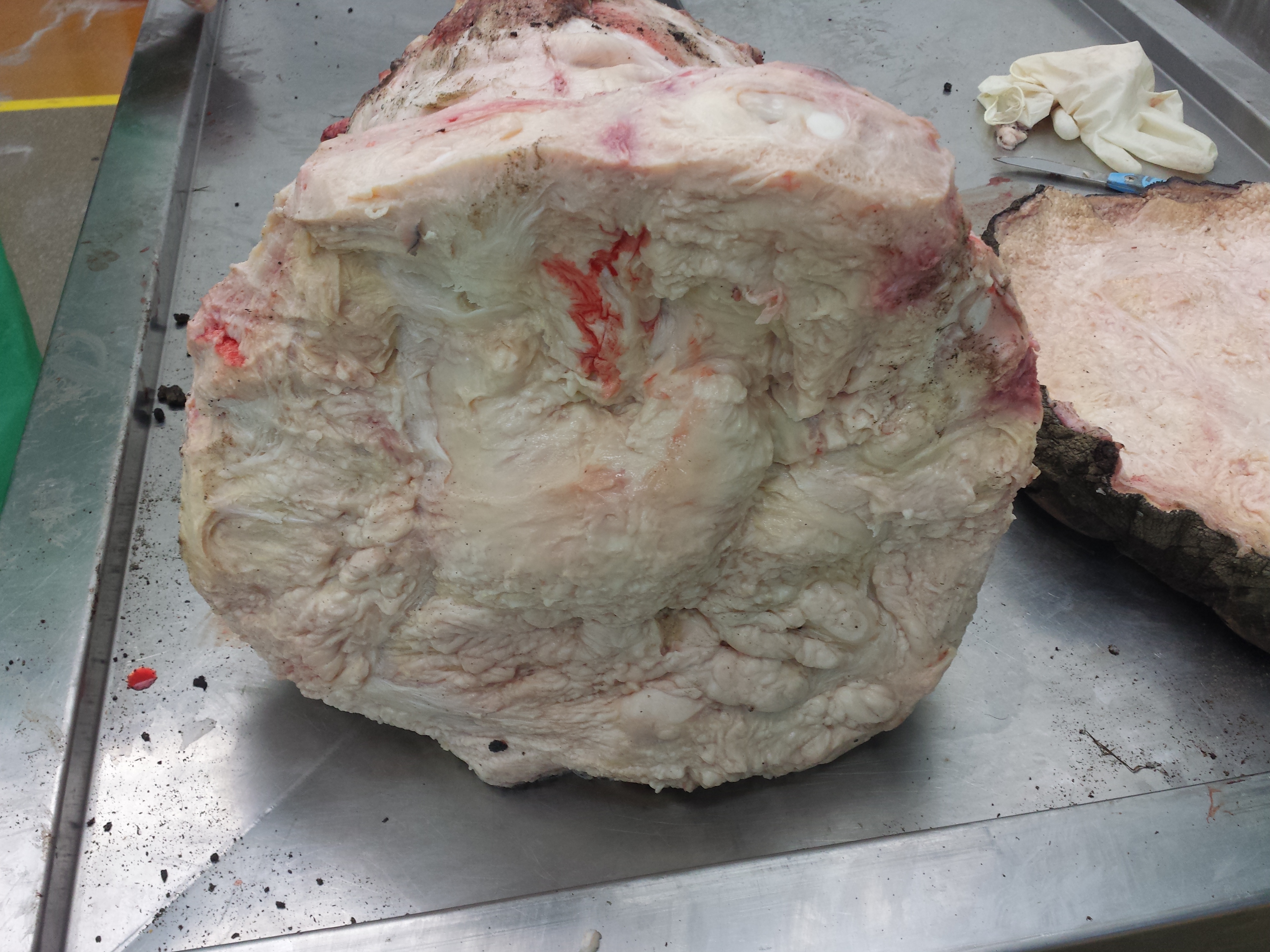
A view down onto the wrist joint. The carpal (wrist) bones are visible at the bottom of the image, whereas the flexor (palmar) tendons and muscles on the back of the “hand” are at the top. There is a LOT of musculotendinous tissue on the back side of an elephant’s foot. As you will see in my dissection of the second foot, further below!
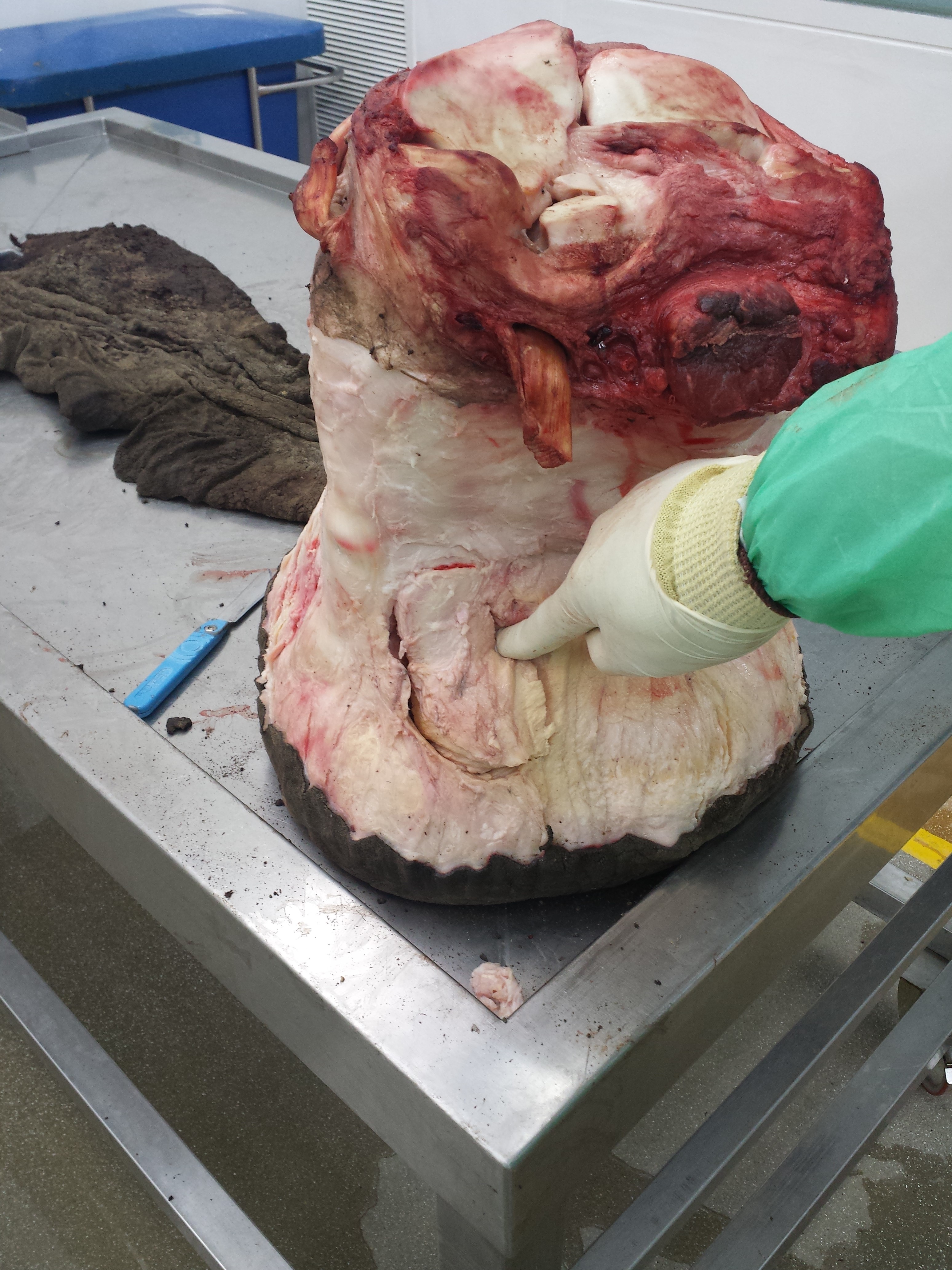
Looking down onto the medial (inner/”thumb”) border of the foot, where I’ve exposed the prepollex, or false “sixth finger”, by removing the first metacarpal (knuckle) bone.

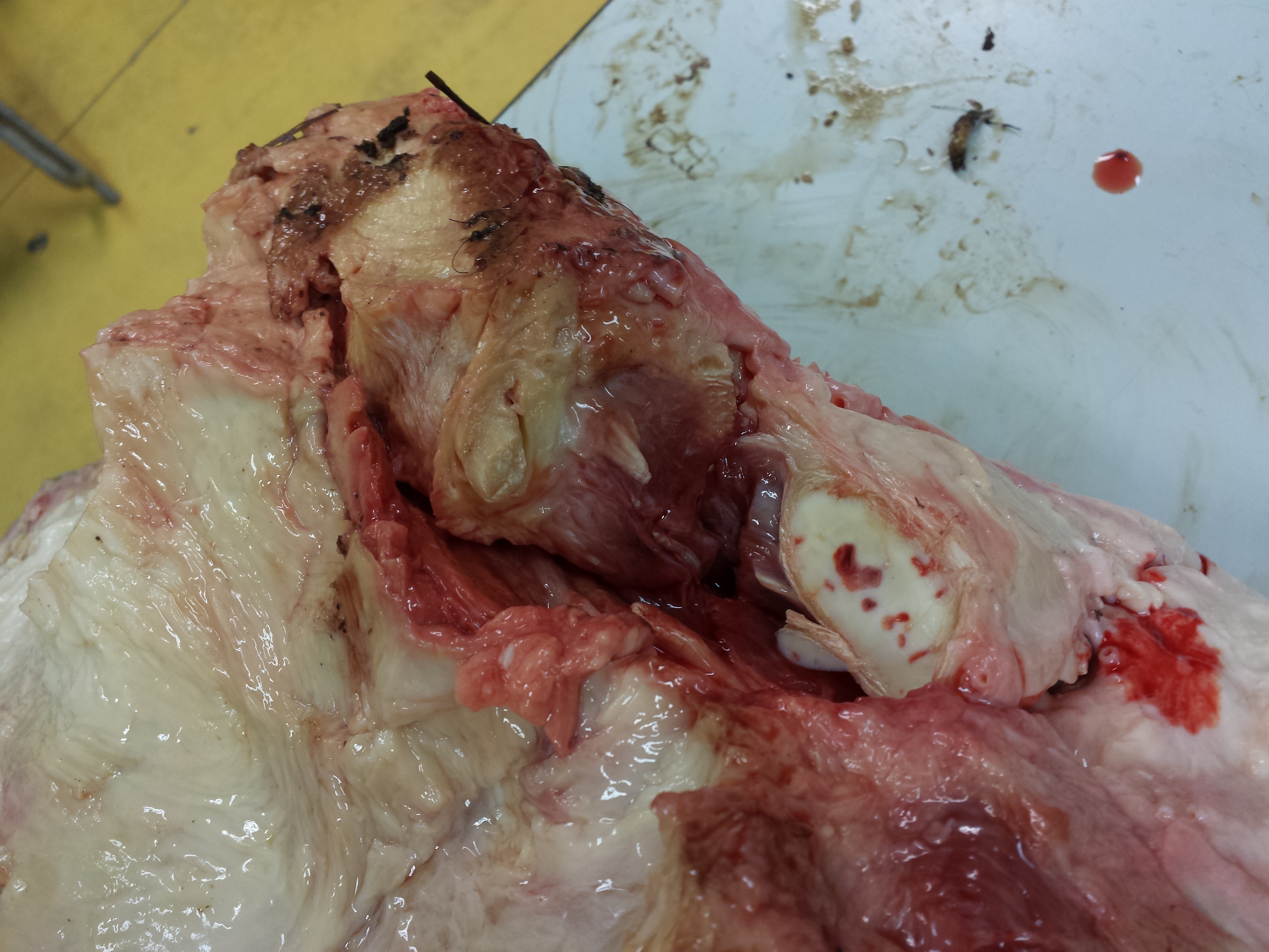
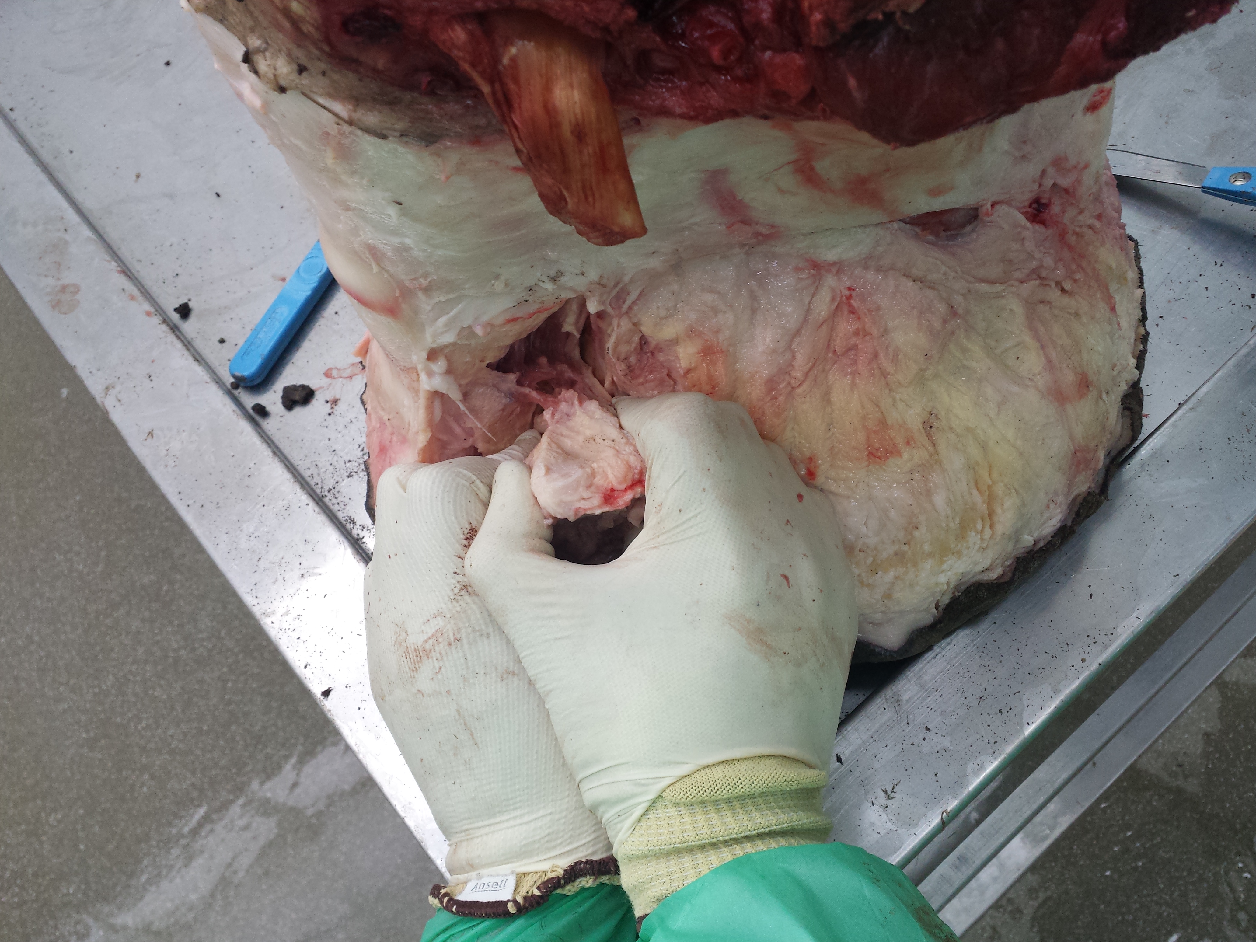
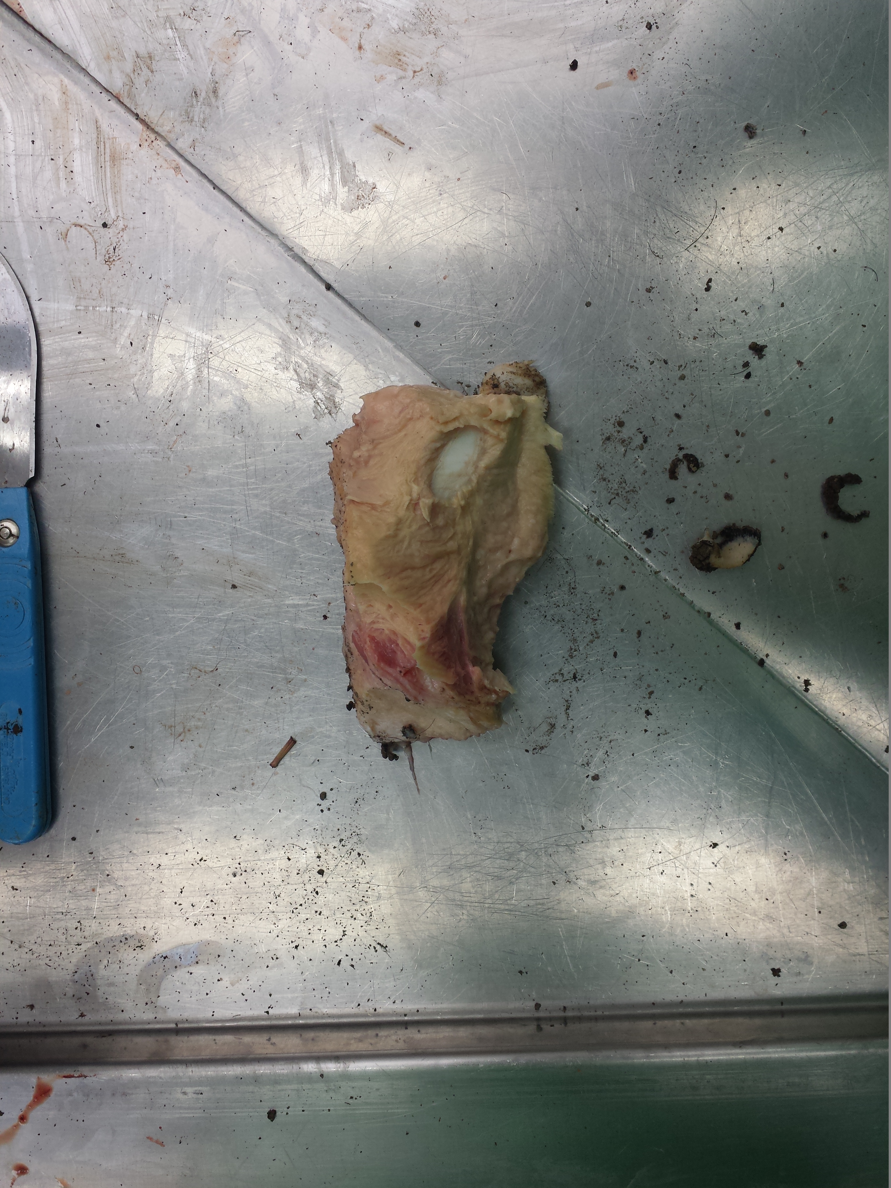
I’ve removed the prepollex from the foot. The white oval structure (bottom right) is the top of the conical prepollex, where it connected to the rest of the foot. White is cartilage, whereas the red “islands” are blood vessels that have invaded the cartilage and are starting to turn it into patches of bone. So this prepollex is at a very early stage of bone formation, still almost entirely cartilaginous, whereas some older elephants have the prepollex largely formed of bone. The fleshy pink tissue adhering to the surface of the prepollex here is a remnant of “abductor” muscle that connects it to the thumb and thus could allow some active control of the prepollex’s mobility.
Well, that was one very pathological elephant’s foot; one of the worst I have ever seen. Every foot I dissect is different and tells me a unique story about that animal’s development, history and health. This one told a very sad tale. What does a somewhat normal elephant’s foot look like? I thawed one out for comparison, and to thin out my overstuffed freezer stock. This one starts off from an intact (if severed) foot so you can witness the stages of dissection:
Whole foot. African elephant (Loxodonta africana). You may spot in later photos that the second and fourth toes’ nails are cracked longitudinally. This happens sometimes in elephants without any obvious health problems such as infection, but if it lasts long enough and conditions are bad enough (e.g. unsanitary conditions getting bacteria into the crack; spreading the crack to let them into the foot tissue), it could worsen.

Nice clean sole. No abscesses or other problems. You can faintly see the cracked toenails here.

Gorgeous white cartilage surfaces of the wrist joints. Nice and healthy-looking. A young animal, in this case.
Removing the skin; nice soft whitish connective tissue underneath.

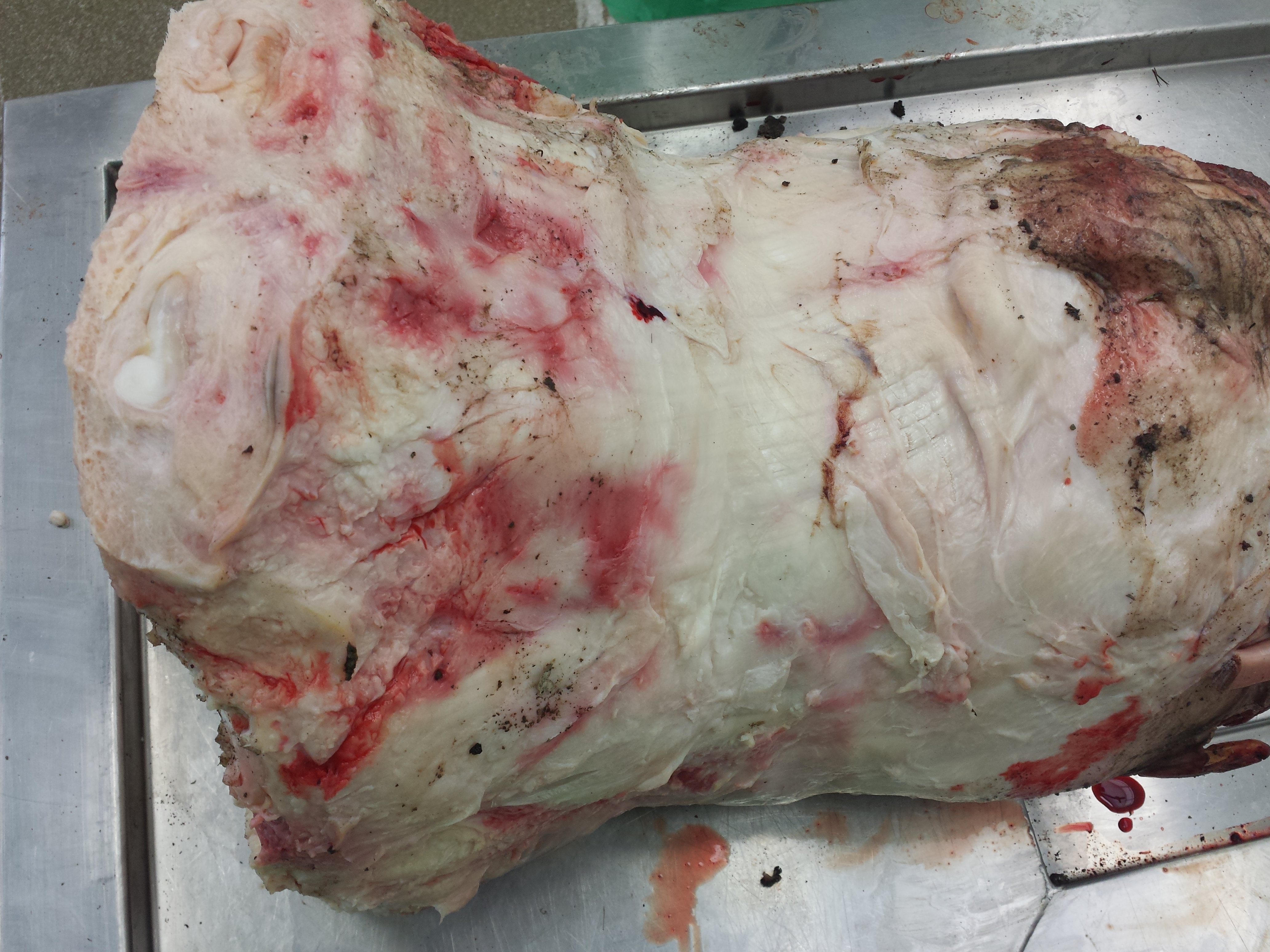
Skinned foot; rear view. The yellowish fat pad is wonderfully visible through the connective tissue sheath.

Skinned foot; front view. The thin, broad extensor tendons that would draw the fingers forward in life are visible here as longitudinal lines along the foot’s surface, running to the toes.
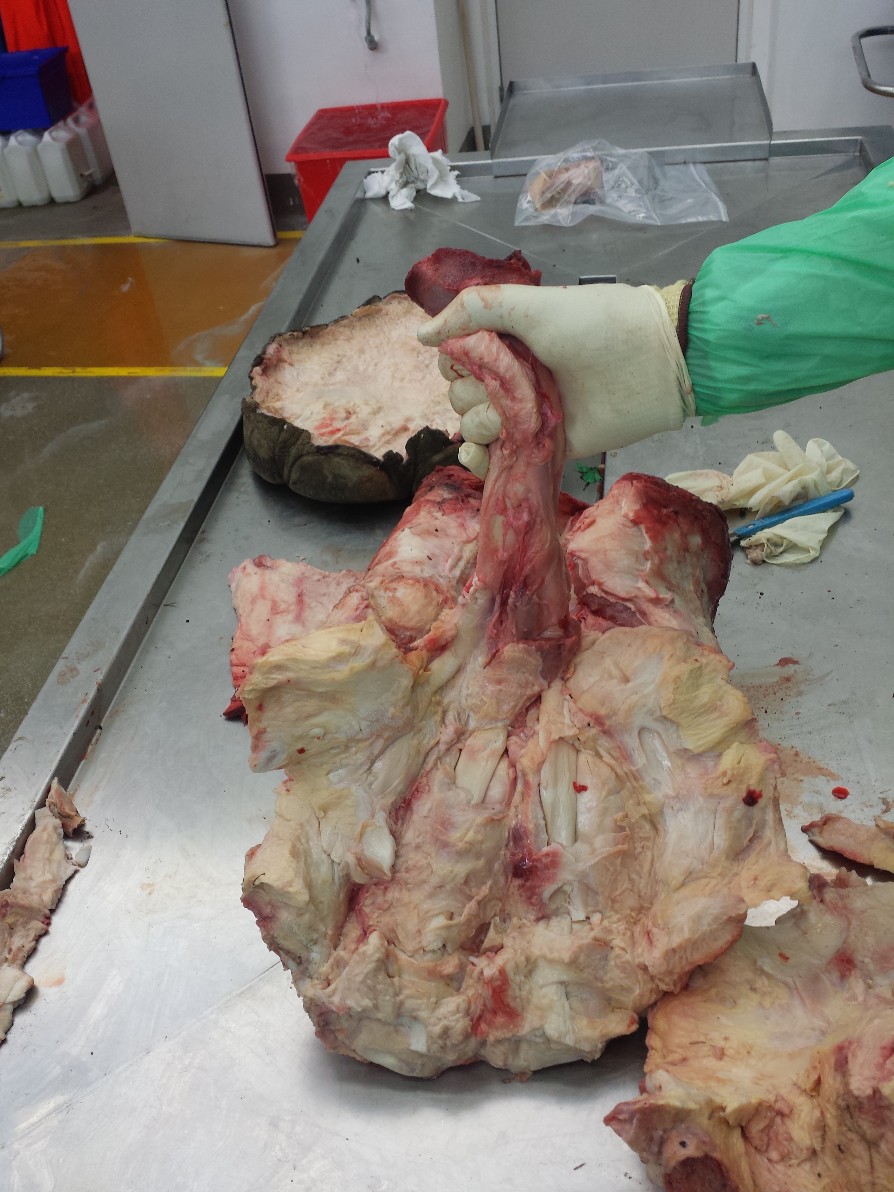
Ahh, my favourite thing! I’ve cut around the prepollex and am pointing at it. It’s almost impossible otherwise to see through all the fatty tissue of the fat pad that surrounds it.
Removing the prepollex. It’s tiny and enmeshed in connective tissue; harder to see than in the first elephant (photos above).
There is the prepollex! Maybe 12cm long. A little bit of cartilage (white) visible where it connected to the foot. These “sesamoid bones” vary tremendously in elephants I’ve inspected. I am still getting my head around that, after >10 years of staring at them in >75 feet!

Gap left by removal of the prepollex, on the median border of the foot; thumb region. Imagine having a little extra thumb growing off the base of your thumb and sticking toward your palm. That’s what elephants have.

Here, removing the slipper/sole of the foot, from the back side forwards. Hard work!

The slipper. Compare with the image above (same orientation). Nothing wrong here that I could see.

Front view of the toes, where they connect to the toenails. This specimen was so fresh that they were surprisingly easy to cut through and remove the foot from the sole.
Looking up at the palm. You can see the bulbous fat pad (yellower tissue) bulging out in the centre of the palm, and segments of it extending between each finger, separated by fibrous tracts. I love this anatomy. I can stare at it for hours and still be fascinated after all these years. So complex!

Looking down onto the inside of the toenails, toes 3 and 4. Healthy, relatively intact tissue; no swelling or bleeding or other pathology.
Skinned foot, oblique front/inside view again, as above.

Fat pad removed, looking up through where it was at the palm of the “hands”, where the tendons and ligaments connect to the five toes. Each arc-like structure is a toe; the “thumb” (first toe) is on the upper left.

Elephant’s-eye-view looking down onto the fat pad, where the palm of the foot in the image below would be placed in life (i.e. the limb would be coming down vertically, perpendicular to the plane of the image). The fat pad of the foot is visibly thicker toward the back of the foot (bottom of the image), as you’d expect, because the toes occupy most of the front parts.
Palmar tendons and muscles; the common digital extensor muscle group, which clenches the toes. Not a small muscle, either!

Tendons of the digital flexor muscle exposed.

I removed the digital flexor muscle so the three major tendons can be seen (the two short side branches to the first and fifth toes have been cut off).
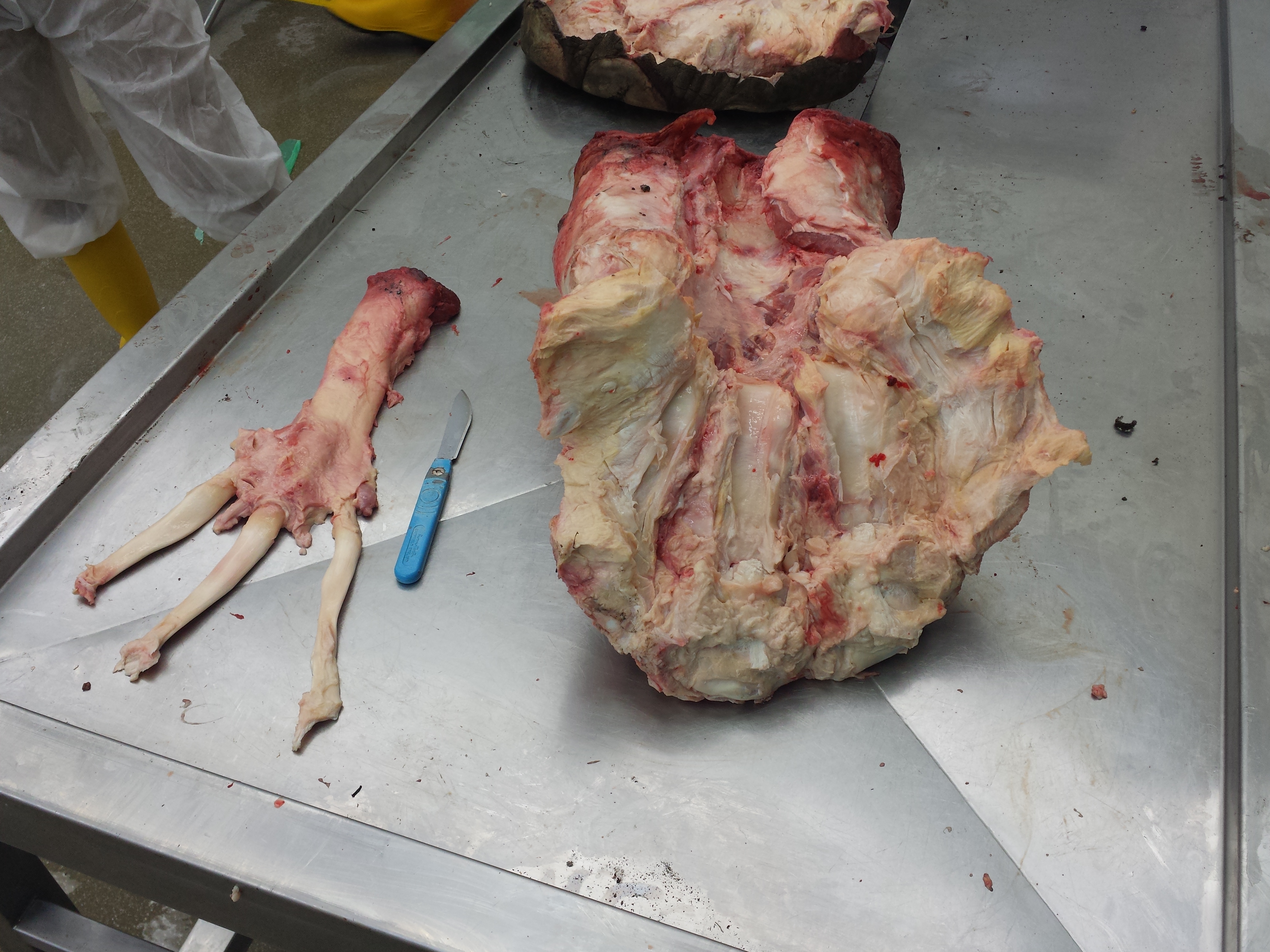
Forefoot with flexor tendons removed, revealing the channels that they coursed through.

Closeup of the glistening channels for the flexor tendons. They are lined with lubricative tissue to help the tendons glide through them. And the tendons do need to be able to glide- although elephant feet look very solid from the outside, and are to an extent, but we’ve done studies showing that they do move if you apply even a moderate load to them in a cadaver, and thus would move in life, too.
Let’s finish off with some osteology, shall we? First the unhealthy Asian elephant, then the healthy African elephant; same front right feet, just the bones (from my CT scans):
Ouch, indeed!
Much better. And that’s the end!
Wow, that was an elephantine post! I wanted to take yet another opportunity to share the amazing anatomy of elephant feet with you. You’re all now qualified experts if you made it this far!
Any questions?


So cool – seeing how everything fits together inside the foot is fascinating. Really interesting seeing the 3D bone models too, those pathological toe bones are wince-inducing
Fascinating. The digital flexor and extensor muscle and tendon dissections are particularly beautiful.
Thanks Anna! I agree, there is deep beauty in this anatomy.