Feeds:
Posts
Comments

Archive for the ‘Conference’ Category

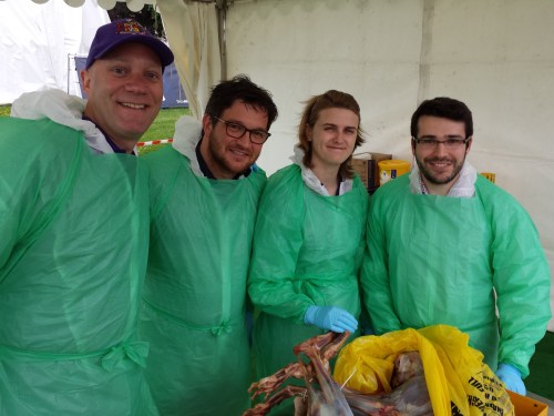
I had the privilege and pleasure of serving for the past 2 years as Chair of the Division of Vertebrate Morphology at the Society for Integrative and Comparative Biology, and that service just ended. So I had the showerthought to briefly post about the broader messages from that experience, with the hope that other scientists might benefit. But first, a little backstory.

Stomach-Churning Rating: 0/10

I’d only done some minor service before this, in scientific societies. At the time I ran for Chair-Elect 5 years ago, I felt it was time to try something new; to give back to science, as one should do. And so I did. It was a challenging and thus rewarding experience of learning the ropes- the Chair position is fairly open-ended to allow one to contribute what new things and leadership one envisions and can manage. In a short 2 years I felt I gained just enough momentum that I could have run the role more smoothly if I’d had a 3rd year, but that’s hindsight. The details don’t matter here but they lead to the messages of this post.

wink-wink musical interlude 1

First, the simple message that service, like the Holy Ghost, is the oft-forgotten third component of the trinity of professional science/academia; teaching and research being the other two (and science communication, to me, bridging all of these). As one moves along in one’s career, service tends to become increasingly expected—and the wisdom accumulated aids its conduct.

Second, service should be done because:

  • It’s the right thing to do
  • You learn things about your professional society, discipline, colleagues, leadership and self (skills and limits)
  • It’s not necessarily just boring bureaucracy (more about that below)
  • It will aid your career (CV, promotion, connections, future service, etc.) and you can aid others along the way

I think a common misconception is that service is boring. Yes, hearing the minutes of prior meetings read to you, or a long screed about minutia of health-and-safety, can be boring at times. But pay attention and find things that interest you and new vistas can open. This depends on the position one serves in and how it fits you. In my case, I found it a fun challenge to run meetings (i.e. try to follow the standard protocol of devising an agenda, checking minutes, etc.; standard bureaucracy) – especially the key activity of raising and voting on issues to consider taking the division in new directions. That allowed some creativity and made for energetic discussions on issues that mattered.

Another contrast to “boring” is resolving crises that arise (in my case, quite a few arose that felt serious to me). Yes, they’re stressful, but they also teach you things about how to handle crises, and you learn about your own ability to do so; and how others interface with that dynamic process. As I tend to emphasize on this blog, doing science is HUGELY about human interactions and foibles, and in service, as everywhere, such things are especially prominent and complex.

wink-wink musical interlude 2

Service comes in many forms. Students and postdocs can and should take part—many societies such as SICB tend to allow or encourage early career researcher participation. At a minimum, scientists should vote on elections (participation tends to be low among student/postdoc members) and attend their societies’ business or other meetings to see how the machine of a professional society works inside. It may even learn to serendipitous outcomes! And lessons learned will serve you well in many forms of future career.

One can do many other forms of service. Minimally in research, one is expected to participate in peer review; and that experience can lead to editor roles at journals, which I’ve found very interesting. Certainly in academic and other departments, there are numerous committee and other roles analogous to those in professional societies that are opportunities to serve.

I’ve surely left out other important lessons learned from serving. I’m still processing my experiences, reflecting and thinking forward. Now I’ve moved on to new service at SICB as Chair of the Student-Postdoc Affairs Committee, so there’s lots more for me to learn and share in that role.

What have you learned from serving? Do you have questions about service in science? Please chime in below.

More links of interest:

DVM-DCB Twitter feed

DVM Facebook page (members+affiliates)

SPDAC Facebook page (anyone!)

SPDAC Twitter feed

Read Full Post »

Who needs “Ice Road Truckers” when you have the “John’s Freezer” team on the road with fossils, amphibians, felids and 3D phenotype fun? No one, that’s who. We’re rocking the Cheltenham Science Festival for our first time (as a group), and pulling out all the stops by presenting two events! Here’s the skinny on them, with updates as the week proceeds.

Stomach-Churning Rating: 2/10 for now (just bones), but it could change once the cheetah dissection is under way… 8/10 bloody cheetah bits but only at the end (updated)

Right now, Lauren Sumner-Rooney (of “Anatomy To You” and other fame) is on-site with a rotating team of others from our lab, in the “Free Activity Tents” area of the Imperial Gardens/Square, inside a marquee where we’ll be showing off our NERC-funded tetrapod research all week. This “First Steps” event features not only our past and present work with Jenny Clack, Stephanie Pierce, Julia Molnar and others on Ichthyostega & its “fishapod” mates, but also our “scampering salamanders” research in Spain, Germany and England. I’ve blogged a lot about all that, and won’t repeat it here, but you can see a 3D-printed Ichthyostega skeleton, view the skeleton in a virtual reality 3D environment, see related specimens and engage in kid-friendly activities, and talk to our team about this and other related research.

Ichthyostega 3D printed backbone is born!

Ichthyostega 3D printed backbone is born!

The central themes of that event are how bone structure relates to function and how we can use such information, along with experimental measurements and computer models of real salamanders, to reconstruct how extinct animals might have behaved as well as how swimming animals became walking ones. How did fins transform into limbs and what did that mean for how vertebrates made the evolutionary transition onto land? If you know my team’s work, that encapsulates our general approach to many other problems in evolutionary biomechanics (e.g. how did avian bipedalism evolve?). Added benefits are that you too can explore this theme in a hands-on way, and you can talk with us about it in person. That continues all week (i.e. until Saturday evening); I’ll be around from Thursday afternoon onwards, too. Kids of all ages are welcome!

Ichthyostega 3D print taking shape!

Ichthyostega 3D print taking shape!

Then, on Saturday for our second free event we join forces with Ben Garrod (master of primate evolution, the secrets of bones, and “Attenborough and the Giant Dinosaur”) and RVC’s forensic pathologist Alexander Stoll as well as Sophie Regnault (“sesamoid street” PhD student w/me). As the “Large Animal Dissection” title hints, it’s not the right kind of gig to bring small kids to. There will be blood and stuff— we’ll be dissecting a cheetah together from 10am-4pm. This will involve walking through all the major organ systems, giving evolutionary anecdotes, and plenty more, with an aim to understand how the magnificent adaptations of cheetahs evolved—but also to investigate what problem(s) this animal faced that led to its sad demise. By the day’s end, there will just be a skeleton left. Get a front row seat early for this event, which serendipitously ties into “Team Cat”’s Leverhulme Trust-funded research project (we wanted a big animal and it just happened to be a cheetah; I had hoped for a giant croc or a shark or something but can’t complain!).

Ichthyostega 3D print is ready!

Ichthyostega 3D print is ready!

If you miss these events, please do cry bitter tears of regret. But don’t despair, there will be another “big cat dissection” in the London area in ~November (watch here for details), and plenty more fossil tetrapod stuff to come, plus a LOT more dinosaurs on the horizon!

Guess the bones! (photo by Zoe Self)

Guess the bones! (photo by Zoe Self)

And please come back to this blog post for more pics and stories as the week carries on… For hashtag afficionados, you can follow the fun on Twitter etc. at #firststepsCSF16. What a world we live in!

Update 1: While you’re here, check out our Youtube playlists of tetrapod-related videos:

Lobe-finned fishes

Ichthyostega‘s awesome anatomy

Tetrapod evolution: Tiktaalik to salamanders!

Update 2: Photos of our main stand (about tetrapod evolution)

csf2016-display

Our poster/banner display looks nice.

20160609_143548

Our tent brings in some punters.

20160609_151508

Our bones excite people young and old, sighted and blind.

20160610_090009

Fun with stickers and lab t-shirts.

And…

Update 3: Cheetah meat & greet
Ben, Alex, Sophie and I tackled the cheetah dissection today and it went GREAT! Much better than I’d optimistically expected. Rain didn’t scare the crowds off and neither did the gore, which there was some of (gelatinous spinal cords, lumpy tumors and at least one flying tiny bit of cheetah flesh that landed on a good-natured audience member!). Photos will tell the tale:

20160611_091345

Peek-a-boo!

20160611_093828

Sophie and Alex help us get set up in our tent.

20160611_094554

Our initial rough schedule- although we ended up improvising more after lunch.

20160611_095500

Dissectors assemble!

20160611_105126

The beast revealed. It was skinned by the museum that loaned it to us.

20160611_110748

Alex showing his talent: removing the viscera in one piece from end to end, starting with the tongue.

20160611_153148

Impressive finding of a surgical fixture (plate and wires) on the tibia, which had been used to hold the shattered bone back together long enough for it to heal. Added to the kidney disease and liver-spleen-lung cancer, this cheetah was in the sorriest shape of any cadaver I’ve seen yet.

20160611_153213

Cheetah coming to pieces: (from bottom) lumbar/pelvic region, hindlimb, thorax, forelimb and other bits.

20160611_155452

Dr Adam Rutherford, an eye expert, did a nice dissection of the cheetah’s eye, here showing the tapetum lucidum (reflective membrane), which shows up as light blue colour. Its small size befits the not-very-nocturnal habits of cheetahs.

20160611_155459

The lens of the cheetah’s eye. Now cloudy because of dehydration and crystalization, but still fascinating to see.

Want to see more images and the enthusiastic responses from the audience (we got some great feedback)? Check out Twitter’s #cheltscifest feed, or more simply my Storify condensation of the cheetah-related tweets here.

Read Full Post »

Hotel Fira Palace: view of the city

Hola from Barcelona, where 500ish of us are telling each other about the latest research in the field of morphology (like anatomy, but broader, deeper, more explanatory; but if you prefer to think of it as anatomy that’s OK by me)!

#ICVM and #ICVM2013 (favoured) are the hashtags, and http://icvm2013.com/ is the website, and there’s Facebook and all that too! You can read the full programme and abstracts here. It’s the best damn conference in the universe and I am not remotely biased. It happens every 3 years somewhere in the world and is always chock full of 5 days of glorious new information on animal form and function and much more, with just too many interesting talks to ever be able to take it all in.

I am speaking a few times and want to share a talk that is about sharing the glory of morphology in public.

Morphology research, that is; please put your clothing back on!

It’s a text-heavier talk than my rules-of-conference-talks normally would allow, but I’m going for it, as that makes it better for sharing because my dulcet tones will not accompany the version I am sharing online. Someday in the future, at a conference venue  that is better set up for reliably live-broadcasting a talk (this is NO FAULT of the excellent organizing committee of ICVM/ISVM!), I would just do it live, but not today, not here.

The point of the talk should be obvious from the first slide (as in my last post). But I’ll presage it by saying that another subtext, which might not come through so strongly in the slides as opposed to my spoken words, is that we need to tell people that we’re doing morphology/anatomy research! We should not be shy of that label because deans or geneticists or conventional wisdom or what/whomever might say (very, very wrongly!) that it is a dead or obsolete science.

While natural history, evolution, palaeontology and other fields allied to morphology do pretty well in the public eye, I don’t see people often reminded that what they are being told about in science communication is a NEW DISCOVERY IN ORGANISMAL MORPHOLOGY and that we are still discovering such new things about morphology all the freaking time! (e.g. my team’s research on elephant false sixth toes, or Nick Pyenson‘s team’s research on whale chin sense organs to name just 2 such studies, both published on the same day in Science!)

Indeed, many of those discoveries such as new fossils/exotic living things with cool features, cool developmental mechanisms that produced complex structures, or insights into how organisms are able to do amazing things are implicitly morphological discoveries, but the fanfare too often goes to natural history, palaeontology, evo-devo or some other area rather than explicitly to morphology.

In contrast, I too often hear people poo-pooing anatomical research as yesterday’s science.

Vesalius's classic skeleton, which is great but to me also conjures misleading connotations of anatomy as a  defunct discipline.

Vesalius’s classic skeleton (from Wikipedia), which is great but to me also conjures misleading connotations of anatomy as a defunct discipline that old dead dudes did.

We need to sell ourselves better not only in that regard, of a renaissance of discoveries and insights in our field, but also in the sense of being in a renaissance that is driven by TOTALLY AWESOME TECHNOLOGICAL AND METHODOLOGICAL ADVANCES, especially computerized tools. We’re just as fancy in terms of techy stuff as any other biologists, but we don’t shout it from the rooftops as much as other disciplines do.

We’re not just primitive scientists armed only with scalpels and maybe a ruler now and then, although that simple approach still has its sublime merits. We’re building finite element models, running dynamic computer simulations, taking high-resolution CT or synchotron scans, manipulating embryos, digging up fossils, sequencing genes– you name it, morphologists may be doing it! (For similar views see Marvalee Wake’s recent review of herpetology & morphology; I’m by far not the first person to make the arguments I’m making in my talk, but I am putting a personal spin on them)

And of course, as the talk is being delivered by me, you might rightly expect that I’ll say that we need to do more of this kind of cheerleading where we have maximal visibility and interaction, which includes online via social media, etc. I’ll discuss one other venue which has featured prominently here on this blog, too: documentaries. Oh I’m not done with that hobby horse, no sirree, not by a long shot!!

ICVM intro

Anyway I should get back to preparing my talk but here is the link to the slideshow (props to Anne Osterrieder for the inspiration to put my slides up here):

Please discuss anything related to this topic in the Comments– I’d love to hear what you think!

I am happy to clarify what my shorthand notes in the slide text mean if needed. There are links in the talk to other sites, which you can click and explore.

Read Full Post »