Feeds:
Posts
Comments

Archive for the ‘Geeking Out’ Category

If you’re in London, you still have almost one week left to hurry to the Valence House in Dagenham and see a great exhibit on Ray Harryhausen’s dinosaurs and other cool “Dynamation” stop-motion models and art!

This blog post is a photo tour of what I saw, in case you cannot go.

Like it? Click it. Bigger pic.

Stomach-Churning Rating: 1/10 nice stop-motion animation models. Medusa won’t hurt you here.

I loooooooooooove Ray Harryhausen’s work, ever since I was a child and saw “Jason and the Argonauts” and many other films, plus “Clash of the Titans” once it came into theatres. There is the attention to detail in anatomy and locomotion, and the wondrous fantastic nature of even the more mundane creatures he animated, and the rich mythology that he drew from to inspire his creations. Modern CGI is great in a different way, but nothing I can think of in recent special effects truly beats (1) the skeleton battle in ‘Jason, and (2) the Medusa encounter in ‘Clash (to name what might be my top two faves). And so when I learned that several of the original (restored) models from those films were on exhibit in northeastern London, I requested to go there with my family for Fathers Day. Results:

Boom! Ole’ stony-gazed, snaky-haired gorgon of yore.

No deadly bow here, but the rattlesnake tail is.

Medusa concept art by Harryhausen; the “bra” was there for American censors but Ray thought it looked wrong and removed it in the final version.

Look out, Jason! Here come the Children of the Hydra! Yep, original (restored) articulated models. Joints are visible. They look ready to kick some Iolcusian butt!

Context of the exhibit- local chap befriended Harryhausen and convinced him to let him restore his models; and so here we are. On with the dinosaurs! (and other palaeo-things)

Gwangi model made in resin; non-poseable but made around time of the “Valley of Gwangi” film to help design the poseable models.

Gwangi climactic scene in church; concept art by Harryhausen.

Other ‘Gwangi characters: “Eohippus” (Hyracotherium), Ornithomimus and boy.

Cowboy lassoing an Ornithomimus as per the movie scene in ‘Gwangi? Yes please. (Harryhausen original)  Jurassic Park had its T. rex lurching out of a forest to grab a Struthiomimus, intentionally mirroring the scene in ‘Gwangi where the titular AllosaurusTyrannosaurus hybrid chomps the Ornithomimus.

Poseable “Eohippus” original- with real fur! Great Dynamation too; very lifelike in the film.

Original Harryhausen concept art of the “Eohippus” show demo.

Suddenly, Ceratosaurus! (from “One Million Years BC”)

Styracosaurus original resin model. (from “One Million Years BC”)

Old school Polacanthus art by Alan Friswell. SPIKEY!

Old school Iguanodon art by Alan Friswell. MUSCLEY!

Panoply of archosaurs by Alan Friswell: pterodactyl, Tenontosaurus (made for the Frame Store special effects company in 2001) and tyrannosaur head (made at age 9).

Pterodactyl made at age 12, so don’t laugh.

Back to the fantastic beasts– original poseable hydra from ‘Jason!

Original Pegasus from ‘Clash! What a seamless blend of fur and feathers.

Original R2, I mean Bubo, from ‘Clash!

I forget the scene (the 1-eyed fates in ‘Clash?) but I like it. Original Harryhausen concept art.

Lunar leader from “First Men in the Moon.” (original)

Non-original (but based thereon) model by Alan Friswell, of nautiloid thingy from “Mysterious Island”.

Fiji mermaid by Alan Friswell.

“Hand of Glory” by Alan Friswell.

Pithecanthropus by Alan Friswell. Very Harryhausen in spirit.

Oddly, but somehow appropriately, there are ?350 year old whale bones on display in the hall next door, with a mysterious history.

WW2 bomb shelter in a “Victory Garden” outside the House. And the house is supposedly haunted. So take care when you visit…

What can I say? I loved it! Almost a religious experience; like seeing holy relics. Awesome in every sense of awesome.

Downside: you cannot grab the precious Dynamation models and play with them hands-on. I wanted to enact a furious Hydra-Gwangi battle. But alas, only in my imagination…

Read Full Post »

Who needs “Ice Road Truckers” when you have the “John’s Freezer” team on the road with fossils, amphibians, felids and 3D phenotype fun? No one, that’s who. We’re rocking the Cheltenham Science Festival for our first time (as a group), and pulling out all the stops by presenting two events! Here’s the skinny on them, with updates as the week proceeds.

Stomach-Churning Rating: 2/10 for now (just bones), but it could change once the cheetah dissection is under way… 8/10 bloody cheetah bits but only at the end (updated)

Right now, Lauren Sumner-Rooney (of “Anatomy To You” and other fame) is on-site with a rotating team of others from our lab, in the “Free Activity Tents” area of the Imperial Gardens/Square, inside a marquee where we’ll be showing off our NERC-funded tetrapod research all week. This “First Steps” event features not only our past and present work with Jenny Clack, Stephanie Pierce, Julia Molnar and others on Ichthyostega & its “fishapod” mates, but also our “scampering salamanders” research in Spain, Germany and England. I’ve blogged a lot about all that, and won’t repeat it here, but you can see a 3D-printed Ichthyostega skeleton, view the skeleton in a virtual reality 3D environment, see related specimens and engage in kid-friendly activities, and talk to our team about this and other related research.

Ichthyostega 3D printed backbone is born!

Ichthyostega 3D printed backbone is born!

The central themes of that event are how bone structure relates to function and how we can use such information, along with experimental measurements and computer models of real salamanders, to reconstruct how extinct animals might have behaved as well as how swimming animals became walking ones. How did fins transform into limbs and what did that mean for how vertebrates made the evolutionary transition onto land? If you know my team’s work, that encapsulates our general approach to many other problems in evolutionary biomechanics (e.g. how did avian bipedalism evolve?). Added benefits are that you too can explore this theme in a hands-on way, and you can talk with us about it in person. That continues all week (i.e. until Saturday evening); I’ll be around from Thursday afternoon onwards, too. Kids of all ages are welcome!

Ichthyostega 3D print taking shape!

Ichthyostega 3D print taking shape!

Then, on Saturday for our second free event we join forces with Ben Garrod (master of primate evolution, the secrets of bones, and “Attenborough and the Giant Dinosaur”) and RVC’s forensic pathologist Alexander Stoll as well as Sophie Regnault (“sesamoid street” PhD student w/me). As the “Large Animal Dissection” title hints, it’s not the right kind of gig to bring small kids to. There will be blood and stuff— we’ll be dissecting a cheetah together from 10am-4pm. This will involve walking through all the major organ systems, giving evolutionary anecdotes, and plenty more, with an aim to understand how the magnificent adaptations of cheetahs evolved—but also to investigate what problem(s) this animal faced that led to its sad demise. By the day’s end, there will just be a skeleton left. Get a front row seat early for this event, which serendipitously ties into “Team Cat”’s Leverhulme Trust-funded research project (we wanted a big animal and it just happened to be a cheetah; I had hoped for a giant croc or a shark or something but can’t complain!).

Ichthyostega 3D print is ready!

Ichthyostega 3D print is ready!

If you miss these events, please do cry bitter tears of regret. But don’t despair, there will be another “big cat dissection” in the London area in ~November (watch here for details), and plenty more fossil tetrapod stuff to come, plus a LOT more dinosaurs on the horizon!

Guess the bones! (photo by Zoe Self)

Guess the bones! (photo by Zoe Self)

And please come back to this blog post for more pics and stories as the week carries on… For hashtag afficionados, you can follow the fun on Twitter etc. at #firststepsCSF16. What a world we live in!

Update 1: While you’re here, check out our Youtube playlists of tetrapod-related videos:

Lobe-finned fishes

Ichthyostega‘s awesome anatomy

Tetrapod evolution: Tiktaalik to salamanders!

Update 2: Photos of our main stand (about tetrapod evolution)

csf2016-display

Our poster/banner display looks nice.

20160609_143548

Our tent brings in some punters.

20160609_151508

Our bones excite people young and old, sighted and blind.

20160610_090009

Fun with stickers and lab t-shirts.

And…

Update 3: Cheetah meat & greet
Ben, Alex, Sophie and I tackled the cheetah dissection today and it went GREAT! Much better than I’d optimistically expected. Rain didn’t scare the crowds off and neither did the gore, which there was some of (gelatinous spinal cords, lumpy tumors and at least one flying tiny bit of cheetah flesh that landed on a good-natured audience member!). Photos will tell the tale:

20160611_091345

Peek-a-boo!

20160611_093828

Sophie and Alex help us get set up in our tent.

20160611_094554

Our initial rough schedule- although we ended up improvising more after lunch.

20160611_095500

Dissectors assemble!

20160611_105126

The beast revealed. It was skinned by the museum that loaned it to us.

20160611_110748

Alex showing his talent: removing the viscera in one piece from end to end, starting with the tongue.

20160611_153148

Impressive finding of a surgical fixture (plate and wires) on the tibia, which had been used to hold the shattered bone back together long enough for it to heal. Added to the kidney disease and liver-spleen-lung cancer, this cheetah was in the sorriest shape of any cadaver I’ve seen yet.

20160611_153213

Cheetah coming to pieces: (from bottom) lumbar/pelvic region, hindlimb, thorax, forelimb and other bits.

20160611_155452

Dr Adam Rutherford, an eye expert, did a nice dissection of the cheetah’s eye, here showing the tapetum lucidum (reflective membrane), which shows up as light blue colour. Its small size befits the not-very-nocturnal habits of cheetahs.

20160611_155459

The lens of the cheetah’s eye. Now cloudy because of dehydration and crystalization, but still fascinating to see.

Want to see more images and the enthusiastic responses from the audience (we got some great feedback)? Check out Twitter’s #cheltscifest feed, or more simply my Storify condensation of the cheetah-related tweets here.

Read Full Post »

Well, that was an eventful week for me, although today’s post will focus on one event: the debut of the film Jurassic World. Briefly though, the awesome “T. rex Autopsy” documentary debuted (I was going to post more about it but all I’d have left to say is that I was very pleased with the result), I also showed up briefly in “Top 10 Biggest Beasts Ever” talking about the giant rhinocerotoid Paraceratherium and the stresses on its feet, our paper on ostrich musculoskeletal modelling was published (more in a future post) after ~12 years of me diddling around with it, and much more happened. Then to cap it all off, very shortly after I hit the “publish” button on my last post, I had four tonic clonic seizures in a row and spent a hazy night in the hospital, then the past week recovering from the damage. Nothing like another near-death (no exaggeration there, I’m afraid) experience to cap off an exciting week. But strangely, what I feel more interested in talking about is, like I said, Jurassic World, but this is not a review, as you will see here.

Stomach-Churning Rating: 0/10; just SPOILERS if you haven’t seen the film yet!

I guess I have to give a brief review of the film and say that I was entertained, to a degree, but it was not a great piece of film-making. It was a far cry from the original but then so were the sequels, and maybe it was better than them. The mosa-star was the most novel, memorable bit. I didn’t care for the Indominus villain, but then when you bring genetic engineering into a film like this, you’ve basically thrown out the rulebook and can make your dinosaurs as magical as you want; we’re already in “X-Men” territory here and almost in “Pacific Rim”-land.

Chris Pratt has signed on for at least another sequel to Jurassic World and the ending of the film already started that ball rolling. So I find it fun to speculate wildly, and certainly incorrectly, on what the sequel might do. What does the Jurassic future have in store?

First of all, who survived to re-appear in the next film? We’re left with the Bryce Dallas Howard character, who probably will return with Pratt to further develop their rather uninteresting social/romantic dynamic, rather than start afresh with someone else. The kids of course survived, as always, and as always they won’t return, as that’s not interesting and they didn’t have much to do except scream and (highly implausibly) hotwire an old car. Much as I’d like the parents from my hometown of Madison, WI to return, they won’t either for the same reasons. But we really only need Pratt and his high-heeled sweetie for the next film. Everyone else memorable(?) seems to have died, although it would be wonderful to bring Goldblum back for some smarmy wit (please!).

Second of all, the next film can’t be set in Jurassic World. There’s not much left to do there (JW already spent much of its time hearkening back to JP), and there’s no way the park would re-open. We need something new. I think by now we’re (very) tired of characters running around islands full of dinosaurs and the Blackfish parallel was milked dry in the latest movie. We need to spend a film with the dinosaurs amongst humanity (as Lost World briefly did), and much as I’d love to see the crazy drug lord/kidnapping plot happen, it won’t. But JW did set one thing up that has to happen now in its sequel: the paramilitary role of engineered, trained dinosaurs. We now know they can sort of train their dinosaurs and they can forge them to be anything they want to in terms of geno/phenotypes. They’ll learn from some mistakes of JW and engineer (or already did by the end of JW, at some remote site) some more compliant, deadlier animals, having largely given up on the public exhibition angle. The naked raptors and T. rex probably have to re-appear (sigh), but enough already of the giant uber-theropods like Spinosaurus and Indominus. The latter was already enough of a reprise of the former (plus psychic talents and chameleon powers etc.). Something truly novel is needed.

Unless they engineer a hyper-aggressive, intelligent sauropod or ceratopsian, which would admittedly be neat, I have this prediction (which is probably wrong but hey!): they have shown they can hybridize anything. There must be fewer and fewer “normal” (1990s…) dinosaurs now in the JW universe. So the next big step, which someone in the JW universe surely would do, is to hybridize dinosaurs and humans. Maybe some raptor-human hybrids, maybe also saving a tyranno-human hybrid for a surprise late appearance. But this is the sensible next step because it allows them to play with the (tired) Frankenstein monster trope but also touch on the hot topic of human cloning and human GM.

Abandoned concept art from JP4; from here

Abandoned concept art from JP4; from here

And by unleashing dino-human hybrids, or at least some freaky clicker-trained and engineered super-dinos, they could also explore the military theme, which the JW universe still hasn’t delved into much. What if those hyper-smart, deadly hybrid dinos, led by Pratt and Howard’s expert training, were used to combat an ISIS-analogue terrorist threat? Dino-Avengers in the badlands of Afghanistan or Iraq? Too predictable perhaps, but that’s a film that the public will want to see. Yeah there’s plenty of stupid there, but there’s no turning back– each film ups the ante, as JW ironically reminds us several times. We’re already in firmly in stupid-land, and the science has largely advanced to the point of magic. My idea is too uncomfortably close to the abandoned John Sayles plot, true. Darn. And (groan) kids have to be involved in some way to make it a family film so it rakes in the $$$ again, so either they get caught in the middle of the paramilitary mess or they are the ones that have to be saved… or the hybrid dinos are cute-ish kids themselves that Pratt and Howard must manage… (shades of Teenage Mutant Ninja Turtles/Kick-Ass?) I’ve found that more fun to think about than dwelling on the flaws of the movie, which is frankly too easy.

(Another good theme that Vivian Allen suggested to me would be climate change and invasive species—i.e. planet warms, dinos are already loose and go feral in waterlogged Central America, ecological disaster is looming and something must be done to round up the dinos… could work in some other bits like ecotourists or drug runners?)

That’s as far as my wildly speculative ruminating has taken me, but I wanted to turn it over to you, Freezerinos. If you were to make the next film (will it be “Jurassic World 2”? “Jurassic Army”? or as I’ve proffered in the post’s title, “Jurassic Future”?), what would it be (A) in your ideal world where you call all the shots (yes, lots of colourful feathery dinos, I know), vs. (B) in a more likely (less daring, more Hollywood) reality, along the lines of what I’ve tried to do here? (but I surely will be wrong, although we’ll see in 2-5 years!)


Read Full Post »

Like many people, I’ve sprung for a personal genomics service lately, in my case “23 and me“. There are deeper reasons for doing it, such as finding out anything more about the genetic basis of my health problems and getting my child advance warning if there’s evidence of heritable risks, but curiosity was a big part of the decision. And hey, as a palaeontology fan I want to know how much Neanderthal is in me, because that’s just cool how sexy our two species were together. Well, here’s what I found out! Part of my obligatory “What’s In John’s [X]” series…

Stomach-Churning Rating: 0/10 unless you hate genes, but that’s pretty futile if you do.

First off, let’s explore my evolutionary history within Homo sapiens:

ancestry


For the benefit of those that don’t want to screen-squint or click to emzoomen, I’m 99.9% European ancestry in terms of modern populations’ genomic similarity. I’m mostly Northern European, with around 3% Southern and <1% Eastern. The <0.1% West African and Native American ancestries (on my chromosomes 6 and 10, I discerned) are just a smidgen, but I’m still happy to hear of them. I like being a mutt, even if mostly (~69%) a British-Irish and French-German mutt. I expected to find a bit more Scandinavian vestiges in my genome than the 0.7%, based on what little I know of my genealogy, but the 25.5% “broadly Northern European” could cover that.

Like maternal haplogroups? Welcome to my clan, D2 (no relation to D-12…):

haplogroupD2

D2’s like to stalk Mammuthus columbi and run from Smilodon fatalis or terror-birds.

Paternal haplogroups (from my freezer-burned, shrivelled little Y chromosome) are fun, too! Especially R1a1a; it’s the hip haplogroup to hang with:

haplogroupR1a1aWe R1a1a’s enjoy the rich flavour of a Megaloceros giganteus.

All that slaughtering of megafauna and perusing phylogenies was tiring. How about we sing the song of my genome?

Well, modern people are boring, even the migratory ass-kicking Ice Age ones. What going on inside me, and outside of Homo sapien? Check it out:

Neanderthal_and_proud

Chest-thumping caveman dance ensues! This was the result that got me the most excited. I’m worthy of wearing this shirt! 95th percentile, W00T!

3.1%NeanderthalYEAH

(then I found out my wife has more Neanderthal, and I was deflated… no fair! LOLZ.)

So anyway, I’m not just a bland European (not that any human’s ancestry is likely “bland” anyhow). Sweet! The ancestry results alone were interesting enough to make me feel like I got my £125 worth.

How about genetic markers for funky traits?

traits

OK, no booze-flushing reaction or lactose issues, I knew that; bitter or asparagus tastes and smells, sure I knew that; blonde and blue-eyed: check; earwax: eew but kinda neat; sprinty muscles, that makes a lot of sense (I love to sprint; not so much endurance running)… baldness: thanks. Thanks a lot, ancestors! Nice try, curly-haired ur-Hutchinsons, but your coiffured efforts were for naught in my case.

Norovirus: OK I’ll try to avoid youse guys. Duly noted. I’m not a fan of vomiting, despite what my college friends might tell you if asked.

Caffeine “fast metabolizer”– hell yes! No doubt about that. I can take about 1 shot of Espresso in the morning and then I’m done; I’ve become extremely sensitive to caffeine. But the good news for that gene marker is that my alleles “didn’t increase subjects’ heart attack risk” with moderate caffeine intake, and indeed some coffee might even be prophylactic. I don’t intend to test that, though. My days of quaffing a pot of coffee before fraternity parties are long gone.

Overall, the traits stuff was intriguing but held no real surprises. “Subjects averaged 0.3 – 0.7 centimeters shorter than typical height” for one genetic marker is a good example, considering my altitudinally-enhanced morphology, of how genes aren’t necessarily simple determinants of fate.

With trepidation, I turn to genomic markers of my health tendencies:

genetics

Not much going on there. But wait… Looking closer…

Alz

D’oh. But not a big surprise; my mother died of Alzheimer’s so it was at least 50/50 for me. And still not a fate set in stone amino acids, but I’m more motivated now to live it up in my youth! There’s genetic destiny, genetic tendency, and then personal choice. I’ll do what I can with the latter.

Gene products can determine how we react to different chemicals, and I take my share, so I was keen to see what 23andme dug up. It was fascinating:

health

Without boring you with my prescription list, I’m sensitive to several hugely important drugs I take or have taken before. My GP doctor was keen to know this! I feel like this was worth the cost of the genome service to know all these caveats about my metabolism of pharmaceuticals.

So, that’s what I’ve found by rummaging around my genome. I’ve also used the ancestry tools in 23andme to find names of some 4th/5th cousins (who also did the 23andme genome service) around the eastern USA, which is where a lot of my ancestors settled in the 18th-19th centuries, I recall being told.

I don’t feel very worried about abuse of my genomic data by corporations, or other privacy issues related to this. Maybe I should. I feel like having my genome data in my possession, and likely insights 23andme or other services will give me using it in the future, are worth the risks.

If you’ve used a personal genome service of any kind and want to share your tales, go for it in the Comments!

Read Full Post »

SupraHoloNet Transmission

Year 277 ABY, Fourth Imperial Age

Hoth System (location classified)

From: Dr. Zhonav Diphyryzas, Imperial Corps for Yesterday’s Misplaced Information; Knowledge Harvesters Unit; New Imperial Science Department

To: Dr. John of the Freezers, Unaligned World Contact #1314, Terran system

Subject: Functional Anatomy of Tatooine Megafauna


Dear Terran Science-Invigilator Dr. Freezers,

I write to you with the detailed correspondence I promised for your “blog carnival, whatever that is, and in honour of our Fourth Empire’s glorious leader Empress Syrrhosyx—may her inestimably wise and orderly rulership soon grace your distant world as it has our not-so-far-away galaxy. I hope that my Galactic translator continues to function properly with your crude technology. Our Empire’s embrace would grant your culture midi-chlorian-powered devices that would make our dialogue far simpler via intermental transmission, with minimal apparent side effects for you. You need not worry about the apocryphal stories that your people told about our first Imperial Age. That Skywalker kid was a terrorist, pure and simple. However, our inside sources reveal that the “documentary” in progress by the Terran named Jjabrams includes a rather accurate portrayal of the perfidious giant muromorph race from planet Dis’snai. “Baby steps”, as you say.

Our communications continue to be crippled by the mynock infestation that has plagued my orbital facility, and moreso by your own barbarian apparati. Thus the resolution of my images included here is a pale reflection of what our holo-imaging can achieve. But your readers can click the images to enhance their magnitude.

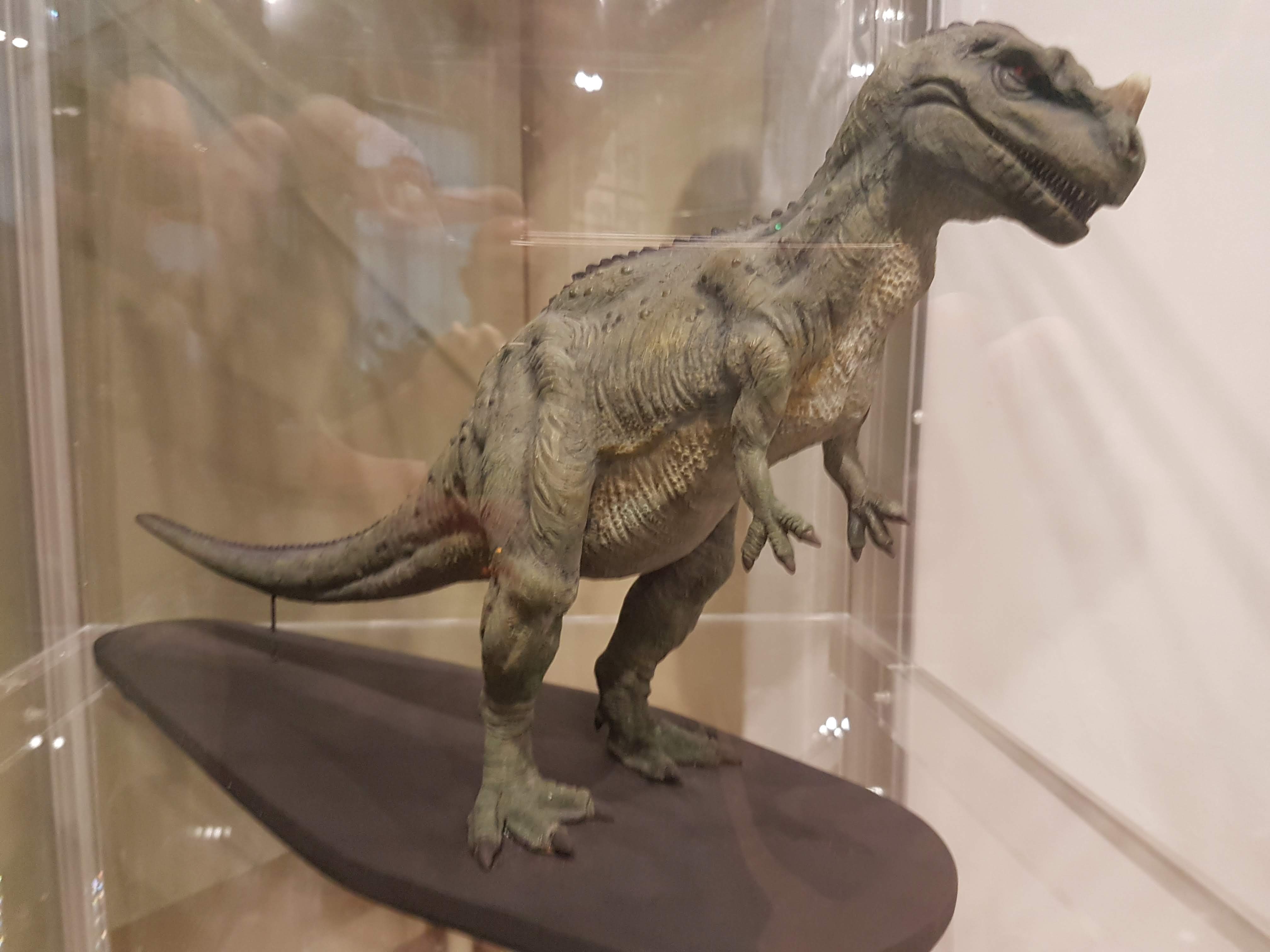
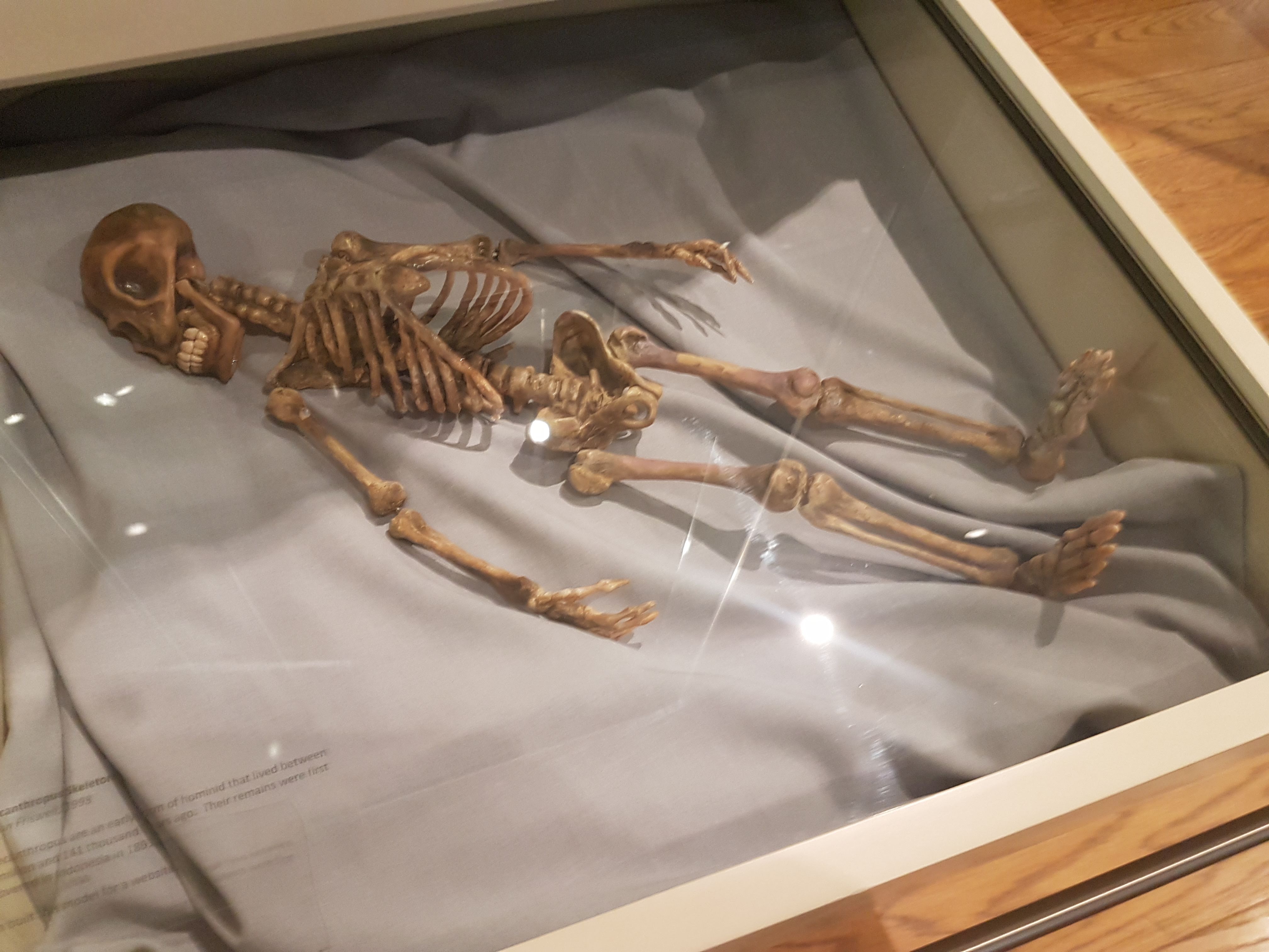
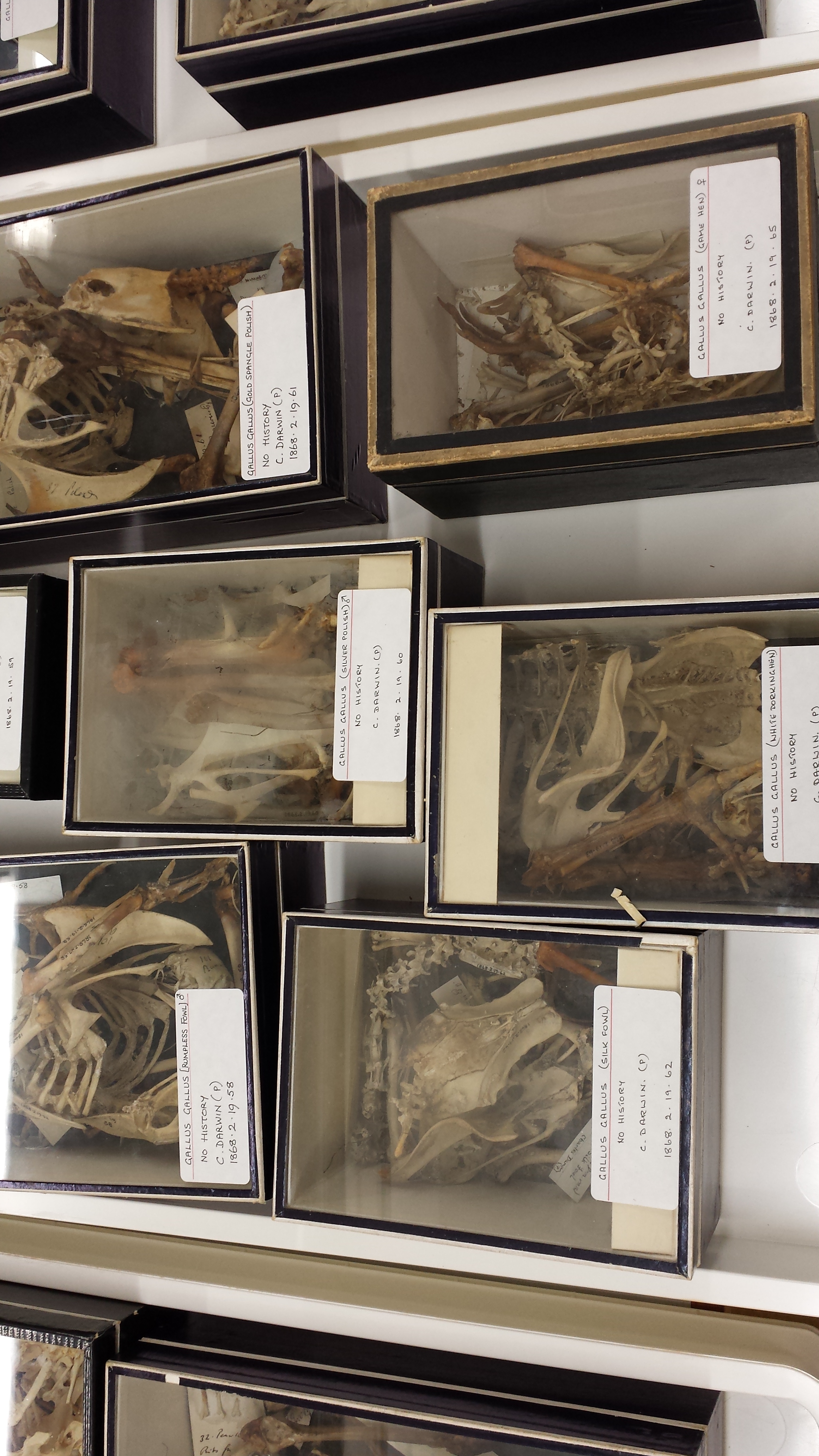
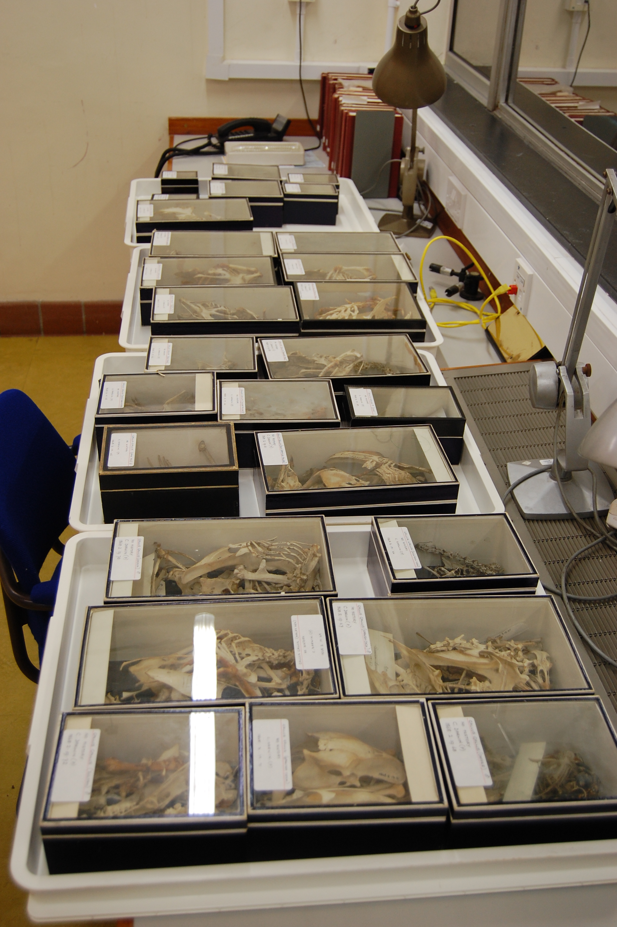
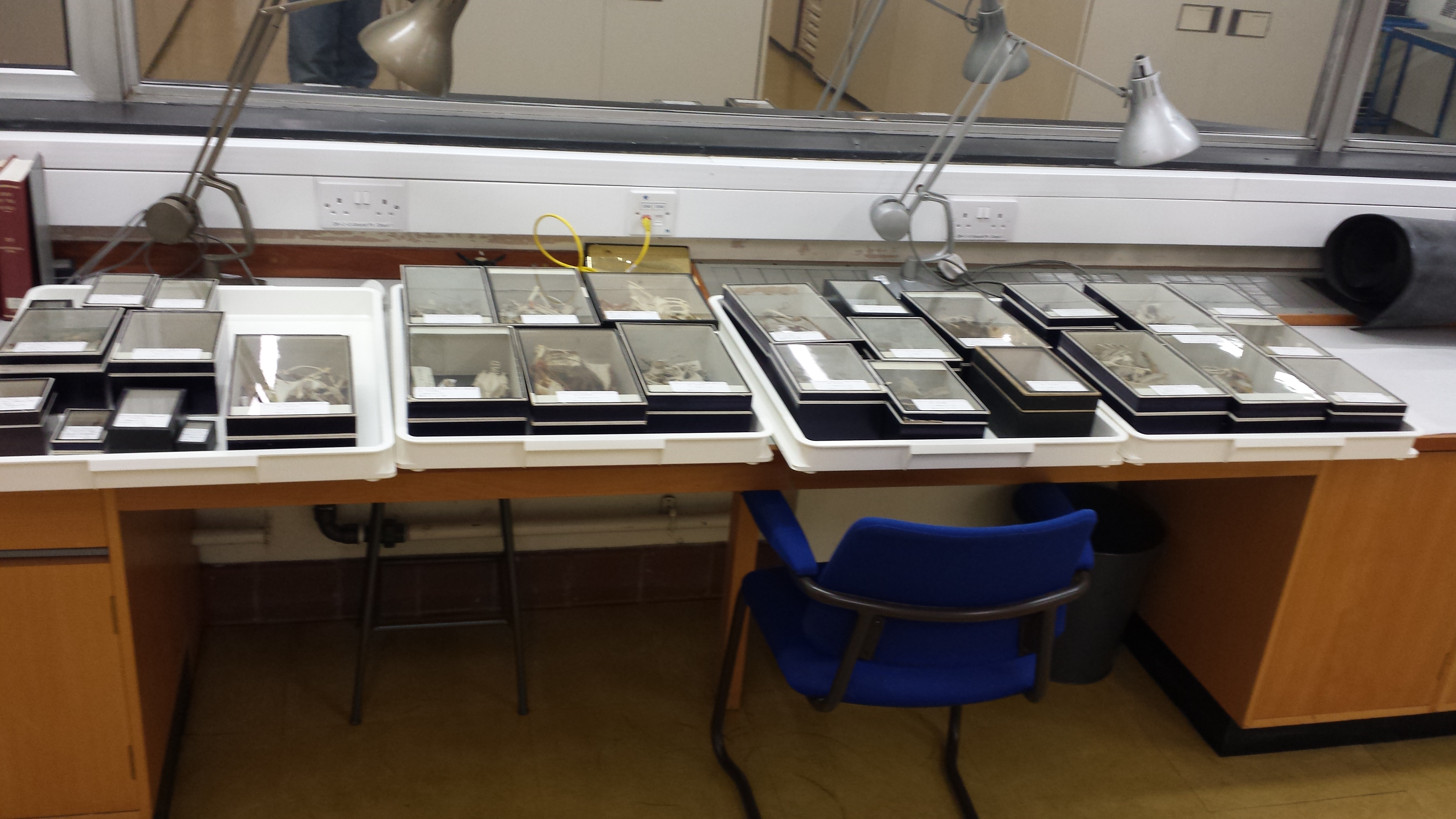
As the subject indicates, the transmission concerns my recent visit to the desert world of Tatooine, stimulated by investigations I conducted in the Corellian Science Museum. In that museum I found rare skeletal remains of the little-studied, reportedly extinct arthroreptile the Krayt Dragon (Tyrannodraconis tatooinensis by your archaic nomenclature). I’ll revisit this further below, because a subsequent discovery changed everything for me. I just wanted to whet your appetite, and this image of museum specimens of krayt dragons may do so:

Two fragmentary skeletons of small Krayt Dragons, from the Corellian Science Museum. (Image source here)

Two fragmentary skeletons of small Krayt Dragons, from the Corellian Science Museum. (Image source here) Note their short necks and quadrupedal limbs.

With growing fascination for the large land vertebratomorphs that are so startlingly diverse on Tatooine, I secured Imperial funding for an expedition to Tatooine, to survey the exotic megafauna and search for fossils of Tyrannodraconis that might further illuminate their evolution. My ensuing report summarizes my trilogy of investigations and discoveries from this “holiday in the suns”:


Stormtrooper on a Dewback in the Eastern Dune Sea (image source here).

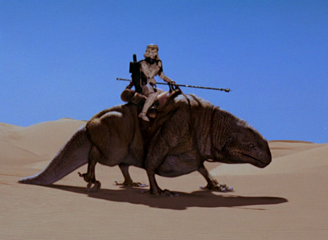
Stormtrooper on a Dewback in the Eastern Dune Sea (image source here). Note how gracile the limbs are below the elbows/knees.

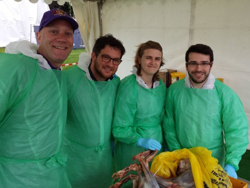
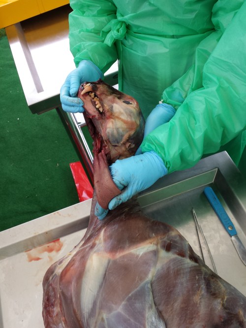
Investigation 1. Dissection of a Dewback, Mos Eisley

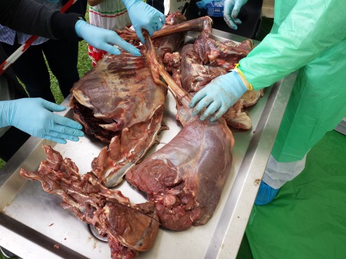
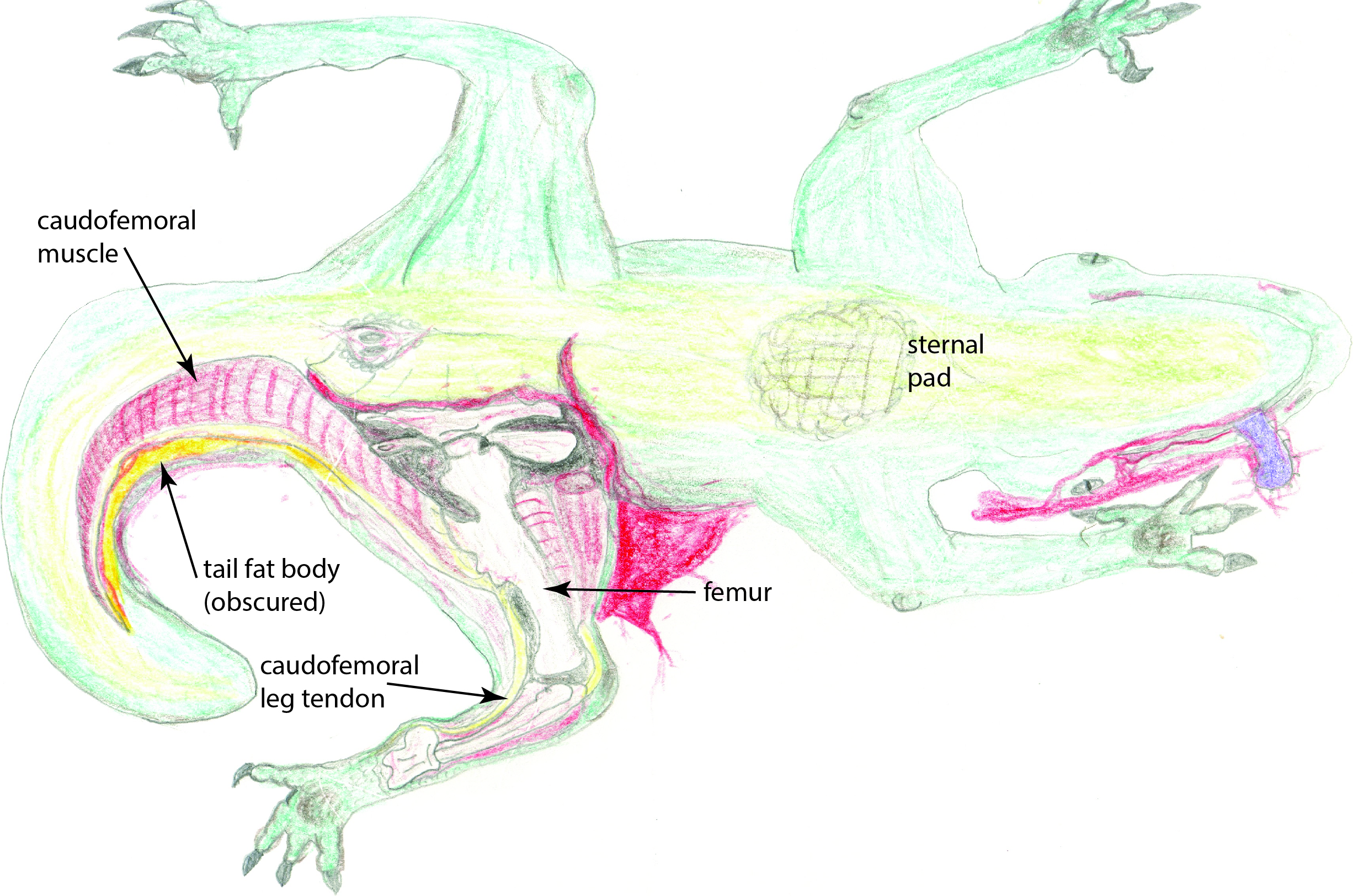
My ample funding (I’m sure you’re jealous) secured and stocked a laboratory for me in the colourful Mos Eisley spaceport, which has seen unprecedented commercial influx in recent years and now largely serves as an adventure park for hyperspace tourists (funded in part by the muromorphs of planet Dis’snai). With coliseum seating for a gathered host of some 1.6 million curiously slavering punters and drunken local yokels, I completed a full dissection of a fresh adult dewback (Iguanomorphus homoplasticus) specimen, illustrated below at its climax: exposure of the great fat body of the tail and the large caudofemoral muscle in the left thigh.  (curse this infernal Jawa 37C-H4 sketching droid’s malfunctions!)

Jawa 37C-H4 sketching droid illustration: My dissection of a common dewback, showing the caudofemoral muscle and tendon, tail fat body, and fibrous pads used while resting on the sand.

Jawa 37C-H4 sketching droid illustration: My dissection of a common dewback, showing (ventral view) the caudofemoral muscle and tendon, tail fat body (obscured by the nearby muscle), and fibrous pads used while resting on the sand.

My main observations support those of prior scholars, even from the Rebel Alliance era (bucking the trend of having to correct all their mistakes!): dewbacks have earned their moniker well by the characteristic water-condensing tissues on their dorsal surfaces. Microdroid explorations of these tissues, which lie within a dimpled midline ridge, house a high density of capillaries in a countercurrent network that surrounds a large number of specialised pores, or stomata, which smooth muscular rings contract to pull open when humidity, temperature and shade are best suited to cooling the surrounding air (via air currents encouraged by the stomata, and by local cooling via the capillary rete).

Previous scholars overlooked this mechanism, which conducts excessive warmth to the heat-emanative fat bodies in the bulky tail and the neck hump (my dissections nicely revealed these; similar tissues are concentrated in the foot pads and sternal pad). The mechanism also allows the body to be up to 20% cooler than the ambient air; an analogous adaptation to that seen in the banthas (below). My peers also failed to realize that the social nature of the dewback is key to its water conservation: while the stomatal rete can draw in some condensed water, it is far more effectively ingested by licking the backs of fellow dewbacks. Lone dewbacks thus are more prone to dehydration. The night-time group-huddling habits of dewbacks to conserve heat that they would otherwise too easily shed in the cool night air is yet another testament to the benefits of their sociality.

As ectotherms, dewbacks are slaves to the hot-cool cycles of the Tatooine wastes, but their sociality liberates them. Further escape comes from their large size (>800 kilograms of Terran mass units), which renders them mostly homeothermic, but never endothermic like some of your otherwise unimpressive Terran reptiles of past or present.

A laser-histology trek by microdroids showed the “scaled” hide around the rest of the body to be composed of siliceous material embedded in the thickly fibrous connective tissue of the skin, forming stereotyped arrowhead-shaped “siliceoderms”, as I term them, shown below.

Curious microstructure of the small "siliceoderms" from dewback skin that I have described-- single 'derm on the left, multiple 'derms surrounding a stomata on the right.

Curious microstructure of the small “siliceoderms” from dewback skin that I have described– single ‘derm on the left, multiple ‘derms surrounding a stoma on the right. To see these structures, one must view the “scales” at high magnification, ideally with microdroids.

I surmise that: (1) these siliceoderms are formed of fused Tatooine sand grains; (2) the grains become embedded into the soft, pliable skin as dewbacks grow, giving them insulation and physical protection; (3) young dewbacks display a previously mysterious behaviour of “sand-rolling” that encourages this embedding during the maturation of a dewback; and (4) the high strength and stiffness of this composite skin not only armours dewbacks but also pressurizes them, ensuring that blood can circulate through their large bodies without backflow or clotting issues, particularly in their gracile lower limbs, which are themselves passively supported by their skin tissues.

With your interest in animal locomotion, you may be curious about tales of how dewbacks can outrun landspeeders, especially in poor weather or terrain conditions. The skin-stiffening agents noted above surely play an important role in this. Indeed, much like your terrestrial varanid lizards, dewbacks do not follow the usual trend of straightening their limbs to support their body more effectively at larger body sizes (improving “effective mechanical advantage” as your field terms it), but they do draw them more closely under the body rather than remain sprawling. I revisit the matter of limb posture toward the end of my transmission.

Furthermore, the huge caudofemoral muscle shown above is able to transmit force from the tail to the thigh, and then its thick tendon transmits the force down the limb to the feet, acting as one strong limb extensor that powers and supports locomotion. No Terran animal does it so well. Banish any thoughts of how the dewback’s wrists and ankles seem implausibly thin– they are pressurized cylinders of dense tendon and bone, more like a Terran horse’s distal limbs than any lizard’s, and linked to far larger tail-to-thigh muscles. The expansive foot pads and reversed first toe (hallux; as in your Terran birds but with no association to arboreality) likewise give dewbacks a stable base of support and spread out their weight over the treacherous desert sands, reducing the work otherwise lost to deforming the sand’s surface and also keeping pressures on their feet at safe levels. Thus dewbacks have many features that explain their reputation for bursts of fast speed (~14 Terran meters/second or 50 kph/30 mph).

Yet whilst during the daytime and over short distances dewbacks can outpace banthas or humanoids on foot, their ectothermic nature causes them to accumulate fatigue too quickly, and thus they must rest. So sans cybernetic enhancements, dewbacks will never be winning any podraces. Nonetheless, I am sure you are awed by how Tatooine’s native reptiliforms, the dewbacks, exceed any living Terran reptile in their size and extreme adaptations to aridity. I have not even described the variations seen in feral, grizzled, cannibal or mountain dewback species, which can surpass the common desert dewback’s. Toward the end of my transmission I will show you animals that exceed even the greatest dinosaurs in sheer glory and ferocity.

Unlike the durable Tauntauns of my home system’s ice planet Hoth, however, dewbacks are ill-suited to cold climates because they are adapted to shed heat, not gain it. But the insulation of the next animal shows a more versatile performance…


Convincing image of a Bantha being ridden by a Sand-Person, from your world's fake documentary "Star Wars Episode IV: A New Hope", from Lucasfilm/Twentieth Century Fox.

Convincing image of a Bantha being ridden by a Tusken Raider/Sand-Person, from your world’s Rebel propaganda film “Star Wars Episode IV: A New Hope”, by Lucasfilm/Twentieth Century Fox.

Investigation 2. Field Dissection of a Bantha Bull

My anatomical study of a large male bantha (Megalingua feteoclunis) was hastened by not only the merciless heat but also by the imminent arrival of a horde of ravenous womp rats. Some quick incisions with my relict lightsaber sped my work. I focused my attention on three issues of scholarly interest: its marvellous tongue and glossopharyngeal adaptations (how does such a tall animal eat in a world that is far below it?), its hirsute integumentary system (what lies under that thick fur and how do banthas cope with the heat while wearing many wookies worth of wooly warmth?) and its peculiar, pillar-like limbs. The spiralling horns that add rings as the bantha grows, the nuchal ligament that supports the heavy head and neck, and the convoluted, multi-partitioned digestive tract that wrenches every last bit of nutrition from the lichens and other flora hidden beneath Tatooine sands are better understood. And with this bull I had no opportunity to study where the famous blue bantha milk comes from, but I have heard stories and no Terran mammal-esque udders are involved, let me tell you that much…

Anatomy of the oral apparatus of the Bantha, which I correct in my report although it is largely right (but how, Terran?). (source)

Anatomy of the oral apparatus of the Bantha, which I correct in my report although it is largely right (but how, Terran authors Terryl Whitlatch and Bob Carrau?). (source)

I don’t know how your Terran science-invigilators managed to get accurate information on bantha tongue anatomy (above) but I have to credit them, they almost got it right. With your can-do attitudes combined with your bungling mistakes, you’d make good Fourth Rebel Alliance members, but don’t get any new hopes. However, as the illustration below shows (and I had to leave the guts in the picture for their sheer impressiveness!), the tongue-projection mechanism extends not around the rear of the skull (occiput) and into the eyes or sinuses, but far back along the giant, spar-like breastbone (sternum) to the hips (pelvis, or propubis).

That mechanism’s powerful projection can extend the tongue as far as 3 Terran meters (10 feet). The tongue is expelled by stretching and then releasing (slowly for precise control, or quickly for a catapult action) a fibrous sac that surrounds the base of the tongue, and this sac then recoils elastically when released to withdraw the tongue. I’ve studied your Terran elephant and chameleon and it combines aspects of both of these, with the tongue having several layers of fine muscle fibres as in the former animal, and the “power amplifier” catch mechanism of the latter, thus providing a superior combination of control and speed. All of these are rightly called muscular hydrostats, but the bantha’s is the best.  You might mention your Terran pangolin as a counter-example, but does that little creature have the spiracle-bearing, ultrasensitive chemosensory tongue and majestic size of the bantha? No. I rest my case.

Jawa 37C-H4 sketching droid illustration: My dissection of a bantha, showing the tongue attachments (note the distal bifurcation), digestive tract and foot structure. The colour variations in the digestive tract seem to be produced by commensal arthroreptiles.

Jawa 37C-H4 sketching droid illustration: My dissection of a bantha, showing the tongue attachments (note the distal bifurcation), digestive tract and foot structure. The colour variations in the digestive tract seem to be produced by commensal arthroreptiles.

A naïve Terran like yourself might wonder why, of all things, a giant desert mammal such as the bantha would evolve to be clothed in thick fur. Here you would reveal your feeble way of grasping about the diversity of pangalactic Nature. First of all, banthas are not mammals as you know them; a Terran word like pseudomammal would suffice. They lack the diagnostic traits of mammary glands, true hair, and inner ear bones that diagnose the Mammalia of your homeworld, but evolution at a giant size in a hot, dry clime has chastened them to become at least superficially similar to a Terran mammal such as an elephant or mammoth. One might be so naïve, even, to think that a bantha is merely a proboscidean in hairy disguise, but drive such thoughts from your rickety cerebral-implant-deprived mind.

Behold, the true nature of bantha fur, as I have seen with microdroid holo-imaging: it is a second, external circulatory system of sorts. Simply put, the hairs have a thermo-conductive submolecular structure that deflects heat (and even, to a degree, the energy of a blaster) and traps cooler air near the body with an intricate network of cross-linking of barbed fibers more like a Terran bird’s feathers than mammalian hair. In this cooler locale, tracts of spongy skin tissue collect condensed water and direct it to absorbent epithelial beds on the chin and lips, belly, and toes, where the bantha imbibes it, or simply sheds it off to carry further heat away. Thus here we have a fascinating case of convergent evolution with the reptiliform dewbacks, but surpassing even that animal’s adaptation and evolving what you would likely call an air-conditioning system. Banthas cool themselves by circulating a slick of cool water around their body inside a heat-resistant fluffy outer mesh. Whether their horn tissues or tails contribute to this system is yet to be investigated.

Lastly, I have conducted holo-viewings of the biomechanics of bantha gaits from numerous remote studies of wild and Sand People-ridden animals, in light of my own dissections of this bull. What strikes me is the phenomenal convergence with giant quadrupeds on your homeworld: like sauropods, elephants and other species, banthas have evolved “graviportal” or weight-bearing adaptations: (1) limbs that are proportionately longest above the elbow and knee, not distally elongated as in “cursorial” animals; (2) heavy, robust bones that lack much of a marrow space; (3) short, thickly padded feet ending in bulky claws or hooves (three toes in the case of banthas); (4) an emphasis on lateral sequence (left hind-left front-right hind-right front) footfalls when walking, extended to a slightly bouncing, rolling “amble” at faster speeds; (5) strongly vertical limbs when walking, using the limbs more like pillars to support the weight more effectively; and (6) slow maximal speeds, limited to ~7 Terran meters/second (24 kph/15mph) at best.

At around 4000 kg of typical body mass, banthas overlap with the masses of your planet’s erstwhile giants that have such features. I did not uncover any “predigits” supporting the feet of banthas as you had in elephants; rather, their “heels” involve dense fibro-elastic cartilage, which works analogously to give shock-absorbing and resilient properties to the feet. This suite of graviportal features reinforces an idea that is now recognized pan-galactically: At huge sizes, land animals must act relatively more constrained by gravity, becoming forced to adapt more aspects of their biology to resist its pull, lest they strain muscles, break bones, snap tendons, or fall and injure themselves. Thus the convergent evolution of banthas and elephants is no surprise. But is there another way to be an imposing giant? Perhaps…


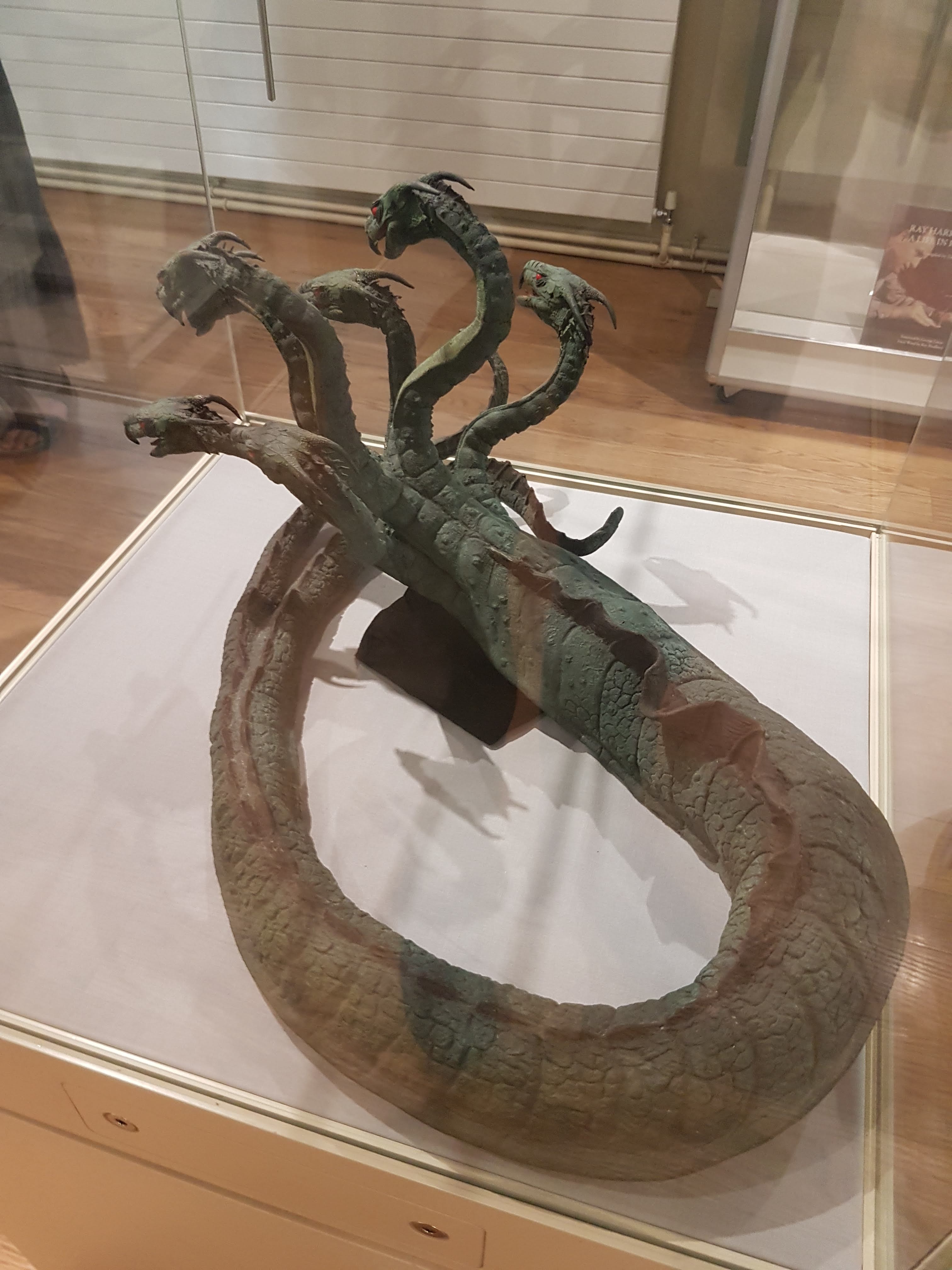
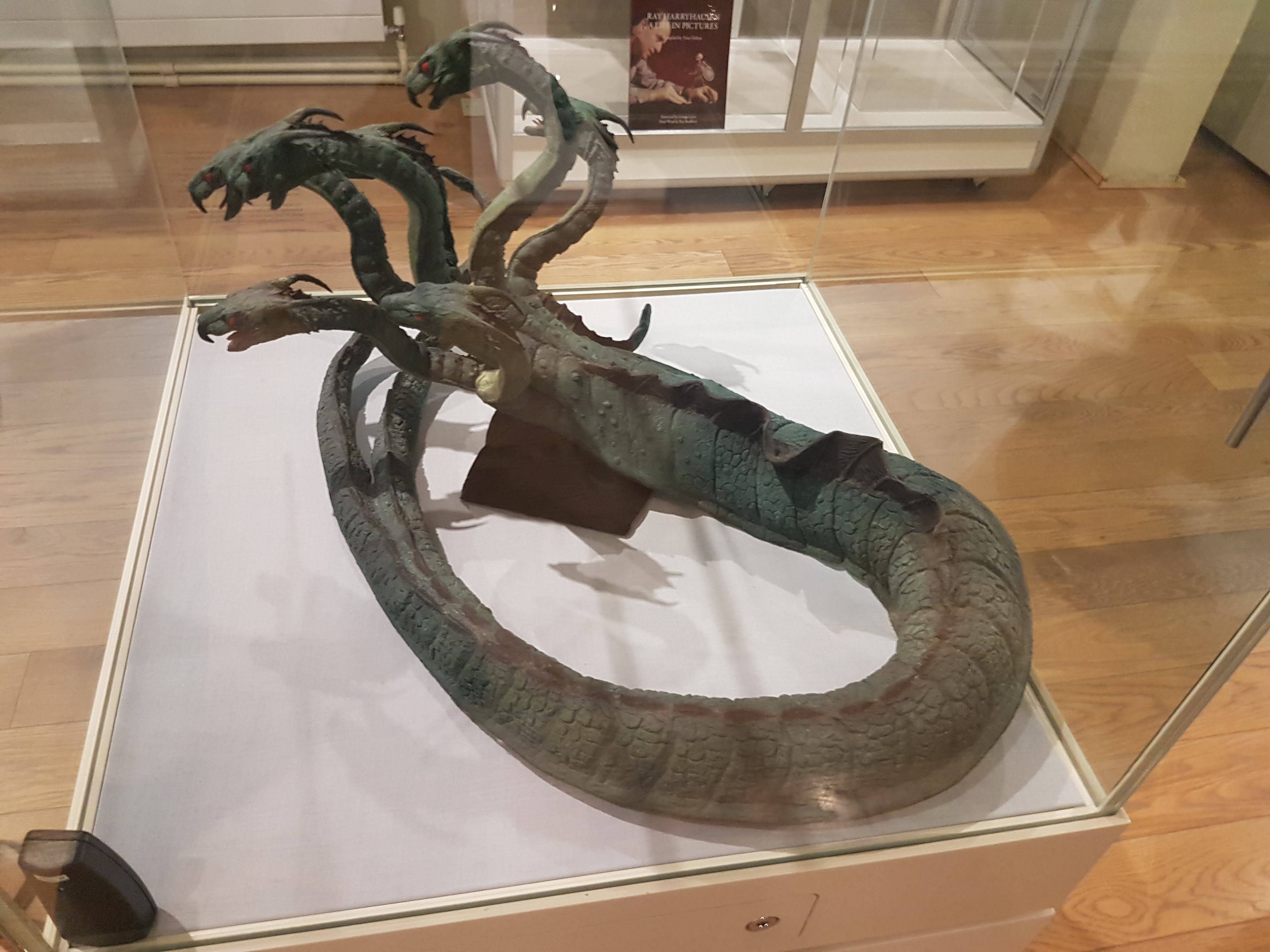
Investigation 3. On some remains of the “extinct” Krayt Dragon

Ever since I left my home system, thoughts kept tumbling through my mind like rocks in an asteroid field, concerning the krayt dragon bones I had viewed in the museum on Corellia. With the krayt (Tyrannodraconis sp.) lineage reported extinct since at least the year 22 ABY, following much publicity of its awesome nature, its menace seemed now but a phantom. Consequently I could only fantasize of deeper study. That is, until a rumour came to me while resupplying in the well-preserved city of Bestine: not far off on the edge of the Jundland Wastes, a stormtrooper patrol had taken down a strange, enormous, multi-legged arthroreptile that had gone after their dewback mounts. A quick skyhopper flight and I was there, giddy with the adrenaline of impending discovery.

Another Terran artist renders a compelling illustration, of a Greater Krayt Dragon in life. Where indeed do they get their information from? Bothan spies, I suspect. (Source)

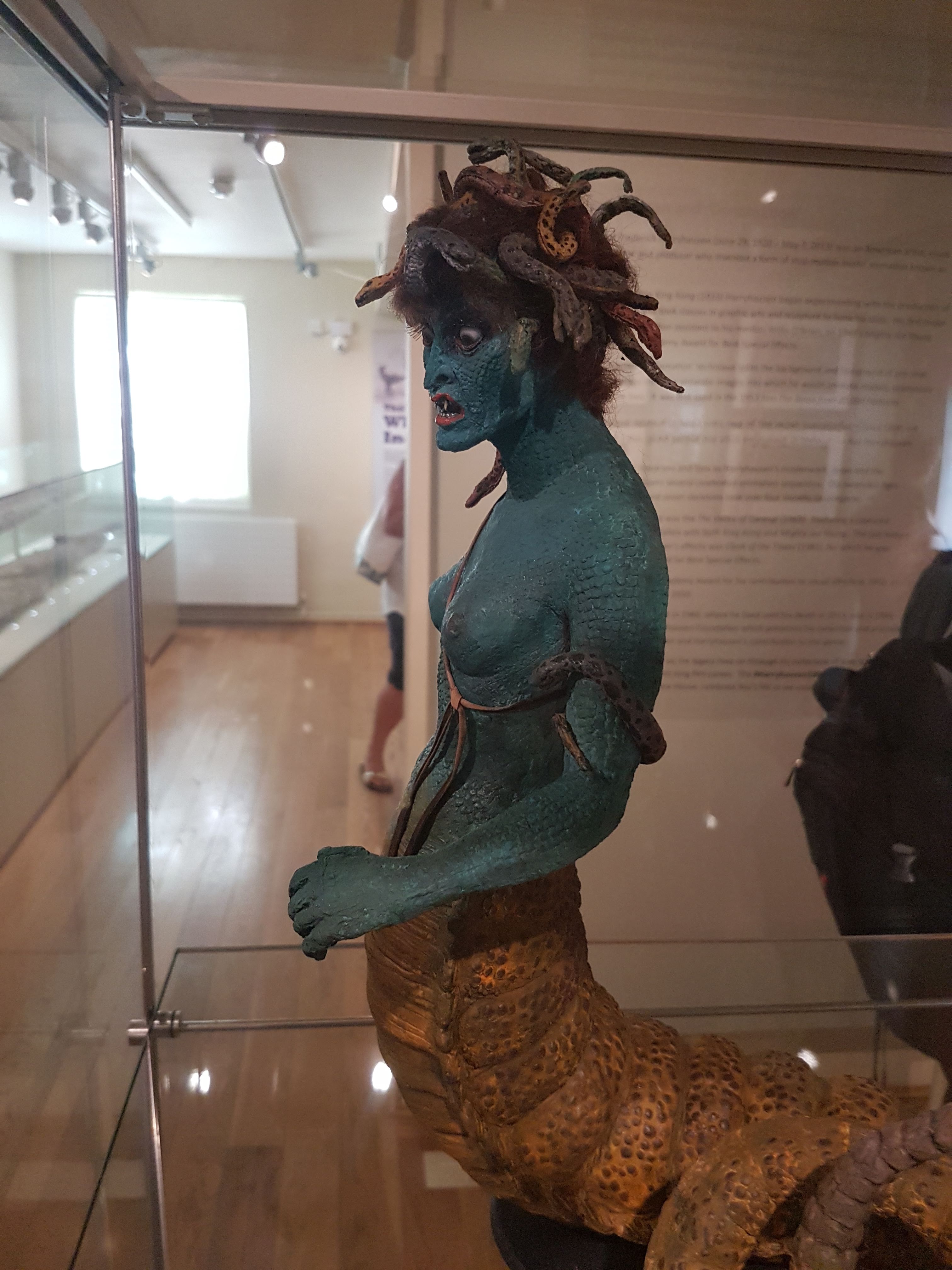
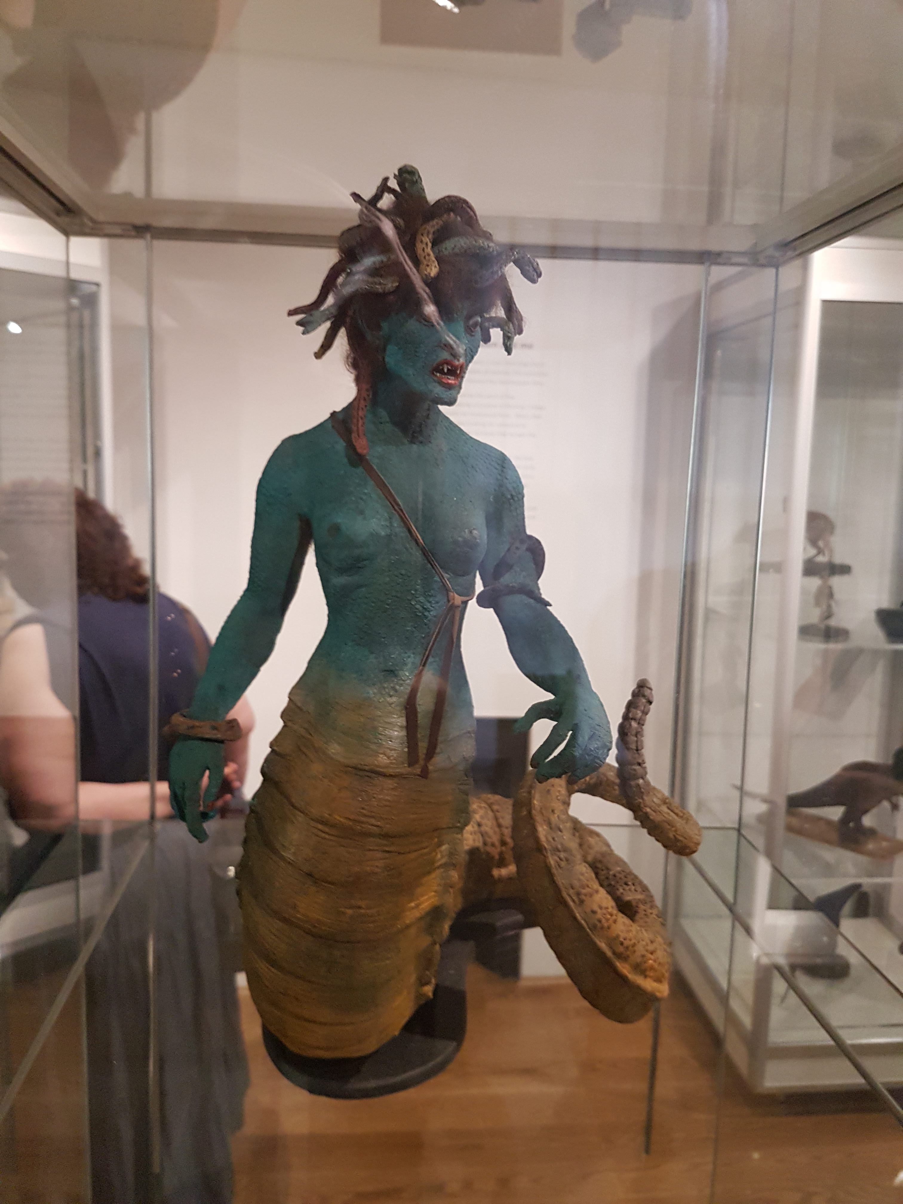
Another Terran artist (one of Terryl Whitlatch and Bob Carrau) renders a compelling illustration, of a Greater Krayt Dragon in life. Where indeed do they get their information from? Bothan spies, I suspect. (Source)

It was a magnificent carcass. Sandworms and scurriers were already attempting to scavenge it, but with little luck and easily driven off with a few shots from my carbine. No stormtroopers remained (alive, anyway), so I didn’t get any details of the fracas that led to this well-timed demise, but the blast points on its body were too precise for sandpeople, and characteristic dewback tracks were everywhere. Even my antique lightsaber seemed poorly up to the task of dissecting this titan: it was over 30 meters (100 feet) long and surely 100 tons of Terran mass if not more; on the scale of your sauropods, but so vastly different in other ways. Right away, from its tracks I could see it had a peculiar mode of movement in life: it had slid up to some rocky cover in these badlands, dragging its belly and bulk along with ten limbs that were slender in comparison to its body, but still each as big as a large bantha’s. I took a deep breath and cut into what was the first Greater Krayt Dragon seen in some 255 years.

Jawa 37C-H4 sketching droid illustration: My dissection of the Greater Krayt Dragon, to extract the Dragon Pearl. The stormtrooper shown forgot the tale that Krayts take 1 hour to die, and so got too close too soon.

Jawa 37C-H4 sketching droid illustration: My dissection of the Greater Krayt Dragon, to extract the Dragon Pearl. The stormtrooper shown forgot the tale that Krayts take 1 hour to die, and so got too close too soon.

If the bantha dissection was a rush job, this one was a sprint. Pockets of gas were forming and erupting while I sliced my way toward the bones and other organs of most interest, with the forces of decomposition slowly winning a race against my science. Oh, if only I’d had a Jawa sandcrawler to repurpose as a mobile freezer! And the sandworms and scurriers were still lurking about, with far nastier things surely soon to be drawn by the carnage out in these remote wastes. Those two days blurred exhaustion and inquiry and disgust and elation into a mire in my mind more pernicious than any on Dagobah. I’m no longer sure of what I saw– you’re probably wondering if I found the fabled krayt dragon pearl in the gizzard, and yes, there was one but I lost it somehow. Same with the venom sacs. Maybe I sipped from one of those; that would explain a lot. I made a sketch that I reproduce here, but then in a crazed, diaphonic state of dehydration and euphoria and frustration I am pretty sure I cut my sketching droid to pieces too, so this is all that remains to bolster my frazzled memories.

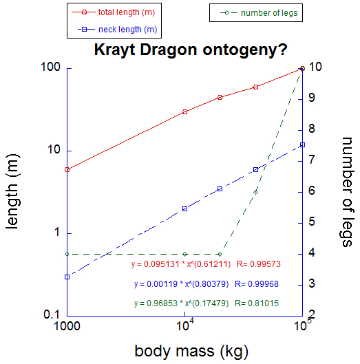
Now that I’ve recovered and ruminated, I have come to some conclusions. First, I am left doubting all the little we know about krayt dragons. It is said that they existed in canyon, normal and greater species, and the immense variation of curved horns, clawed limbs and flanged tails lent this taxonomy much credibility in the past. But, call it chronic heatstroke or inspiration as you may, what if all krayt “species” are just stages of a long and repeatedly metamorphic developmental sequence? As my graph below shows, and this is admittedly pieced together from what few museum specimens and documents I have since marshalled to test my hypothesis, krayt traits change uniformly with their body size. As they get bigger, krayt dragons get more multi-legged and longer-necked, diverging from the form of their relatives (in the evolutionary sense of your sciences, sister group or outgroup) from Ruutan, the Kell dragons. The genus Tyrannodraconis, more so than the Kell, betrays its arthroreptile ancestry with their spines, exoskeletal plates, and tendency for polypedality. Their sternum also elongates to support their chest as they change from lumbering, bantha-chasing quadrupeds to slithering, sarlacc-snatching octa- or decapedal behemoths.

Although based on little concrete data, my analysis of known Krayt and related specimens suggests that they change continuously during ontogeny, although leg number may shift more suddenly (I predict this happens during their first metamorphosis at sexual maturity). Strong allometric scaling of neck and total length is evident- if the two lengths scaled as mass^0.33 they would be maintaining shape across the proposed growth series. But they don't.

Although based on little concrete data, my analysis of known Krayt and related specimens suggests that they change continuously during ontogeny, although leg number may shift more suddenly (I predict this happens during their first metamorphosis at sexual maturity). Strong allometric scaling of neck and total length is evident- if the two lengths scaled as body mass0.33 they would be maintaining shape across the proposed growth series. But they don’t.

I return to the best-documented krayt dragon remains: those that even Terrans have seen in the Rebel propaganda film you call “Episode IV”. Dr. Freezers, even your fellow blog-invigilators at SV-POW! discussed it. Witness the large size and long neck of the typical Krayt; whether horns existed or not in that form from the film is uncertain, and I note that these could even be a sexually dimorphic feature, but this is beside the point. Remnants of the body and limbs were never found. But this specimen fits well with my idea that all krayts are one species, or two at most—and how many top predatory megafaunal species could coexist on a desolate arid planet like Tatooine anyway?

What still strikes me is the phenotypic variation in krayts: some large or small varieties have from two to four toes, and different scythe-like horns on their tail tips. This leads me to heap speculation atop my precarious pile of hypotheses: what if krayts are simply phenotypically labile, varying their traits almost stochastically between individuals due to relatively flexible ontogenetic programming, but still following strong overall trends as size increase, like those I have plotted above? Those stronger trends might be more tightly regulated by homeobox-like genes similar to those that have shaped so much of your Terran metazoan diversity, influencing features along the body axis like those I have mentioned (neck, limbs) across growth? I like this idea too much for it to be true, I admit. But if one krayt dragon existed just a short time ago, it is not simply fodder for the cryptoxenozoologists. And so, sooner or later, someone will answer my scientific salvo. I predict that burrows where the krayt dragons metamorphose between life stages, growing new legs and longer bodies, will be found in due time.

However, I have a stronger inference that I present to you as part of our common interest. On Terra and Tatooine alike, larger animals tend to adopt more straight-legged limb poses to improve their leverage, as I outlined with the dewbacks above. I plot existing data for Terran animals with my best estimates (for dewbacks and banthas, quite reliable; for krayts, my guesses) for this “effective mechanical advantage” below. What this shows is that dewbacks and Banthas both fall below the “normal” curve for Terran land mammals, as I explain:

In the case of dewbacks, this decrease of limb leverage seems offset by passive support from their pressurized scaly legs and enlarged whole-limb extensor muscles of their hindlegs, so they are overall about as well adapted to bursts of speed as large mammals from your world, such as buffalo or large antelope, even if their endurance suffers (a tradeoff, perhaps, for their reptile-like adaptations to desert life).

In the case of banthas, they do no better or worse than elephants; all are slow due to their size and “graviportal” focus of adaptations. Like elephants, but unlike dewbacks, banthas do not “invest” more body mass into supportive leg muscle, and so they are slower than they might otherwise be.

Effective mechanical advantage of the limbs, with Terran data for mammals (red+blue) (source 1 and source 2), and my new data for Tatooine megafauna. Past a moderate size, EMA either declines or remains constant. Once the limbs are fairly straight (near the size of a Terran horse), EMA cannot be much improved.

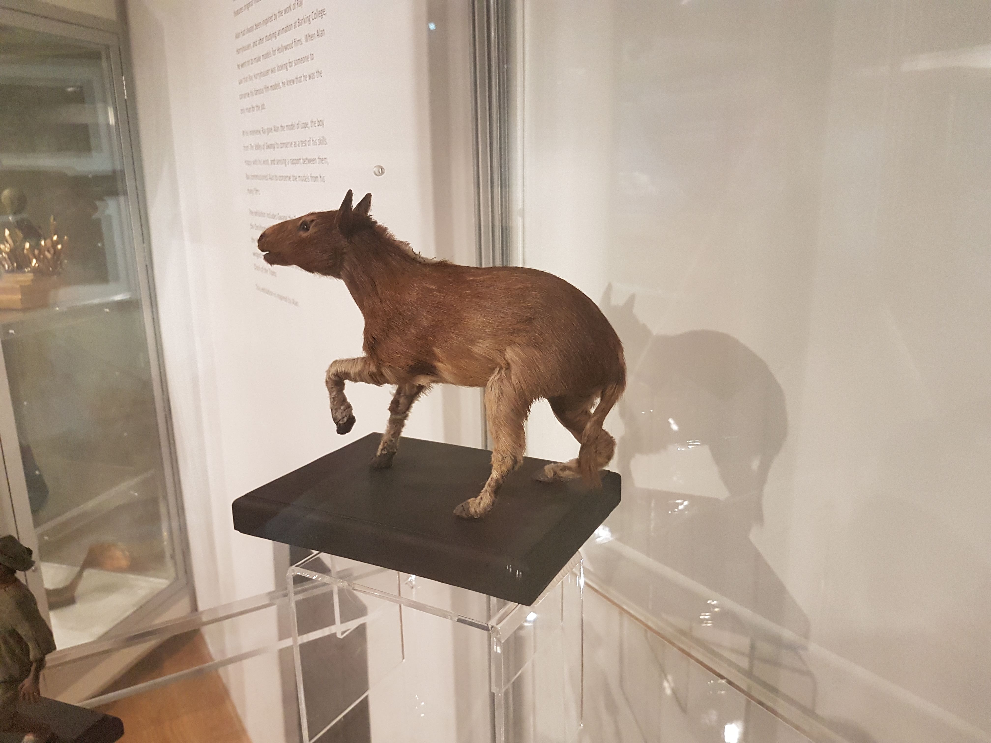
Effective mechanical advantage (EMA) of the limbs, with Terran data for mammals (red+blue) (source 1 and source 2), and my new data for Tatooine megafauna (green). Past a moderate size, EMA either declines or remains constant. Once the limbs are fairly straight (near the size of a Terran horse, or Tatooine eopie; vertical dashed line), EMA cannot be much improved.

But the krayts (young or smaller species aside) suffer more from their size than other Tatooine megafauna, as they do not increase their limbs’ mechanical advantage any more than the others do, and so they must become slower as they grow. This explains, however, why their ecology shifts from being a mobile predator when smaller (feeding on dewback, then bantha-sized prey) to being more of an ambush predator or specialist on slow/immobile prey like sarlaccs as they attain titanic sizes. Their limbs, despite becoming more numerous, must become less able to support them as size increases, as in other Terran and Tatooine megafauna, and thus they are destined to benefit from giant size (in many ways, including near-invulnerability and capacity to take the largest prey) at a cost of athleticism (but with prey like sarlaccs, who needs it?). In the greater, or fully mature, krayt dragons, I suggest that the limbs each become less supportive and more of a stabilizer to prevent their slug-like bulk from rolling over, or a set of “oars” to help them navigate through sandy environments like the Dune Seas. They support their weight not so much with limbs and levers, but with a larger, cuirass-like breastbone system, rings of muscles and fibrous tissue, and their whole elongate body.

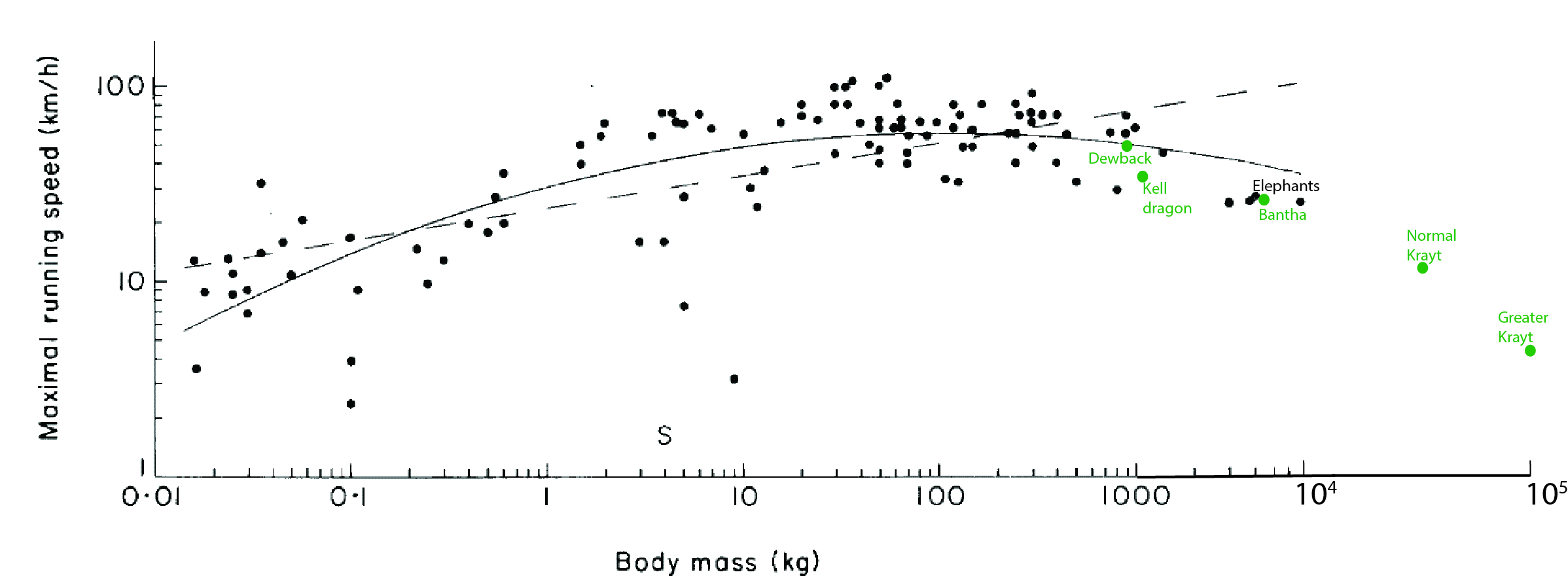
The ultimate implications of my biomechanical research are summarized below—I am sure you will agree with my reasoning.

Maximal speed vs. body mass data from (black) Terran animals (source), and (green) Tatooine megafauna (plus non-native Kell dragons for comparison). As size increases past ~100 kg mass, speed inevitably declines.

Maximal speed vs. body mass data from (black) Terran animals (source), and (green) Tatooine megafauna (plus non-native Kell dragons for comparison). As size increases past ~100 kg mass (when EMA in the other graph above is already maximal), speed inevitably declines.

As for those that have said that Greater Krayt Dragons and such are thereby confined to a life as scavengers and nothing more, I would welcome them to explore the Jundland Wastes locales armoured by all the security that this foolish notion provides. I, for one, would enjoy viewing such a visit, but only remotely via a probe droid’s holo-feed.

One of your Terran artists (jeddbub on deviantart) produced a provocative imagining of a Greater Krayt Dragon facing a Jedi. I'd wager for the former.

One of your Terran artists (jeddibub on deviantart) produced a provocative imagining of a Greater Krayt Dragon facing a Jedi. I’d wager for the former.

I submit this report in honour of Empress Syrrhosyx and the Fourth Empire– may you find the contents enlightening and may her rule grace your benighted homeworld before you, too, have nothing left of your megafauna but stories of dragons.

I welcome your comments, and perhaps some of your lauded “freezerinos” would care to comment below—but they must behave themselves, lest I find cause to deposit them in carbonite for hyperspace shipping to a lonely suffering on a lonely planet!

I shall shortly return this “blog” to your control, when the mood strikes me. That is the deal for this correspondence. Pray I don’t alter it any further.

Enjoy your little blog carnival, Terrans…

Pangalactically,

Dr. Zhonav Diphyryzas

Read Full Post »

This post was just published yesterday in a shorter, edited form in The Conversation UK, with the addition of some of my latest thoughts and the application of the editor’s keen scalpel. Check that out, but check this out too if you really like the topic and want the raw original version! I’ve changed some images, just for fun. The text here is about 2/3 longer.

Recently, the anatomy of animals comes up a lot, at least implicitly, in science news stories or internet blogs. Anatomy, if you look for it, is everywhere in organismal and evolutionary biology. The study of anatomy has undergone a renaissance lately, in a dynamic phase energized by new technologies that enable new discoveries and spark renewed interest. It is the zombie science, risen from what some had assumed was its eternal grave!

Stomach-Churning Rating: 4/10; there’s a dead elephant but no gore.

My own team has re-discovered how elephants have a false “sixth toe” that has been a mystery since it was first mentioned in 1710, and we’ve illuminated how that odd bit of bone evolved in the elephant lineage. This “sixth toe” is a modified sesamoid kind of bone; a small, tendon-anchoring lever. Typical mammals just have a little nubbin of sesamoid bone around their ankles and wrists that is easily overlooked by anatomists, but evolution sometimes co-opts as raw material to turn into false fingers or toes. In several groups of mammals, these sesamoids lost their role as a tendon’s lever and gained a new function, more like that of a finger, by becoming drastically enlarged and elongated during evolution. Giant pandas use similar structures to grasp bamboo, and moles use them to dig. We’ve shown that elephants evolved these giant toe-like structures as they became larger and more terrestrial, starting to stand up on tip-toe, supported by “high-heels” made of fat. Those fatty heels benefit from a stiff, toe-like structure that helps control and support them, while the fatty pads spread out elephants’ ponderous weight.

Crocodile lung anatomy and air flow, by Emma Schachner.

Crocodile lung anatomy and air flow, by Emma Schachner.

I’ve also helped colleagues at the University of Utah (Drs. Emma Schachner and Colleen Farmer) reveal, to much astonishment, that crocodiles have remarkably “bird-like” lungs in which air flows in a one-way loop rather than tidally back and forth as in mammalian lungs. They originally discovered this by questioning what the real anatomy of crocodile lungs was like- was it just a simple sac-like structure, perhaps more like the fractal pattern in mammalian lungs, and how did it work? This question bears directly on how birds evolved their remarkable system of lungs and air sacs that in many ways move air around more effectively than mammalian lungs do. Crocodile lungs indicate that “avian” hallmarks of lung form and function, including one-way air flow, were already present in the distant ancestors of dinosaurs; these traits were thus inherited by birds and crocodiles. Those same colleagues have gone on to show that this feature also exists in monitor lizards, raising the question (almost unthinkable 10-20 years ago) of whether those bird-like lungs are actually a very ancient and common feature for land animals.

Speaking of monitor lizards, anatomy has revealed how they (and some other lizards) all have venom glands that make their bites even nastier, and these organs probably were inherited by snakes. For decades, scientists had thought that some monitor lizards, especially the huge Komodo dragons, drooled bacteria-laden saliva that killed their victims with septic shock. Detailed anatomical and molecular investigations showed instead that modified salivary glands produced highly effective venom, and in many species of lizards, not just the big Komodos. So the victims of numerous toothy lizard species die not only from vicious wounds, but also from worsened bleeding and other circulatory problems promoted by the venomous saliva. And furthermore, this would mean that venom did not evolve separately in the two known venomous lizards (Gila monster and beaded lizard) and snakes, but was inherited from their common ancestor and became more enhanced in those more venomous species—an inference that general lizard anatomy supports, but which came as a big surprise when revealed by Bryan Fry and colleagues in 2005.

There’s so much more. Anatomy has recently uncovered how lunge-feeding whales have a special sense organ in their chin that helps them detect how expansive their gape is, aiding them to engulf vast amounts of food. Scientists have discovered tiny gears in the legs of leafhoppers that help them make astounding and precise leaps. Who knew that crocodilians have tiny sense organs in the outer skin of their jaws (and other parts of their bodies) that help them detect vibrations in the water, probably aiding in communication and feeding? Science knows, thanks to anatomy.

Just two decades or so ago, when I was starting my PhD studies at the University of California in Berkeley, there was talk about the death of anatomy as a research subject; both among scientists and the general public. What happened? Why did anatomy “die” and what has resuscitated it?


TH Huxley, anatomist extraordinaire

TH Huxley, anatomist extraordinaire, caricatured in a lecture about “bones and stones, and such-like things” (source)

Anatomy’s Legacy

In the 16th through 19th centuries, the field of gross anatomy as applied to humans or other organisms was one of the premier sciences. Doctor-anatomist Jean Francois Fernel, who invented the word “physiology”, wrote in 1542 that (translation) “Anatomy is to physiology as geography is to history; it describes the theatre of events.” This theatric analogy justified the study of anatomy for many early scientists, some of whom also sought to understand it to bring them closer to understanding the nature of God. Anatomy gained impetus, even catapulting scientists like Thomas Henry Huxley (“Darwin’s bulldog”) into celebrity status, from the realisation that organisms had a common evolutionary history and thus their anatomy did too. Thus comparative anatomy became a central focus of evolutionary biology.

But then something happened to anatomical research that can be hard to put a finger on. Gradually, anatomy became a field that was scoffed at as outmoded, irrelevant, or just “solved”; nothing important being left to discover. As a graduate student in the 1990s, I remember encountering this attitude. This apparent eclipse of anatomy accelerated with the ascent of genetics, with anatomy reaching its nadir in the 1950s-1970s as techniques to study molecular and cellular biology (especially DNA) flourished.

One could argue that molecular and cellular biology are anatomy to some degree, especially for single-celled organisms and viruses. Yet today anatomy at the whole organ, organism or lineage level revels in a renaissance that deserves inspection and reflection on its own terms.


Anatomy’s Rise

Surely, we now know the anatomy of humans and some other species quite well, but even with these species scientists continue to learn new things and rediscover old aspects of anatomy that laid forgotten in classic studies. For example, last year Belgian scientists re-discovered the anterolateral ligament of the human knee, overlooked since 1879. They described it, and its importance for how our knees function, in novel detail, and a lot of media attention was drawn to this realisation that there are some things we still don’t understand about our own bodies.

A huge part of this resurgence of anatomical science is technology, especially imaging techniques- we are no longer simply limited to the dissecting knife and light microscope as tools, but armed with digital technology such as 3-D computer graphics, computed tomography (series of x-rays) and other imaging modalities. Do you have a spare particle accelerator? Well then you can do amazing synchrotron imaging studies of micro-anatomy, even in fairly large specimens. Last year, my co-worker Stephanie Pierce and colleagues (including myself) used this synchrotron approach to substantially rewrite our understanding of how the backbone evolved in early land animals (tetrapods). We found that the four individual bones that made up the vertebrae of Devonian tetrapods (such as the iconic Ichthyostega) had been misunderstood by the previous 100+ years of anatomical research. Parts that were thought to lie at the front of the vertebra actually lay at the rear, and vice versa. We also discovered that, hidden inside the ribcage of one gorgeous specimen of Ichthyostega, there was the first evidence of a sternum, or breastbone; a structure that would have been important for supporting the chest of the first land vertebrates when they ventured out of water.

Recently, anatomists have become very excited by the realization that a standard tissue staining solution, “Lugol’s” or potassium iodide iodine, can be used to reveal soft tissue details in CT scans. Prior to this recognition, CT scans were mainly used in anatomical research to study bone morphology, because the density contrast within calcified tissues and between them and soft tissues gives clearer images. To study soft tissue anatomy, you typically needed an MRI scanner, which is less commonly accessible, often slower and more expensive, and sometimes lower resolution than a CT scanner. But now we can turn our CT scanners into soft tissue scanners by soaking our specimens in this contrast solution, allowing highly detailed studies of muscles and bones, completely intact and in 3D. Colleagues at Bristol just published a gorgeous study of the head of a common buzzard, sharing 3D pdf files of the gross anatomy of this raptorial bird and promoting a new way to study and illustrate anatomy via digital dissections- you can view their beautiful results here. Or below (by Stephan Lautenschlager et al.)!

Buzzard-head

These examples show how anatomy has been transformed as a field because we now can peer inside the bodies of organisms in unprecedented detail, sharing and preserve those data in high-resolution digital formats. We can do this without the concern that a unique new species from Brazilian rainforests or exciting fossil discovery from the Cambrian period would be destroyed if we probed certain questions about its anatomy that are not visible from the outside– a perspective in which science had often remained trapped for centuries. These tools became rapidly more diverse and accessible from the 1990s onward, so as a young scientist I got to see some of the “before” and “after” influences on anatomical research—these have been very exciting times!

When I started my PhD in 1995, it was an amazing luxury to first get a digital camera to use to take photographs for research, and then a small laser scanner for making 3D digital models of fossils, with intermittent access to a CT scanner in 2001 and now full-time access to one since 2003. These stepwise improvements in technology have totally transformed the way I study anatomy. In the 1990s, you dissected a specimen and it was reduced to little scraps; at best you might have some decent two-dimensional photographs of the dissection and some beetle-cleaned bones as a museum specimen. Now, we CT or MRI scan specimens as routine practice, preserving many mega- or gigabytes of data on its internal and external, three-dimensional anatomy in lush detail, before scalpel ever touches skin. Computational power, too, has grown to the point where incredibly detailed 3D digital models produced from imaging real specimens can be manipulated with ease, so science can better address what anatomy means for animal physiology, behaviour, biomechanics and evolution. We’re at the point now where anatomical research seems no longer impeded by technology– the kinds of questions we can ask are more limited by access to good anatomical data (such as rare specimens) than by the ways we acquire and use those data.

My experience mirrors my colleagues’. Larry Witmer at Ohio University in the USA, past president of the International Society for Vertebrate Morphologists, has gone from dissecting bird heads in the 1990s to becoming a master of digital head anatomy, having collected 3D digital scans of hundreds of specimens, fossil and otherwise. His team has used these data to great success, for example revealing how dinosaurs’ fleshy nostrils were located in the front of their snouts (not high up on the skull, as some anatomists had speculated based on external bony anatomy alone). They have also contributed new, gorgeous data on the 3D anatomy of living animals such as opossums, ostriches, iguanas and us, freely available on their “Visible Interactive Animal” anatomy website. Witmer comments on the changes of anatomical techniques and practice: “For extinct animals like dinosaurs, these approaches are finally putting the exploration of the evolution of function and behavior on a sound scientific footing.

I write an anatomy-based blog called “What’s in John’s Freezer?” (haha, so meta!), in which I recount the studies of animal form and function that my research team and others conduct, often using valuable specimens stored in our lab’s many freezers. I started this blog almost two years ago because I noticed a keen interest, or even hunger for, stories about anatomy amongst the general public; and yet few blogs explicitly were about anatomy for its own sake. This interest became very clear to me when I was a consultant for the BAFTA award-winning documentary series “Inside Nature’s Giants” in 2009, and I was noticing more documentaries and other programmes presenting anatomy in explicit detail that would have been considered too risky 10 years earlier. So not only is anatomy a vigorous, rigorous science today, but people want to hear about it. Just in recent weeks, the UK has had “Dissected” as two 1-hour documentaries and “Secrets of Bones” as back-to-back six 30-minute episodes, all very explicitly about anatomy, and on PRIME TIME television! And PBS in the USA has had “Your Inner Fish,” chock full of anatomy. I. Love. This.

Before the scalpel: the elephant from Inside Nature's Giants

Before the scalpel: the elephant from Inside Nature’s Giants

There are many ways to hear about anatomy on the internet these days, reinforcing the notion that it enjoys strong public engagement. Anatomical illustrators play a vital role now much as they did in the dawn of anatomical sciences– conveying anatomy clearly requires good artistic sensibilities, so it is foolish to undervalue these skills. The internet age has made disseminating such imagery routine and high-resolution, but we can all be better about giving due credit (and payment) to artists who create the images that make our work so much more accessible. Social media groups on the internet have sprung up to celebrate new discoveries- watch the Facebook or Twitter feeds of “I F@*%$ing Love Science” or “The Featured Creature,” to name but two popular venues, and you’ll see a lot of fascinating comparative animal anatomy there, even if the word “anatomy” isn’t necessarily used. I’d be remiss not to cite Emily Graslie’s popular, unflinchingly fun social media-based explorations of gooey animal anatomy in “The Brain Scoop”. I’d like to celebrate that these three highly successful disseminators of (at least partly) anatomical outreach are all run by women—anatomical science can (and should!) defy the hackneyed stereotype that only boys like messy stuff like dissections. There are many more such examples. Anatomy is for everyone! It is easy to relate to, because we all live in fleshy anatomical bodies that rouse our curiosity from an early age, and everywhere in nature there are surprising parallels with — as well as bizarre differences from — our anatomical body-plans.


Anatomy’s Relevance

What good is anatomical knowledge? A great example comes from gecko toes, but I could pick many others. Millions of fine filaments, modified toe scales called setae, use micro-molecular forces called van der Waals interactions to help geckos cling to seemingly un-clingable surfaces like smooth glass. Gecko setae have been studied in such detail that we can now create their anatomy in sufficient detail to make revolutionary super-adhesives, such as the product “Geckskin”, 16 square inches of which can currently suspend 700 pounds aloft. This is perhaps the most famous example from recent applications of anatomy, but Robert Full’s Poly-Pedal laboratory at Berkeley, among many other research groups excelling at bio-inspired innovation in robotics and other fields of engineering and design, regularly spins off new ideas from the principle that “diversity enables discovery”, as applied to the sundry forms and functions found in organisms. By studying the humble cockroach, they have created new ways of building legged robots that can scour earthquake wreckage for survivors or explore faraway planets. By asking “how does a lizard use its big tail during leaping?” they have discovered principles that they then use to construct robots that can jump over or between obstacles. Much of this research relates to how anatomical traits determine the behaviours that a whole, living, dynamic organism is capable of performing.

Whereas when I was a graduate student, anatomists and molecular biologists butted heads more often than was healthy for either of them, competing for importance (and funding!), today the scene is changing. With the rise of “evo devo”, evolutionary developmental biology, and the ubiquity of genomic data as well as epigenetic perspectives, scientists want to explain “the phenotype”—what the genome helps to produce via seemingly endless developmental and genetic mechanisms. Phenotypes often are simply anatomy, and so anatomists now have new relevance, often collaborating with those skilled in molecular techniques or other methods such as computational biology. One example of a hot topic in this field is, “how do turtles build their shells and how did that shell evolve?” To resolve this still controversial issue, we need to know what a shell is made of, what features in fossils could have been precursors to a modern shell, how turtles are related to other living and extinct animals, how a living turtle makes its shell, and how the molecular signals involved are composed and used in animals that have or lack shells. The first three questions require a lot of anatomical data, and the others involve their fair share, too.

Questions like these draw scientists from disparate disciplines closer together, and thanks to that proximity we’re inching closer to an answer to this longstanding question in evolutionary biology and anatomy, illustrated above in the video.  As a consequence, the lines between anatomists and molecular/cellular biologists increasingly are becoming blurred, and that synthesis of people, techniques and perspectives seems to be a healthy (and inevitable?) trend for science. But there’s still a long way to go in finding a happy marriage between anatomists and the molecular/cellular biologists whose work eclipsed theirs in past decades. Old controversies like “should we use molecules or morphology to figure out how animals are related to each other?” are slowly dying out, as the answer becomes evident to be “Yes. Both.” (especially when fossils can be included!) Such dwindling controversies contribute to the healing of disciplinary rifts and the unruffling of parochial feathers.

Yet many anatomists would point to lingering obstacles that give them concern for their future; funding is but one of them (few would argue that gross anatomical research is as well off in provision of funding as genetics is, for example). There are clear mismatches between the hefty importance, vitality, popularity and rigour of anatomical science and its perception or its role in academia.

Romane 1892, covering Haeckel's classic, early evo-devo work (probably partly faked, but still hugely influential)

Romane 1892, covering Haeckel’s classic, early evo-devo work (probably partly faked, but still hugely influential) (source)


Anatomy’s Future

One worry the trend that anatomy as a scientific discipline is clearly flourishing in research while it dwindles in teaching. Fewer and fewer universities seem to be teaching the basics of comparative anatomy that were a mainstay of biology programmes a century ago. Yet anatomy is everywhere now in biology, and in the public eye. It inspires us with its beauty and wonder—when you marvel at the glory of beholding a newly discovered species, you are captivated by its phenotypic pulchritude. Anatomy is still the theatre in which function and physiology are enacted, and the physical encapsulation of the phenotype that evolution moulds through interactions with the environment. But there is cause for concern that biology students are not learning much about that theatre, or that medical schools increasingly seem to eschew hands-on anatomical dissection in favour of digital learning. Would you want a doctor to treat you if they mainly knew human anatomy from a CGI version on an LCD screen in medical school, and hence were less aware of all the complexity and variation that a real body can house?

Anatomy has an identity problem, too, stemming from decades of (Western?) cultural attitudes (e.g. the “dead science” meme) and from its own success—by being so integral to so many aspects of biology, anatomy seems to have integrated itself toward academic oblivion, feeding the perception of its own obsolescence.  I myself struggled with what label to apply to myself as an early career researcher- I was afraid that calling myself an “anatomist” would render me quaint or unambitious in the eyes of faculty job interview panels, and I know that many of my peers felt the same. I resolved that inner crisis years ago and came to love identifying myself at least partly as an anatomist. I settled on the label “evolutionary biomechanist” as the best term for my speciality. In order to reconstruct evolution or how animals work (biomechanics), we first often need to describe key aspects of anatomy, and we still discover new, awesome things about anatomy in the process. I still openly cheer on anatomy as a discipline because its importance is so fundamental to what I do, and I am far from alone in that attitude. Other colleagues that do anatomical research use other labels for themselves like “biomechanist”, “physiologist,” or “palaeontologist”, because those words better capture the wide range of research and teaching that they do, but I bet also because some of them likely still fear the perceived stigma of the word “anatomy” among judgemental scientists, or even the public. At the same time, many of us get hired at medical, veterinary or biology schools/departments because we can teach anatomy-based courses, so there is still hope.

Few would now agree with Honoré de Balzac’s 19th century opinion that “No man should marry until he has studied anatomy and dissected at least one woman”, but we should hearken back to what classical scientists knew well: it is to the benefit of science, humanity and the world to treasure the anatomy that is all around us. We inherit that treasure through teaching; to abscond this duty is to abandon this trove. With millions of species around today and countless more in the past, there should always be a wealth of anatomy for everyone to learn from, teach about, and rejoice.

X-ray technology has revolutionized anatomical studies; what's next? Ponder that as this ostrich wing x-ray waves goodbye.

X-ray technology has revolutionized anatomical studies; what’s next? Ponder that as this ostrich wing x-ray waves goodbye.

Like this post? You might also find my Slideshare talk on the popularity of anatomy interesting- see my old post here for info!

Read Full Post »

Bovids to the right of me, pinnipeds above, what's a guy to do but squee?

Bovids to the right of me, pinnipeds above, what’s a guy to do but squee?

I’ve been doing some osteological studies of the patella (bone in the major tendon in front of the knee; termed a sesamoid) that have included frequent visits to the Natural History Museum’s avian skeleton collection at Tring. It’s a cute little town, northeast of London, in the green county of Hertfordshire where I live and work. The museum at NHM-Tring is a great old school multi-storey display packed with skeletons and stuffed animals in dark wood cabinets, with many critters hanging from wrought iron railings or other suspensions above (see above). I blogged about the Unfeathered Bird exhibit (and book) that just finished up its tour there yesterday. And I’ll be blogging later, as I keep promising, about the cool things I’ve learned during the past year of my studies of the form, function, development and evolution of the patella.

As an aside, I heartily recommend doing research at the NHM-Tring. It’s away from the bustle (and arduous Tube trip) of the South Kensington NHM, and the curatorial staff are immensely helpful… and there is something else that makes the trip even more enjoyable, but you must read more below to find out about it.

Stomach-Churning Rating: 2/10; 150-year-old dry bones. But an advance warning to (1) diabetics and (2) pun-haters, for reasons that will become evident.

Dr Heather Paxton and Dr Jeffrey Rankin, postdoc researchers working on our collaborative BBSRC chicken biomechanics grant (see thechickenofthefuture.com), explain their science to an attentive Darwin.

Dr Heather Paxton and Dr Jeffery Rankin, postdoc researchers working on our collaborative BBSRC chicken biomechanics grant (see thechickenofthefuture.com), use the Structure & Motion Lab whiteboard to explain their science to an attentive Darwin.

Today I have a short pictorial exhibit of something wonderful I ran into while patellavating in the NHM collections. As often happens while doing museum research, I had a serendipitous encounter with a bit of history that blew my mind a little, and had me geeking out. These things happen because museum collections are stuffed with specimens that, to the right eyes or the right mindset, pack a profound historical whallop. As a scientist who is pretty keen on chickens (Gallus gallus), there are probably no museum specimens of chickens that would get me more excited about than the chickens Darwin studied in his investigations of artificial selection. In fact, most museum specimens of domestic chickens would not be that interesting to me, especially after seeing these ones.

Darwin wielded the analogy between artificial selection and his conceptual mechanism of natural selection in the first ~4 chapters of On the Origin of Species to clobber the reader with facts and try to leave them with no doubt that, over millennia, nature could craft organisms in vastly more complex and profound ways than human breeders could mould them over centuries. While people most often speak of Darwin’s pigeons when referring to Darwin and avians or artificial selection and variation, his chickens appear in The Origin and other writings quite often, too (most prominently, The Variation of Animals and Plants Under Domestication in 1868– more about that here). For example, from my 1st edition facsimile of The Origin from Harvard University Press, pp. 215-216:

Natural instincts are lost under domestication… It is not that chickens have lost all fear, but fear only of dogs and cats, for if the hen gives the danger-chuckle, they will run… and conceal themselves in the surrounding grass or thickets; and this is evidently done for the instinctive purpose of allowing, as we see in wild ground-birds, their mother to fly away. But this instinct retained by our chickens has become almost useless under domestication, for the mother-hen has almost lost by disuse the power of flight.”

Well told, Mr D!

I am also reminded of how chickens and Darwin have had darker relationships, such as this sad story. Or how evolution via Darwinian mechanisms crosses paths with pop culture in fowl ways, such as how tastes-like-chicken evolved, or how some say that chickens, over great periods of time, have been naturally selected in such a way that they are now heritably predisposed to cross roads, or that the amniote egg preceded the evolution of the genus Gallus by some 325+ million years. I see I am drifting and drifting further away from the topic at hand, so let me segue back to Darwin’s chickens. We’ll take this corridor there:

Inside the avian osteology collection at Tring. Sterlie at it might seem, places like this are  fertile breeding grounds for scientific discovery.

Inside the avian osteology collection at Tring. Sterile at it might outwardly seem, places like this are fertile breeding grounds for scientific discovery. And a sterile-looking collection means well cared-for specimens that will persevere for future discoveries.

So anyway, when museum curator Jo Cooper said to me something like “I have some of Darwin’s chickens out over on the other counter, do you want to have a look or shall I put them away?” my answer was quick and emphatic. YES! But only after lunch. I was hungry, and nothing stops me from sating that hunger especially when the sun is out and there are some fine pubs within walking distance! I settled on the King’s Arms freehouse, and had a delicious cheeseburger followed by a spectacularly good apple-treacle-cake with ice cream, expediently ingested while out on their sunny patio. Yum! I cannot wait to have that cake again. What a cake! Darwin’s bushy eyebrows would have been mightily elevated by the highly evolved flavour, which would have soothed his savage stomach ailments. He would have been like:

Damn, Emma! Holy s___ this is great apple-cake; here, try some! There is grandeur in this tasty cake, with its several flavours, having been originally cooked into a few baking trays or into one; and that, whilst this pub has gone on serving fine food according to the fixed hygiene laws of Tring, from so simple a beginning endless foods most beautiful and most wonderful have been, and are being, devoured.” And Emma, cake then firmly in hand, would have said something like, “My dear Charles, I shall try this enticing dessert, and I am glad to see you so enthused about something other than barnacles. Write a letter to Huxley or Lyell about that cake later. You need to focus on concocting an ending to that big species book of yours, not cakes. It’s been 20 bloody years, dude; cake can wait. End the book on a high note.” And so it must have happened.

Working at a museum collection is like having an extra home/office for a day or more. You get familiar with the environment while working there, and start to settle in and enjoy the local environs while taking work breaks. Or I do, anyway. So this post is also partly about how cake and other provisions are an important part, or even a perk, of life as a visiting museum researcher. Put in some good solid work, then it’s cake time, but where are the cakes? You explore, and you discover them– opening the door of an unfamiliar shop or pub near a museum can be like opening a museum cabinet to discover the goodness inside. Just don’t get them mixed up. Museum specimens: for research; subjects for science. Cakes: for eating; fuel for scientists. Got it?

But I digest digress. This post is not about my lunch. Not so much, anyway, although I did enjoy the cake quite a bit. Back to the chickens. Here, try some!

Darwins-chickens (1)Darwins-chickens (6)

Darwins-chickens (5) Darwins-chickens (4)

Above: Views of Darwin’s chickens laid out at the NHM-Tring.  (all photos in this post can be clucked to emchicken them)

The chickens, much like the pub lunch, did not disappoint in the least. Here I had before me Darwin’s own personal specimens, which I envisioned him dissecting and defleshing himself, studying them in deep introspection, then handing them over to the museum for curation once his lengthy researches were complete (all the ones I studied dated back to around 1863-1868, so they were curated shortly after The Origin was published (1859)). Perhaps the museum gave him some fine sponge-cake in return. There was at least one male and female adult of each of numerous breeds, many of them still bearing the dried flesh of centuries past. This was great for me, as the patella often gets removed and clucked chucked in the bin with its tendon when museum specimens of birds are prepared (much as elephant “sixth toe” sesamoids are). All of the specimens had their honking huge patellae on display, so that’s what a lot of my photos feature. I do so lament that I did not take a photo of the cake. Did I tell you about that cake? Oh… Check out these examples of Darwin’s chickens:

XXXX breed in right side view, with the patella indicated by a red arrow. It is still attached to the tibiotarsus by the patellar tendon (often misnamed the patellar "ligament", but it is just a continuation of the proximal tendon).

African rooster (wild variety? Darwin’s label was not clear) in right side view, with the patella indicated by a red arrow. That patella is still attached to the tibiotarsus by the patellar tendon (often misnamed the patellar “ligament”, but it is just a continuation of the proximal tendon).

Darwin's handwritten label and the well-endowed patella of the Spanish Cock. What? Oh, you. Stop it.

Darwin’s handwritten label and the well-endowed patella of the Spanish Cock. What? Oh, you. Stop it. That has nothing to do with cake, and only cake-related humour is allowed in this post.

Some other fascinating features exhibited by Darwin’s chickens, which he doubtless mulled over while nibbling on fine cakes, included the following:

The hindlimb of a Polish Silver Laced breed, nicely showing the ossified tendons (red arrow) along the tarsometatarsus. Why these tendons turn into bone is one of the great unsolved mysteries of bone biology/mechanics and avian evolution. Check out the famed feather crest here.

The hindlimb of a Polish Silver Laced breed, nicely showing the ossified tendons (red arrow) along the tarsometatarsus. Why these tendons turn into bone is one of the great unsolved mysteries of bone biology/mechanics and avian evolution.

Check out the famed feather crest of the Silver (Laced) Polish here; it gets so extreme in males that they have a hard time seeing, and can get beaten up by other cockerels when kept in mixed breed flocks.

Here on this blog, and of course on the companion blog “Towards the Chicken of the Future,” domestic chickens and wild junglefowl have often come up, most recently with the Dorking Chicken (another of Darwin’s own specimens that I studied) in the “Mystery Museum Specimen” poetry round of late. Dorkings are HUGE chickens; easily twice the weight of even a broiler chicken, up to 4-5kg. The Dorking-characteristic polydactyly featured in that post is also observed at a relatively high incidence in Silkie and Sultan breeds, I’ve learned. Like this one! (I was so patella-focused, or cake-somnolescent, that I missed it while studying at the museum and only noticed it now while browsing through my photos, bereft of cake)

Nice leg of a Sultan hen. There is an extra toe here as in the Dorking chicken; a duplicate hallux (first toe). This is not, as it might at first seem, a pathological condition as in modern "twisted toe"-suffering domestic chickens.

Nice leg of a Sultan hen. There is an extra toe here as in the Dorking chicken; a duplicate hallux (first toe). This is not, as it might at first seem, a pathological condition as in modern “twisted toe”-suffering domestic chickens.

Malays are another giant breed like the Dorking, but with longer and more muscular legs and longer necks, looking much more like a classic, badass wild junglefowl than a fancy, pampered chicken. But here, undressed to the bare bones, it just looks like a skinny chicken leg, albeit perhaps a bit svelte compared to the Dorking or Sultan.

Hindlimb of a Malay breed of chicken, which Wikipedia nicely tells the story of its misnomer (it may originate from Pakistan, not Malaysia!). Can you find the nice patella? Check out Darwin's lovely label, too.

Hindlimb of a Malay breed of chicken, which Wikipedia nicely tells the story of its misnomer (it may originate from Pakistan, not Malaysia!). Can you find the nice patella? Check out Darwin’s lovely label, too.

You may have come across wild-eyed news stories 5 years ago about “OMG Darwin was sooooooo wrong about chickens!”, referring to his writings on the origin of domestic chickens from Red junglefowl. As Greg Laden adeptly wrote, Darwin (say it with me) didn’t really get it very wrong after all. He did quite well, in fact. Some media outlets did get it more wrong, probably inspired by this press release. Oh well; the science they were reporting about definitely was interesting- modern chickens seem to have some of their yellow skin pigmentation-related genes from Grey junglefowl, although they are still largely descendants of Red junglefowl.

Here, have a JUMBLE-fowl, or rather a junglefowl cockerel, with another Darwin label:

Darwin's example of a wild-type chicken; a Red Junglefowl. As he suspected, these Asian birds were the ancestors of domestic chickens, but today evidence suggests that domestication occurred multiple times in Asia and with different wild varieties of junglefowl bred/mixed in different regions.

Darwin’s example of a wild-type chicken; a Red junglefowl. As he suspected, these Asian birds were the ancestors of domestic chickens, but today evidence indicates that domestication may have occurred multiple times in Asia and with different wild varieties of junglefowl bred/mixed in different regions.

Some breeds aren’t so funky inside, of course, but just have cool feather patterns on the outside, like the “pencilling” (dark streaks on white feathers) evident in pencil breeds; also called triple-laced. Like this fine chap below once would have had, before Darwin tore off his feathers and reduced him to a research-friendly naked skeleton:

A Golden Pencil Hamburgh breed of chicken (cockerel), whose skeleton features the leg and a fine articulated patella.

A Golden Pencil(led) Hamburg breed of chicken (cockerel), whose skeleton features the leg and a fine articulated patella.

Also known as the Holland Fowl, several European countries including the UK claim the Hamsburg as an original breed from their respective realm, and no surprise they do- it’s a lovely spangled chicken. Then, later in the 1800’s the Americans got involved in breeding them, too, and it’s all a big mess. They should get together, have some delectable cakes, and just sort it out.

Scaly, still-greasy foot and hindlimb of what Darwin labelled as the male of a "Game" breed.

Scaly, still-greasy foot and hindlimb of what Darwin labelled as the male of a “Game” breed.

We thus close with another leg of another chicken. Darwin was a bit naughty here, or else terminology of breeds has changed a lot since the 1850’s (very possible), as he just labelled this as a “Game” cockerel. Now, Gamefowl is a big category of breeds. I’m guessing this one was either (1) a Cornish/Indian Game variety or (2) an Old or Modern English Game Fowl. Maybe a person who knows their chicken breeding far better than me (that’s not hard!) will opine differently. The latter varieties were popular in Darwin’s time — the (Muffed) Old English version was mated with other breeds (Malay?) to produce the Modern English form as cockfighting “sports” became banned in 1849 and breeder attentions shifted to the polar opposite of producing showy, fancy birds instead. And thus the bufante, feathered-hair-adorned 1980s pop-rock group was created, to sing about mating or moulting or melting with people or something terribly disgusting and probably having nothing at all to do with chickens,  cake, or cockfighting, or other more seemly pursuits.

So, we have come to the end of my photos of Darwin’s chicken leg bones and such. If you’ve learned something here about chicken breeds, patellae, cake, or Darwin, that’s simply frabjous. Enough of those poncey pigeons, already! I’m crying fo… no, I won’t use that pun. Nevermind. Not even remotely cake-related. Let’s give Darwin’s chickens their just desserts, is the point– and a much better pun, too. Darwin’s chickens are an important part of Darwiniana, and an interesting evolutionary study in and of themselves. I’ve certainly become impressed during my researching for this blog post by the diverse, fascinating biology of chicken breeds. My copy of the “Complete Encyclopedia of Chickens” will be getting some more thorough reading shortly.

Today, however, I am off to return to the NHM-Tring and peruse their other, non-chickeny Galliformes and Anseriformes, with a detour to the mythical hoatzin. But… but… there may be a cake detour involved, too. I shall report back in due course. Off I go!

No, hopefully not that cake.

Read Full Post »

Title is so meta?

OK Londoners, and Olympics visitors, and anatomy (or just science/biology) buffs, and those not lucky enough to see other versions of the animal Body Worlds show. You have a mission. And that mission is to go see “Animal Inside Out”, a special (£9 for adults is well worth it!) exhbit at the Natural History Museum, open until September 16. This blog will self destruct, very messily, by turning itself inside out in 5 seconds… Boom.

Hippopotamus attempting to outdo elephant guts.

Anatomy to me is beautiful even when it’s “ugly” (messy, wet, mucosal, intestinal, asymmetrical, unlike human, whatever), and that’s a major theme of this blog. Hence I am embarrassed that I hadn’t yet gone to see this Body Worlds spinoff exhibit until now, but can begin to shake off that shame by means of an almost exclusively effusive gushing of blood love for said exhibit. Wow, wow, wow! I went in with no particular expectations, having seen some pictures and knowing some of what to expect, and having other things on my mind. I came out very pleased; the NHM exhibits folks and von Hagens’s crew have created an inspirational spectacle that could do wonders for anatomical sciences and natural history. More about that at the end.

(Warning: possibility of spoilers, but the exhibit is so visual that I don’t think my descriptions can spoil it)

The entrance

No photos are allowed as usual, so all I have to show you is the entrance and some anatomy pics I’ve interspersed from my team’s research to lighten up the text. I suppose I could have asked for special permission to take photos for review usage but this was a very impromptu visit, and with ~4 months of showing left I may well be back again.

Weighing a hippo; spot on at 1600 kg!

There is a brief panel on homology and why it is the major concept underlying comparative anatomy (and a key part of evolution, co-opted from the not-so-evolutionary ideas of Sir Richard Owen, whom the NHM rightly mentions here). Another panel rightly brings up the issue of ethics, which has plagued Body Worlds before. It comforts the visitors that animals were not slaughtered just for this display and that the NHM applied its strict collections criteria to them. Convincing enough for me, and absolutely necessary to bring up early on.

The entry hall then presents you with about five cephalopods (labelled “squid” and “octopus”—a gripe is that species names/details are not given for most specimens on show) prominently occupying the view. The cephalopods, like basically everything else, are plastinated (by a now US-patented set of procedures, I learned from the exhibit book detailed later). They are stunningly frozen in lifelike poses or with gaping cuts to show their interior anatomy, although there was very little explanation here about cephalopod biology and anatomy (about 1 smallish panel). No mention of Cthulhu. Damn. He’d approve of the Grand Guignol scenery.

Toward the back of the first corridor of specimens and cases, there is a stunning scarlet haze outlining the body of a “shark” (species not given) with its huge liver lying below it. The haze, a technique used repeatedly throughout the exhibit, is some kind of corrosion cast of the circulatory system, I gather. A bunch of cross/longitudinal sections of cephalopods, crocodiles, fish, horse hooves and other animals decorate blank spaces on the walls, some with labels showing basic features and some just hung like paintings. Fair enough, but a missed opportunity for a bit more educational content here.

Gratuitious Melanosuchus (black caiman) shot.

A smallish whole shark confronts you as you turn the corner from the crimson chondrichthyan; again of unknown classification. One would think a museum exhibit would care about classification beyond “shark,” but oh well, I am banging the same drum here too much and missing the point, that the exhibit is really a visual, visceral expose rather than a deep prose-driven intellectual dissection. On one of the shark panels it is noted that sharks have red and white kinds of muscle used for slower and faster swimming, but not clarified that this is a very widespread vertebrate (chordate?) feature. This forms my second gripe, that a truly evolutionary approach, such as that taken by dozens of the museum’s research staff as their major paradigm of phylogenetic systematics, could have helped the public grasp the evolutionary, hierarchical nature of homology and depart with accurate information about what features characterize groups at which levels. I’m not asking for cladograms laid out on the floor as at the American Museum of Natural History, although maybe that could work, but the exhibit tended to fall back on an outmoded “this animal has this feature, and that animal has that feature, and these are cool adaptations” shopping list approach rather than a modern comparative approach. Granted, almost all museum exhibits fall into this trap, for various reasons and some of them justified. But with a spare word or phrase here or there, this could have been done better without drowning the visitors in that dreaded sea of bloodprose.

Passing the sharks, we come to one of several thematic sections about body systems, this first one on the skeleton (later, brain/nerves, circulation, muscles, etc.). A few small skeletal specimens of the type that are seen throughout the museum are presented, with a scallop reminding us that skeletons can come in many types among multicellular organisms. There is a horse skull and a stark white whole skeleton of a young-ish ostrich, which was very nicely mounted. However, I was caught off guard by the pelvis, which lacked the curved, ventral “boot” like connection of the pubic bones that ostriches have—presumably explained by its juvenile status although I wasn’t 100% sure it was even an ostrich pelvis. OK, I am having a serious pelvis-nerd moment here; forgive me as my PhD was on this stuff.

Ostrich in the midst of disassembling.

BUT, once again the small interpretive panel had a moment of Fail. The ostrich was explained to have two toes, in contrast to normal birds which have “five”.  HUH? Birds have three main toes and variably also a fourth, inner (first) toe called the hallux, used for perching and other activities including walking. None have a fifth toe; indeed their dinosaurian forebears lost that feature some 230ish million years ago. Just an embryonic vestige of the base of the fifth toe is visible in bird embryos today. Furthermore, the panel said that two toes in ostriches can grip the ground more strongly than more toes in other birds. I know of no evidence that shows this, and suspect that the contrary might be true. The standard explanation for toe reduction in ostriches is that it is a lightening feature characteristic of “cursorial” (long-legged, sometimes fleet/efficient) animals, to make swinging the long legs easier. These errors really should have been caught by involving experts in polishing the scientific content of the exhibit.

But I don’t want this post to grumble too much; wrong message. There was so much to celebrate in this exhibit, which was felt impressively spacious and full of cool specimens! Visitors pass some plastinated whole sheep and goats, with panels nicely explaining that goats and sheep look quite similar on the inside and are evolutionary relatives. Having “four stomachs” (technically, a four-chambered stomach; not four distinct organs that were duplicated) is attributed as a sheep trait, then being a ruminant is said to be a goat trait; this might get a little confusing for non—anatomists (both are ruminants and have similar stomachs).

I learned that goats have an extra tail muscle that allows them to swing up/down as well as side-to-side. Hey, I teach veterinary anatomy and I don’t know that!? I must tuck my tail between my legs in shame, but I am no goat so I do not think I can (do satyrs count?). But I wasn’t so sure that goats, as described, were the first animals to be domesticated—I thought that was dogs? Ahh, Wikipedia says dogs, then sheep, then pigs, then goats? I’m outside my expertise here, I admit, and resorting to Wikipedia out of ignorant desperation. Anyway, here, another instance of coulda-been-more-phylogenetically-specific presented itself: the forelimb of goats was said to be connected to the thorax by muscles and ligaments, not a joint, but this is a feature common to most Mammalia. Although audience attentions might be wandering at this point, waiting for the next big spectacle (goats and sheep are not a big crowd draw, even plastinated), some more care as to what was written would be good. Some reindeer and horses and other animals join in the fun later on. Good, but mostly ‘filler’ (wise to put these in the middle of the exhibit, after sharks/cephalopods and before climax) unless you’re a big fan of fairly familiar ungulates with fairly homogeneous postcrania. OK, my bias is showing…

Gratuitious image of emu curled up for CT scan.

Next along the path, a longitudinal section of a whole ostrich caught my attention. Wow again! I had no idea that one could make a section like this of such a large animal, all in one plastic sheet like a giant microscope slide! I stared at this for a while, wondering how both legs could be fit in a ~1cm thick panel, and gave up trying to understand the technology. Von Hagens, you got me there; I’m stumped. Were multiple sections glued together somehow to produce a pseudo-2D slice from many thin 3D sections? I could not tell, and felt humbled and deeply impressed by the technical skill shown in the exhibits so far…

And then the punches kept coming, one-two-three! The exhibit approaches its climax with a crescendo of great specimens in the final hall. First, another maroon marvel. A whole ostrich, standing with wings askew, showing off its entire circulatory system (plus a few wing plumes for aesthetics) from head to toes! Gorgeous, technically brilliant, and well worth at least a 5 minute walk around (you can stroll around many of the displays in 360 degrees- very good move!). A plastinated whole ostrich stands next to it, and for a muscular anatomy geek like me, it was nirvana. However, in a churlish moment I had to look away from a panel explaining that an ostrich is “too heavy to fly” (I admit some younger visitors may need reminding of this). But then I looked into the big open space of this main hall, and the climax was before me. I think I’d had my climax a few times since this, but wow this was enormous in so many ways. All the ways. Mind-blowingly, vastly, geektastically kewl.

Gratuitious rhinoceros leg.

Across from the two posed ostriches and flanked by numerous smaller specimens, the elephant and giraffe stand frozen in vigil. There is also a lovingly detailed dissection of a huge male gorilla by the back wall and exit, with a panel reminding us that gorillas are (among) “our closest relatives.” The giraffe is precariously poised on one front toe-tip, in mid-gallop. What a great pose! There is the requisite explanation of how they solve the blood pressure problem in their neck (e.g. arterial valves), but also the statement, news to me, that they are the only animals able to ruminate while running. Who figured that out and how? I really want to know! Must be hard to check. (or was walking intended? Are my notes wrong?) Across from the full-fleshed plastinated giraffe (which I could see with my eyes closed after all our dissections from a month ago), there was another visually arresting and technically monumental giraffe on exhibit: one represented completely by small, reddish cross-sectional slices, from head to toes in a standing pose. That took me a while to absorb, it was so lovely, almost like a hanging mobile of morphological splendour.

There is a panel about genes and variation and inheritance. It is brief. (and it belongs there) Thank you. Let’s celebrate anatomy for anatomy’s sake for once!

“But John,” you might say, “What about the elephant? No love for the elephant? The star of the show?”

Zoinks! I want one! Stoic and triumphant (except against death and plastination), the Asian elephant is the centrepiece of the collection. (The book explains it was “Samba” from Neunkirchen Zoo, Germany, dead of some circulatory problem in 2005 and the first one plastinated, plus the inspiration for the animal show). I was speechless and paralyzed for a moment. I didn’t even know how to start looking at the partly-exploded-to-show-its-insides elephant. I actually avoided it for a while, looking closely at the other specimens, and building up anticipation, before stepping up and taking a long, intense look at this tall drink of water.

Go see the elephant. If you know basic anatomy, look at its leg muscles. Check out the huge triceps, still attached to the elbow; I like to say it is the size of a graduate student. Same for the analogous superficial gluteal and somewhat-fused biceps femoris muscles on the rear end, around the thigh/knee joint. Huge! I’ve never been able to view a standing dissected elephant, so this really impressed me more than a table full of giant muscle slabs like I normally deal with. And best of all, for me, the “false sixth toes”; the prepollex and prehallux; are visible in all four feet (but not noted anywhere, even in the book; too bad, these things were widely known by anatomists before my work on them). So much to marvel at here. It is an anatomical treasure. I wish I had a 3D image of it to use for anatomical studies- it was so easy to identify every single muscle group (except for a few missing around the shoulder/neck), even in the distal limbs. Hmm, photogrammetry might be possible (nugget of idea begins to crawl around John’s brain like a Zimmerian parasite)…

Behold, the triceps muscle of an elephant!

Behind that gorgeous elephant, don’t miss the wall mountings of two cross-sectional slices: through the head/neck of a moderate-sized elephant (How!?!?) and distal leg (no predigits but good features). And definitely don’t miss the stool (non-fecal, furniture form). I almost did. A wooden stool is shaped like a newborn elephant and a cross-section of the body is adhered on top of it. I assume you cannot sit there, and I am very glad that it was not, as I first imagined, an actual plastinated baby elephant turned into a stool. That would be bad taste.

The exhibit is in very good taste, without exception, and although I am gore-desensitized to say the least, it is not gory in my view. The plastination process preserves the reality and even some of the colour faithfully, but renders it just unreal enough (past uncanny valley territory?) that it should not be very disturbing to most viewers.

You can’t leave with your own photographs, but you can be schnookered into buying the exhibit book (£12.99) and a couple of packages of nice colour postcards (£4 for six; excellent quality images and cardstock IMO). The book and postcards show many of the exhibit specimens but not all, and include some others that are not on exhibit. I was saddened that the bear was left out—very cool image of that in the book. I’ve only skimmed the book a bit. I was annoyed by a few mistruths about elephants (25mph running speed, “have no ankle joints, which is one of the reasons why elephants cannot jump”, the bones “do not contain any marrow”—wrong, 15mph and there are ankles, they just are not very flexible (but not immobile either); also the bones do contain marrow (how could a large vertebrate survive entirely without it???) but just not as much of it per unit volume, due to lots of spongy bone). But I am still very happy with the 139 pages chock fulla pretty images, which is all I really wanted. Indeed, the book is a great pictorial anatomical reference- some of the species such as elephants and giraffe lack a really good anatomical resource in the modern, or any, literature! The exhibit shop also sells some good anatomy texts, mostly on humans but I recommend “Animal Anatomy for Artists” very strongly; I use that regularly in my own work.

So, £29.99 of schnookering later (haha, poor victimized me!), I emerged and reflected more on what I’d seen. I’m still a bit giddy about it all. I like the minimalism in most aspects- black backgrounds, minimal signage (but just enough to make it educational—when they got the facts right), focus kept on the specimens. Well done there. The spectacle of the specimens I’ve raved plenty about- it is not at all disappointing. It is AWESOME in every sense. I feel I easily got £9 of value from the ticket, and would (probably will!) pay it again. It is a profound experience to see the rich anatomical detail exposed, and be able to circumnavigate the specimens to absorb multiple perspectives. If you know some anatomy, you’ll be doubly rewarded at least, and if you bring your own phylogenetic perspective that can be trebled.

Baby white rhinoceros. Sad infant mortality.

What makes me happiest after my visit is realizing that we are in an anatomical renaissance for science and public interest therein. Exhibits like this and documentaries like “Inside Nature’s Giants” have tapped a public interest and curiosity in the wonders of basic anatomy. Anatomy is at the core of so many biological sciences and is so immediately accessible to people, because we all have anatomy. Anatomy is at the crossroads of art and science; it is visual, variable and complex, yet concrete, objective and easy to relate to. “Animal Inside Out” is a spectacular blend of art and science. They nail the artistic aspect, and the science is done reasonably well (despite my few gripes)—the exhibit’s science speaks for itself, in a way, although many visitors will need a nudge to grasp that.

I’d like to make a call for a permanent exhibit of the likes of “Animal Inside Out” in the UK. We deserve this! Museum exhibits could use something new, other than lame, quickly broken digital pushbuttons and bland skeletons devoid of soft tissue context (although the latter can be sufficient, e.g. at the Paris NMNH). That’s what makes “Animal Inside Out” (and Body Worlds) such a hit- as Hagens is quoted on the book dustcover, animal anatomy that goes beyond digitized abstractions and dusty bones is able “to sharpen our sense of the extraordinary by looking at the self-evident.” I could not say it better myself. This exhibit is extraordinary; that is self-evident after even a peek. It is a loving tribute to how fantastic the totality of animal structure is. Go! Enjoy. Absorb. Gape. Stare. Thrill. Revel. Think. Question. IT’S BEAUTIFUL.

Impressive hippo mouth says “Farewell for now.”

Edit: @samjamespearson on Twitter has kindly posted some photos (for free NHM/AIO publicity) of the exhibits and here are the links, now that they’re out there– SPOILERS! And thanks, Sam! I don’t think these really spoil the intense visual experience of actually being there and walking around the specimens, not at all.

octopus, whelk, squid, needlefish, scarlet haze of shark, hare brain, cat nerves,  bactrian camel, another camel,  bull (I forgot to mention it; this one was pretty great!)

Read Full Post »

This post will walk through the basic steps we take to do some of the major, ongoing research in my team. It comes from our lengthy project aiming to determine how elephant legs work at the level of individual muscle/tendon/bone organs. We need fancy computer simulations because anatomy, mechanics, physiology, neural control etc. are all very complex and not only impossible to completely measure in a living, moving animal but also extremely unethical and unjustified in the case of a rare, fragile animal like an Asian elephant. We want to do such complex things to test hypotheses about how animals work. For example, we want to estimate how fast an elephant could run if it wanted to, or why they cannot (or will not) jump or gallop like smaller mammals do— even as baby elephants (~100 kg or 220 lbs), which is an ancillary question we’re tackling. That’s cool basic science, and that’s enough for me. But the applications once such models and simulations are established are manifold– human clinical research now routinely employs such approaches to help treat “crouch gait” in patients with cerebral palsy, plan corrective surgeries, aid in rehabilitation strategies, and even potentially optimize athletic performance. Non-human research is pretty far behind this kind of confident application, because there are too damn many interesting non-humans out there to study and not many people using these approaches to study them (but it’s catching on).

Breaking up the monotony of the text with a baby elephant we met during our research in Thailand (Chiang Mai, here) in 2001. It was just a few days old and VERY cuddly and playful (chewing on everything!) but it’s mother did not want us playing with it so we only gave a quick hello.

I use the term model to refer to a simple abstraction of reality (such as an anatomically realistic computer graphic of a limb), and a simulation as a more complex process that is more open-ended and generally uses a model to ask a question (such as what level of extreme athletic behaviour a modelled limb could support). We use models and simulations to test how all the structures of the limb work together to produce movement. This also reciprocally gives us insight into the question, as I like to say it, of why is there anatomy? What is anatomy for? Why does it vary so much within so many groups and not so much in others? This can more easily be addressed by focusing on the consequences of a given anatomy rather than the more tricky question of why it evolved.

These approaches also can answer the frightening question of “Does anatomy really matter?” Sometimes it does not. And those “sometimes” can be impossible to predict- although sometimes they can be easy to predict, too. I think we are not at a point in the maturity of biomechanics/functional morphology to usually know a priori when either is the case.  Many factors in addition to anatomy determine function, behaviour, or performance; that’s why; and biomechanics aims to unravel those relationships. A lot of anatomists, palaeontologists, etc. assume that form can be reliably used to predict function, but plenty of studies have shown already (and if you peer deeply into the details, it comes from first principles) that one cannot be sure without either measuring what anatomy is doing in a particular behaviour or estimating that function in a computer model or simulation.

Anyway, I’ve covered my perspective on this in a paper which you can read if you want to go into deep philosophical details of the science (and read me blabbering on more about this particular hobby horse of mine?). This post will proceed mostly with pretty images and simple explanations, although I welcome comments and queries at the end. As part of this post, I’ll try to give an idea of the timespans involved in doing the research. Some steps are quick and easy; others can take dauntingly long — especially to do well, without building a digital house of cards.

I’ll start, as my posts often do, with a deceased animal, and in this case it will again be an Asian elephant. Incidentally it is the same animal from the “Inside Nature’s Giants” series (see previous post).

Above: the hindlimb viewed from the rear, showing the medial (inside) region of the thigh skinned down to the superficial musculature. The hip is toward the left of the screen, and the knee is to the far right (whitish rounded area), with the shank (still bearing most of its grey hide) heading to the bottom right corner of the picture. Muscles pictured include ST (semitendinosus) and SM (semimembranosus); major hamstring muscles; as well as the thin, sheet-like gracilis, the straplike sartorius, and the massive adductors toward the top of the image.

When collecting data from dissections for functional analysis including computer models and simulations, we dissect the muscles one by one as we identify and photograph/sketch them, then remove them and do a suite of measurements to characterize how their form relates to some basic functional parameters. From the mass (weight) of the muscle and the length and angulation (pennation) of its fibres (bundled as fascicles) we can estimate what is called the physiological cross-sectional area (PCSA) of each muscle, which is known to strongly correlate with the force the muscle can produce. Different muscles have different PCSAs; for example check out these pictures of a long-fibred, lower-PCSA muscle and a short-fibred, highly pennate and high PCSA muscle:

Above: the long muscle fibres (bands running from left to right, somewhat diagonally from the bottom left corner toward the top right) of a hip adductor muscle in our specimen. The adductors are fairly simple muscles that run from the underside of the pelvis to the inside of the thigh (femur).

Above: the tensor fasciae latae (TFL; pretty sure of ID but going from memory) hip muscle of our specimen, cut open to show the short, angled fibres (each leading at around a 45 degree angle to attach onto a thick central internal tendon). The TFL is just out of view at the top of the screen in the whole leg anatomy picture above; it is on the front outer, upper margin of the hip/thigh and runs down to the outer side of the knee, invested with thick sheets of connective tissue (fascia).

The maximal isometric force (Fmax) of a muscle is computed as the PCSA times the muscle stress (force/unit area), which is fairly conservative in vertebrates. A square meter of PCSA can produce around 200-300 kilonewtons of force, or about 60,000 cheeseburger-weights (the standard unit of force on this blog). That’s a lot of quarter pounders! And an elephant has pretty close to that many cheeseburgers worth of leg muscle (around 150 kg mass, very close to a square meter of PCSA; total Fmax would be around 80,000 cheese-burger weights!). That much muscle is important because an Asian elephant like this one weighed 3550 kg or about 9000 cheeseburger-weights. So if all the muscles in one elephant hindlimb could push in one direction at once, in theory they could hold about 9 elephants aloft. However, as the picture above shows, they do not all act in the same direction. Furthermore, there are many other factors involved in determining how hard a leg can push, such as the leverage of the muscle forces versus the actions of gravity and inertia (mechanical advantage). All those factors, again, are why we need computer models to address the complexity. But the end result is that elephants cannot support 9 times their body weight on one hind leg.

Enough talk about cheeseburgers and enough possibly savory pictures of giant steak-like leg muscles. I don’t want to be blamed for hunger-induced health problems in my beloved blog-readership, dear Freezerinos! The above steps take about a week to complete for 2 legs of a big elephant, rushing against decomposition to try to get the best quality data we can. On to the digital stuff- let’s turn the geekitude dial up to 11 with some videos of computer modelling.

Our next step, often featured on this blog because I do this so often, is to take CT (and/or MRI) scans of the specimen that we wisely did before we cut it to bits, and use those to make a computer model. That’s the easy step; a scan nowaways takes me less than an hour to complete, including moving the specimen back and forth between the freezer and imaging centre. MRI scans can take quite a bit longer. Here is a CT scan of a similar hindlimb (right leg for the toes up to the knee, from a juvenile elephant; the above leg was too big for our scanner!). See what you can identify here:

And then here is a resulting computer model of the same animal (just knee down to toes), showing how we took each CT slice of even the muscles and turned them into fully or partially 3D digital organs, in our case using commercial software that makes this procedure (a step called segmentation) very easy:

The segmentation step for bones is usually incredibly simple; it can take anywhere from an hour to a day or so, depending on anatomical complexity and image quality. For muscles, this is harder because the images are often more hazy and muscles tend to interweave with each other, segue into tiny tendons, take sudden turns through bones or other narrow spaces, or even fuse with other muscles. So when we do this kind of musculoskeletal modelling, it gets pretty laborious, and can take weeks or months to finish.

Ahh, but once you’re done with the basic anatomy, the real fun begins! We take the 3D images of bones, muscles, etc. and import them into our biomechanics software. We use two packages: one commericial item called SIMM (Software for Integrative Musculoskeletal Modeling) for making models, and a nice freebie called OpenSim for doing simulations (although actually we’re finding SIMM is often better at doing both modelling and simulation for more unusual animals). Quite a bit more anatomical work is required to get the joints to move properly, then position the muscles in accurate or at least realistic 3D paths (depending on segmented image quality), then check the muscles to ensure they move properly throughout the joints’ ranges of motion, then import all the PCSA and Fmax and other data we need from dissections, then do a lot more debugging of the model… this takes months, at least.

But the greatest joy and pain comes in getting the biomechanics done with the models and simulations. You can get quite simple data out of the models alone; such as the leverages (moment arms) of individual muscles and how these change with limb joint position, across a gait cycle, etc… That’s pretty interesting to us, and can just take a few days to crank out from a finished model. Yet the ultimate goal is to do either a tracking simulation, in which we make the model try to follow forces and motions that we measured in experiments from the same or a similar animal (standard, harmless gait analyses), or a theoretical simulation, in which we set the model a task and some rules (‘optimization criteria’) and then set it to run (for hours, days or weeks) to solve that task while following the rules. In both cases, the simulations estimate the muscle activation timings (on/off and intensity) and forces, as well as the kinematics (motions) and kinetics (forces) of the limbs. Then we check the results, play around with the inputs (unknown parameters) as part of a sensitivity analysis, and re-run the analyses again, and again, and again… Here is a draft of a tracking simulation we’ve run for our elephant’s hindlimb:

Above: again, a right hindlimb of an Asian elephant. This test of our tracking simulation is replicating real experimental data (from motion capture and force platform analysis) of an elephant running at near its top speed; over 4 meters/second (>10 mph/16kph). The red lines are the individual muscles, and the green arrow is the ground reaction force, equal and opposite to the force that the limb applies to the ground. In a fast elephant that force can exceed the elephant’s body weight, so the muscles need to crank out kilo-cheeseburger-units of force!

And that’s about as far as I’ll get today. My team’s previous research (explore links for some fun videos) has shown that elephants can run about 7 meters/second (~15mph; 24kph) and that they have pretty poor mechanical advantage when they do run, so their muscles must have to work pretty hard (about 6 times more cheeseburger units in a fast run vs. a slower walk). So how do they do it? And what prevents them from going faster? What would happen if they jumped? What limits speed more; muscles, tendons or bones? Stay tuned. I’m still not sure how much longer this final step of the research will take… (presumably will precede the heat death of the universe by a long shot) But overall, the whole process when everything works nicely can take a year or so to do, proceeding from whole limbs to a simulated limbs.

As a final teaser, here is work we’ve done on using a different kind of model, called finite element analysis (FEA), to estimate how many cheeseburgers it would take to break an elephant’s femur (thigh bone), for example. How “overbuilt” are bones vs. muscles or tendons? This is still a poorly resolved question in biology. We’ve established some rigorous methodology for doing this, now we just need to see what answers it gives us…

(the colour shows the strain (deformation) in the bone in a simple bending experiment; “hot” colours are higher strain. The visualization of the strain is greatly exaggerated; in the real results they are barely visible, as bone only bends a tiny amount before fracturing)

Read Full Post »

Much prettier now!

With the help of Heinrich Mallison of dinosaurpalaeo and his student Sebastian Marpmann (now having finished the Great Giraffe Deconstruction), I did a quick cleanup and reorganization of the big walk-in freezer we’ve all come to know and love as Freezersaurus. This had to be done because some of the big stuff was becoming a terrible obstacle to cross in order to get anything from inside; cue health-and-safety paperwork nightmares. And yes, the ice penis is now gone. End of an era…

All photos henceforth by Doktor Mallison:

Emerging from Freezersaurus; most big stuff removed (note our giraffe's metacarpus trying to escape, on right side)

The big stuff. Emus, elephant feet, too much giraffe (metacarpus continuing to sidle away), and horse legs.

Carnage remaining inside; needed to be taken out gradually and reorganized, to make more space on floor for intermittent human presence (i.e. walking in).

Elephant feet, mystery giraffe pelvis, oh my!

For ME!?!? Aww shucks, you shouldn't have. Reorganizing the small stuff on shelves. PhD student Mike Pittman makes a guest appearance, delivering crocodile vertebrae.

Sebastian poses with giraffe buddy. We emphasize the Buddy System when dealing with Freezersaurus; she is a treacherous hostess. Note that Sebastian has also cunningly halted the abscondence of the giraffe metacarpus.

Job done! Farewell Freezersaurus! You look mahvelous! Wall of archosaurs on the left; wall of synapsids on the right, and sundry giant mammals in the middle.

And so we finished, and so you’ve now had a very intimate look at Freezersaurus too! Don’t you feel lucky? 🙂

This post was brought to you by the wildly popular Nü-Folk-Metal post-bluegrass band Mystery Giraffe Pelvis, and the letter K.

Read Full Post »

Older Posts »