
A Confuciusornis fossil; not the one from our study but prettier (more complete).
Today almost three years of collaboration come together in a publication that is a fun departure from my normal research, but also makes sense in light of it. Professor Baoyu Jiang from Nanjing University in China has been being working on the taphonomy of the Early Cretaceous Jehol biota from northeastern China (Manchuria) for a while, and he found a lovely Confuciusornis (early bird) fossil; one of thousands of them; from the volcanic pyroclastic flow-based lake deposits there.
Although at first glance the skeletal remains of that fossil are not fabulous compared with some other Confuciusornis, what makes this one lovely is that, on peering at it with multiple microscopic and other imaging techniques, he (and me, and a China-UK collaboration that grew over the years) found striking evidence of very well-preserved fossil soft tissues. Our paper reporting on these findings has gone live in Nature Communications so I can blog about it now.
Reference: Jiang, B., Zhao, T., Regnault, S., Edwards, N.P., Kohn, S.C., Li, Z., Wogelius, R.A., Benton, M., Hutchinson, J.R. 2017. Cellular preservation of musculoskeletal specializations in the Cretaceous bird Confuciusornis. Nature Communications 8:14779. doi: 10.1038/NCOMMS14779
Stomach-Churning Rating: 3/10; gooey, but fossil gooey, except for some colourful, gastrically-tolerable histology of bird tissue.

Front view of the ankle/foot of our specimen.

Back view of the ankle/foot of our specimen.
What has been fun about this collaboration is that, for one, it fits in perfectly with my prior work. Ever since my PhD thesis I’d been wondering about odd bones in the legs of birds, including a very puzzling and very, very neglected bit of bone called the tarsal sesamoid, on the outside of the upper end of the ankle joint. Furthermore, a tunnel of tissue called the tibial cartilage sits next to that sesamoid bone, and then across the ankle joint there is a bony prominence with grooves and tunnels that vary highly among bird species; that is called the hypotarsus. These structures are all known in living birds and, to a degree, in extinct fossil cousins. Our specimen seems to reveal an earlier stage in how these little features of bird ankles originated, which we concluded to be a step along the transition to the more crouched legs that modern birds have.
This study has also challenged me to broaden my horizons as a scientist. Although this was a big collaboration and thus we had several specialists to apply supercharged technological techniques to our fossil, I had to learn something about what all that meant. My kind colleagues helped me learn more about tissue histology, scanning electron microscopy, synchrotron mapping, FTIR and mass spectrometry and more. I won’t go through all of these techniques but there are some pretty pictures sprinkled here and in the paper, and a lot more detail in the paper for those who want the gory techno-detail. Basically we threw the kitchen sink of science at the fossil to crack open some of its secrets, and what we found inside was nifty.
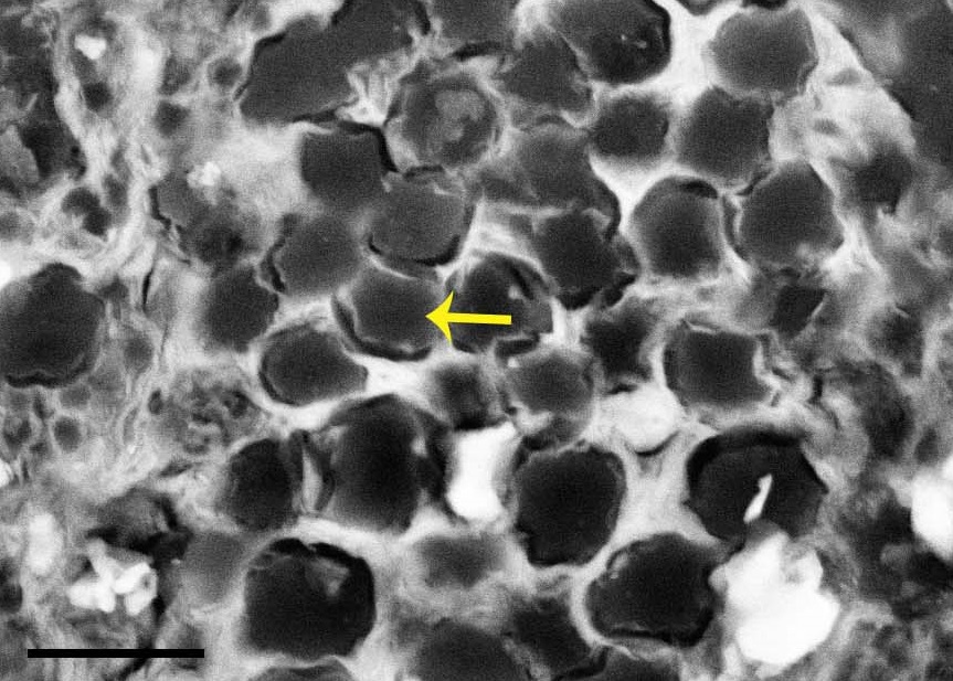
Scanning electron micrograph image of probable tendon or ligament fibres (arrow) in cross-section, from near the ankle joint.
We found preserved cells and other parts of connective tissues including tendons and/or ligaments, fibrocartilage (the tougher kind) and articular cartilage (the softer joint-padding kind). That’s great, although not unique, but the kitchen sink also flushed out even more reductionist data: those tissues included some organic residues, including what appear to be bits of proteins (amino acids); particularly the collagen that makes up tendons.

Fibrocartilage (“fc”) from the ankle joint region.
Hopefully we’re right, and we included as much of the data as we could manage so that others can look at our findings. The specimen is crushed into nearly two dimensions, like all Jehol biota organisms, so its anatomy was hard to interpret but we think we got it right. All of the other kitchen-sinky tools have their own nuances and pitfalls but we did our best with a superb team of experts. We’ve had to wait 125 million years to uncover this specimen and a few more years to find out if we’ve looked at the right way is no greater test of patience.
I thank my coauthors, especially Baoyu Jiang for the kind invitation to participate and the very fun experience of collaborating. I think I’ll remember this study for a long time because, for me, it takes a step beyond just describing Another Case of Jaw-Dropping Fossilization (can you hear the hipsters recounting the excitement and cynicism of the 1990s when this all was dawning? I was there and maybe now I’m one of them). By combining all of those methods we learned new things about the palaeobiology of birds and the evolution of traits within birds. Confuciusornis, not shockingly, had ankles that should have functioned in ways intermediate between those of bog-standard non-avian theropods and modern birds.

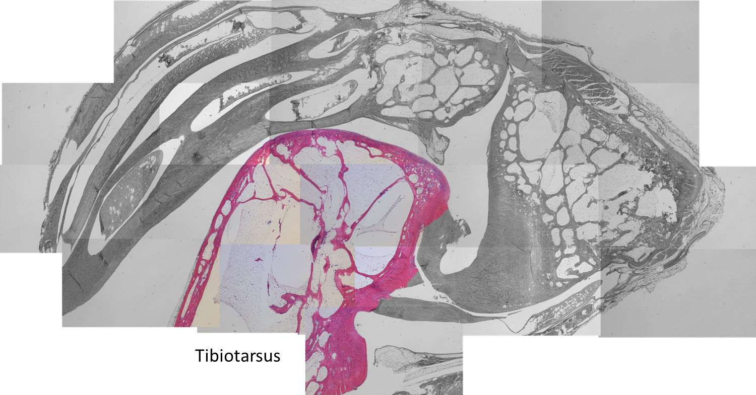
Same anatomical regions in an extant bird as in the main fossil specimen. Left distal tibiotarsus (TT; below) and proximal tarsometatarsus (TMT; above) from an adult helmeted guineafowl (Numida meleagris) after formalin fixation. (from our paper’s Supp Info)
I’m hopeful that more synthesis of molecular/cellular, imaging, biomechanical and other tools (not to mention good old palaeontology and anatomy!) can wash away some more of this mystery. And it was fun to be a part of a study that adds to overwhelming evidence that was heretical ~25 years ago: some hardy biomolecules such as collagen and keratin can survive hundreds of millions of years, not just thousands. Pioneers such as Prof. Mary Schweitzer led the original charge that made reporting on discoveries like ours much easier today.
“I know how the birds fly, how the fishes swim, how animals run. But there is the Dragon. I cannot tell how it mounts on the winds through the clouds and flies through heaven. Today I have seen the Dragon.“– Confucius, ca. 500 BCE.
Let’s finish with some images of a living bird’s ankle region, by co-author and PhD student Sophie Regnault. We considered these for inclusion in the paper but they didn’t fit quite right. I love them anyway so here they are:
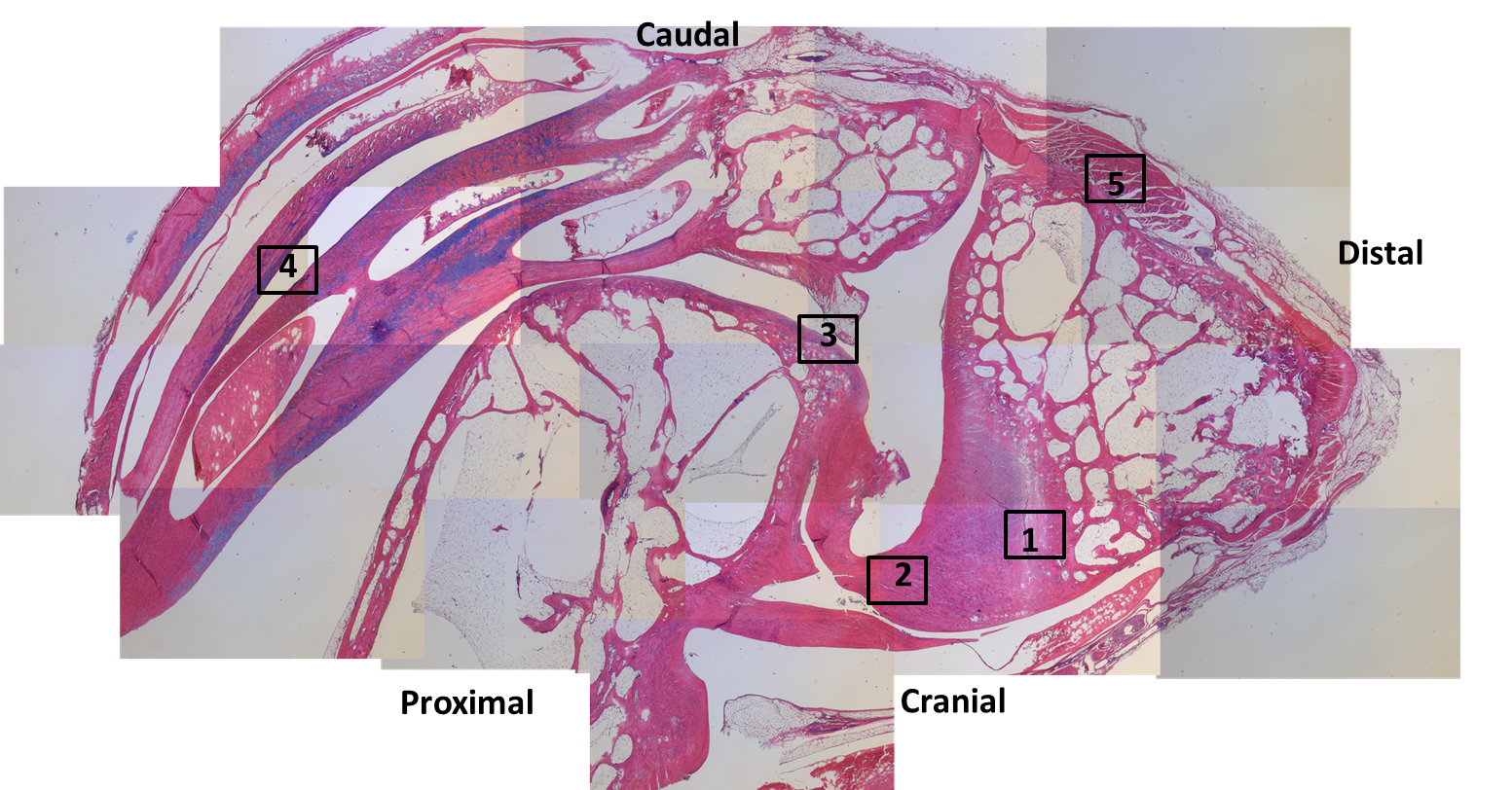
Patchwork of histology slide images, from a guineafowl’s ankle (as per photo above). The numbered squares correspond to zoomed-in images below. The tibiotarsus is on the proximal end (bottom left); the tarsometatarsus is on the distal end (right side); and the enigmatic tarsal sesamoid is at the top. Magnification: 20x overall.


Region 1. nice (fibro)cartilage-bone inferface at ligament insertion.
Region 2: longitudinal slice through ligaments connecting the tibiotarsus to the tarsometatarsus across the ankle joint.

Region 3: front (bottom) of the tibiotarsus/upper ankle.

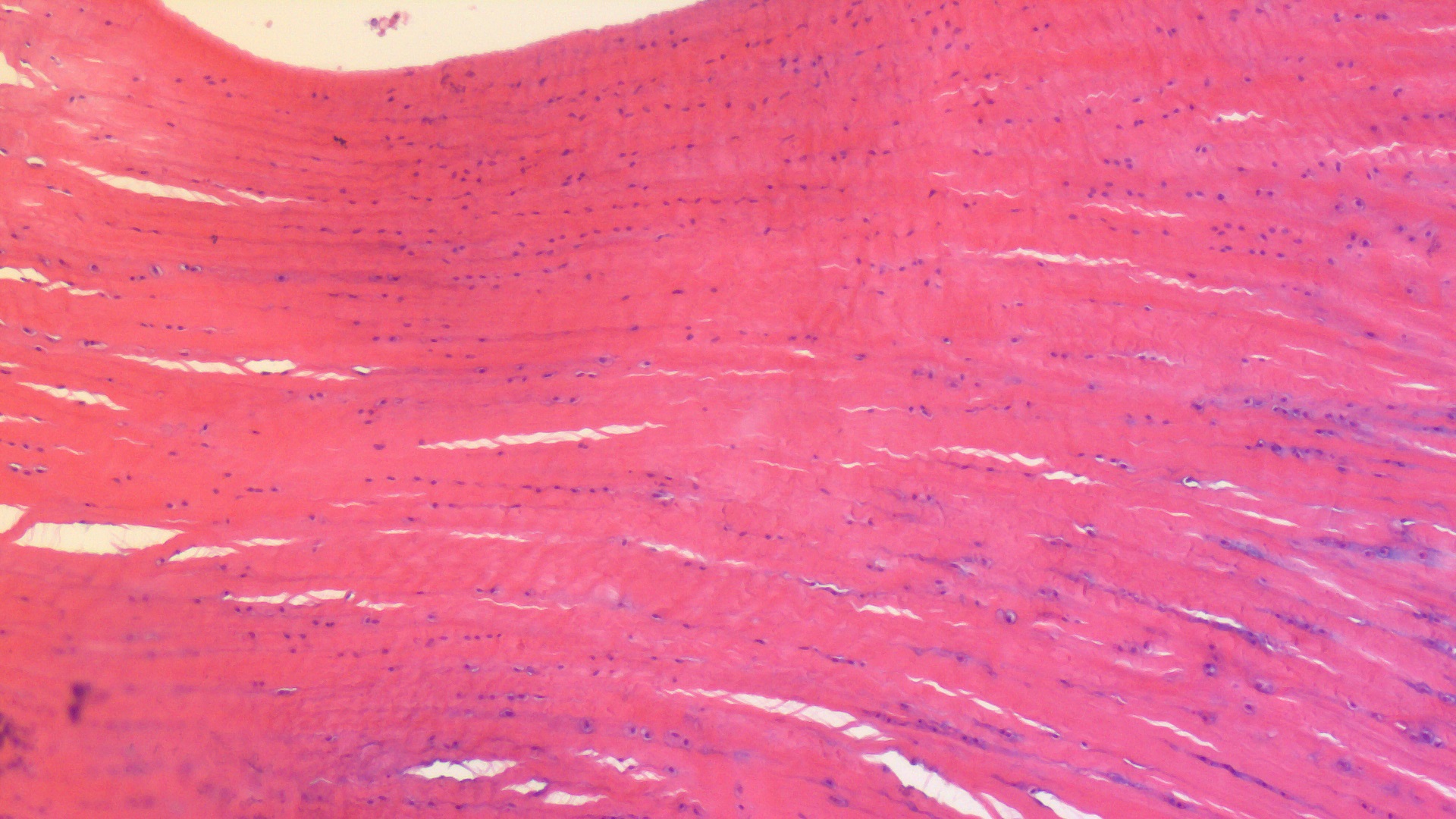
Region 4: tendon fibres in longitudinal section; on the back of the tibiotarsus. Some show mineralization into ossified tendons (“metaplasia”); another curious feature of modern birds.

Region 5: muscle attachment to the back of the upper tarsometatarsus bone. Small sesamoid fragment visible.


Leave a comment