Feeds:
Posts
Comments

Archive for the ‘Better Know A Muscle’ Category

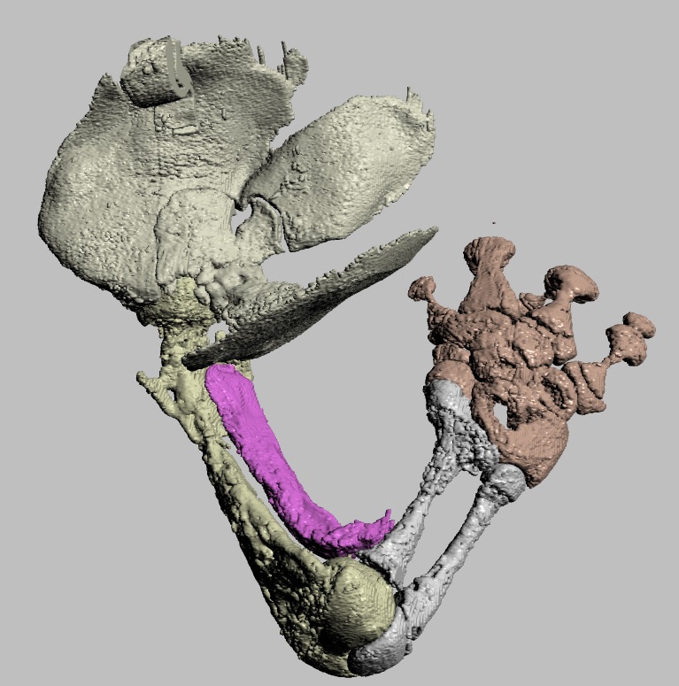
The blog is back! Briefly. With dinosaurs. Back in 2005, I published a paper in which I used a “SIMM” 3D musculoskeletal biomechanical model of Tyrannosaurus rex to analyse its muscle actions and infer a relatively upright hindlimb pose. This was an outcome from my NSF-funded postdoctoral research at Stanford University, in which engineers kindly taught me how to use SIMM (handing me a loaded gun?). Part of my plan all along was to build multiple such models along a rough evolutionary sequence to revisit old questions I had with past, qualitative functional morphology papers from 2000 onwards, and see if biomechanics could quantitatively reveal more about the functional evolution of dinosaur hindlimb muscles. So I got data for modelling some extinct dinosaurs (theropods Dilophosaurus, Allosaurus, Velociraptor) and living birds (Struthio, others) and published nuggets of that but held others back…

Stomach-Churning Rating: -1/10; dinosaurs!

I handed these 3D model data off to my PhD student Vivian Allen in ~2007, charging him with the task of making more models to flesh out the phylogeny and finish what I’d started. And he sure did. He graduated, did a couple of postdocs with me, and we gradually massaged his thesis chapter on this topic into a draft paper. Easier said than done, though! That’s why 14 more years have passed.

Viv came up with some clever tools in MATLAB software code (from which he became a very competent programmer and went on to a successful career in that!) to boil complex data on muscle leverages (moment arms) across a wide range of joint motion for the hindlimbs for each taxon.

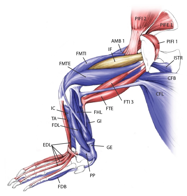
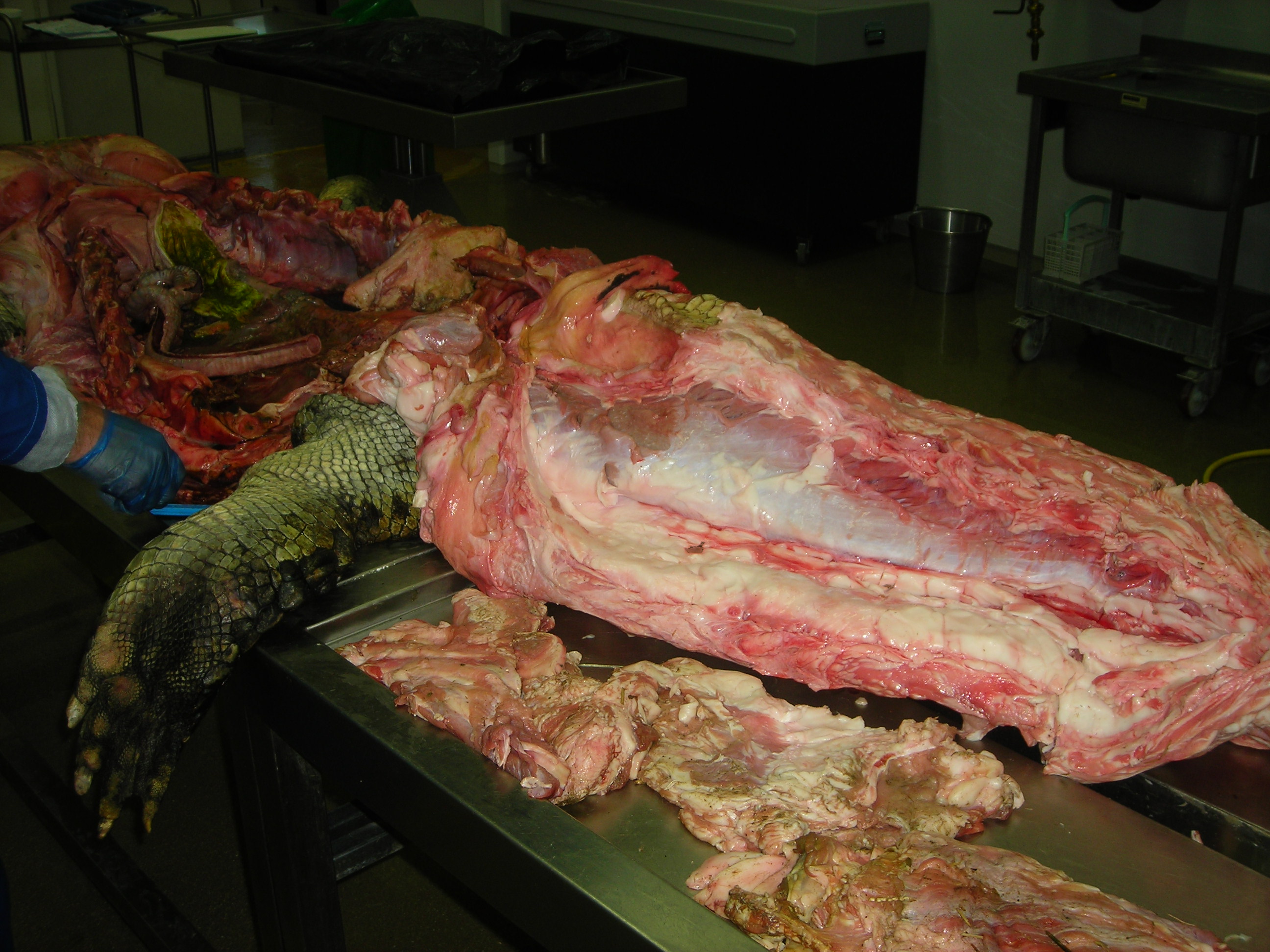
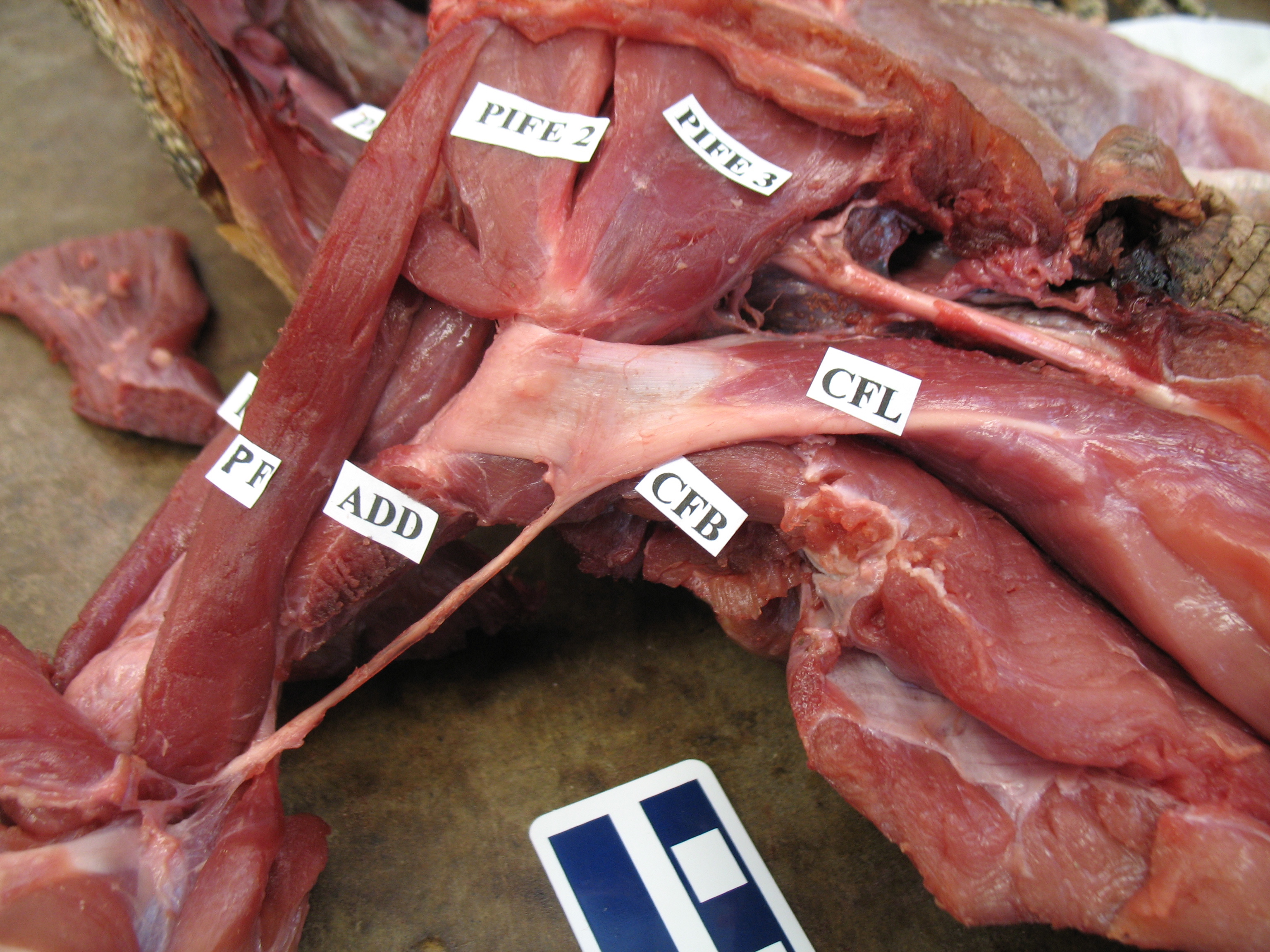
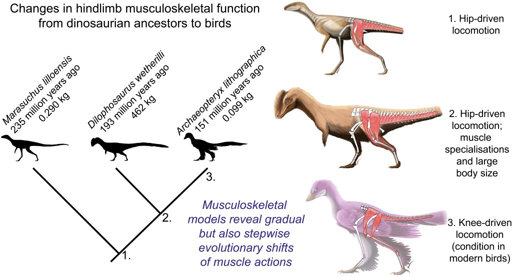
These data then were fed into further code that took the results from all models, ultimately 13 of them from an Australian freshwater crocodile to two living birds and 10 extinct dinosaurs plus close cousin Mara/Lagosuchus (Figure 1). The code expressed these leverages as changes in ancestral values along the main branch of the evolutionary tree from early (Triassic) “ruling reptile” Archosauria (represented here just by the croc as a proxy) to modern birds, and 9 main ancestral “nodes” in between. Our code tracked both how each of 35 hindlimb muscles we modelled evolved in its leverage, as well as overall “average” leverage of functional groups around the hip, knee and ankle joints.

So, back and forth we went for some 10 years playing with the models (see Video below), data and code, and the paper describing the whole thing, slowly closing in on a final version but also sometimes distracted by our other projects and Real Life Stuff like health and children, and concerns about how we conducted this study (i.e. a lot of fiddling).

Figure 1: Evolutionary tree of dinosaurs and their relatives as used in the study, showing all 13 models, species names, and names of groups along the bottom (red nodes) of the tree. Averostra and Avetheropoda were ancestral groups of theropod dinosaurs that the study inferred had particular specialisations of the hindlimb muscles. Right hindlimbs in side view. The limbs are all straightened vertically into a baseline reference posture but the study investigated variation in muscle function across a wide range of limb poses.

Then I got a new grant “DAWNDINOS” that changed the scene for me, refocusing my team’s energies onto the Triassic (and early Jurassic) and the evolutionary biomechanics of diverse archosaurs’ locomotion, assessed with both LOTS of experimental studies of living crocs and birds, and LOTS of predictive simulations of locomotion. Stay tuned for much more on that from our team, but we’ve already published some key steps here. Most notably, we developed an improved protocol for modelling and simulating our animals, as shown by Bishop et al.’s 2021 study of the early theropod Coelophysis bauri (also appearing in the current paper). Awkwardly for me, that new method rendered our old models and methods a bit obsolete (although still fine), so I pushed to publish this current paper with Viv, and brought collaborator Dr. Brandon Kilbourne on board to aid in some final stats, figures and more. That finally did it, and now we’ve published the paper in Science Advances. Deep breath.

Video: Rotating movies of 3 musculoskeletal models from this study. Models have been posed into representative limb orientations illustrating a gradual or stepwise transformation from more upright to more crouched.

Well what’s the paper about, then? We used our 13 models and processed evolutionary functional patterns to test three main questions (hypotheses) about muscle leverage, making educated guesses at what might prevail from early Archosauria to Aves:

  1. Hip extensor / flexor (i.e. femur retractor/protractor) moment arm ratios remained constant. We weren’t sure what to expect, as these antagonists both seem to change a lot on the whole lineage, so we went with this prediction.
  2. Knee flexor / extensor ratios decreased; i.e. the flexors (“hamstrings” etc.) weakened and/or extensors (equivalent of our quadriceps) strengthened their leverage. Anatomy of the knee joint and muscles around it suggests this, plus since Gatesy’s 1990-onwards studies we’ve expected archosaurs to shift from more ‘hip-based’ to more ‘knee-based’ locomotion as we get closer to avian ancestry.
  3. Hip medial (internal) long-axis rotator / abductor (i.e. pronators of the limb vs. those that draw the leg away from the body) ratios increased. This idea comes right from my paper w/Gatesy in 2000, where we surmised that archosaurs shifted from relying on hip adductors (in crocs/other quadrupeds) to abductors (in bipedal dinosaurs; like humans) to medial rotators (‘torsional control’ as in birds today) during weight support.

Moreover, we reconstructed the evolution of 35 muscles’ actions across ~250 million years, which was a new step.

Here’s a summary of what we found (Figure 2):

Figure 2: Short visualization/explanation of the study’s main insights. Pictures by palaeoartist Jaime Headden: https://qilong.wordpress.com/about/ in left side view, including “muscled” and silhouette images. Right side images include representative hip, knee and ankle muscles from the study. Changes such as the enlargement of muscles in front of the hip that straighten the knee, and reduction of the caudofemoralis longus muscle that runs from the tail to the back of the thigh, are evident.

So, overall hypothesis 1 about hip extensors/flexors ended up complicated; rejected because hip flexor leverage actually increased. Furthermore, we found that around the ancestral nodes for early theropod dinosaurs (Neotheropda through Avetheropoda; around 200 Mya), there were peaks in muscle leverage (size-normalized) that surprised us, and persisted despite many different analyses we threw at them over the years. As far as we could tell, these peaks that kept appearing for various muscles’ actions were “real” (estimates). Which meant these ancestors may have had specialised high leverage relative to both their own ancestors and descendants; the peaks got reversed in evolution. These ancestors had some other weird anatomical and functional traits, such as tightly articulated hip joints early on (which they lost later), increased body size in the later forms, more ‘macropredatory’ ecology (e.g. eating sauropods?), and a centre of mass of the body that was shifted forwards (due to big arms and heads/necks). This weirdness is a cool unexpected finding that showed up for the other hypotheses too, and it needs some more investigating. A ‘failed’ hypothesis test led to neat insights.

Figure 3. From the paper– showing our main results for changes in moment arm ratios across archosaurian ancestors. Hip extensors/flexors decreased then increased; knee flexors/extensors decreased; and hip medial rotators/abductors decreased then had a series of increases.

Hypotheses 2 and 3 found good support, on the contrary, overall (Figure 3). We seem to have been able to quantify the shifts from hip-based to knee-based, and abductor-based to medial-rotator-based, muscle actions. I find that very satisfying. Ankle weight support (extension) capacity also increased, which fits morphological changes fairly well. If you’re into archosaur limb muscle form and function, there’s a lot more food for thought in the paper.

Funnily enough, ~20 years has been sufficient time that we could have had plenty more models in this study if we’d delayed it even longer and re-re-re-analysed our data. But we had to draw the line somewhere and not infinitely revise with every new model we’ve been creating. With the current state of musculoskeletal modelling in my group, we could have more than doubled our sample size and fleshed out the most important gaps such as in the crocodile-lineage (extinct Pseudosuchia) and other Triassic forms plus elsewhere. A big challenge remains having some nice 3D-preserved early fossil birds beyond Archaeopteryx; e.g. so many nice Chinese ones are too flat (e.g. joints we need) to reliably model here. It’s something that can still be done and is worth doing, but I suspect the general trends we’ve found along the dinosaur lineage are “correct”.

What’s personally important to me about this paper is (1) how it not only bridges a huge morphofunctional gap across archosaur evolution in scientific terms, and (2) how we’ve completed a long-delayed project with stubbornness (and during a pandemic!), but also (3) how it bridges my past career from my PhD and postdoc to the present work with DAWNDINOS. We’re now forging well beyond what this new paper has done in terms of truly testing, as best we can (estimate) so far, how limb muscles of archosaurs functioned and evolved, and how these contributed to particular behaviours and performance (maybe even palaeoecology and evolutionary success/extinction?). The current paper is just simple modelling of muscle leverage, but leverage is only one (very important!) piece of muscle function and performance. With fully dynamic, anatomically integrative, physiologically and physically representative biomechanical computer simulations that predict what living and extinct archosaurs could or could not do, we can do even better. So watch for that! Hopefully it won’t all take 20 years, or 250 million.

Read Full Post »

Nice GIF of the human biceps in action- By Niwadare - Own work, CC BY-SA 4.0, https://commons.wikimedia.org/w/index.php?curid=38718790

GIF of the human biceps (above) and its antagonist triceps (below) in action- By “Niwadare” – own work, CC BY-SA 4.0, https://commons.wikimedia.org/w/index.php?curid=38718790

Last year on Darwin Day I debuted “Better Know A Muscle” (BKAM), which was intended to be a series of posts focusing on one cool muscle at a time, and its anatomical, functional and evolutionary diversity and history. A year later, it’s another post on another muscle! Several dozen more muscles to go, so I’ve got my work cut out for me… But today: get ready to FLEX your myology knowledge! Our subject is Musculus biceps brachii; the “biceps” (“two-headed muscle of the arm”). Beloved of Arnie and anatomists alike, the biceps brachii is. Let’s get pumped up!

Stomach-Churning Rating: 7/10. Lots of meaty elbow flexion!

While the previous BKAM’s topic was a hindlimb muscle with a somewhat complex history (and some uncertainties), the biceps brachii is a forelimb muscle with a simpler, clearer history. Fish lack a biceps, just having simple fin ab/adductor muscles with little differentiation. Between fish and tetrapods (limb-bearing vertebrates), there was an explosion in the number of muscles; part of transforming fins into limbs; and the biceps is thenceforth evident in all known tetrapods in a readily identifiable anatomical form. In salamanders and their amphibian kin, there is a muscle usually called “humeroantebrachialis” that seems to be an undivided mass corresponding to the biceps brachii plus the brachialis (shorter humerus-to-elbow) muscle:

Most of the humerobrachialis muscle (purplish colour), in dorsal (top) view of the right forelimb of the fire salamander Salamandra salamandra (draft from unpublished work by my team).

Most of the humerobrachialis muscle (purplish colour), in dorsal (top) view of the right forelimb of the fire salamander Salamandra salamandra (draft from unpublished work by my team).

In all other tetrapods; the amniote group (reptiles, mammals, etc.); there is a separate biceps and brachialis, so these muscles split up from the ancestrally single “humeroantebrachialis” muscle sometime after the amphibian lineage diverged from the amniotes. And not much changed after then– the biceps is a relatively conservative muscle, in an evolutionary (not political!) sense. In amniote tetrapods that have a biceps, it develops as part of the ventral mass of the embryonic forelimb along with other muscles such as the shorter, humerus-originating brachialis, from which it diverges late in development (reinforcing that these two muscles are more recent evolutionary divergences, too).

Biceps brachialis or humerobrachialis, the “biceps group” tends to originate just in front of the shoulder (from the scapula/coracoid/pectoral girdle), running in front of (parallel to) the humerus. It usually forms of two closely linked heads (hence the “two heads” name), most obviously in mammals; one head is longer and comes from higher/deeper on the pectoral girdle, whereas the other is closer to the shoulder joint and thus is shorter. The two heads fuse as they cross the shoulder joint and we can then refer to them collectively as “the biceps”. It can be harder to see the longer vs. shorter heads of the biceps in non-mammals such as crocodiles, or they may be more or less fused/undifferentiated, but that’s just details of relatively minor evolutionary variation.

The biceps muscle then crosses in front of the elbow to insert mainly onto the radius (bone that connects your elbow to your wrist/thumb region) and somewhat to the ulna (“funny bone”) via various extra tendons, fascia and/or aponeuroses. The origin from the shoulder region tends to have a strong mark or bony process that identifies it, such as the coracoid process in most mammals (I know this well as I had my coracoid process surgically moved!). The insertion onto the radius tends to have a marked muscle scar (the radial tuberosity or a similar name), shared with the brachialis to some degree. A nice thing about the biceps is that, because it may leave clear tendinous marks on the skeleton, we sometimes can reconstruct how its attachments and path evolved (and any obvious specializations; even perhaps changes of functions if/when they happened).

Here are some biceps examples from the world of crocodiles:

Crocodile's right forelimb showing the huge pectoralis, and the biceps underlying it on the bottom right.

Crocodile’s right forelimb showing the huge pectoralis, and the biceps underlying it; on the bottom right (“BB”- click to embiceps it).

Crocodile left forelimb with biceps visible (

Crocodile left forelimb with biceps visible (“BB”) on the left.

Crocodile biceps muscle cut off, showing the proximal and distal tendons (and long parallel muscle fibres) for a typical amniote vertebrate.

Crocodile biceps muscle cut off, showing the proximal (to right) and distal (to left) tendons (and long parallel muscle fibres) for a typical amniote vertebrate.

What does the biceps muscle do? It flexes (draws forward) the shoulder joint/humerus, and does the same for the elbow/forearm while supinating it (i.e. rotating the radius around the ulna so that the palm faces upwards, in animals like us who can rotate those two bones around each other). In humans, which have had their biceps muscles studied by far the most extensively, we know for example that the biceps is most effective at flexing the elbow (e.g. lifting a dumbbell weight) when the elbow is moderately straight. These same general functions (shoulder and elbow flexion; with some supination) prevail across the biceps muscle of [almost; I am sure there are exceptions] all tetrapods, because the attachments and path of the biceps brachii are so conservative.

And this flexor function of the biceps brachii stands in contrast to our first BKAM muscle, the caudofemoralis (longus): that muscle acts mainly during weight support (stance phase) as an antigravity/extensor muscle, whereas the flexor action of biceps makes it more useful as a limb protractor or “swing phase” muscle used to collapse the limb and draw it forwards during weight support. However, mammals add some complexity to that non-supportive function of the biceps…

Hey mammals! Show us your biceps!

Jaguar forelimb with biceps peeking out from the other superficial muscles, and its cousin brachialis nicely visible.

Jaguar forelimb with biceps peeking out from the other superficial muscles, and its cousin brachialis nicely visible, running along the front of the forearm for a bit.

Elephant's left forelimb with the biceps labelled.

Elephant’s left forelimb with the biceps labelled.

Longitudinal slice thru the biceps of an elephant, showing the internal tendon.

Longitudinal slice thru the biceps of an elephant, showing the internal tendon that helps identify where the two bellies of the biceps fuse.

In certain mammals; the phylogenetic distribution of which is still not clear; the biceps brachii forms a key part of a passive “stay apparatus” that helps keep the forelimb upright against gravity while standing (even sleeping). The classic example is in horses but plenty of other quadrupedal mammals, especially ungulate herbivores, show evidence of similar traits:

Giraffe biceps cut away proximally to show the

Giraffe biceps cut away proximally to show the “stay apparatus” around the shoulder joint (upper right).

Zooming in on the

Zooming in on the “stay apparatus”; now in proximal view, with the biceps tendon on the left and the humeral head (showing some arthritic damage) on the right, with the groove for the biceps in between.

Hippo's humerus (upper left) and biceps muscle cut away proximally, displaying the same sort of

Hippo’s humerus (upper left) and biceps muscle cut away proximally, displaying the same sort of “stay apparatus” as in the giraffe. Again, note the stout proximal and distal tendons of the biceps. The proximal tendon fits into the groove of the humerus on the far left side of the image; becoming constrained into a narrow circular “tunnel” there. It’s neat to dissect that region because of its fascinating relationships between bone and soft tissues.

The biceps brachii, in those mammals with a stay apparatus, seems to me to have a larger tendon overall, especially around the shoulder, and that helps brace the shoulder joint from extending (retracting) too far backward, whilst also transmitting passive tension down the arm to the forearm, and bracing the elbow (as well as distal joints via other muscles and ligaments). It’s a neat adaptation whose evolution still needs to be further inspected.

Otherwise, I shouldn’t say this but the biceps is sort of boring, anatomically. Whether you’re a lizard, croc, bird or mammal, a biceps is a biceps is a biceps; more or less-ceps. But the biceps still has a clear evolutionary history and Darwin would gladly flex his biceps to raise a pint in toast to it.

So now we know a muscle better. That’s two muscles now. And that is good; be you predator or prey. Let’s shake on it!

Read Full Post »

Happy Darwin Day from the frozen tundra sunny but muddy, frosty lands of England! I bring you limb muscles as peace offerings on this auspicious day. Lots of limb muscles. And a new theme for future blog posts to follow up on: starting off my “Better Know A Muscle” (nod to Stephen Colbert; alternative link) series. My BKAM series intends to walk through the evolutionary history of the coolest (skeletal/striated) muscles. Chuck Darwin would not enjoy the inevitable blood in this photo-tour, but hopefully he’d like the evolution. Off we go, in search of better knowledge via an evolutionary perspective!

There is, inarguably, no cooler muscle than M. caudofemoralis longus, or CFL for short. It includes the largest limb muscles of any land animal, and it’s a strange muscle that confused anatomists for many years– was it a muscle of the body (an axial or “extrinsic” limb muscle, directly related to the segmented vertebral column) or of the limbs (an “abaxial” muscle, developing with the other limb muscles from specific regions of the paraxial mesoderm/myotome, not branching off from the axial muscles)? Developmental biologists and anatomists answered that conclusively over the past century: the CFL is a limb muscle, not some muscle that lost its way from the vertebral column and ended up stranded on the hindlimb.

The CFL is also a muscle that we know a fair amount about in terms of its fossil record and function, as you may know if you’re a dinosaur fan, and as I will quickly review later. We know enough about it that we can even dare to speculate if organisms on other planets would have it. Well, sort of…

Stomach-Churning Rating: 8/10. Lots of meaty, bloody, gooey goodness, on and on, for numerous species. This is an anatomy post for those with an appetite for raw morphology.

Let’s start from a strong (and non-gooey) vantage point, to which we shall return. The CFL in crocodiles and most other groups is (and long was) a large muscle extending from much of the front half or so of the tail to the back of the femur (thigh bone), as shown here:

Julia Molnar's fabulous illustration of Alligator's limb muscles, from our 2014 paper in Journal of Anatomy.

Julia Molnar’s fabulous illustration of Alligator‘s limb muscles, from our 2014 paper in Journal of Anatomy. Note the CFL in blue at the bottom right.

As the drawing shows, the CFL has a friend: the CFB. The CFB is a shorter, stumpier version of the CFL restricted to the tail’s base, near the hip. The “B” in its name means “brevis”, or runty. It gets much less respect than its friend the CFL. Pity the poor CFB.

But look closer at the CFL in the drawing above and you’ll see a thin blue tendon extending past the knee to the outer side of the lower leg. This is the famed(?) “tendon of Sutton“, or secondary tendon of the CFL. So the CFL has two insertions, one on the femur and one (indirectly) onto the shank. More about that later.

Together, we can talk about these two muscles (CFL and CFB) as the caudofemoralis (CF) group, and the name is nice because it describes how they run from the tail (“caudo”) to the femur (“femoralis”). Mammal anatomists were late to this party and gave mammal muscles stupidly unhelpful names like “gluteus” or “vastus” or “babalooey”. Thanks.

But enough abstract drawings, even if they rock, and enough nomenclature. Here is the whopping big CFL muscle of a real crocodile:

Huge Nile crocodile, but a relatively small CFL.

Huge Nile crocodile, but a relatively small CFL.

Bigger crocs have smaller legs and muscles.

Bigger crocs have smaller legs and thus smaller leg muscles, relatively speaking. CFL at the top, curving to the left.

The giant Nile croc's CFL muscle removed for measurements.

The giant Nile croc’s CFL muscle removed for measurements. 2.35 kg of muscle! Not shabby for a 278 kg animal.

However, maybe crocodile and other archosaur CFL muscles are not “average” for leggy vertebrates? We can’t tell unless we take an evolutionary tack to the question.

Where did the CFL come from, you may ask? Ahh, that is shrouded in the fin-limb transition‘s mysteries. Living amphibians such as salamanders have at least one CF muscle, so a clear predecessor to the CFL (and maybe CFB) was present before reptiles scampered onto the scene.

But going further back through the CF muscles’ history, into lobe-finned fish, becomes very hard because those fish (today) have so few fin muscles that, in our distant fishy ancestors, would have given rise eventually to the CF and other muscle groups. With many land animals having 30+ hindlimb muscles, and fish having 2-8 or so, there obviously was an increase in the number of muscles as limbs evolved from fins. And because a limb has to do lots of difficult three-dimensional things on land while coping with gravity, more muscles to enable that complex control surely were needed.

OK, so there were CF muscles early in tetrapod history, presumably, anchored on that big, round fleshy tail that they evolved from their thin, finned fishy one — but what happened next? Lizards give us some clues, and their CFL muscles aren’t all that different from crocodiles, so the CFL’s massive size and secondary “tendon of Sutton” seems to be a reptile thing, at least.

Courtesy of Emma Schachner, a large varanid lizard's very freshly preserved CFL and other hindlimb muscles.

Courtesy of Emma Schachner, a large varanid lizard’s very freshly preserved CFL and other hindlimb muscles.

Courtesy of Emma Schachner, zoomed in on the tendons and insertions of the CFL muscle and others.

Courtesy of Emma Schachner, zoomed in on the tendons and insertions of the CFL muscle and others. Beautiful anatomy there!

Looking up at the belly of a basilisk lizard and its dissected right leg, with the end of the CFL labelled.

Looking up at the belly of a basilisk lizard and its dissected right leg, with the end of the CFL labelled. It’s not ideally dissected here, but it is present.

An unspecified iguanid(?) lizard, probably a juvenile Iguana iguana, dissected and showing its CFL muscle at its end. The muscle would extemd about halfway down the tail, though.

An unspecified iguanid(?) lizard, probably a juvenile Iguana iguana, dissected to reveal its CFL muscle near its attachment to the femur. The muscle would extend further, about halfway down the tail, though.

Let’s return to crocodiles, for one because they are so flippin’ cool, and for another because they give a segue into archosaurs, especially dinosaurs, and thence birds:

A moderate-sized (45kg) Nile crocodile with its CFL muscle proudly displayed.

A moderate-sized (45kg) Nile crocodile with its CFL muscle proudly displayed. Note the healthy sheath of fat (cut here) around the CFL.

American alligator's CFL dominates the photo. Photo by Vivian Allen.

American alligator’s CFL dominates the photo [by Vivian Allen].

Black caiman, Melanosuchus, showing off its CFL muscle (pink "steak" in the middle of the tail near the leg).

Black caiman, Melanosuchus, showing off its CFL muscle (pink “steak” in the middle of the tail near the leg), underneath all that dark armour and fatty superficial musculature.

A closer look at the black caiman's thigh and CFL muscle.

A closer look at the black caiman’s thigh and CFL muscle.

Like I hinted above, crocodiles (and the anatomy of the CFL they share with lizards and some other tetrapods) open a window into the evolution of unusual tail-to-thigh muscles and locomotor behaviours in tetrapod vertebrates.

Thanks in large part to Steve Gatesy’s groundbreaking work in the 1990s on the CFL muscle, we understand now how it works in living reptiles like crocodiles. It mainly serves to retract the femur (extend the hip joint), drawing the leg backwards. This also helps support the weight of the animal while the foot is on the ground, and power the animal forwards. So we call the CFL a “stance phase muscle”, referring to how it mainly plays a role during ground contact and resisting gravity, rather than swinging the leg forwards (protracting the limb; i.e. as a “swing phase muscle”).

The “tendon of Sutton” probably helps to begin retracting the shank once the thigh has moved forward enough, facilitating the switch from stance to swing phase, but someone really needs to study that question more someday.

And thanks again to that same body of work by Gatesy (and some others too), we also understand how the CFL’s anatomy relates to the underlying anatomy of the skeleton. There is a large space for the CFL to originate from on the bottom of the tail vertebrae, and a honking big crest (the fourth trochanter) on the femur in most reptiles that serves as the major attachment point, from which the thin “tendon of Sutton” extends down past the knee.

Femur bones (left side) from an adult ostrich (Left) and Nile crocodile (Right).

Femur bones (left side; rear view) from an adult ostrich (left) and Nile crocodile (right). Appropriate scale bar is appropriate. The fourth trochanter for the CFL is visible in the crocodile almost midway down the femur. Little is left of it in the ostrich but there is a bumpy little muscle scar in almost the same region as the fourth trochanter, and this is where the same muscle (often called the CFC; but it is basically just a small CFL) attaches.

That relationship of the CFL’s muscular anatomy and the underlying skeleton’s anatomy helps us a lot! Now we can begin to look at extinct relatives of crocodiles; members of the archosaur group that includes dinosaurs (which today we consider to include birds, too), and things get even more interesting! The “tendon of Sutton”, hinted at by a “pendant” part of the fourth trochanter that points down toward the knee, seems to go away multiple times within dinosaurs. Bye bye! Then plenty more happens:

A large duckbill dinosaur's left leg, with a red line drawn in showing roughly where the CFL would be running, to end up at the fourth trochanter. Many Mesozoic dinosaurs have skeletal anatomy that indicates a similar CFL muscle.

A large duckbill dinosaur’s left leg, with a red line drawn in showing roughly where the CFL would be running, to end up at the fourth trochanter. Many Mesozoic dinosaurs have skeletal anatomy that indicates a similar CFL muscle.

We can even go so far as to reconstruct the 3D anatomy of the CFL in a dinosaur such as T. rex ("Sue" specimen here; from Julia Molnar's awesome illustration in our 2011 paper), with a fair degree of confidence.

We can even go so far as to reconstruct the 3D anatomy of the CFL in a dinosaur such as T. rex (“Sue” specimen here; from Julia Molnar’s awesome illustration as part of our 2011 paper), with a fair degree of confidence. >180kg steak, anyone?

As we approach birds along the dinosaur lineage, the tail gets smaller and so does the fourth trochanter and thus so must the CFL muscle, until we’re left with just a little flap of muscle, at best. In concert, the hindlimbs get more crouched, the forelimbs get larger, flight evolves and voila! An explosion of modern bird species!

Ozburt (72)

Left femur of an ostrich in side view (hip is toward the right side) showing many muscles that attach around the knee (on the left), then the thin strap of CF muscle (barely visible; 2nd from the right) clinging near the midshaft of the femur.

Another adult ostrich's CF muscle complex, removed for study.

Another adult ostrich’s CF muscle complex, removed for study. Not enough ostrich myology for you yet? Plenty more in this old post! Or this one! Or this one… hey maybe I need to write less about ostriches? The CF muscle complex looks beefy but it’s no bigger than any other of the main hindlimb muscles, unlike the CFL in a crocodile or lizard, which puts everything else to shame!

STILL not enough ostrich for you yet? Take a tour of the major hindlimb muscles in this video:

And check out the limited mobility of the hip joint/femur here. No need for much femur motion when you’re not using your hip muscles as much to drive you forwards:

But I must move on… to the remainder of avian diversity! In just a few photos… Although the CF muscles are lost in numerous bird species, they tend to hang around and just remain a long, thin, unprepossessing muscle:

Chicken's right leg in side view. CFC (equivalent of CFL) muscle outlined and labelled.

Chicken’s right leg in side view. CFC muscle (equivalent of CFL; the ancestral CFB is confusingly called the CFP in birds, as it entirely resides on the pelvis) outlined and labelled.

A jay (species?) dissected to show some of the major leg muscles, including the CF. Photo by Vivian Allen.

A jay (species? I forget) dissected to show some of the major leg muscles, including the CFL-equivalent muscle; again, smallish. [Photo by Vivian Allen]

Finally, what’s up with mammals‘ tail-to-thigh CF-y muscles? Not much. Again, as in birds: smaller tail and/or femur, smaller CF muscles. Mammals instead depend more on their hamstring and gluteal muscles to support and propel themselves forward.

But many mammals do still have something that is either called the M. caudofemoralis or is likely the same thing, albeit almost always fairly modest in size. This evolutionary reduction of the CF muscle along the mammal (synapsid) lineage hasn’t gotten nearly as much attention as that given to the dinosaur/bird lineage’s CFL. Somebody should give it a thoroughly modern phylogenetic what-for! Science the shit outta that caudofemoralis…

Yet, oddly, to give one apparent counter-example, cats (felids) have, probably secondarily, beefed up their CF muscle a bit:

Cats have a pretty large CF muscle in general, and this jaguar is no exception! But mammals still tend to have fairly wimpy tails and thus CF muscles, or they even lose them (e.g. us?).

Cats have a pretty large CF muscle in general, and this jaguar is no exception! But mammals still tend to have fairly wimpy tails and thus CF muscles, or they even lose them (e.g. us?). [photo by Andrew Cuff, I think]

In summary, here’s what happened (click to embeefen):

Better Know A Muscle: The Evolution of M. caudofemoralis (longus)

Better Know A Muscle: the evolution of M. caudofemoralis (longus).

I hope you enjoyed the first BKAM episode!
I am willing to hear requests for future ones… M. pectoralis (major/profundus) is a serious contender.

P.S. It was Freezermas this week! I forgot to mention that. But this post counts as my Freezermas post for 2016; it’s all I can manage. Old Freezermas posts are here.

Read Full Post »