(John: here’s a guest post from my former PhD student, soon to be 100% legit PhD, Dr., and all that jazz, Julia Molnar!)
This is my first guest post, but I have been avidly following what’s in John’s freezer (and the blog too) for quite a while. I joined the lab in 2009 and left a month ago on the bittersweet occasion of surviving my PhD viva (oral exam/defense), so I’d like to take a moment here to thank John and the Structure & Motion Lab for a great 4 years!
Moving on to freezer-related matters; specifically, a bunch of frozen crocodile spines. It was late 2011, and the reason for the spines in John’s freezer was that John, Stephanie Pierce, and I were trying to find out more about crocodile locomotion. This was anticipated to become my first major, first-author research publication (but see my Palaeontologia Electronica paper on a related subject), and I was about to find out that these things seldom go as planned; for example, the article would not be published for more than three years (the research took a long time!). Before telling the story of how it lurched and stumbled toward eventual publication, I’ll give you some background on the project.
Stomach-Churning Rating: 3/10; x-ray of dead bits and nothing much worse.
A stumbly sort-of-bounding crocodile. They can do better.
First of all, why crocodiles? For one thing, they’re large, semi-terrestrial animals, but they use more sprawling postures than typical mammals. Along with alligators and gharials, they are the only living representatives of Crocodylomorpha, a 200+ million year-old lineage that includes wolf-like terrestrial carnivores, fish-like giants with flippers and a tail fin, even armored armadillo-like burrowers. Finally, crocodiles are interesting in their own right because they use a wide variety of gaits, including bounding and galloping, which are otherwise known only in mammals.
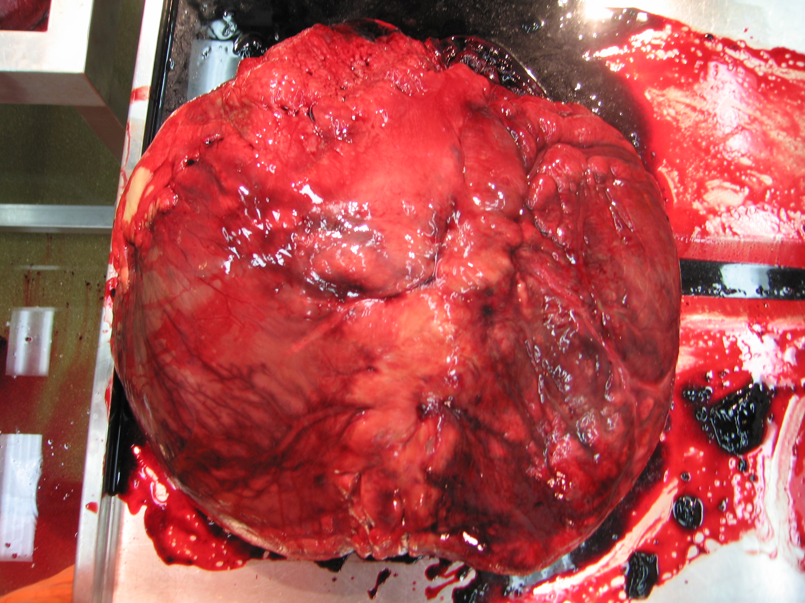
Nile crocodile skeletal anatomy
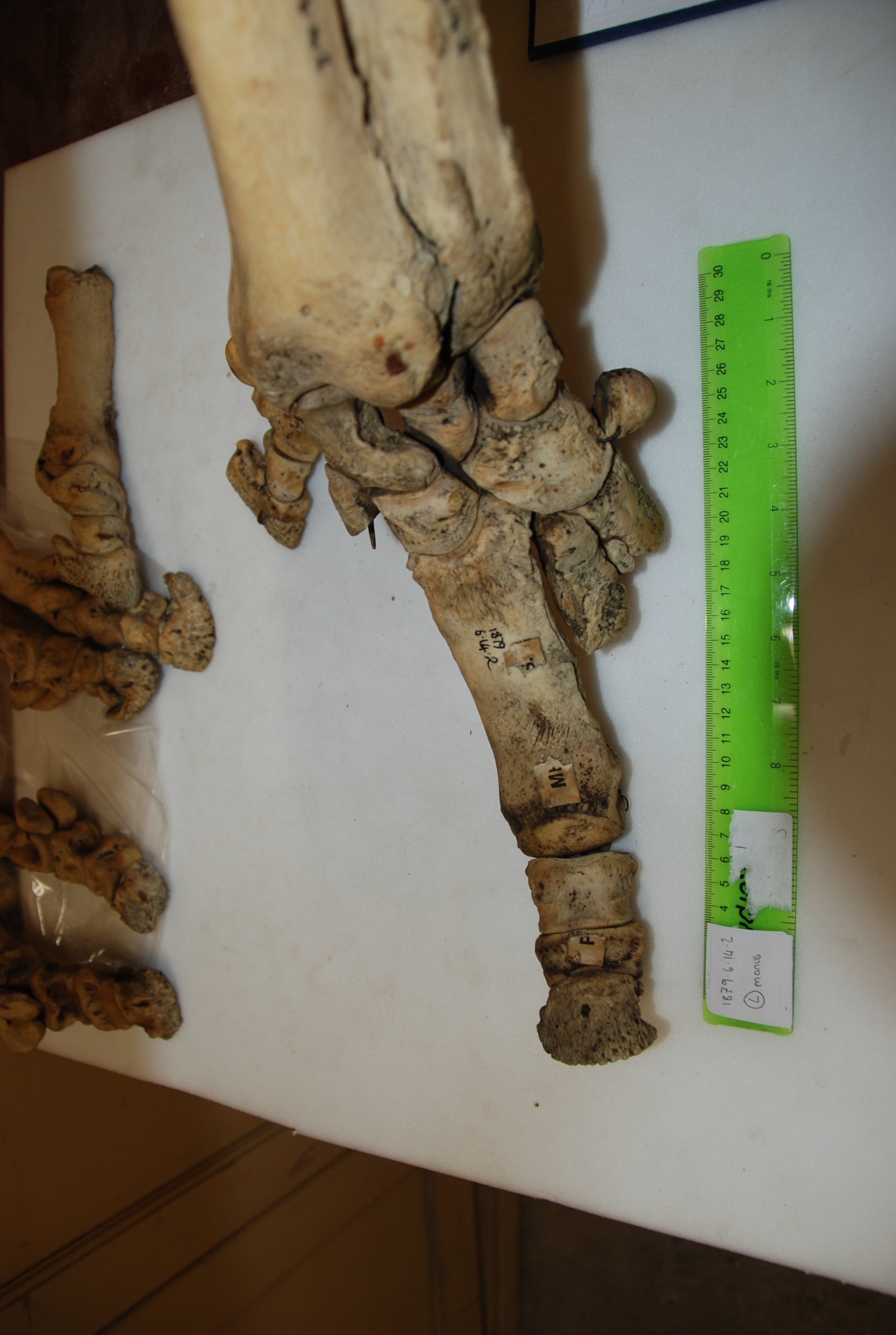
OK, so why spines? Understanding how the vertebral column works is crucial to understanding locomotion and body support on land, and inter-vertebral joint stiffness (how much the joints of the backbone resist forces that would move them in certain directions) in particular has been linked to trunk movements in other animals. For this reason, vertebral morphology is often used to infer functional information about extinct animals, including dinosaurs. However, vertebral form-function relationships have seldom been experimentally tested, and tests on non-mammals are particularly scarce. So we thought the crocodile spines might be able to tell us more about the relationship between vertebral morphology, mechanics, and locomotion in a broader sample of vertebrate animals. If crocodile spine morphology could be used to predict joint stiffness, then morphological measurements of extinct crocodile relatives would have some more empirical heft to them. Several skeletal features seem to play roles such as levers to mechanically stiffen crocodile spines (click to emcroc’en):
Anatomy of a crocodile vertebra
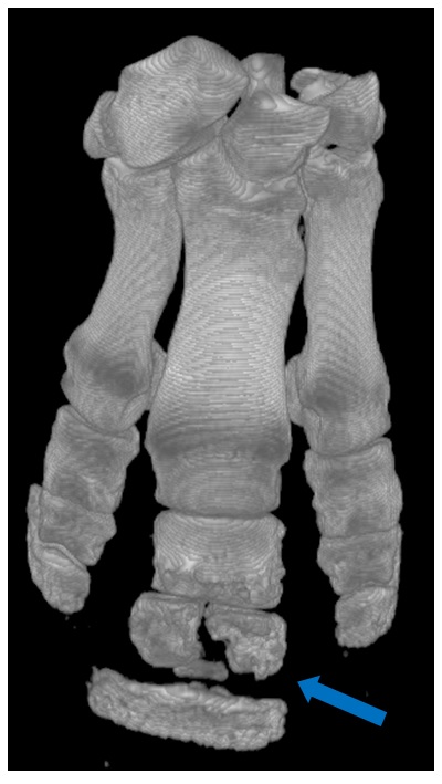
We decided to use a very simple technique that could be replicated in any lab to measure passive stiffness in crocodile cadavers. We dissected out individual joints were and loaded with known weights. From the movement of the vertebrae and the distance from the joint, we calculated how much force takes to move the joint a certain number of degrees (i.e. stiffness).
Me with crocodile vertebra and G-clamp
X-ray of two crocodile vertebrae loaded with a metric weight to calculate their joint’s stiffness
Afterwards, we boiled the joints to remove the soft tissues – the smell was indescribable! We took 14 measurements from each vertebra. All of these measurements had been associated with stiffness or range of motion in other studies, so we thought they might be correlated with stiffness in crocodiles also.
Some of the vertebral measurements that were related to stiffness
Despite my efforts to keep it simple, the process of data collection and analysis was anything but. I recall and exchange with Stephanie Pierce that went something like this:
Stephanie: “How’s it going?”
Me: “Well, the data are messy, I’m not seeing the trends I expected, and everything’s taking twice as long as it was supposed to.”
Stephanie: “Yes, that sounds like science.”
That was the biggest lesson for me: going into the project, I had been unprepared for the amount of bumbling around and re-thinking of methods when the results were coming up implausible or surprising. In this case there were a couple of cool surprises: for one thing, crocodiles turn out to have a very different pattern of inter-vertebral joint stiffness than typical mammals: while mammals have stiff thoracic joints and mobile lumbar joints, crocodiles have stiffer lumbar joints. Many mammals use large lumbar movements during bounding and galloping, so crocodiles must use different axial mechanics than mammals, even during similar gaits. While that’s not shocking (they did evolve their galloping and bounding gaits, and associated anatomy, totally independently), it is neat that this result came out so clearly. Another unexpected result was that, although several of our vertebral measurements were correlated with stiffness, some of the best predictors of stiffness in mammals from previous studies were not correlated with stiffness in crocodiles. The study tells a cautionary tale about making assumptions about extinct animals using data from only a subset of their living relatives or intuitive ideas about form and function.
Finally, the experience of doing the experiments and writing the paper got me interested in other aspects of crocodilian functional anatomy. For instance, how does joint stiffness interact with other factors, such as muscle activity and properties of the ribs, skin, and armor in living crocodiles? Previous studies by Frey and Salisbury had commented on this, but the influence of those factors is less tractable to experiment on or model than just naked backbones with passively stiff joints. In the future, I’d like to study vertebral movements during locomotion in crocodiles – especially during bounding and galloping – to find out how these patterns of stiffness relate to movement. In the meantime, our study shows that, to a degree, crocodile backbone dimensions do give some clues about joint stiffness and locomotor function.
To find out more, read the paper! It was just featured in Inside JEB.
Julia Molnar, Stephanie Pierce, John Hutchinson (2014). An experimental and morphometric test of the relationship between vertebral morphology and joint stiffness in Nile crocodiles (Crocodylus niloticus). The Journal of Experimental Biology 217, 757-768 link here and journal’s “Inside JEB” story
Why should you care
If you have to trim my hooves?
I’ve got to move with good feet
Or be put down fast.
I know I should trot
But my old vet she cares a lot.
And I’m still living on stone
Even though these feet won’t last.
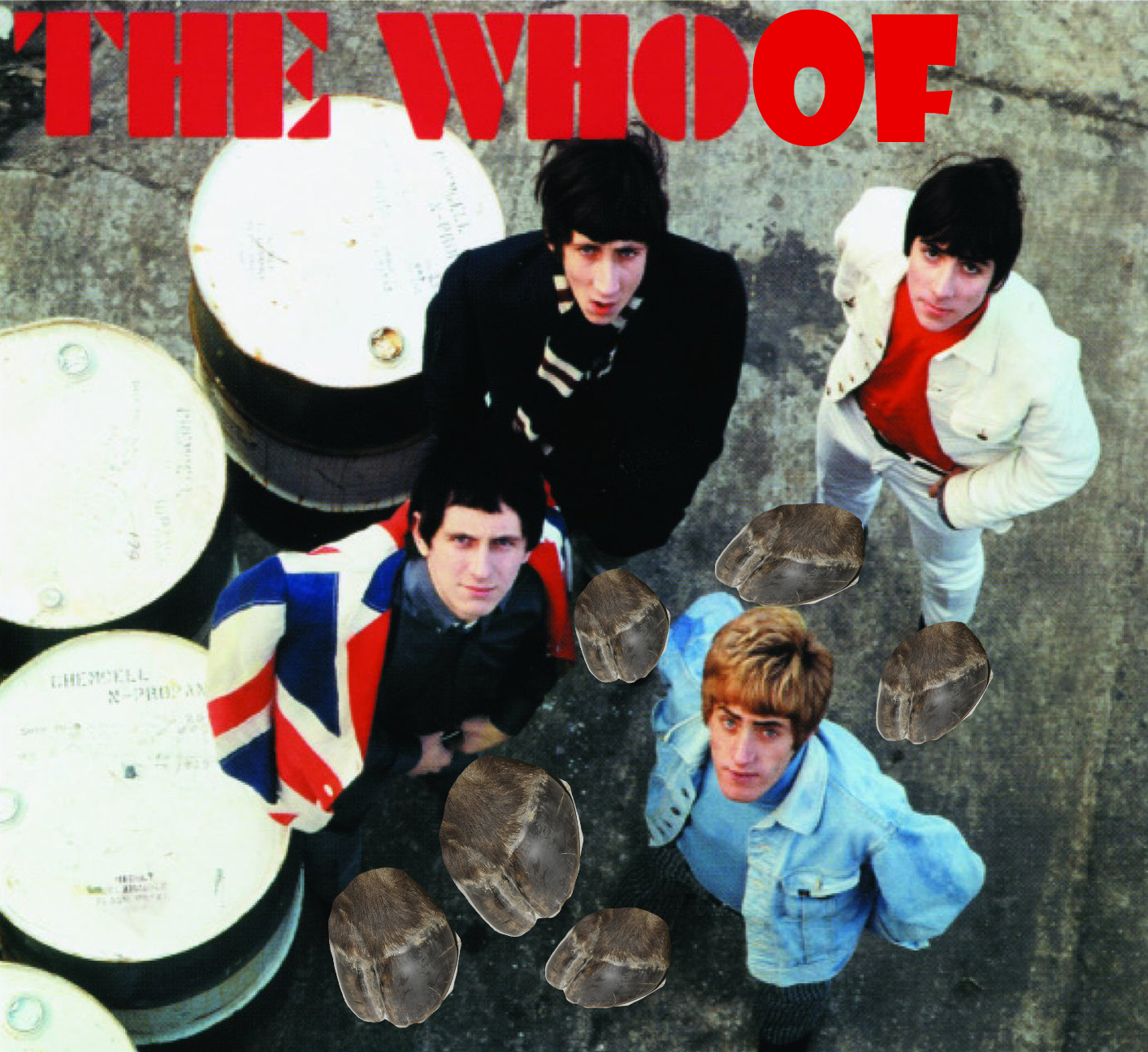
(mutated from The Who, “Cut My Hair“, Quadrophenia… from the heyday of concept albums and grandiose rock!)
Talkin’ bout my osteitis
Day Four of Freezermas. Four posts to go. I can see through time… Hence the silly title for today’s concept album track. Quadrupedophilia did not have a good ring to it, anyway.
Stomach-Churning Rating: 4/10. Reasonably tame; bones and hooves. Some pathologies of those, but not gory.
If Quadrophenia was the story of a man with four personalities (metaphor for the four band members), then quadrupedopheniaphilia is the story of how diverse forms of four-legged animals have lots of problems because of our exploitation of them, which leaves a crisis to resolve: Who are we? Are we caring enough to fix a bad situation we’ve created for our four-legged ungulate comrades?
Four legs good, two legs bad? Not really. I featured ostriches earlier this week and two legs are indeed pretty good. Four-legged cats are great, too. But four-footed big beasties with deformed hooves: those are bad all around. That leads to today’s topic…
But hey, happy 205th funkin’ birthday Charles freakin’ Robert Darwin!
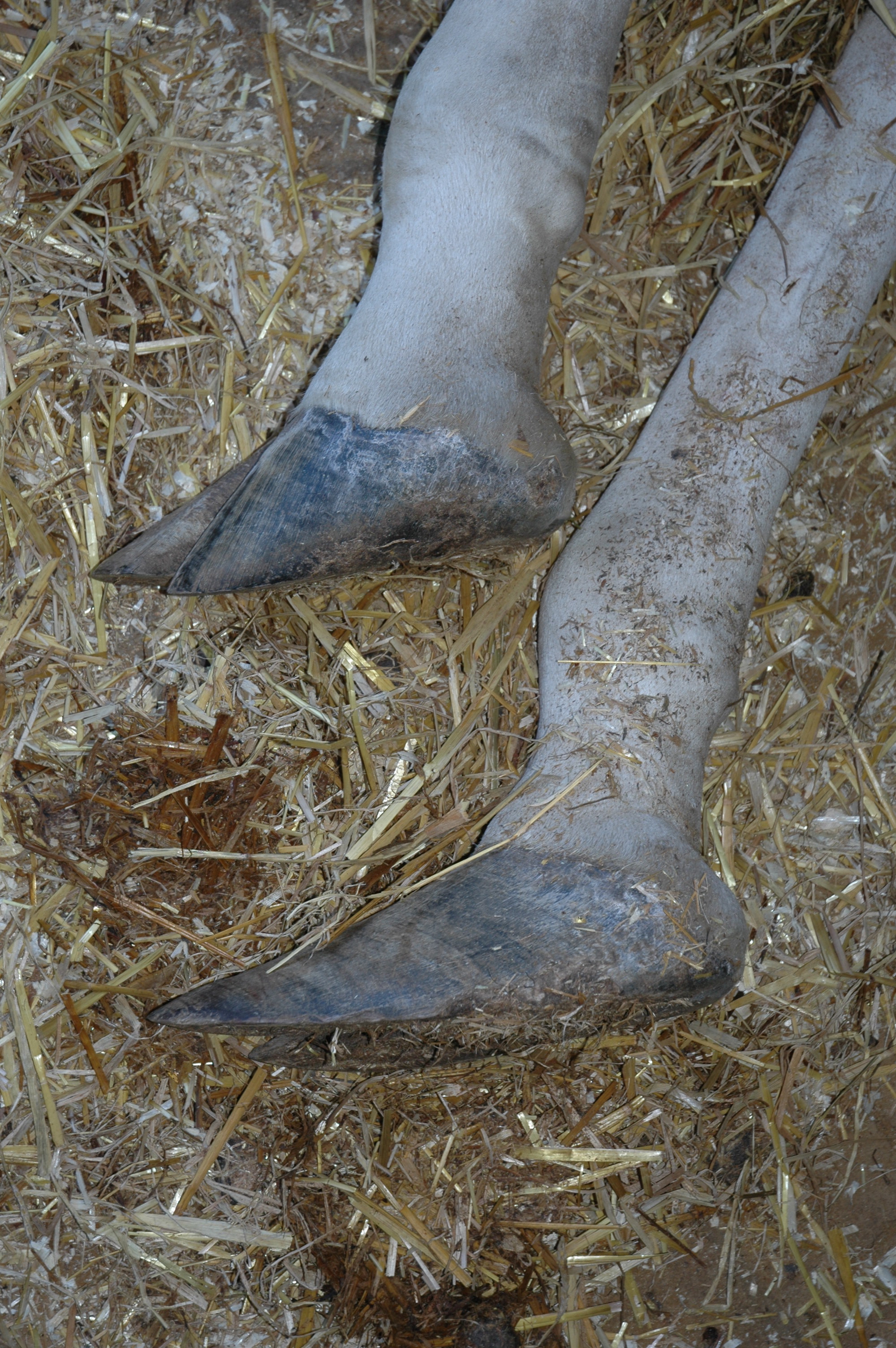
Today’s post concerns a phenomenon that (Western) civilization has wrought with large hoofed mammals, and evolution is a big part of it(as well as biomechanics and anatomy). Cynical perspective, with some truth to it: We’ve evolved larger and heavier animals to either do harder and harder work on tough surfaces like concrete floors and tarmac roads, or to stand around while we gawk at them or wait for them to get fat and tasty. Either way, the outcome should come as no surprise: their feet, the interface of that hard ground and their body, eventually start falling apart.
I’ve posted about this several times with respect to rhinos and elephants (here and here and here and here and here), but this post hits closer to home: what goes wrong with the humble hoof of our friend the horse, cow, sheep or other ungulate. It’s where the rubberkeratin hits the road. Ungulates have not evolved to live on dirty, wet concrete floors; to be obese and inactive; or to have hooves that don’t get worn down. So they suffer when they do encounter those modern conditions.
“No foot no horse,” they say, and it’s so true- once the feet start to go (due to hoof overgrowth or cracks, abscesses or other trouble), it’s hard to reverse the pathologies that ensue (arthritis, osteomyelitis, infections, fractures, etc.) and the animals start going lame, then other limbs (supporting greater loads than the affected limb) start to go, too, sometimes.
Jerry the obese, untrimmed-hoof-bearing horse. “Turkish slippers” is an apt description. DM has more here.
We can do plenty about these problems, and the title track above explains one of them: trimming hooves. Hooves often get overgrown, and if animals are tame enough (requires training!) or are sedated (risky!), hoof care experts (farriers) can rasp/file/saw them down to a more acceptable conformation. If we don’t, and the animals don’t do the trimming themselves by digging or walking around or living on varied surfaces, then the feet can suffer. But there’s still not much evidence for most common species kept in captivity by humans that indicates what the best methods are for avoiding or fixing foot problems.
What we’ve been trying to do at the RVC is use our expertise in evolution, anatomy and biomechanics to find new ways to prevent, detect, monitor or reverse these foot problems. We had BBSRC grant funding from 2009-2012 to do this, and the work continues, as it behooves us to do… Past posts have described some of this research, which spun off into other benefits like re-discovering/illuminating the false sixth toes of elephants. We’re working with several zoos in the UK to apply some of the lessons we’re learning to their animals and management practices.
Above: Thunderous hoof impacts with nasty vibrations, and large forces concentrated on small areas, seem to contribute to foot problems in hoofed mammals. From our recent work published in PLOS ONE.
Foot health check on a white rhino at a UK zoo; one of the animals we’ve worked with. Photo by Ann & Steve Toon, http://www.toonphoto.com/
If it works, it’s the most satisfying outcome my research will have ever had, and it will prevent my freezers from filling up with foot-influenced mortality victims.
Again, I’ll tell this tale mainly in photos. First, by showing some cool variations evolved in the feet of hoofed mammals (artiodactyls and perissodactyls; mostly even/odd-toed ungulates of the cow/sheep and horse lineages, respectively). Second, by showing some pretty amazing and shocking images of how “normal” hooves go all wonky.
Two ways to evolve a splayed hoof for crossing soft ground: 2 toes that are flexible and linked to big pads (camel), and 2 main toes that allow some extra support from 2 side toes when needed (elk). At Univ. Mus. Zoology- Cambridge.
Diversity of camelid foot forms: big clunky, soft Old World camel feet and dainty, sharp highland New World camelids. [Image source uncertain]
Moschus, Siberian musk deer with remarkable splayed hooves/claws; aiding it in crossing snowy or swampy ground. At Univ. Mus. Zoology- Cambridge.
Tragulus, or mouse-deer, with freaky long “splint bones” (evolutionarily reduced sole bones or metatarsals) and dainty hooved feet. At Univ. Mus. Zoology- Cambridge.
Overgrown giraffe hooves. An all-too-common problem, and one we’re tacking with gusto lately, thanks to PhD student Chris Basu’s NERC-funded giraffe project!
Wayyyyyyyyy overgrown hooves of a ?sheep, from the RVC’s pathology collection.
Craaaaaaazy overgrown ?sheep hooves, from the RVC’s pathology collection.
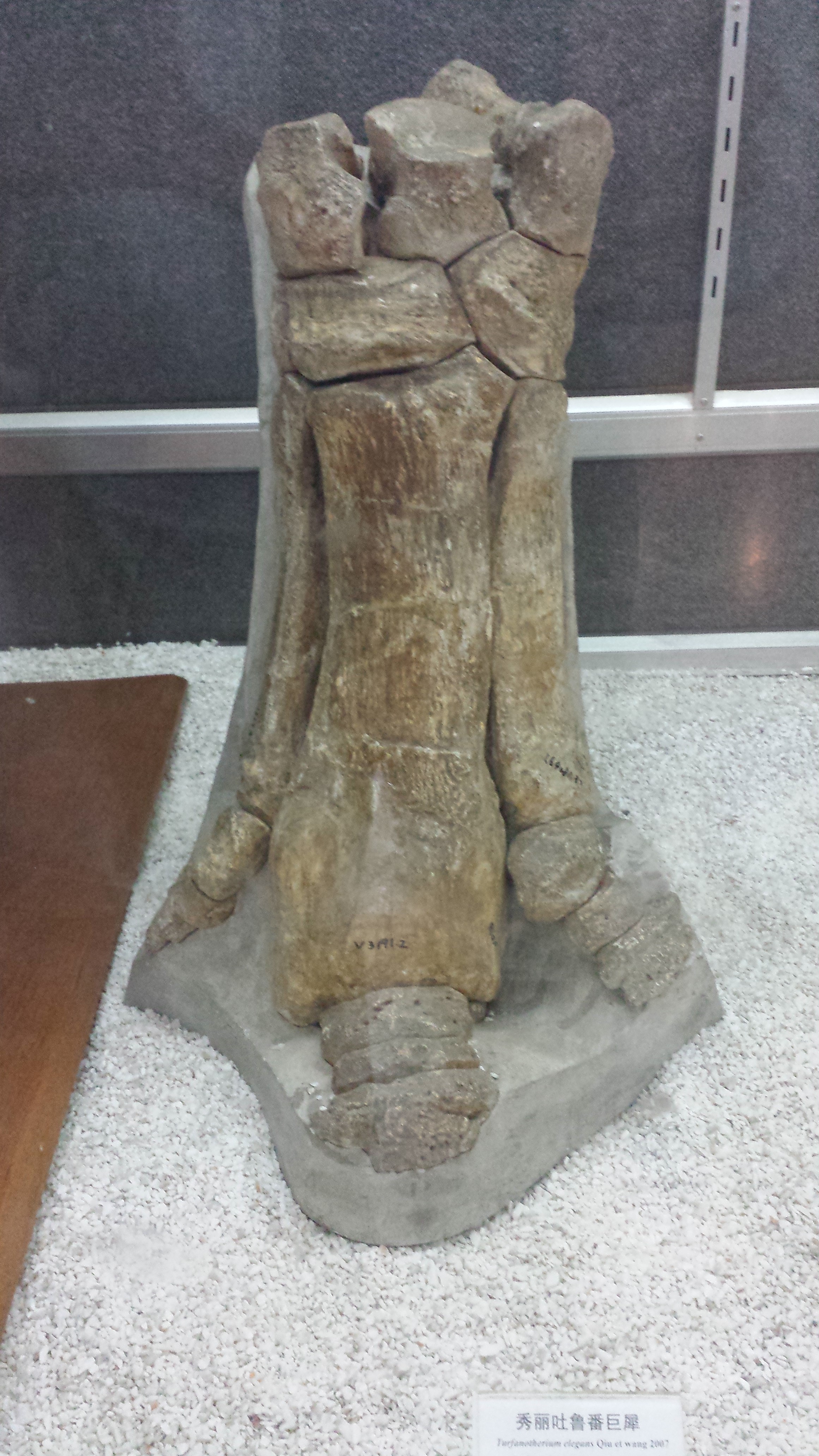
If we understand how foot form, function and pathology relate in diverse living hoofed mammals, we can start to piece together how extinct ones lived and evolved- like this giant rhinoceros! At IVPP museum in Beijing.
So, what do we do now? If we love our diverse hoofed quadrupeds, we need to exert that quadrupedopheniaphilia and take better care of them. Finding out how to do that is where science comes in. I’d call that a bargain. The best hooves ever had?
Today, to help thaw you poor Americans out of that Arctic Vortex, we have a guest post bringing the heat, by my PhD student Sophie Regnault! This relates to some old posts about rhinos, which are a mainstay here at the WIJF blog- I’ve posted a lot about the rhino extinction crisis, feet, skin, big and bigger bones, and more, but this is our first rhinoceros-focused, actual published scientific paper! Take it away, Sophie! (We’re planning a few more “guest” blog posts from my team, so enjoy it, folks!)
Almost a year ago to the day, I submitted my first paper written with John Hutchinson and Renate Weller at the RVC and it has (finally!) just been published. To celebrate, I have been allowed to temporarily hijack ‘What’s in John’s Freezer?’ for my first foray into the world of blogging. I started the paper back as an undergraduate veterinary student. It was my first experience of proper research, and so enjoyable that I’m now doing a PhD, studying sesamoid bones like the patella!
We wanted to discover more about the types of bony disease rhinos get in their feet, of which there isn’t much known. Rhinos, of course, are big, potentially dangerous animals – difficult enough to examine and doubly difficult to x-ray clearly because of their thick skin. Unlike diseases which are fairly easy to spot (like abscesses or splitting of the nails and footpad), there is hardly anything out there in the scientific literature on bony diseases in rhino feet. It’s no small issue, either. When your feet each need to support over 900kg (typical for a large white rhino), even a relatively minor problem can be a major pain. Progressing unseen under their tough hide, lesions in the bone can eventually become so serious than the only solution is euthanasia, but even mild conditions can have negative consequences. For example, foot problems in other animals are known to have knock-on effects on fertility, which would be a big deal for programs trying to breed these species in captivity.
Hidden treasures abound! (Photos can be clicked to embiggen)
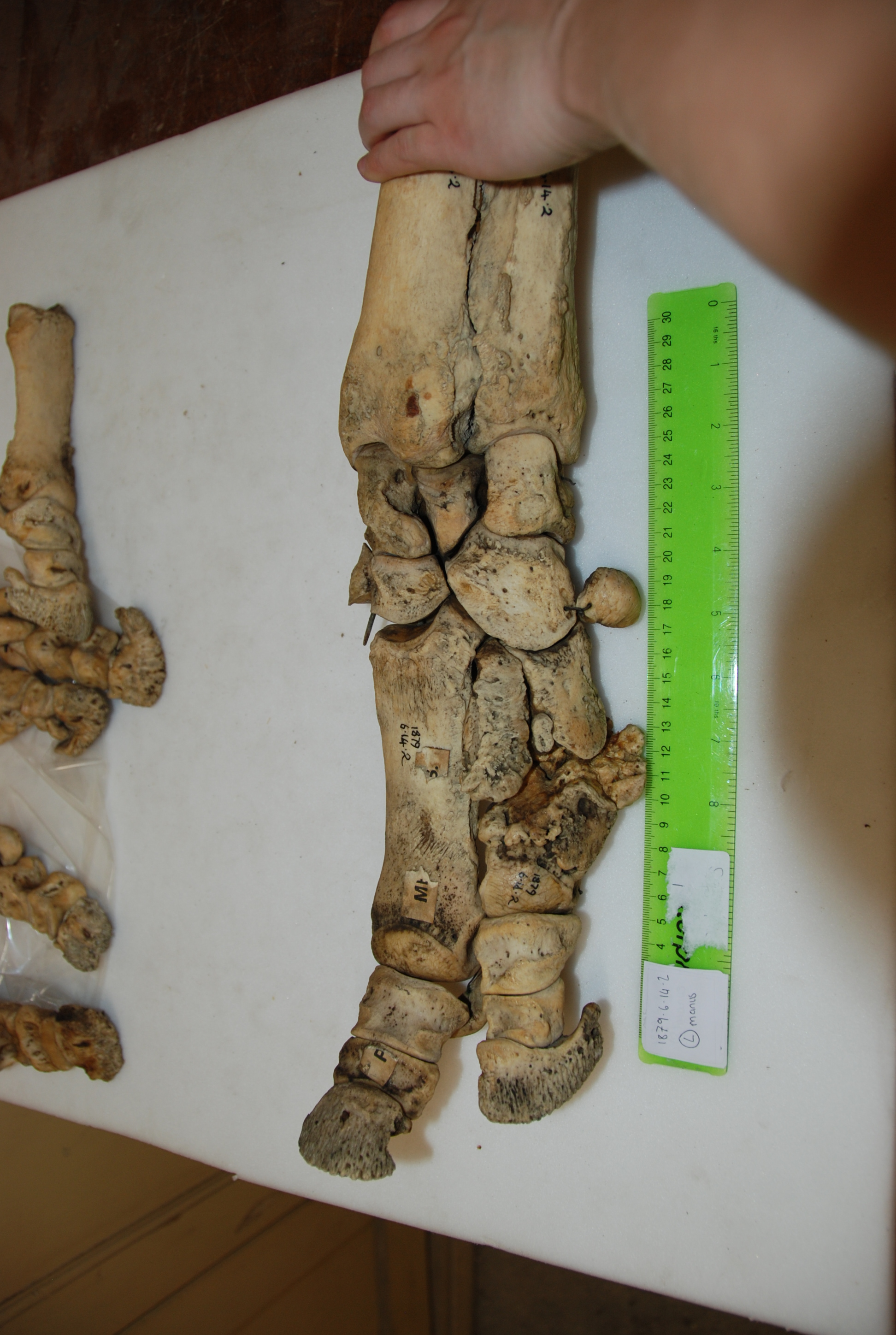
Data gathering was a blast. I got to travel to Cambridge, Oxford, and London during one of England’s better summers, and these beautiful old museums were letting me snoop around their skeleton collections. I’d been there often as a visitor, but it was anatomy-nerd-heaven to go behind the scenes at the Natural History Museum, and to be left alone with drawers and drawers of fantastic old bones. Some of the specimens hadn’t been touched for decades – at Cambridge University Museum of Zoology, we opened an old biscuit tin filled with the smallest rhinoceros foot bones, only to realise they were wrapped in perfectly preserved 1940’s wartime Britain newspaper.
Osteomyelitis… (3 clickable pics above) the toe’s probably not meant to come off like that!
In addition to my museum studies, I had another fun opportunity to do hands-on research. John (of course!) had freezers full of rhino legs (looking disconcertingly like doner kebabs, but maybe that’s just me!), which we CT scanned to see the bones. Although it is a pretty standard imaging technique, at this point I had only just started my clinical studies at the vet hospital, and being able to flick through CT scans felt super badass. Most vet students just get to see some horse feet or dog/cat scans, at best.
Another osteomyelitis fracture, visible in a CT scan reconstruction.
We expected to find diseases like osteoarthritis (a degenerative joint disease) and osteomyelitis (bone infection and inflammation). Both had previously been reported in rhinoceroses, although it was interesting that we saw three cases of osteomyelitis in only 27 rhinos, perhaps making it a fairly common complication. It’s an ugly-looking disease, and in two of the cases led to the fat, fluffy bones fracturing apart.
We also had several unexpected findings, like flakes of fractured bone, mild dislocations, tons of enthesiophytes (bone depositions at tendon/ligament attachments) and lots of holes in the bones (usually small, occasionally massive). For me, writing up some of these findings was cool and freaky paranoid in equal measures. They hadn’t been much described before, and we were unsure of their significance. Was it normal, or pathological? Were we interpreting it correctly? Discussions with John and Renate (often involving cake) were reassuring, as was the realisation that in science (unlike vet school at the time, where every question seemed to have a concrete answer) you can never be 100% sure of things. Our study has a few important limitations, but has addressed a gap in the field and found some neat new things. Six months into my PhD, I’m enjoying research more than ever, and hoping that this paper will be the first of many (though I promise I won’t keep nicking John’s blog for my own shameless self-promotion if that happens! EDIT BY JOHN: Please do!).
Nasty osteoarthritis wearing away the bone at the joint surface. Most cases occurred in the most distal joint.
Deep holes in some of the bones: infection, injury?
The paper:
Sophie Regnault, Robert Hermes, Thomas Hildebrandt, John Hutchinson, and Renate Weller (2013) OSTEOPATHOLOGY IN THE FEET OF RHINOCEROSES: LESION TYPE AND DISTRIBUTION. Journal of Zoo and Wildlife Medicine: December 2013, Vol. 44, No. 4, pp. 918-927.
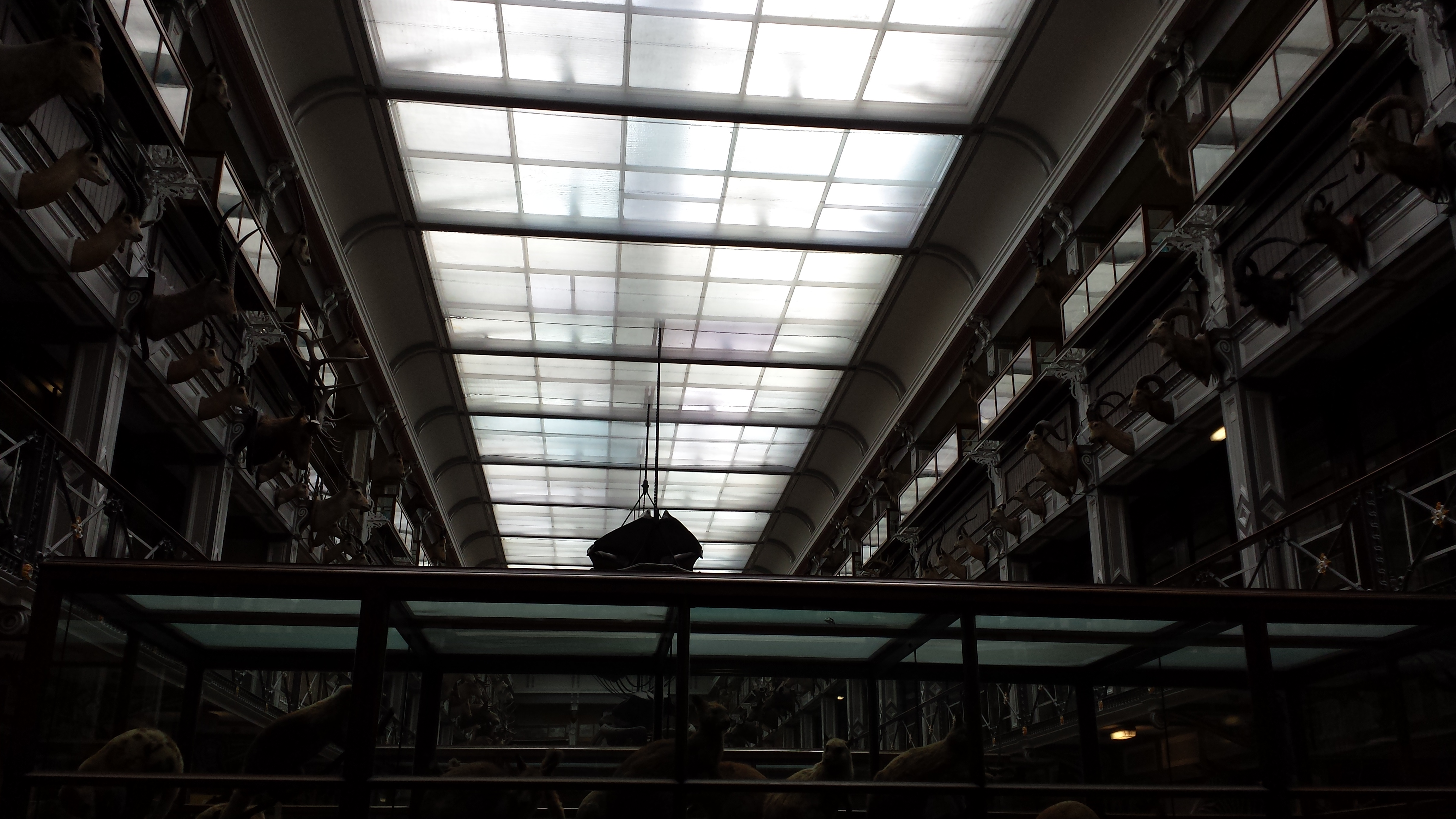
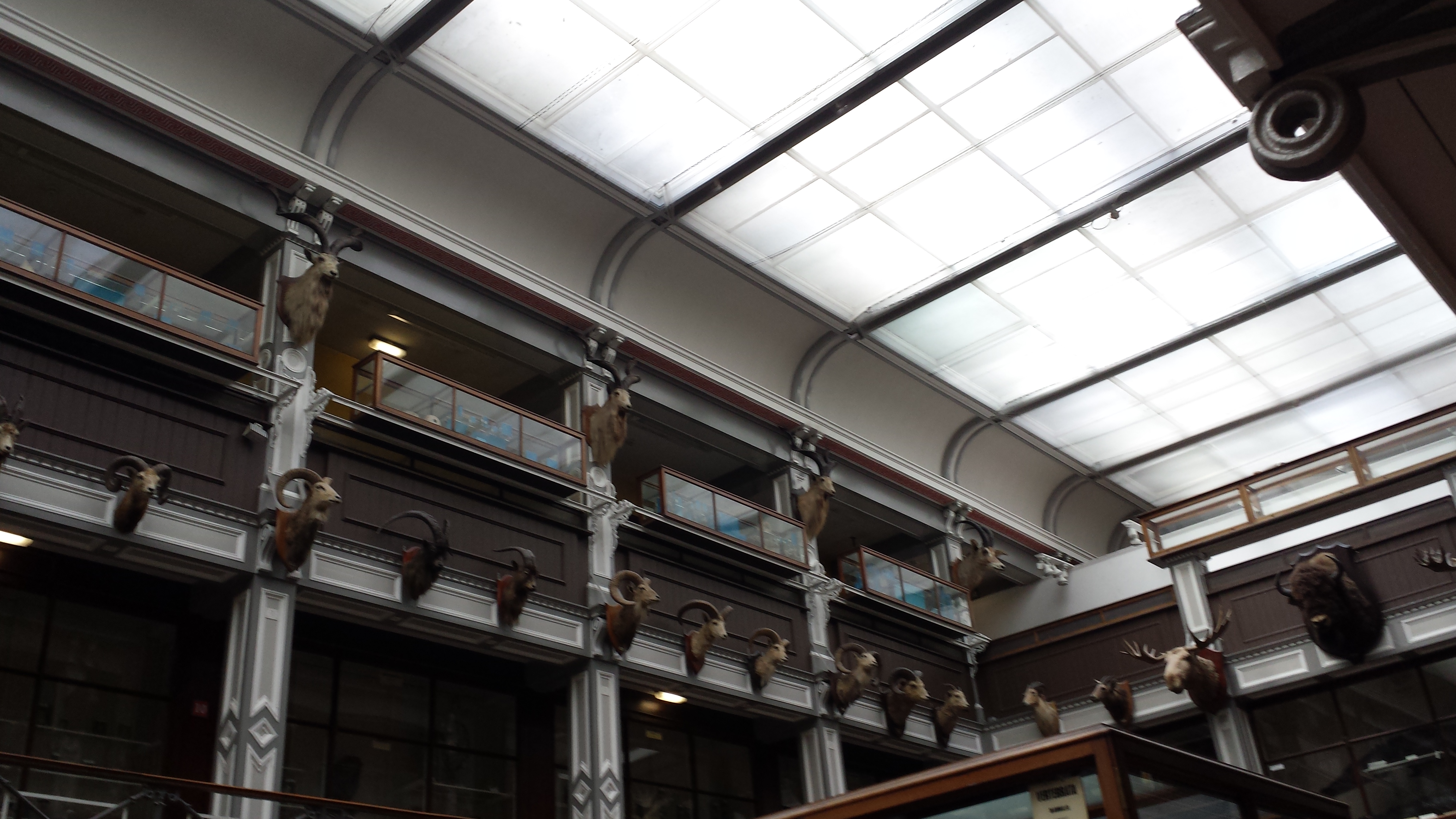
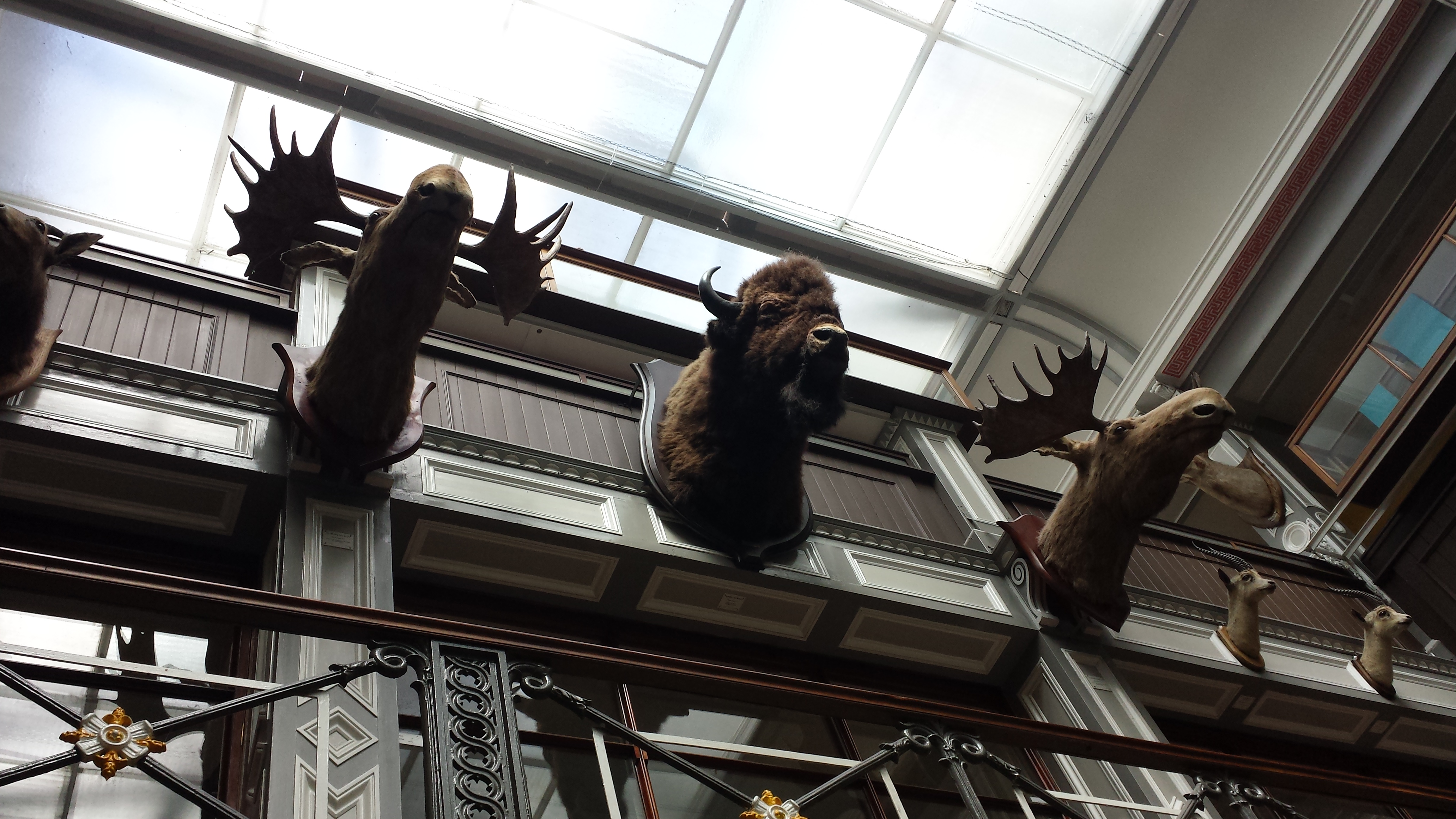
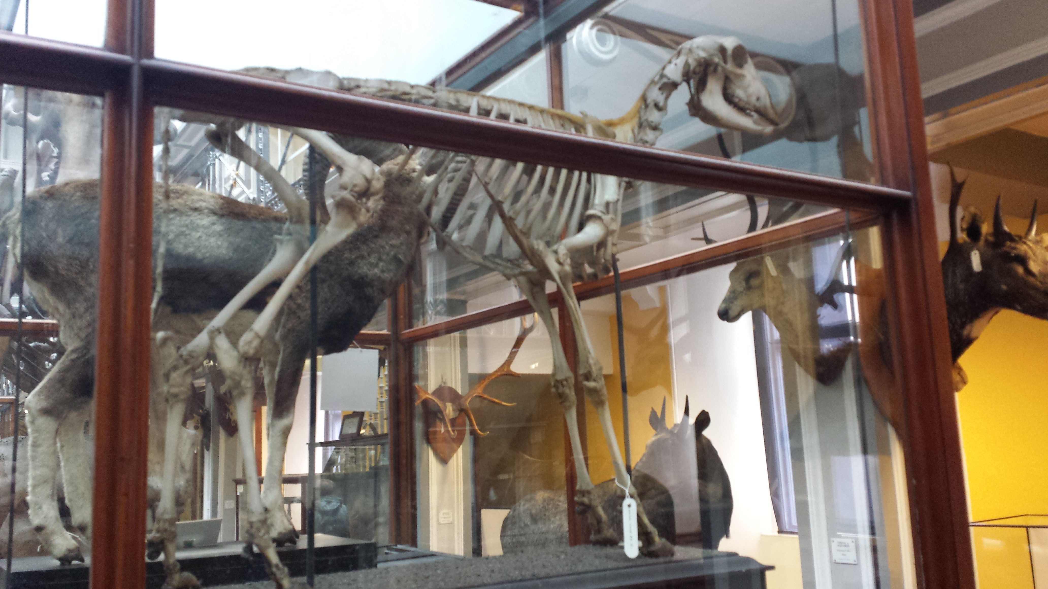
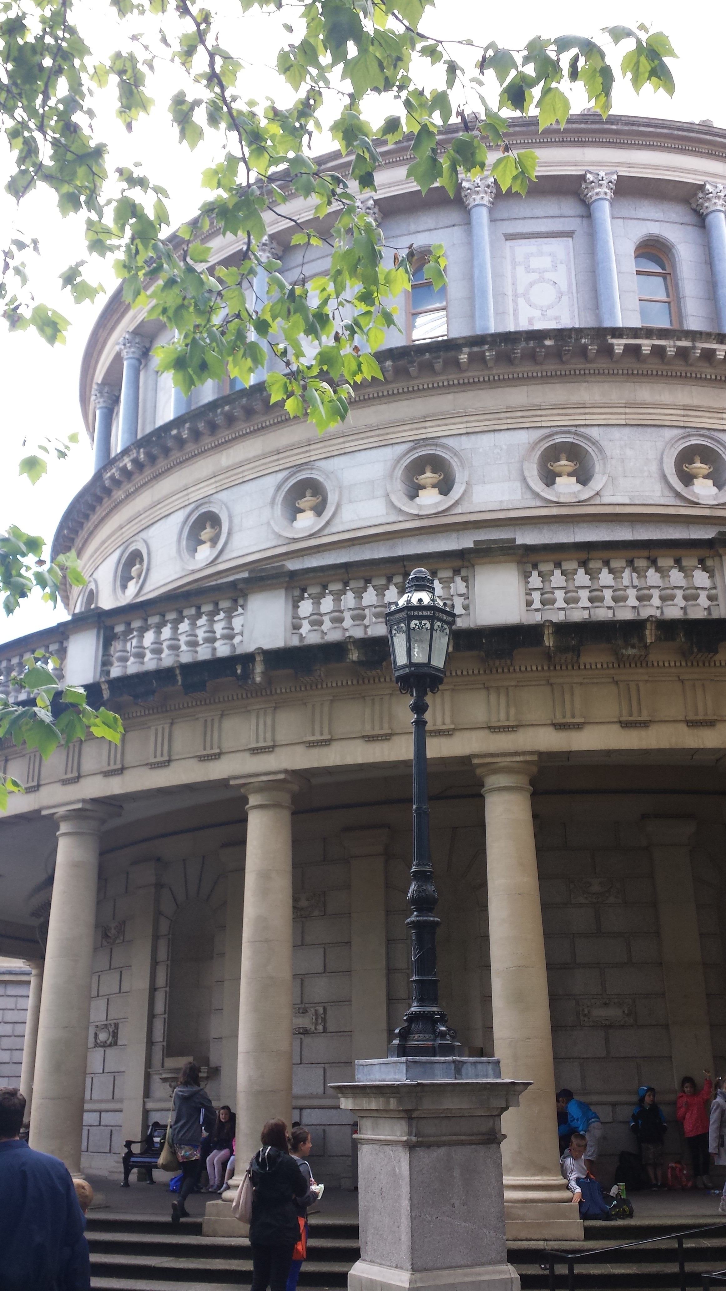
A photo blog post for ya here! I went to Dublin on a ~28 hour tour, for a PhD viva (now-Dr Xia Wang; bird feather/flight evolution thesis) earlier this month. And I made a beeline for the local natural history museum (National Museum of Ireland, Natural History building) when I had free time. So here are the results!
Stomach-Churning Rating: Tame; about a 1/10 for most, but I am going to break my rule about showing human bodies near the end. Just a warning. The bog bodies were too awesome not to share. So that might be 4/10-8/10 depending on your proclivities. They are dry and not juicy or bloody, and don’t look as human as you’d expect.
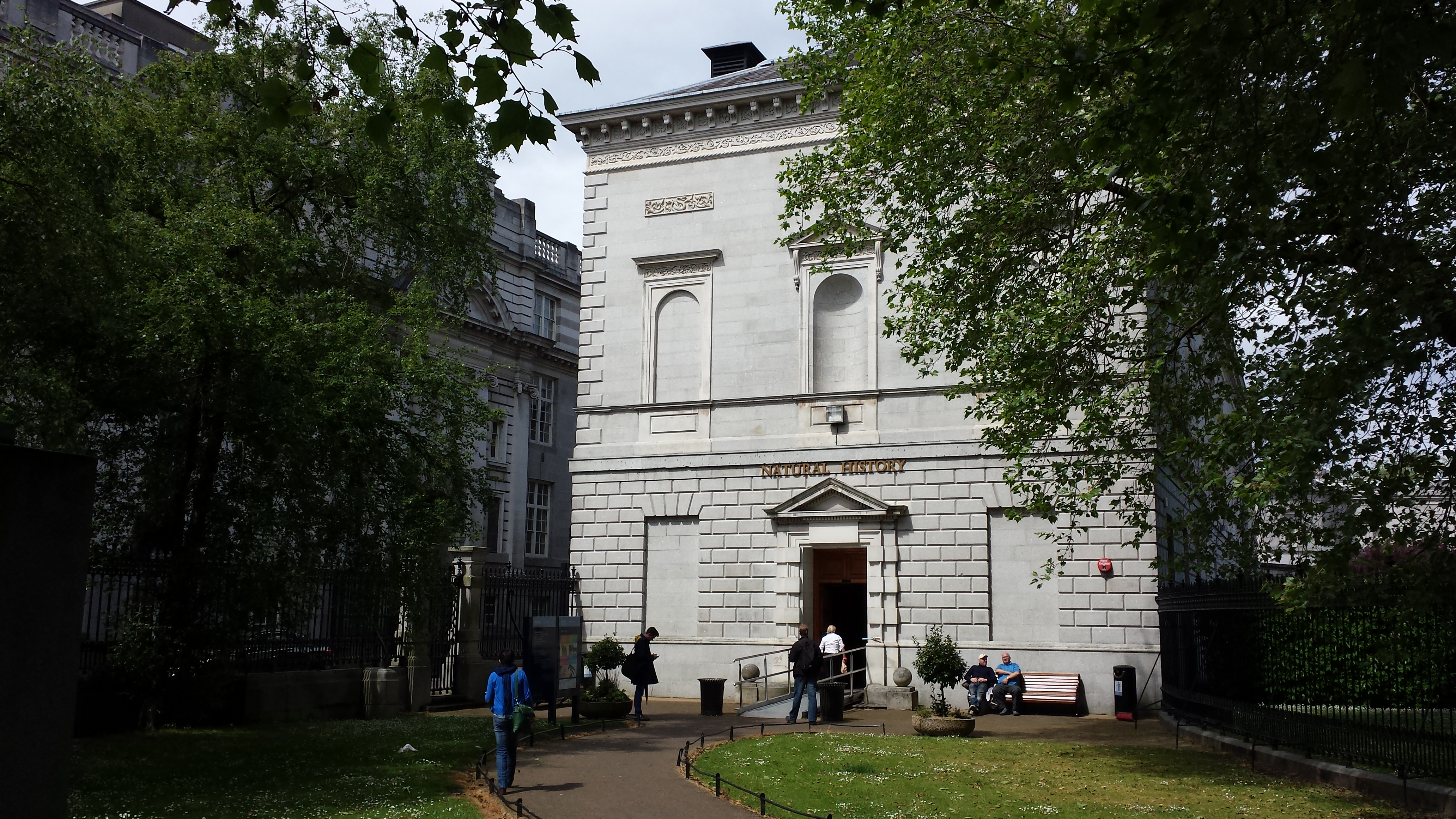
Simple Natural History museum entrance area.
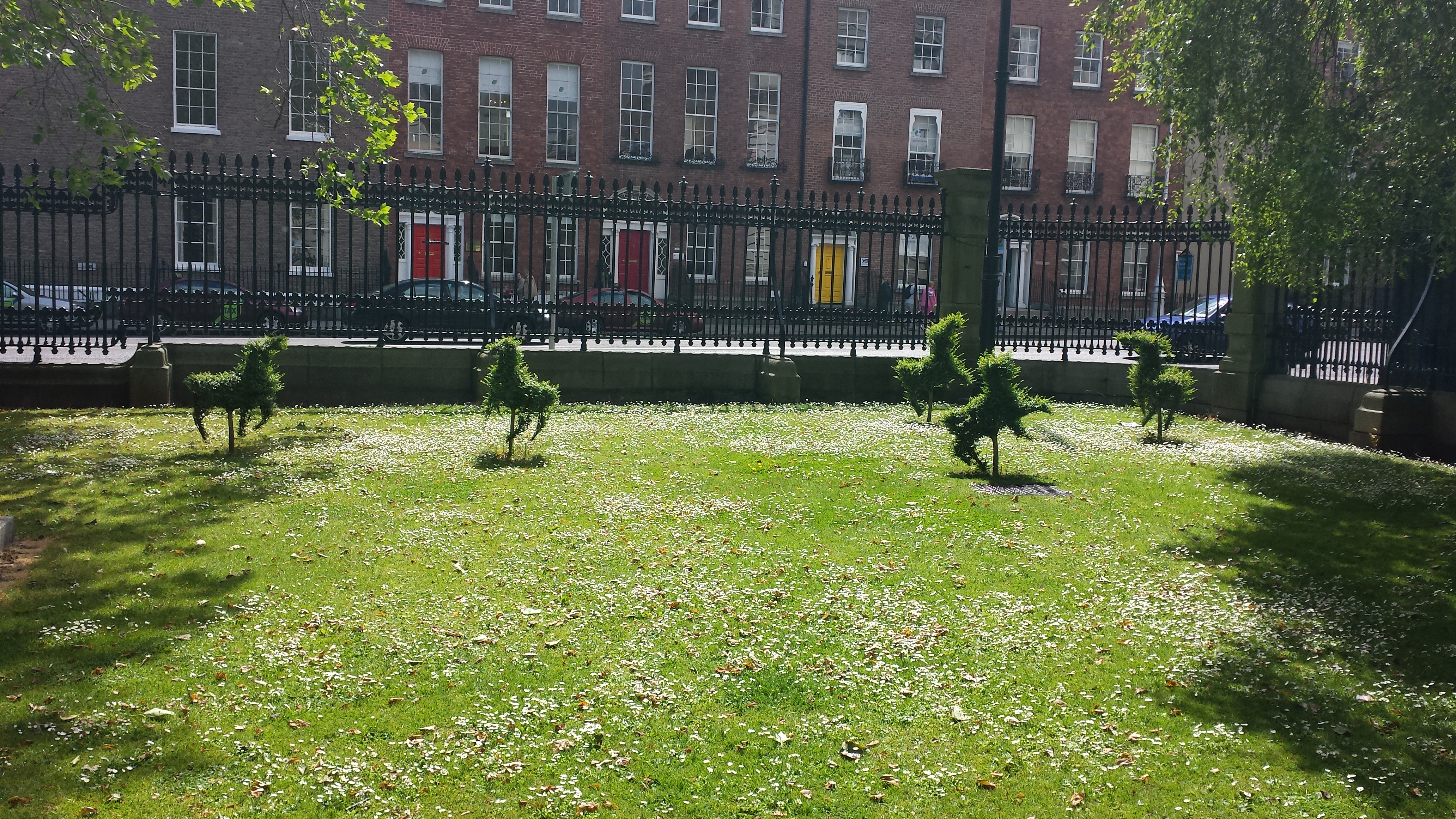
Adorable frolicking topiaries outside the NHM.
Inside, it was a classical Victorian-style, dark wood-panelled museum stuffed with stuffed specimens. It could use major refurbishment, but I do love old-fashioned exhibits. Get on with it and show us the animals; minimize interpretive signage and NO FUCKING INTERACTIVE COMPUTER PANELS! So by those criteria, I liked it. Some shots of the halls: And on to the specimens!
Giant European deer (“Irish elk”). I looked at these and thought, “why don’t we see female deer without antlers ever? then noticed one standing next to these (you can barely see it in back); too bad my photo is crappy.
Superb mounted skeleton of giraffe (stuffed skin was standing near it).
A sheep-y or a goat-y beastie; I dunno but it shows off a nice example of the nuchal ligament (supports the head/neck).
Yarr, narwhals be internet gold!
Giant blown glass models of lice!
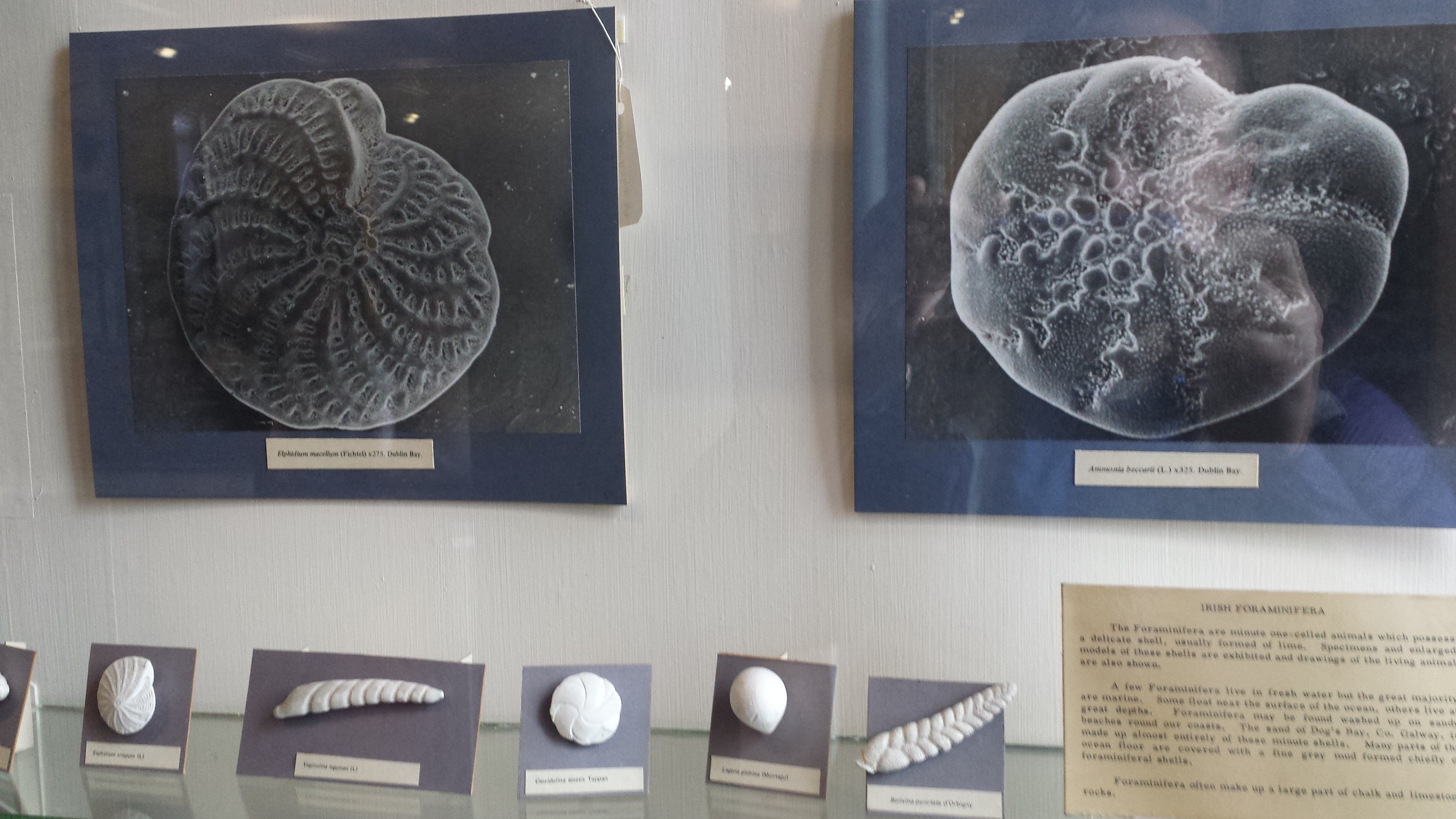
Who doesn’t like a good giant foramanifera image/model?
“That’s one bigass skate,” I murmured to myself.
“That’s one bigass halibut,” I quipped.
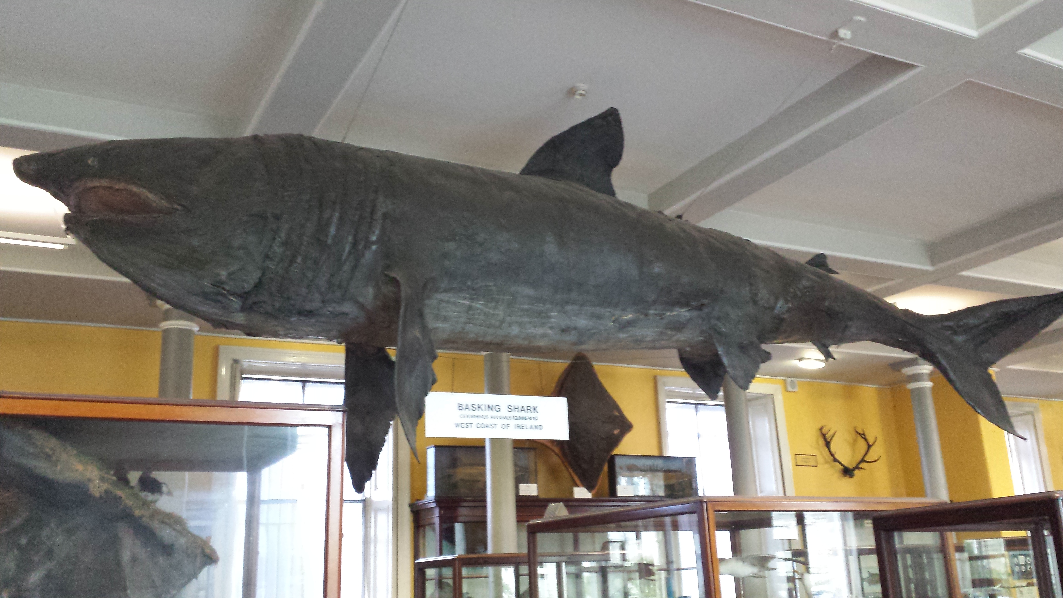
Tatty basking shark in entry hall.
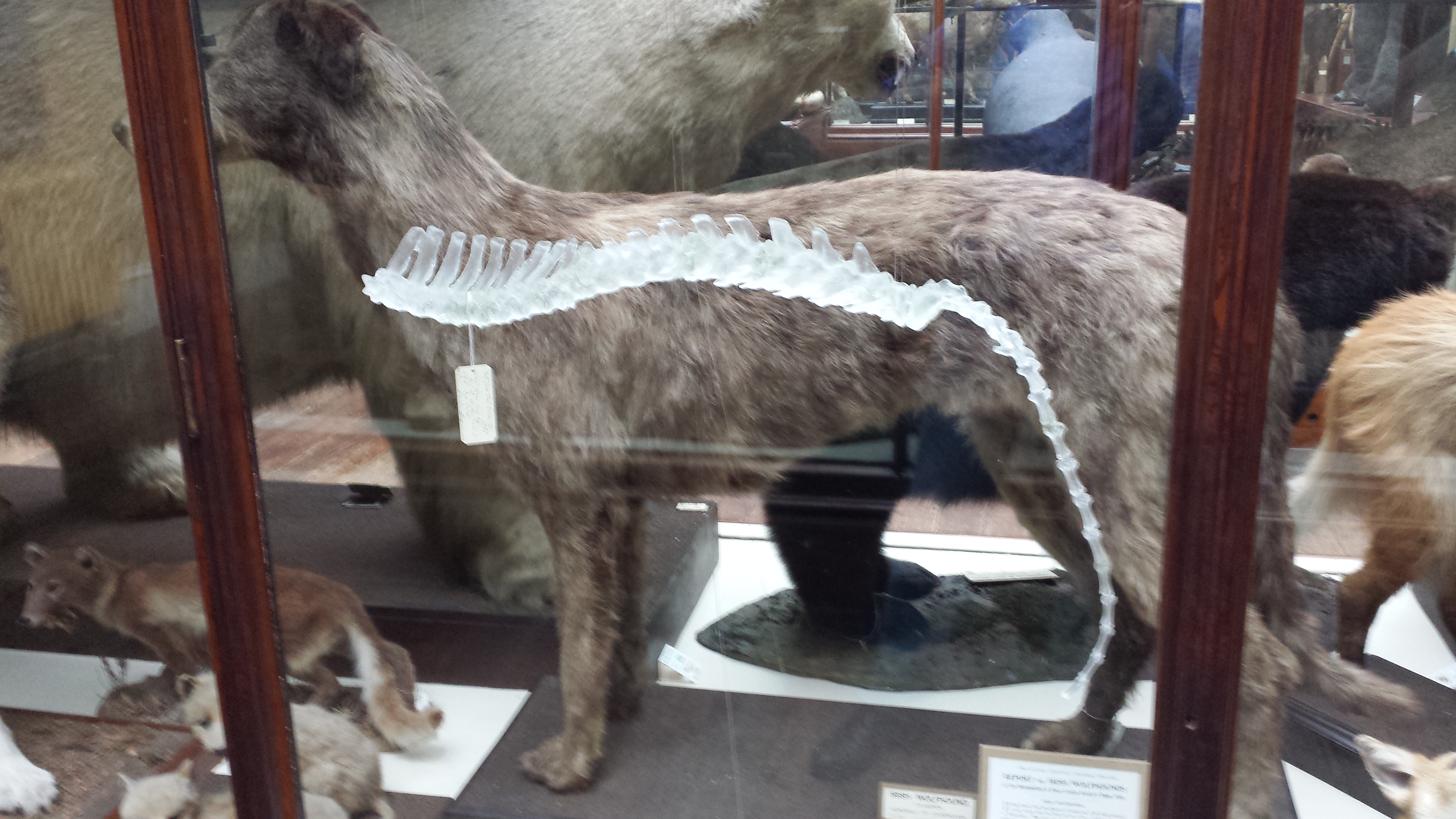
Irish wolfhound, with a glass sculpture of its spine hanging near it, for some reason.
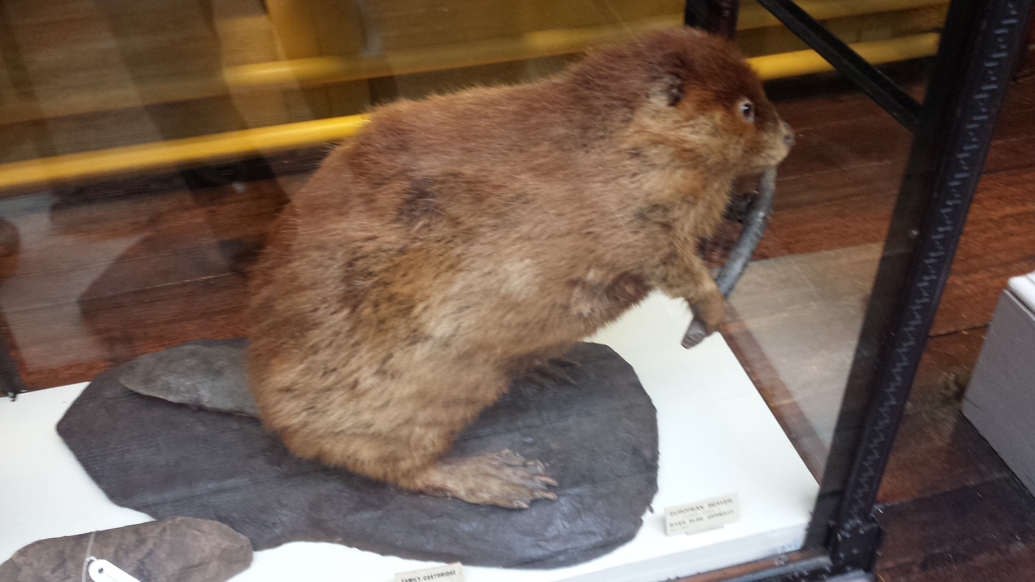
Stand back everyone! That beaver has a club!
Skull of a pilot whale/dolphin.
Nice anteater skeleton and skin.
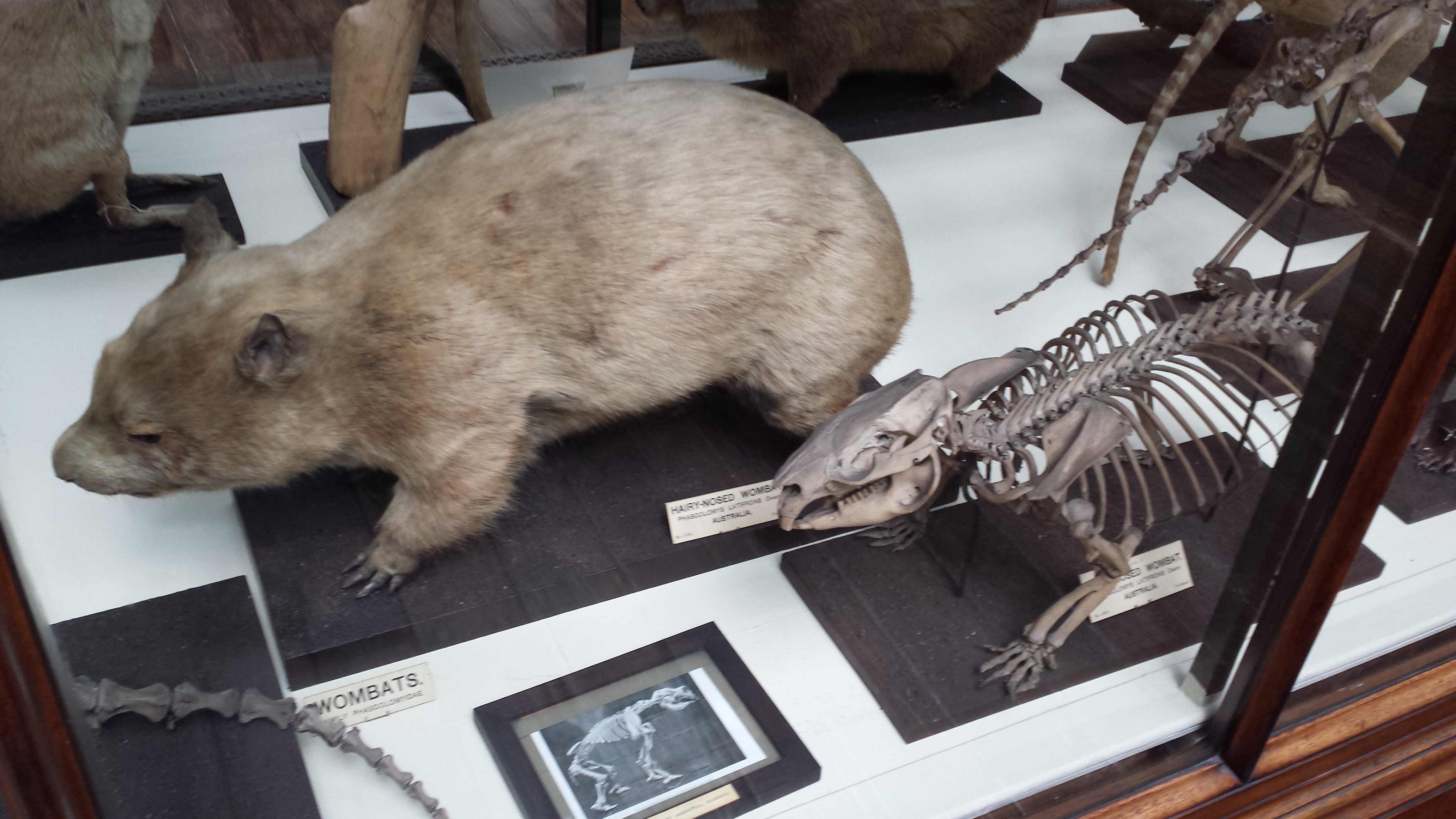
Nice wombat skeleton and skin.
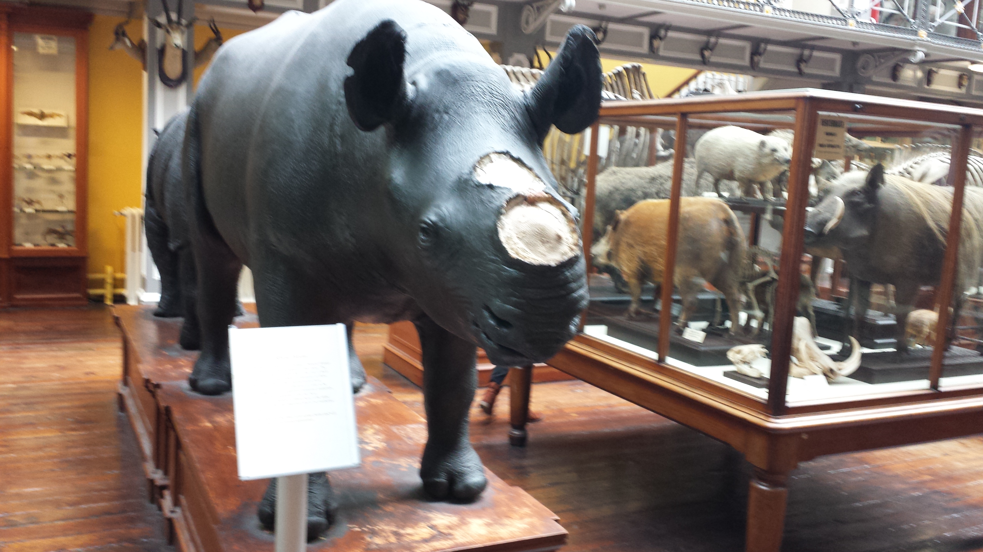
Sad display of a stuffed rhino with the horn removed, and signage explaining the problem of thefts of those horns from museum specimens of rhinos worldwide.
But then the stuffed animals started to get to me. Or maybe it was the hangover. Anyway, I saw this…
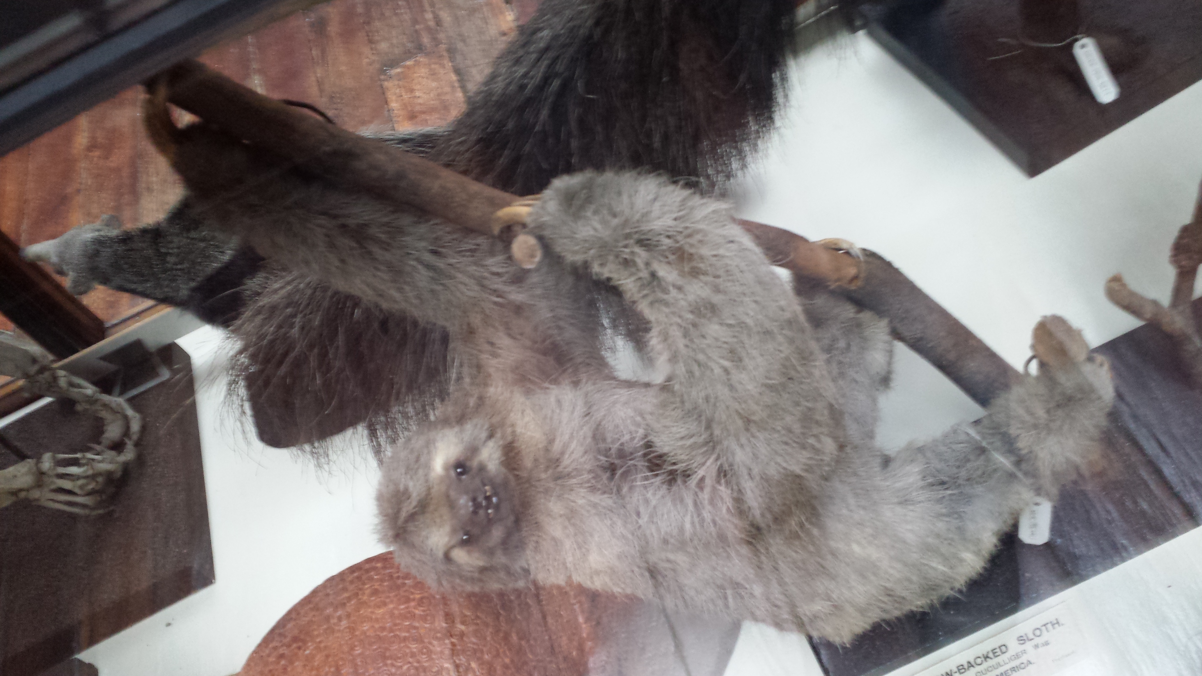
A proboscis monkey mother who seemed to be saying “Hey kid, you want this yummy fruit? Tough shit. I’m going to hold it over here, out of reach.” with a disturbing grimace. That got me thinking about facial expressions in stuffed museum specimens of mammals more, and I couldn’t help but anthropomorphize as I toured the rest of the collection, journeying deeper into surreality as I progressed. What follows could thus be employed as a study of the Tim-Burton-eseque grimaces of stuffed sloths. Click to emslothen.
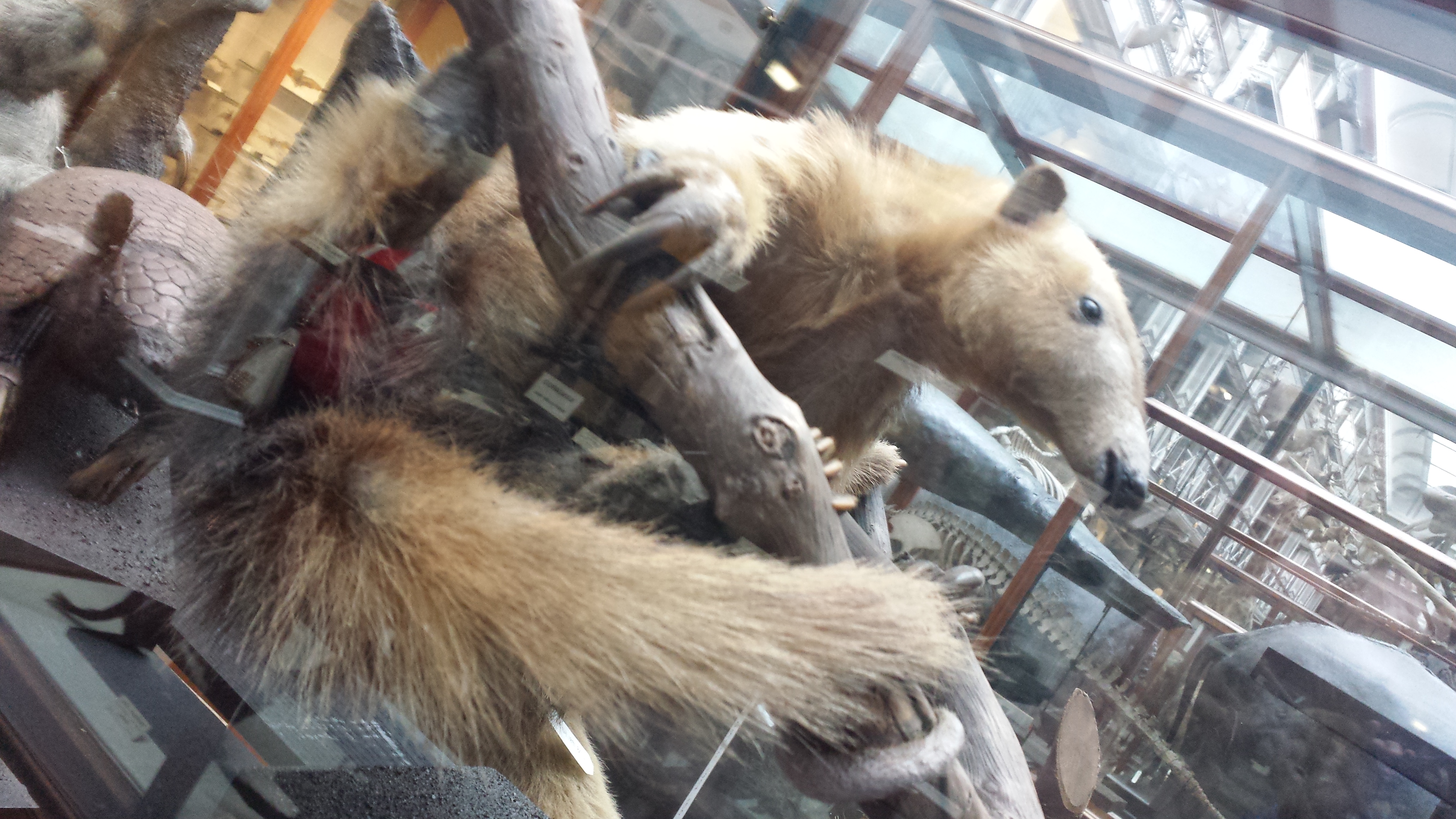
Tree anteater has a go at the awkward expression game.
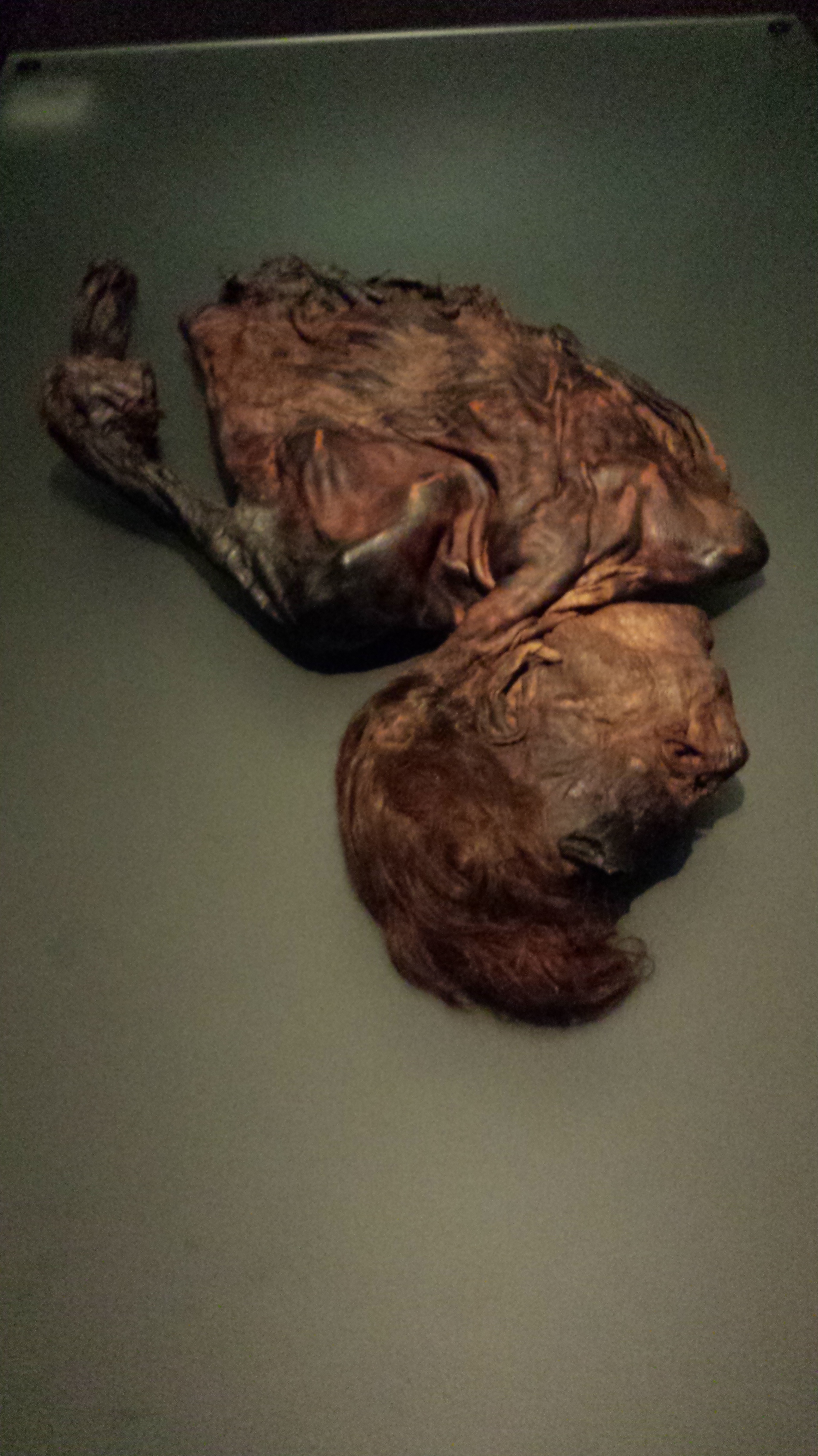
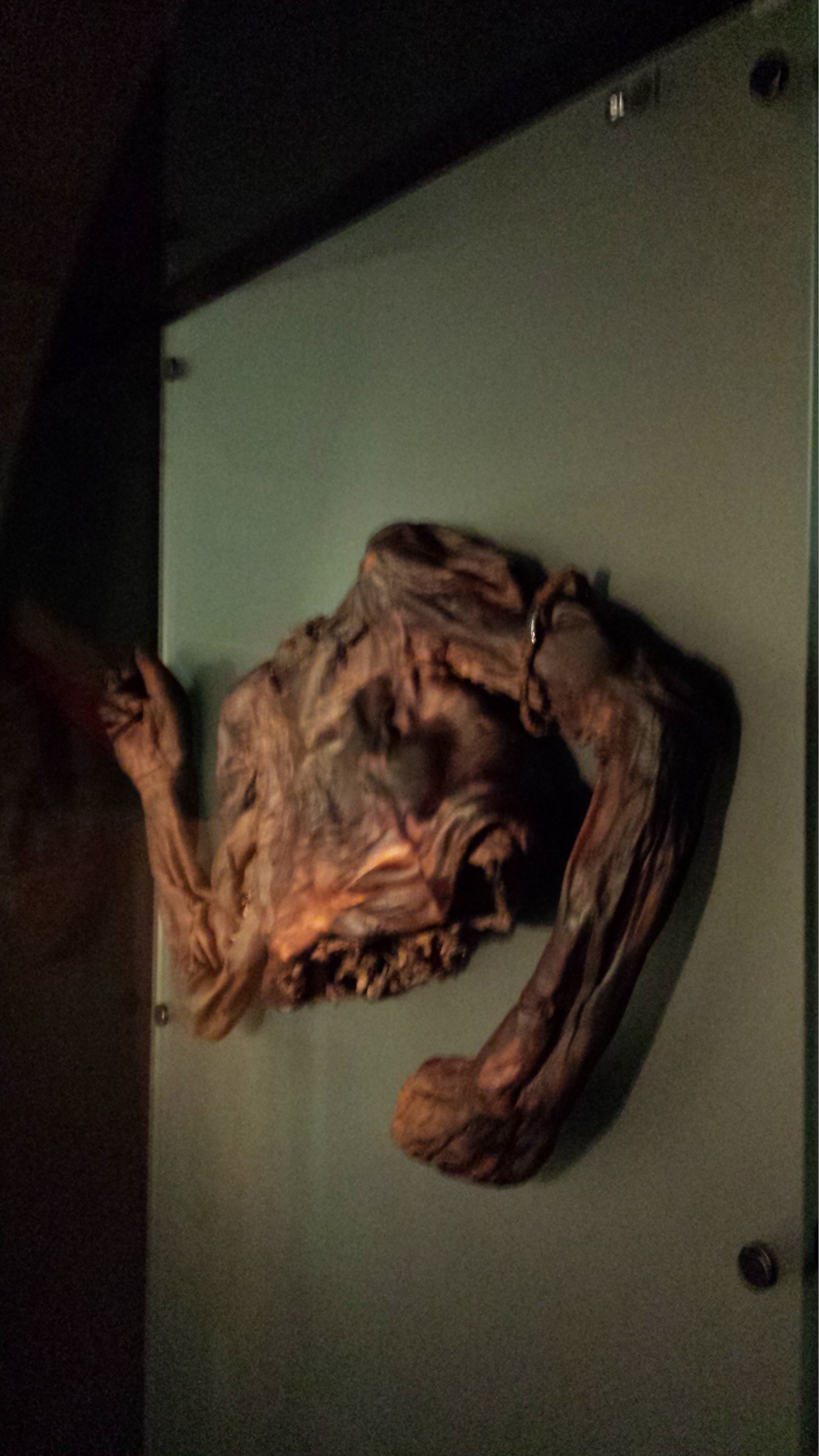
This completed my tour of the museum; there were 2 more floors of specimens but they were closed for, sigh, say it with me… health and safety reasons. Balconies from which toddlers or pensioners or drunken undergrads could accidentally catapult themselves to their messy demise upon the throngs of zoological specimens below. But the National Museum’s Archaeology collection was just around the block, so off I went, following whispered tales of bog bodies. There will be a nice, calm, pretty photo, then the bodies, so if peaty ~300 BCE cadavers are not your cup of boggy tea, you can depart this tour now and lose no respect.
Impressive entrance to the National Museum’s Archaeology building.
The bog bodies exhibit is called “Kingship and Sacrifice“. It is packed with cylindrical chambers that conceal, and present in a tomb-like enclosed setting, the partial bodies of people that were killed and then tossed in peat bogs as honoraria for the ascension of a new king. The peaty chemistry has preserved them for ~2300 years, but in a dessicated, contorted state. The preservation has imparted a mottled colouration and wrinkled texture not far off from a Twix chocolate bar’s. Researchers have studied the bejesus out of these bodies (including 3D medical imaging techniques) and found remarkable details including not just wounds and likely causes of death (axes, strangling, slit throats etc) but also clothing, diet, health and more.
Here they are; click to (wait for it)… emboggen:
Did you find the Celtic armband on one of them?
Finally (actually this happened first; my post is going back in time), I visited UCD’s zoology building for the PhD viva and saw a few cool specimens there, as follows:
Giant deer in UCD zoology building foyer, with a lovely Pleistocene landscape painted on the wall behind it.
Sika deer in an awkward posture (what is it supposed to be doing?) in Univ Coll Dublin zoology building’s foyer.
The pose of this ?baboon?mandrill struck me as very peculiar and menacing- reminiscent of a vampire bat’s pose.
A whole lotta chicken skeletons in a UCD teaching lab.
After the viva we went out for some nice Chinese food and passed some Dublin landmarks like this:
Trinity College entrance, I think.Former Irish Parliament; now the Bank of Ireland.
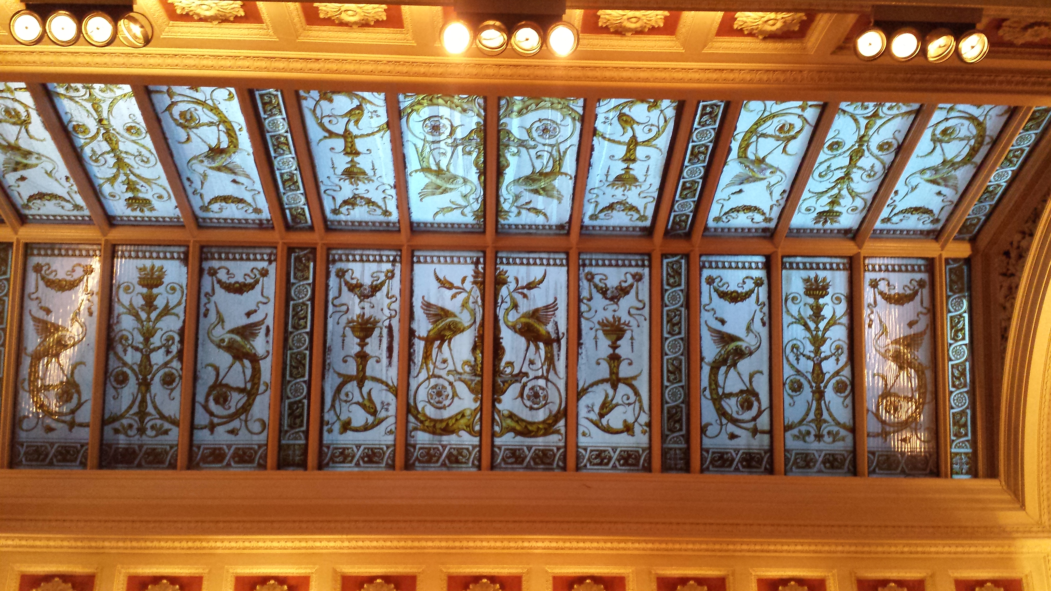
And we wandered into a very posh Irish pub called the Bank (on College Green), which displayed this interesting specimen, as well as some other features shown below:
Replica of illuminated 9th Century gospel manuscript “The Book of Kells”, with gorgeous Celtic art.
Vaults near toilets in the Bank pub. Almost as cool as having giant freezers down there.
Nice glass ceiling of the Bank pub.
And Irish pub means one big, delicious thing to me, which I will finish with here– much as I finished that night off:
Less words, more pictures in this post, and I’ll get the one lame cake joke out of the way early. I’ve nearly finished my research blitz through the postcranial material of the NHM-Tring’s osteological collection and have made some pit-stops for cake skulls now and then when I see one that pleases me. Now I shall present a survey of some of the species I’ve examined. I’ll proceed up from the base of the crown clade of living birds (Neornithes/Aves; the most recent common ancestor of living birds and all its descendants) and first take a tour of Palaeognathae; the ratites and kin; then move another step up into the Neognathae, first featuring the lineage featuring the ground fowl (Galliformes) and then the waterfowl (Anseriformes). If all this taxonomy and phylogeny is a bit much, check out this page for a brush-up on the bushy branches of bird biodiversity.
First, lots of bones of our cast of currasows, chachalacas, cassowaries and other kooky characters. And then, perhaps, a stop to the excessive alliteration. Finally, I will finish with some examples of species oddity (hat tip to Chris Hadfield).
Stomach-Churning Rating: 2/10- some bony pathologies but still just dry bones. Minimal cake jokes, and no filthy swearing this time.
BRING ON THE BONES:
My photographs are shown with kind permission from the Natural History Museum, London.
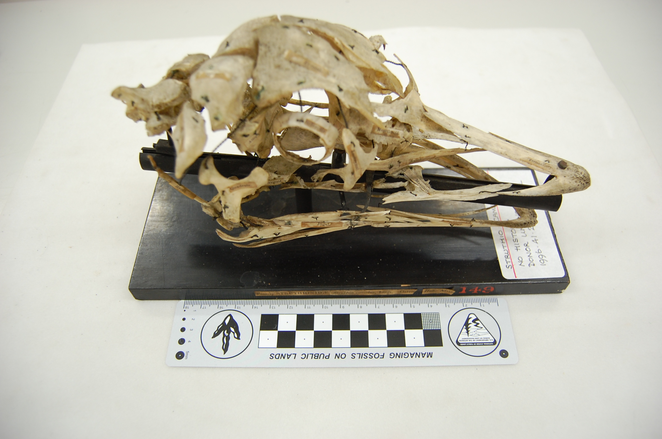
Exploded skull of an ostrich, Struthio camelus. This kind of careful preparation takes crazy skill, and creates a thing of rare beauty.
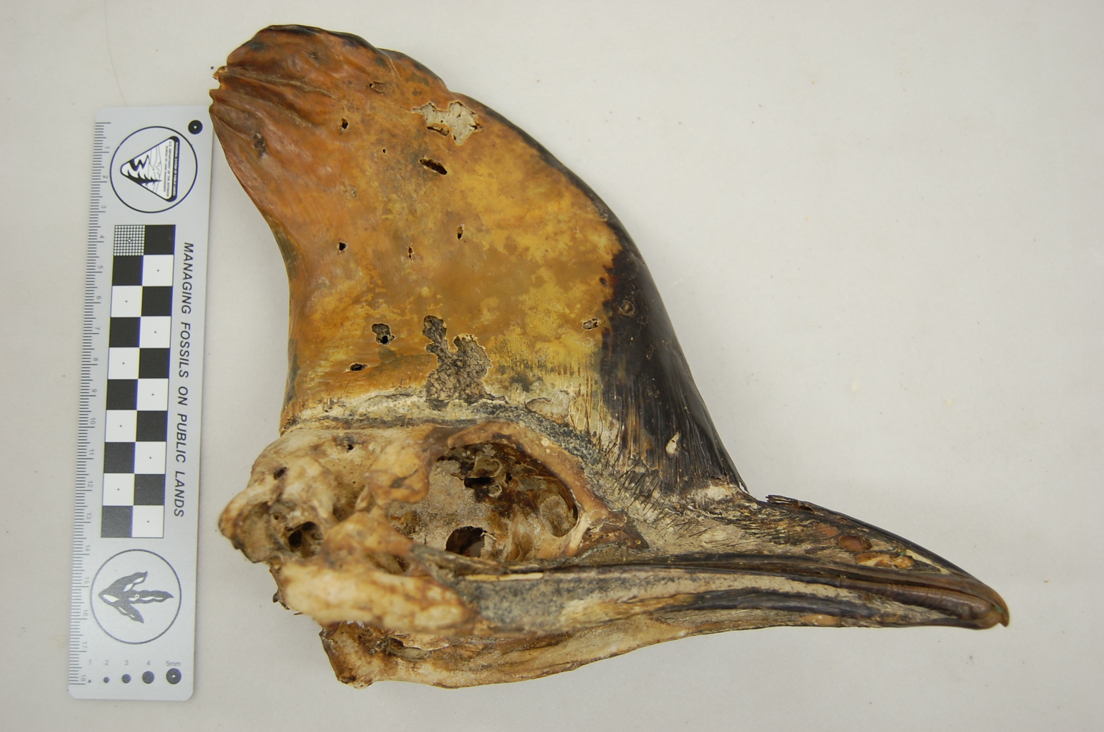
Imposing skull of a cassowary, Casuarius casuarius, with a rather worn head casque.
Mummified Owen’s Little Spotted Kiwi, Apteryx owenii. The feathers were still soft and fluffy, but I would not call this specimen cuddly.
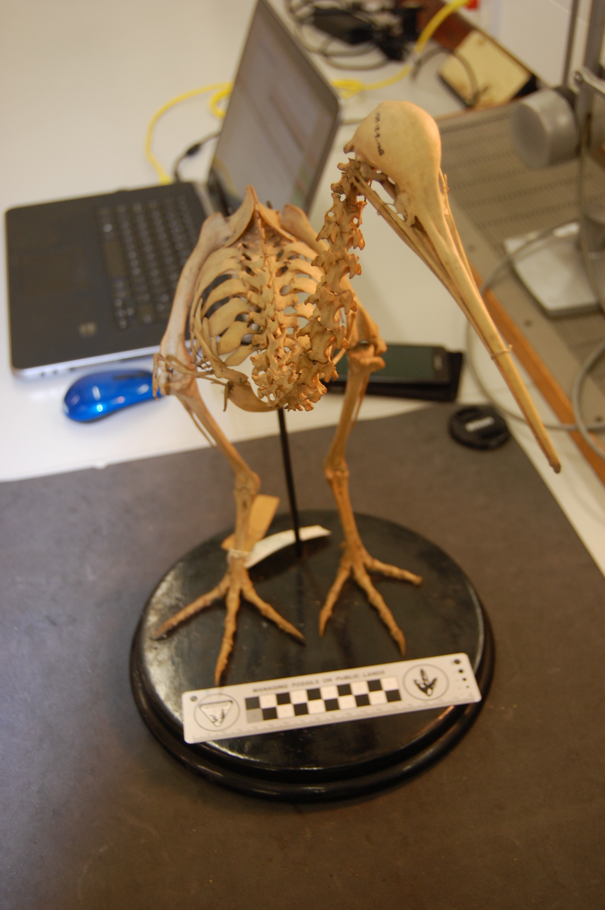
Dorsal view of the back/hips of the Great Spotted Kiwi, Apteryx haasti. I like this photo and am not sure why. The symmetry and shading pleases me, I guess.
Front view of the back/hips of the Great Spotted Kiwi, Apteryx haasti, watching over my laptop and watching me while I write this blog on my laptop… so meta(ornithine)!
Wing of a kiwi, showing the fragile bones and feather attachments. “Apteryx” = “no wings”… well not quite. Click to emkiwi(?) so you can identify the individual bones, from the humerus right down to the fingers! I love this specimen.
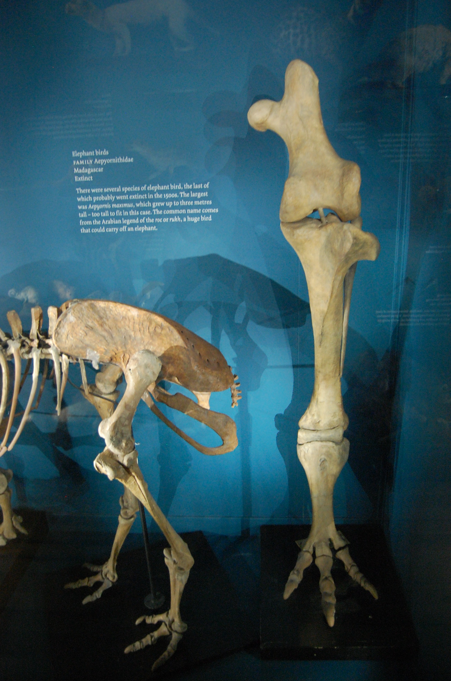
The titanic left leg (in front view) of the Elephant Bird, Aepyornis maximus, from Madagascar, with a small moa nearby in left side view. There’s so much awesomeness about elephant birds I don’t know where to start, but this is one good place to do so.
The smaller end of the palaeognath scale: a mummified Undulated Tinamou, Crypturellus undulatus. Somehow the head got stuck into the abdominal cavity underneath the sternum, so this tinamou almost had its head up its arse. A tinamou with head in its proper position looks and sounds like this (video).
And now we take a left turn into the Galloanseres, most basal branch of the neognath birds, to see some of the neglected, strange early branches off from the “main line” that led to the modern diversity of ducks, geeses and swans (Anatinae, Anserinae).
Screamers (Anhimidae) are to Anseriformes as megapodes (see below; brush turkeys) are to Galliformes. By that I mean that both screamers and megapodes are very early branches off the main line of their respective lineages’ evolution, and both are quite strange when seen in that context… an unfair one, frankly; over-focused on the most familiar, “modern” or most speciose group. More about this issue further below.
This was my first hands-on experience with screamer anatomy; I was familiar from reading Tetrapod Zoology and other material about them. Check out the sound that gives them their name here! I’m now a big fan- they have so many strange features: oddly chunky but often very light bones, big feet with long toes, and then these switchblade-wrists, which would make Batman jealous:
Crested Screamer, Chauna torquata, showing the wicked spur (and smaller one) on the carpometacarpus.
Horned Screamer, Anhima cornuta; similar carpometacarpal spurs as in Chauna.
Torso of a screamer seen in top view. Nice narrow body, and no uncinate processes (spur-like bony struts that cross the ribs and act as levers for the muscles that move the ribcage during breathing)
The long, gracile, clawed toes of a screamer. Those toes, especially as they belong to an animal called a screamer, are spooky for me. Note also: very little toe-webbing for a “waterfowl.”
Not to be outdone, on the Galliformes side of Galloanserae, we have some funky headgear in the Maleo (a megapode bird/Megapodiidae; a very basal branch of “brush turkeys” and kin) and curassows (part of the Cracidae; odd South American birds whose males make booming sounds, presumably using their head-casques as resonating chambers?):
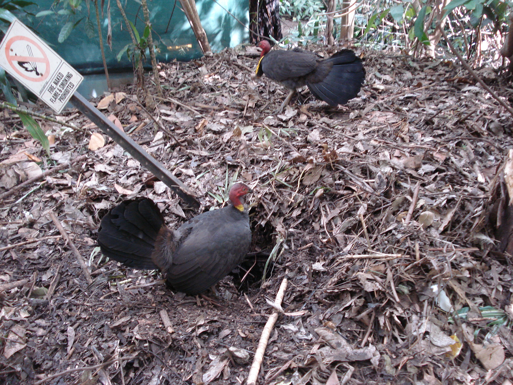
Australian brush-turkeys, Alectura lathami, at the Alma Park Zoo near Brisbane, Australia; they run wild there. Here they are doing what they are best known for: making a mound-like nest. We were doing kangaroo biomechanics experiments and they were everywhere. I was in awe to see such exotic (to me) birds; locals seemed not so enthused (the birds are loud and make a lot of mess).
Skull of Helmeted Curassow, Crax/Pauxi pauxi, showing that resonating chamber. Along with this boom-boom-room, the male uses a piece of food that he holds to draw in the female; if she takes it, then it’s sexy time.
Foot of a Siberian Black Grouse, Tetrao tetrix (nothing to do with a certain videogame), with and without flesh. Regard the broad, feathered feet, well insulated and with plenty of surface area for prancing around in the snow or moorlands. Tetrao engage in a cool display pattern called lekking, in which the males group together and show off to watching females.
A theme in the section above that is not to be missed is that there is some amazing disparity of anatomical forms in these basal lineages of poultry-relatives. Don’t dismiss the Galloanserae as just boring food-birds! Heaps of not-so-well-studied species exist here, surely with a treasure trove of cool neontological and evolutionary questions waiting for the right person to ask! Darwin’s chickens may get their share of neglect, but that pales in comparison to how little we understand about many basal Galloanserae.
What a lot of people think of as a “ground fowl” or galliform way of life is more of a way of life somewhat typical of the Phasanidae- chickens, pheasants and their familiar kin. Megapodes, curassows, guans, grouse and other Galliformes do not necessarily do things in the “typical” ground fowl way, much as the earlier branches of the Anseriformes don’t always look/act like “proper water fowl” (i.e. Anatidae). The phenomenon at play here is one of the great bugaboos in biology: essentialism— the often implicit misconception that variation away from some abstract ideal is negligible, uninteresting or just not conceivable due to mental blinders. When we say something like “the chicken is a fascinating species” we are sliding down the essentialistic slope. There is no “the chicken.” Not really. Oh dear, speaking of slippery slopes, I’d best stop here before I start talking about species concepts. And no one wants that to happen! Anyway, essentialism still pervades a lot of modern scientific thinking, and has its place as a conceptual crutch sometimes. But in biology, essentialism can be very insidious and misleading. It burrows in deep into the scientific mind and can be hard to root out. Unfortunately, it is entrenched in a lot of science education, as it makes things easier to teach if you sweep aside the exceptions to the essentialist “rules” in biology. I catch myself thinking in static, essentialist ways sometimes. The punishment is no cake for a week; so awful. 🙂
And speaking of “normal” or “typical,” morphology is of course often not that way even within a species, age class or gender. Pathology is a great example; by definition it is abnormal. It is a shattering of the “essence” of animals, brought on by some malady.
Next I’ve highlighted some of the amazing pathologies I’ve seen in the Tring skeletons. There have been so many I’ve been unable to keep track of them– some of these birds had the stuffing beaten out of them, and I’m not talking about Thanksgiving turkeys. Some were captive animals, in which the pathology might be blamed on living an inappropriate environment, but some were wild-caught — given the extreme pathologies, it’s a wonder those even survived to be found, but perhaps less a surprise that they were caught.
BONES GONE BONKERS:
View of left knee of a specimen of the Highland Guan, Penelopina nigra, showing some nasty osteoarthritis around the whole joint. Eew. A happier Guan sounds like this.
Femora and tibiotarsi of the Blue-throated Piping Guan, Aburria cumanensis. Amazing pathology involving the left femur (broken, rehealed) and tibiotarsus (secondary infection?). Interestingly, the non-fractured limb also showed some pathology, perhaps indicating general infection and/or arthritis in reaction to the severe damage to the other leg, or just increased load-bearing on that leg.
Little Chachalaca, Ortalis motmot, showing a broken and rehealed right femur and the tibiotarsus. As in the guan above, this animal was not walking for many weeks; its femur had snapped in two, but somehow melted back together. The tibiotarsus didn’t look too great, either; lumpy and bendy. In better times, the Chachalaca does the cha-cha like this.
These two specimens blew my mind. On the left is a normal Tetrao tetrix (Black Grouse); on the right is one hybridized with another (unknown) species.
In the picture above, what amazed me first was the very unusual flattened pelvis/synsacrum of Tetrao, which characteristically is light and wide. But in the hybrid this morphology was completely gone; the pelvis had a more standard “galliform” (read: Phasianid)-like shape, deeper and narrower and more solid in build. I am guessing that the hybrid was a cross with a pheasant like Phasianus itself, whose anatomy would be more like this. Somewhere in here there is a fantastic evo-devo/morphometrics project waiting to happen.
That’s my quick specimen-based tour of “basal birds”. Beyond these two clades of Palaeognathae and Galloanseres, there lies the forebidding territory of Neoaves: much of living avian diversity, and extremely contentious in its phylogenetic relationships. I’m tackling them next for my research on the evolution of the patella/kneecap. But first, I’ll be at the NHM-Tring today for a whirlwind tour through the respectably speciose “normal” Galloanseres clades of Phasianidae and Anserinae+Anatidae, so off I go! (It’s my wife’s birthday celebration, so cake may have to wait for later this time)
So what do you think? What’s your favourite neglected “primitive” bird group (more apropos: early branching avian lineage that may still be very specialized, rare and poorly understood), or cool factoid about palaeognaths and basal neognaths?
No quaggas were harmed during the writing of this post. Polly wanna quagga?
Bovids to the right of me, pinnipeds above, what’s a guy to do but squee?
I’ve been doing some osteological studies of the patella (bone in the major tendon in front of the knee; termed a sesamoid) that have included frequent visits to the Natural History Museum’s avian skeleton collection at Tring. It’s a cute little town, northeast of London, in the green county of Hertfordshire where I live and work. The museum at NHM-Tring is a great old school multi-storey display packed with skeletons and stuffed animals in dark wood cabinets, with many critters hanging from wrought iron railings or other suspensions above (see above). I blogged about the Unfeathered Bird exhibit (and book) that just finished up its tour there yesterday. And I’ll be blogging later, as I keep promising, about the cool things I’ve learned during the past year of my studies of the form, function, development and evolution of the patella.
As an aside, I heartily recommend doing research at the NHM-Tring. It’s away from the bustle (and arduous Tube trip) of the South Kensington NHM, and the curatorial staff are immensely helpful… and there is something else that makes the trip even more enjoyable, but you must read more below to find out about it.
Stomach-Churning Rating: 2/10; 150-year-old dry bones. But an advance warning to (1) diabetics and (2) pun-haters, for reasons that will become evident.
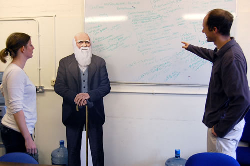
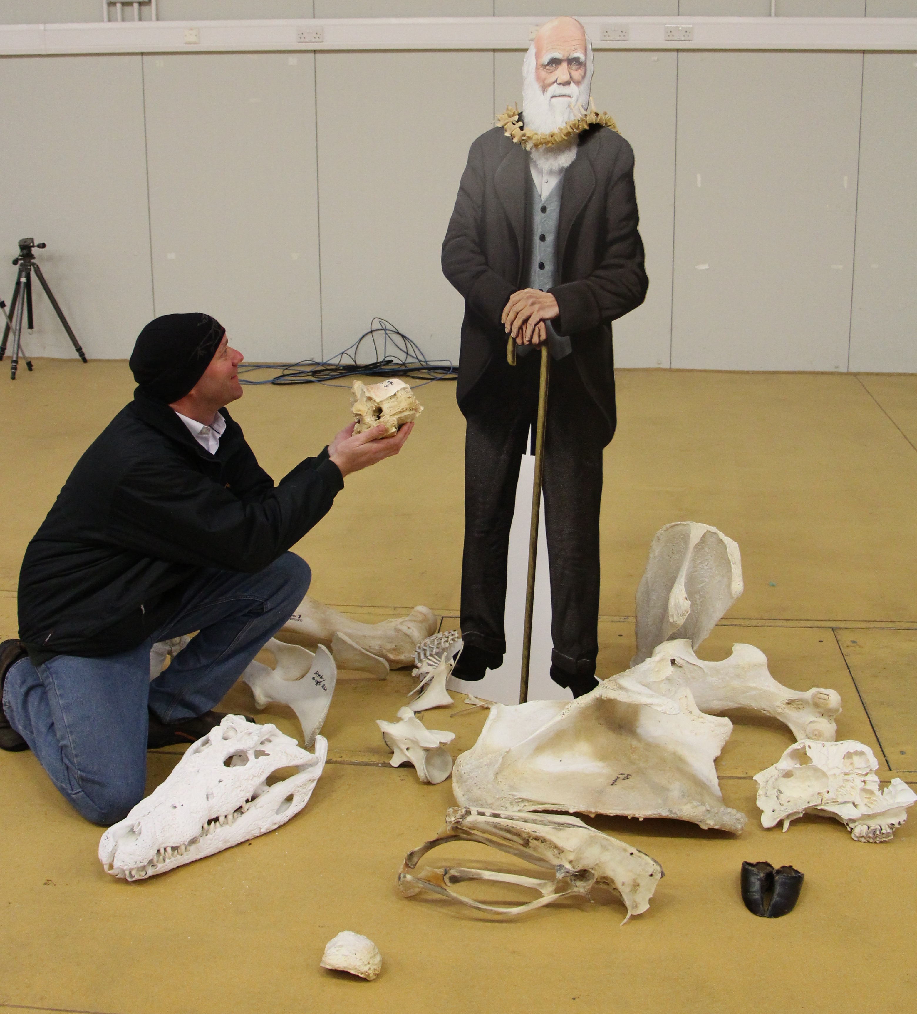
Dr Heather Paxton and Dr Jeffery Rankin, postdoc researchers working on our collaborative BBSRC chicken biomechanics grant (see thechickenofthefuture.com), use the Structure & Motion Lab whiteboard to explain their science to an attentive Darwin.
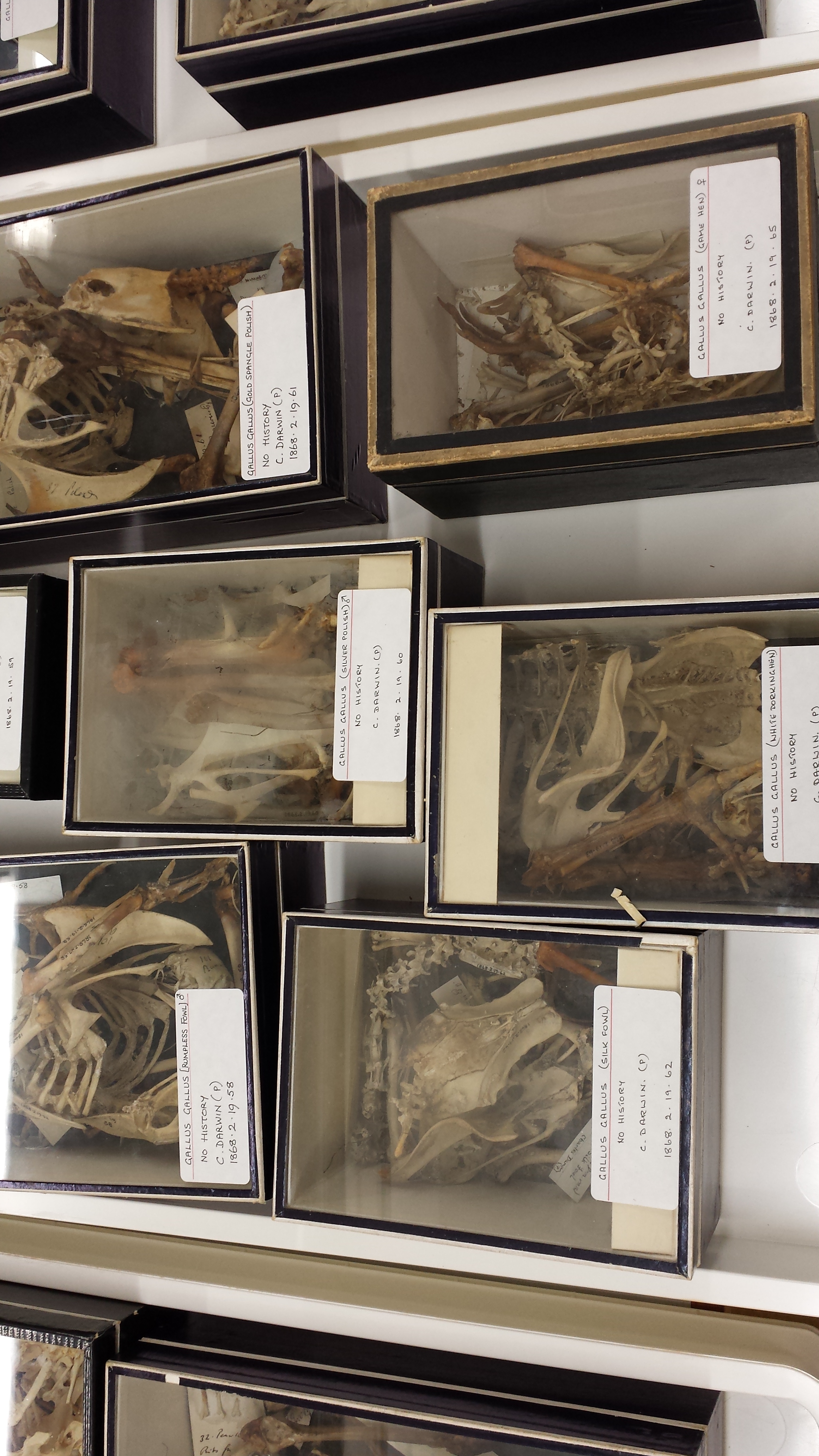
Today I have a short pictorial exhibit of something wonderful I ran into while patellavating in the NHM collections. As often happens while doing museum research, I had a serendipitous encounter with a bit of history that blew my mind a little, and had me geeking out. These things happen because museum collections are stuffed with specimens that, to the right eyes or the right mindset, pack a profound historical whallop. As a scientist who is pretty keen on chickens (Gallus gallus), there are probablyno museum specimens of chickens that would get me more excited about than the chickens Darwin studied in his investigations of artificial selection. In fact, most museum specimens of domestic chickens would not be that interesting to me, especially after seeing these ones.
Darwin wielded the analogy between artificial selection and his conceptual mechanism of natural selection in the first ~4 chapters of On the Origin of Species to clobber the reader with facts and try to leave them with no doubt that, over millennia, nature could craft organisms in vastly more complex and profoundways than human breeders could mould them over centuries. While people most often speak of Darwin’s pigeons when referring to Darwin and avians or artificial selection and variation, his chickens appear in The Origin and other writings quite often, too (most prominently, The Variation of Animals and Plants Under Domesticationin 1868– more about that here). For example, from my 1st edition facsimile of The Origin from Harvard University Press, pp. 215-216:
“Natural instincts are lost under domestication… It is not that chickens have lost all fear, but fear only of dogs and cats, for if the hen gives the danger-chuckle, they will run… and conceal themselves in the surrounding grass or thickets; and this is evidently done for the instinctive purpose of allowing, as we see in wild ground-birds, their mother to fly away. But this instinct retained by our chickens has become almost useless under domestication, for the mother-hen has almost lost by disuse the power of flight.”
Well told, Mr D!
I am also reminded of how chickens and Darwin have had darker relationships, such as this sad story. Or how evolution via Darwinian mechanisms crosses paths with pop culture in fowl ways, such as how tastes-like-chicken evolved, or how some say that chickens, over great periods of time, have been naturally selected in such a way that they are now heritably predisposed to cross roads, or that the amniote egg preceded the evolution of the genus Gallus by some 325+ million years. I see I am drifting and drifting further away from the topic at hand, so let me segue back to Darwin’s chickens. We’ll take this corridor there:
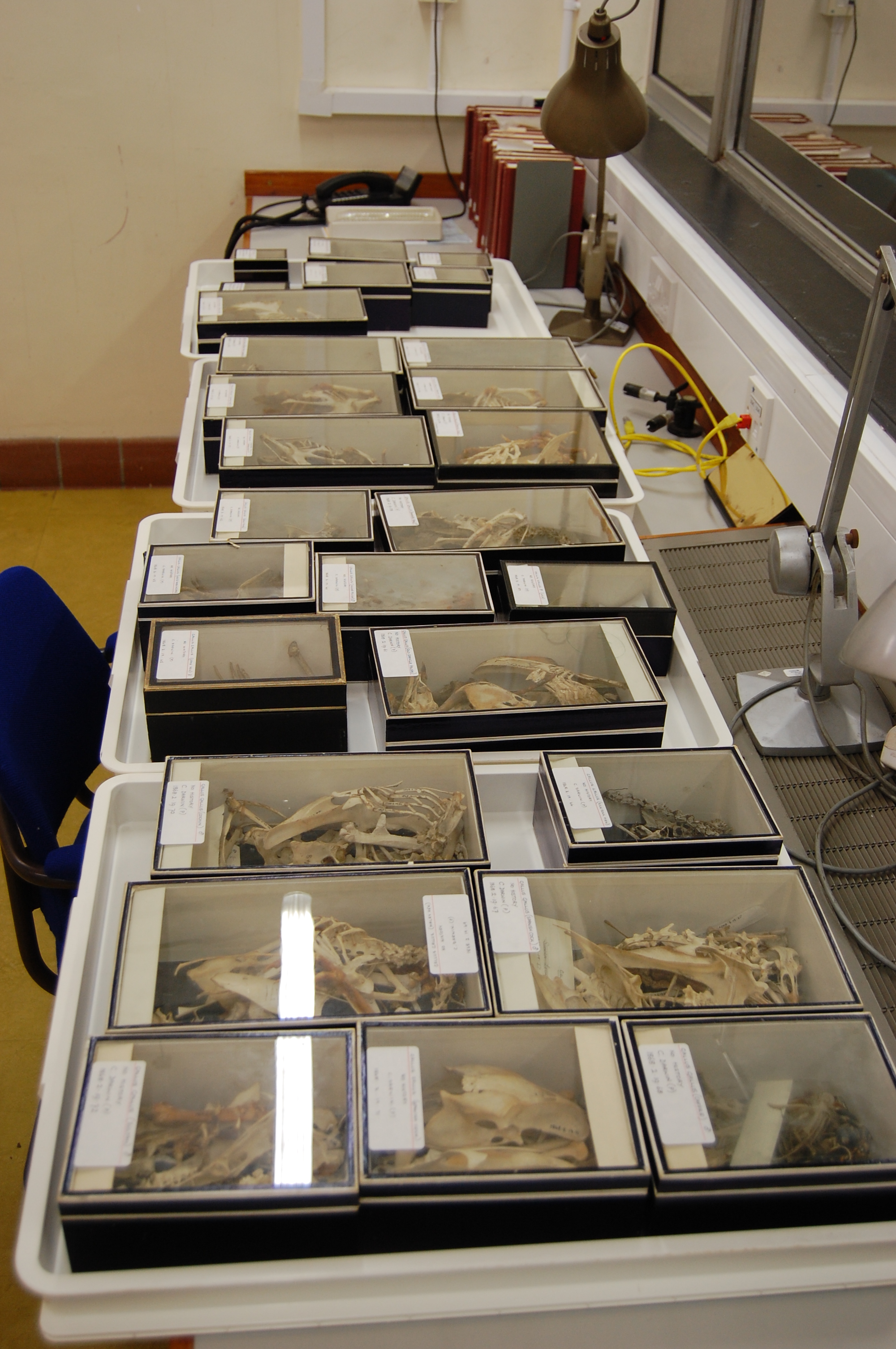
Inside the avian osteology collection at Tring. Sterile at it might outwardly seem, places like this are fertile breeding grounds for scientific discovery. And a sterile-looking collection means well cared-for specimens that will persevere for future discoveries.
So anyway, when museum curator Jo Cooper said to me something like “I have some of Darwin’s chickens out over on the other counter, do you want to have a look or shall I put them away?” my answer was quick and emphatic. YES! But only after lunch. I was hungry, and nothing stops me from sating that hunger especially when the sun is out and there are some fine pubs within walking distance! I settled on the King’s Arms freehouse, and had a delicious cheeseburger followed by a spectacularly good apple-treacle-cake with ice cream, expediently ingested while out on their sunny patio. Yum! I cannot wait to have that cake again. What a cake! Darwin’s bushy eyebrows would have been mightily elevated by the highly evolved flavour, which would have soothed his savage stomach ailments. He would have been like:
“Damn, Emma! Holy s___ this is great apple-cake; here, try some! There is grandeur in this tasty cake, with its several flavours, having been originally cooked into a few baking trays or into one; and that, whilst this pub has gone on serving fine food according to the fixed hygiene laws of Tring, from so simple a beginning endless foods most beautiful and most wonderful have been, and are being, devoured.” And Emma, cake then firmly in hand, would have said something like, “My dear Charles, I shall try this enticing dessert, and I am glad to see you so enthused about something other than barnacles. Write a letter to Huxley or Lyell about that cake later. You need to focus on concocting an ending to that big species book of yours, not cakes. It’s been 20 bloody years, dude; cake can wait. End the book on a high note.” And so it must have happened.
Working at a museum collection is like having an extra home/office for a day or more. You get familiar with the environment while working there, and start to settle in and enjoy the local environs while taking work breaks. Or I do, anyway. So this post is also partly about how cake and other provisions are an important part, or even a perk, of life as a visiting museum researcher. Put in some good solid work, then it’s cake time, but where are the cakes? You explore, and you discover them– opening the door of an unfamiliar shop or pub near a museum can be like opening a museum cabinet to discover the goodness inside. Just don’t get them mixed up.Museum specimens: for research; subjects for science. Cakes: for eating; fuel for scientists. Got it?
But I digest digress. This post is not about my lunch. Not so much, anyway, although I did enjoy the cake quite a bit. Back to the chickens. Here, try some!
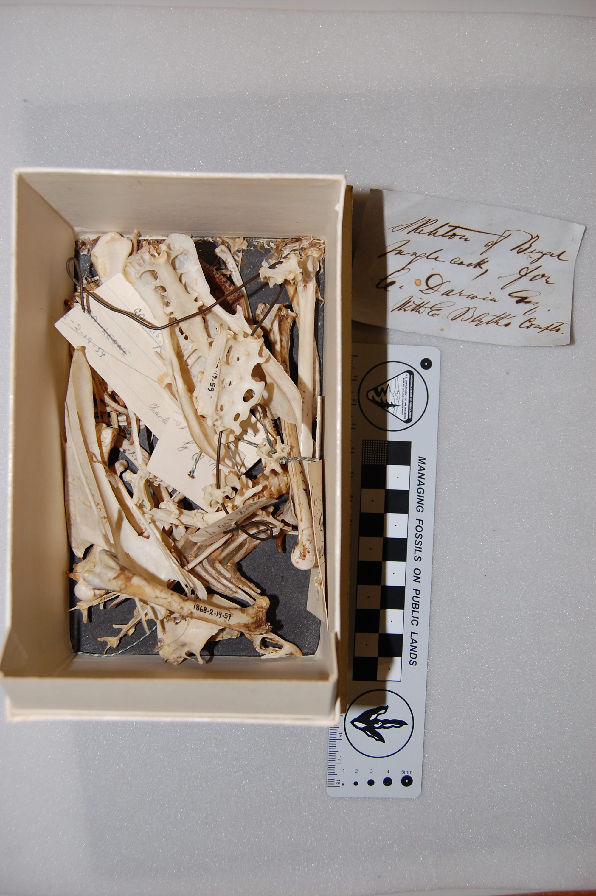
Above: Views of Darwin’s chickens laid out at the NHM-Tring. (all photos in this post can be clucked to emchicken them)
The chickens, much like the pub lunch, did not disappoint in the least. Here I had before me Darwin’s own personal specimens, which I envisioned him dissecting and defleshing himself, studying them in deep introspection, then handing them over to the museum for curation once his lengthy researches were complete (all the ones I studied dated back to around 1863-1868, so they were curated shortly after The Origin was published (1859)). Perhaps the museum gave him some fine sponge-cake in return. There was at least one male and female adult of each of numerous breeds, many of them still bearing the dried flesh of centuries past. This was great for me, as the patella often gets removed and clucked chucked in the bin with its tendon when museum specimens of birds are prepared (much as elephant “sixth toe” sesamoids are). All of the specimens had their honking huge patellae on display, so that’s what a lot of my photos feature. I do so lament that I did not take a photo of the cake. Did I tell you about that cake? Oh… Check out these examples of Darwin’s chickens:
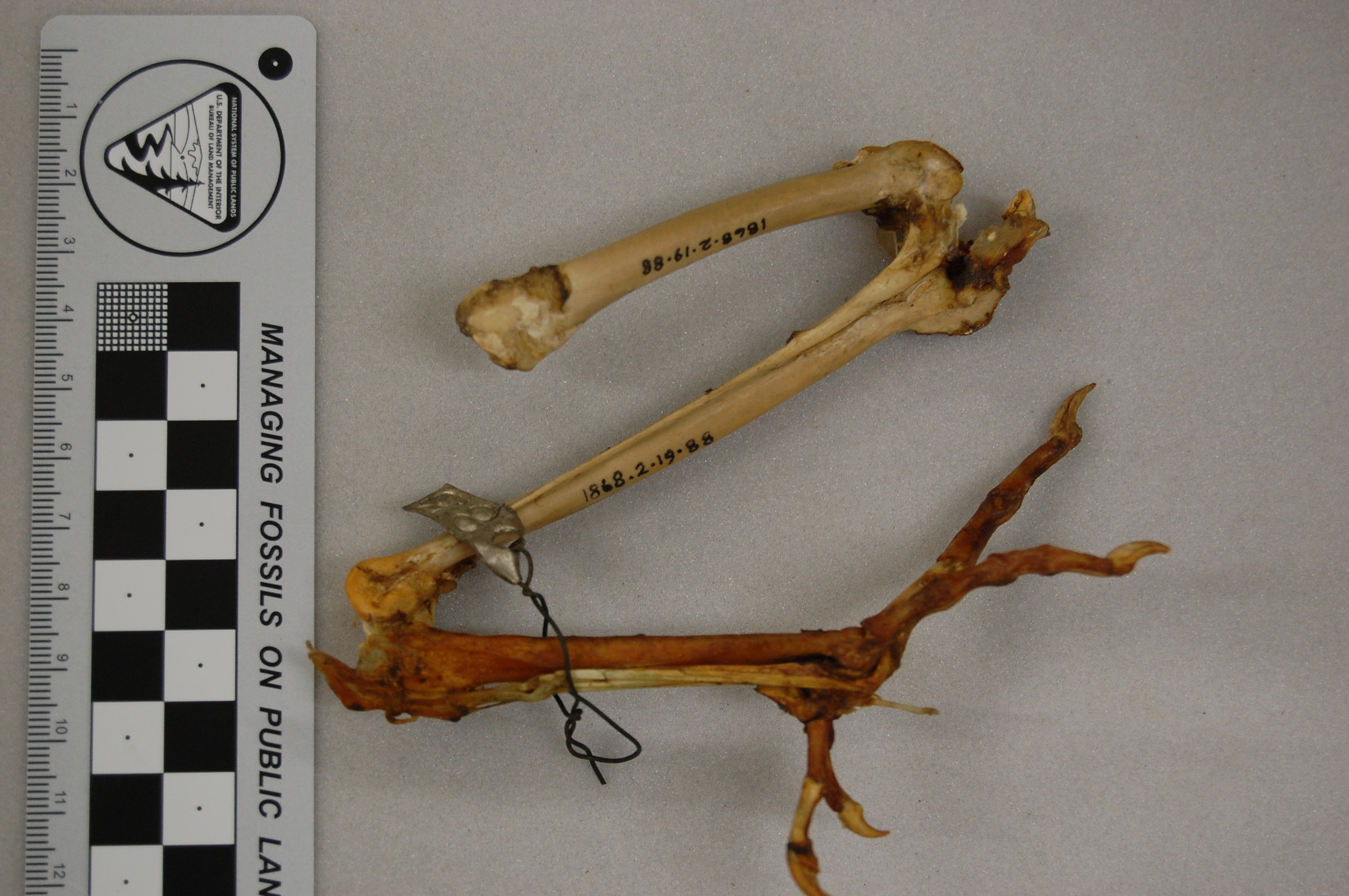
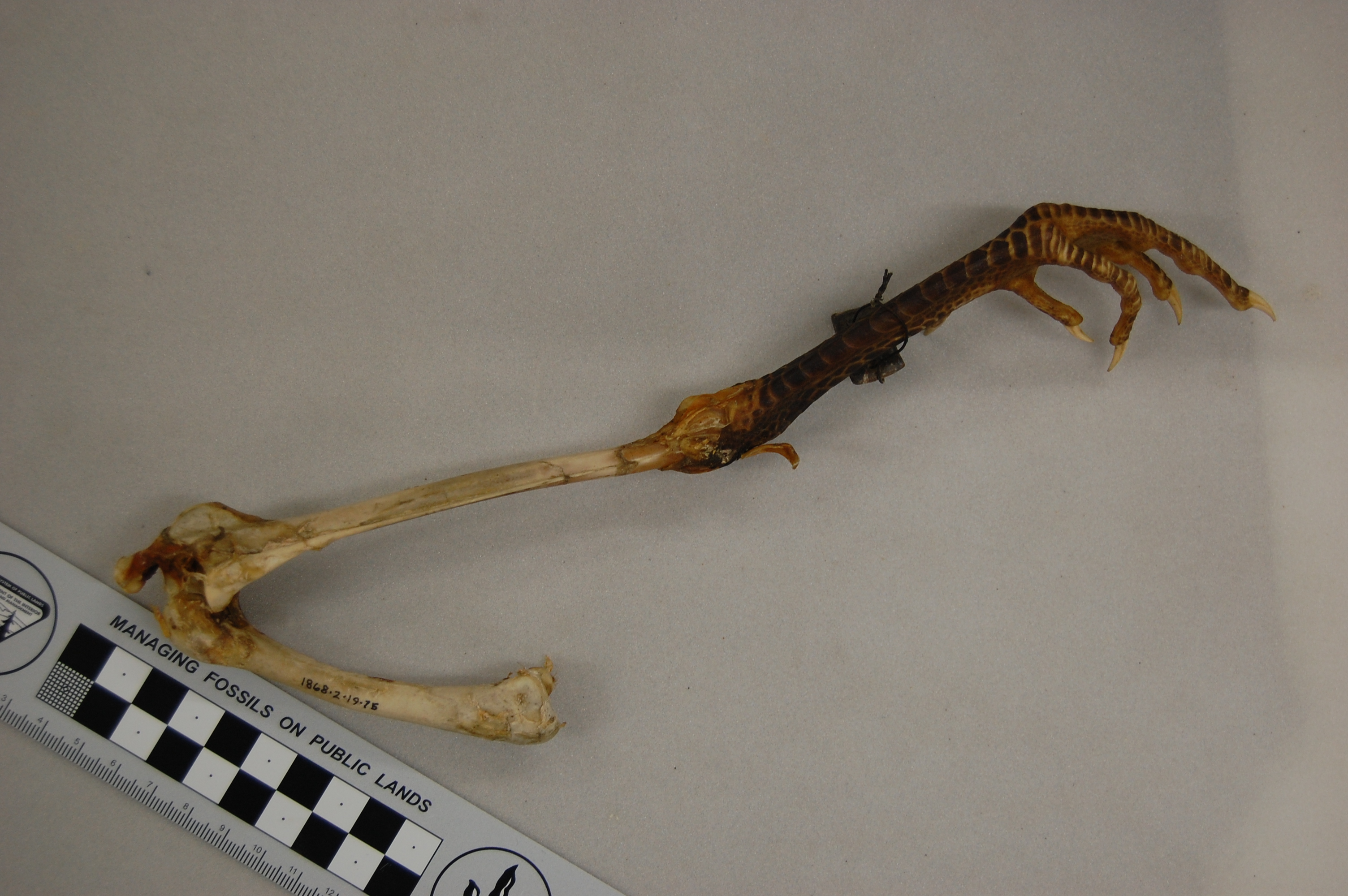
African rooster (wild variety? Darwin’s label was not clear) in right side view, with the patella indicated by a red arrow. That patella is still attached to the tibiotarsus by the patellar tendon (often misnamed the patellar “ligament”, but it is just a continuation of the proximal tendon).
Darwin’s handwritten label and the well-endowed patella of the Spanish Cock. What? Oh, you. Stop it. That has nothing to do with cake, and only cake-related humour is allowed in this post.
Some other fascinating features exhibited by Darwin’s chickens, which he doubtless mulled over while nibbling on fine cakes, included the following:
The hindlimb of a Polish Silver Laced breed, nicely showing the ossified tendons (red arrow) along the tarsometatarsus. Why these tendons turn into bone is one of the great unsolved mysteries of bone biology/mechanics and avian evolution.
Check out the famed feather crest of the Silver (Laced) Polish here; it gets so extreme in males that they have a hard time seeing, and can get beaten up by other cockerels when kept in mixed breed flocks.
Here on this blog, and of course on the companion blog “Towards the Chicken of the Future,” domestic chickens and wild junglefowl have often come up, most recently with the Dorking Chicken (another of Darwin’s own specimens that I studied) in the “Mystery Museum Specimen” poetry round of late. Dorkings are HUGE chickens; easily twice the weight of even a broiler chicken, up to 4-5kg. The Dorking-characteristic polydactyly featured in that post is also observed at a relatively high incidence in Silkie and Sultan breeds, I’ve learned. Like this one! (I was so patella-focused, or cake-somnolescent, that I missed it while studying at the museum and only noticed it now while browsing through my photos, bereft of cake)
Nice leg of a Sultan hen. There is an extra toe here as in the Dorking chicken; a duplicate hallux (first toe). This is not, as it might at first seem, a pathological condition as in modern “twisted toe”-suffering domestic chickens.
Malays are another giant breed like the Dorking, but with longer and more muscular legs and longer necks, looking much more like a classic, badass wild junglefowl than a fancy, pampered chicken. But here, undressed to the bare bones, it just looks like a skinny chicken leg, albeit perhaps a bit svelte compared to the Dorking or Sultan.
Hindlimb of a Malay breed of chicken, which Wikipedia nicely tells the story of its misnomer (it may originate from Pakistan, not Malaysia!). Can you find the nice patella? Check out Darwin’s lovely label, too.
You may have come across wild-eyed news stories 5 years ago about “OMG Darwin was sooooooo wrong about chickens!”, referring to his writings on the origin of domestic chickens from Red junglefowl. As Greg Laden adeptly wrote, Darwin (say it with me) didn’t really get it very wrong after all. He did quite well, in fact. Some media outlets did get it more wrong, probably inspired by this press release. Oh well; the science they were reporting about definitely was interesting- modern chickens seem to have some of their yellow skin pigmentation-related genes from Grey junglefowl, although they are still largely descendants of Red junglefowl.
Here, have a JUMBLE-fowl, or rather a junglefowl cockerel, with another Darwin label:
Darwin’s example of a wild-type chicken; a Red junglefowl. As he suspected, these Asian birds were the ancestors of domestic chickens, but today evidence indicates that domestication may have occurred multiple times in Asia and with different wild varieties of junglefowl bred/mixed in different regions.
Some breeds aren’t so funky inside, of course, but just have cool feather patterns on the outside, like the “pencilling” (dark streaks on white feathers) evident in pencil breeds; also called triple-laced. Like this fine chap below once would have had, before Darwin tore off his feathers and reduced him to a research-friendly naked skeleton:
A Golden Pencil(led) Hamburg breed of chicken (cockerel), whose skeleton features the leg and a fine articulated patella.
Also known as the Holland Fowl, several European countries including the UK claim the Hamsburg as an original breed from their respective realm, and no surprise they do- it’s a lovely spangled chicken. Then, later in the 1800’s the Americans got involved in breeding them, too, and it’s all a big mess. They should get together, have some delectable cakes, and just sort it out.
Scaly, still-greasy foot and hindlimb of what Darwin labelled as the male of a “Game” breed.
We thus close with another leg of another chicken. Darwin was a bit naughty here, or else terminology of breeds has changed a lot since the 1850’s (very possible), as he just labelled this as a “Game” cockerel. Now, Gamefowl is a big category of breeds. I’m guessing this one was either (1) a Cornish/Indian Game variety or (2) an Old or Modern English Game Fowl. Maybe a person who knows their chicken breeding far better than me (that’s not hard!) will opine differently. The latter varieties were popular in Darwin’s time — the (Muffed) Old English version was mated with other breeds (Malay?) to produce the Modern English form as cockfighting “sports” became banned in 1849 and breeder attentions shifted to the polar opposite of producing showy, fancy birds instead. And thus the bufante, feathered-hair-adorned 1980s pop-rock group was created, to sing about mating or moulting or melting with people or something terribly disgusting and probably having nothing at all to do with chickens, cake, or cockfighting, or other more seemly pursuits.
So, we have come to the end of my photos of Darwin’s chicken leg bones and such. If you’ve learned something here about chicken breeds, patellae, cake, or Darwin, that’s simply frabjous. Enough of those poncey pigeons, already! I’m crying fo… no, I won’t use that pun. Nevermind. Not even remotely cake-related. Let’s give Darwin’s chickens their just desserts, is the point– and a much better pun, too. Darwin’s chickens are an important part of Darwiniana, and an interesting evolutionary study in and of themselves. I’ve certainly become impressed during my researching for this blog post by the diverse, fascinating biology of chicken breeds. My copy of the “Complete Encyclopedia of Chickens” will be getting some more thorough reading shortly.
Today, however, I am off to return to the NHM-Tring and peruse their other, non-chickeny Galliformes and Anseriformes, with a detour to the mythical hoatzin. But… but… there may be a cake detour involved, too. I shall report back in due course. Off I go!
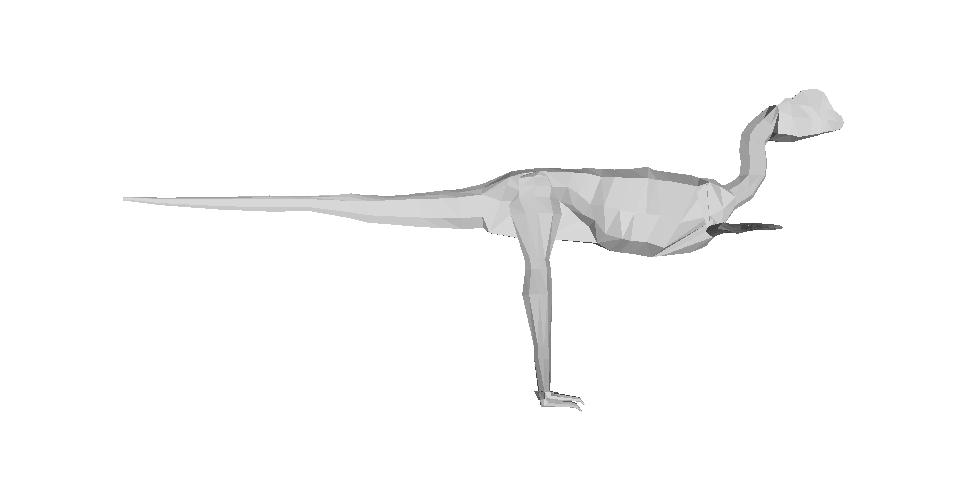
Our 3D computer models of a basal dinosaur and bird, showing methods and key differences in body shape. The numbers at the bottom are museum specimen numbers.
At about the moment I’m posting this, our Nature paper (our more formal page here, and the actual article here) embargo is ending, drawing a 14+ year gestation to a close. The paper is about how dinosaur 3D body shape changed during their evolution, and how that relates to changes in hindlimb posture from early dinosaurs/archosaurs to birds; “morpho-functional evolution” sums up the topic. We used the 3D whole-body computational modelling that I, Allen and Bates (among others) have developed to estimate evolutionary changes in body dimensions, rather than focusing on single specimens or (as in our last study) tyrannosaur ontogeny. We’ve strongly supported the notion (dating back to Gatesy’s seminal 1990 Paleobiology paper) that the centre of mass of dinosaurs shifted forwards during their evolution, and that this shift gradually led to the more crouched (flexed) hind leg posture that characterizes living birds. Here is a movie from our paper showing how we did the modelling:
And here is a summary of our 17 computer models of archosaur bodies, shown as one walks along the tips of the phylogeny shown in the video (the models are not considered to be ancestral to one another; we used a common computer algorithm called squared-change parsimony to estimate ancestral state changes of body dimensions between the 16 numbered nodes of the tree):
But we’ve done much more than just put numbers on conventional wisdom.
We’ve shown, to our own surprise, that the shift of the centre of mass was largely driven by evolutionary enlargements of the forelimbs (and the head and neck, and hindlimbs, to a less strong degree), not the tail as everyone including ourselves has assumed for almost 25 years. And the timing of this shift occurred inside the theropod dinosaur group that is called Maniraptora (or Maniraptoriformes, a slightly larger group), so the change began in animals that were not flying, but not long before flight evolved (depending on whom you ask, what taxonomy they favour and what evidence one accepts, either the smaller clade Eumaniraptora/Paraves or the bird clade Aves/Avialae).
Now, if you don’t like the cliche “rewriting the textbooks”, do have a look through texts on dinosaur/early avian palaeobiology and you probably will find a discussion of how the tail shortened, the centre of mass moved forwards as a consequence, the caudofemoral musculature diminished, and theropod dinosaurs (including birds) became more crouched as a result. We did that to confirm for ourselves that it’s a pretty well-accepted idea. Our study supports a large proportion of that idea’s reasoning, but modifies the emphasis to be on the forelimbs more than the tail for centre of mass effects, so the story gets more complex. The inference about caudofemoral muscles still seems quite sound, however, as is the general trend of increased limb crouching, but our paper approximates the timing of those changes.
Figure 3 from our paper, showing how the centre of mass moved forwards (up the y-axis) as one moves toward living birds (node 16); the funny dip at the end is an anomaly we discuss in the paper.
A final implication of our study is that, because the forelimbs’ size influenced the centre of mass position, and thus influenced the ways the hindlimbs functioned, the forelimbs and hindlimbs are more coupled (via their effects on the centre of mass) than anyone has typically considered. Thus bipedalism and flight in theropods still have some functional coupling– although this is a matter of degree and not black/white, so by no means should we do away with helpful concepts like locomotor modules.
And in addition to doing science that we feel is good, we’ve gone the extra mile and presented all our data (yes, 17 dinosaurs’ worth of 3D whole body graphics!) and the critical software tools needed to replicate our analysis, in the Dryad database (link now working!), which should have now gone live with the paper! It was my first time using that database and it was incredibly easy (about 1 hour of work once we had all the final analysis’s files properly organized)– I strongly recommend others to try it out.
That’s my usual general summary of the paper, but that’s not what this blog article is about. I’ll provide my usual set of links to media coverage of the paper below, too. But the focus here is on the story behind the paper, to put a more personal spin on what it means to me (and my coauthors too). I’ll take a historical approach to explain how the paper evolved.
Embarassing picture of me before I became a scientist. Hardee’s fast food restaurant cashier, my first “real job”, from ~1999- no, wait, more like 1986. The 1980s-style feathered (and non-receding) hair gives it away!
Rewind to 1995. I started my PhD at Berkeley. I planned to use biomechanical methods and evidence to reconstruct how Tyrannosaurus rex moved, and started by synthesizing evidence on the anatomy and evolution of the hindlimb musculature in the whole archosaur group, with a focus on the lineage leading to Tyrannosaurus and to living birds. As my PhD project evolved, I became more interested and experienced in using 3D computational tools in biomechanics, which was my ultimate aim for T. rex.
In 1999, Don Henderson published his mathematical slicing approach to compute 3D body dimensions in extinct animals, which was a huge leap for the field forward beyond statistical estimates or physical toy models, because it represented dinosaurs-as-dinosaurs (not extrapolated reptiles/mammals/whatever) and gave you much more information than just body mass, with a lot of potential to do sensitivity analysis.
I struggled to upgrade my computer skills over the intervening years. I was developing the idea to reconstruct not only the biomechanics of T. rex, but also the evolutionary changes of biomechanics along the whole archosaur lineage to birds– because with a series of models of different species and a working phylogeny, you could do that. To me this was far more interesting than the morphology or function of any one taxon, BUT required you to be able to assess the latter. So Tyrannosaurus became a “case study” for me in how to reconstruct form and function in extinct animals, because it was interesting in its own right (mainly because of its giant size and bipedalism). (Much later, in 2007, I finally finished a collaboration to develop our own software package to do this 3D modelling, with Victor Ng-Thow-Hing and F. Clay Anderson at Honda and Stanford)
Me and a Mystery Scientist (then an undergrad; now a very successful palaeontologist!), measuring up a successful Cretaceous hypercarnivore at the UCMP; from my PhD days at Berkeley, ~2000 or so.
In all this research, I was inspired by not only my thesis committee and others at Berkeley, but also to a HUGE degree by Steve Gatesy, a very influential mentor and role model at Brown University. I owe a lot to him, and in a sense this paper is an homage to his trailblazing research; particularly his 1990 Paleobiology paper.
In 2001, I got the NSF bioinformatics postdoc I badly wanted, to go to the Neuromuscular Biomechanics lab at Stanford and learn the very latest 3D computational methods in biomechanics from Prof. Scott Delp’s team. This was a pivotal moment in my career; I became partly-an-engineer from that experience, and published some papers that I still look back fondly upon. Those papers, and many since (focused on validating and testing the accuracy/reliability of computer models of dinosaurs), set the stage for the present paper, which is one of the ones I’ve dreamed to do since the 1990s. So you may understand my excitement here…
Stanford’s Neuromuscular Biomechanics Lab, just before I left in 2003.
But the new paper is a team effort, and was driven by a very talented and fun then-PhD-student, now postdoc, Dr Vivian Allen. Viv’s PhD (2005-2009ish) was essentially intended to do all the things in biomechanics/evolution that I had run out of time/expertise to do in my PhD and postdoc, in regards to the evolution of dinosaur (especially theropod) locomotor biomechanics. And as I’d hoped, Viv put his own unique spin on the project, proving himself far better than me at writing software code and working with 3D graphics and biomechanical models. He’s now everything that I had hoped I’d become by the end of my postdoc, but didn’t really achieve, and more than that, too. So huge credit goes to Viv for this paper; it would never have happened without him.
We also got Karl Bates, another proven biomechanics/modelling expert, to contribute diverse ideas and data. Furthermore, Zhiheng Li (now at UT-Austin doing a PhD with Dr Julia Clarke) brought some awesome fossil birds (Pengornisand Yixianornis) from the IVPP in Beijing in order to microCT scan them in London. Zhiheng thus earned coauthorship on the paper — and I give big thanks to the Royal Society for funding this as an International Joint Project, with Dr Zhonghe Zhou at the IVPP.
That’s the team and the background, and I’ve already given you the punchlines for the paper; these are the primitive and the derived states of the paper. The rest of this post is about what happened behind the scenes. No huge drama or anything, but hard, cautious work and perseverance.
Me shortly after I moved to the RVC; video still frame from a dinosaur exhibit (c. 2004) I was featured in. Embarassingly goofy pic, but I like the blurb at the bottom. It’s all about the evolutionary polarity, baby!
The paper of course got started during Viv’s PhD thesis; it was one of his chapters. However, back then it was just a focus on how the centre of mass changed, and the results for those simple patterns weren’t very different from those we present in the paper. We did spot, as our Nature supplementary information notes, a strange trend in early theropods (like Dilophosaurus; to a lesser degree Coelophysis too) related to some unusual traits (e.g. a long torso) and suggested that there was a forward shift of centre of mass in these animals, but that wasn’t strongly upheld as we began to write the Nature paper.
On the urging of the PhD exam committee (and later the paper reviewers, too), Viv looked at the contributions of segment (i.e. head, neck, trunk, limbs, tail) mass and centre of mass to the overall whole body centre of mass. And I’m glad he did, since that uncovered the trend we did not expect to find: that the forelimb masses were far more important for moving the centre of mass forwards than the mass (or centre of mass) of the tail was– in other words, the statistical correlation of forelimb mass and centre of mass was strong, whereas changes of tail size didn’t correlate with the centre of mass nearly as much. We scrutinized those results quite carefully, even finding a very annoying bug in the 3D graphics files that required a major re-analysis during peer review (delaying the paper by ~6 months).
The paper was submitted to Nature first, passing a presubmission inquiry to check if the editor felt it fit the journal well enough. We had 3 anonymous peer reviewers; 1 gave extensive, detailed comments in the 3 rounds of review and was very fair and constructive, 1 gave helpful comments on writing style and other aspects of presentation as well as elements of the science, and 1 wasn’t that impressed by the paper’s novelty but wanted lots more species added, to investigate changes within different lineages of maniraptorans (e.g. therizinosaurs, oviraptorosaurs). That third reviewer only reviewed the paper for the first round (AFAIK), so I guess we won them over or else the editor overruled their concerns. We argued that 17 taxa were probably good enough to get the general evolutionary trends that we were after, and that number was ~16 more species than any prior studies had really done.
Above: CT scan reconstruction of the early extinct bird Yixianornis in slab conformation, and then Below: 3D skeletal reconstruction by Julia Molnar, missing just the final head (I find this very funny; Daffy Duck-esque) which we scaled to the fossil’s dimensions from the better data in our Archaeopteryx images. There is also the concern, which the reviewers didn’t focus on but I could see other colleagues worrying about, that some of the specimens we used were either composites, sculpted, or otherwise not based on 100% complete, perfectly intact specimens. The latter are hard to come by for a diversity of extinct animals, especially in the maniraptoran/early bird group. We discussed some of these problems in our 3D Tyrannosaurus paper. The early dinosauromorph Marasuchus that we used was a cast/sculpted NHMUK specimen based on original material, as was our Coelophysis, Microraptor and Archaeopteryx; and our Carnegie ??Caenagnathus??Anzu (now published) specimen was based more on measurements from 1 specimen than from direct scans, and there were a few other issues with our other specimens, all detailed in our paper’s Supplementary Information.
But our intuition, based on a lot of time spent with these models and the analysis of their data, is that these anatomical imperfections matter far, far less than the statistical methods that we employed– because we add a lot of flesh (like real animals have!) outside of the skeleton in our method, the precise morphology of the skeleton doesn’t matter much. It’s not like you need the kind of quality of anatomical detail that you need to do systematic analyses or osteological descriptive papers. The broad dimensions can matter, but those tend to be covered by the (overly, we suspect) broad error bars that our study had (see graph above). Hence while anyone could quibble ad infinitum about the accuracy of our skeletal data, I doubt it’s that bad– and it’s still a huge leap beyond previous studies, which did not present quantitative data, did not do comparative studies of multiple species, or did not construct models based on actual 3D skeletons as opposed to artists’ 2D shrinkwrapped reconstructions (the “Greg Paul method”). We also did directly measure the bodies of two extant archosaurs in our paper: a freshwater crocodile and a junglefowl (CT scan of the latter is reconstructed below in 3D).
One thing we still need to do, in future studies, is to look more carefully inside of the bird clade (Aves/Avialae) to see what’s going on there, especially as one moves closer to the crown group (modern birds). We represented modern birds with simply 1 bird: the “wild-type chicken” Red junglefowl, which isn’t drastically different in body shape from other basal modern birds such as a tinamou. Our paper was not about how diversity of body shape and centre of mass evolved within modern birds. But inspecting trends within Palaeognathae would be super interesting, because a lot of locomotor, size and body shape changes evolved therein; ostriches are probably a very, very poor proxy for the size and shape of the most recent common ancestor of all extant birds, for example, even though they seem to be fairly basal within that whole lineage. And, naturally, our study opens up opportunities for anyone to add feathers to our models and investigate aerodynamics, or to apply our methods to other dinosaur/vertebrate/metazoan groups. If the funding gods are kind to us, later this year we will be looking more closely, in particular, at the base of Archosauria and what was happening to locomotor mechanics in Triassic archosaurs…
Clickum to embiggum:
Australian freshwater crocodile, Crocodylus johnstoni; we CT scanned it in 3 pieces while visiting the Witmer lab in Ohio.
A Red junglefowl cockerel, spotted in Lampang, Thailand during one of my elephant gait research excursions there. Svelte, muscular and fast as hell. This photo is here to remind me to TAKE BLOODY PICTURES OF MY ACTUAL RESEARCH SPECIMENS SO I CAN SHOW THEM!
I’d bore you with the statistical intricacies of the paper, but that’s not very fun and it’s not the style of this blog, which is not called “What’s in John’s Software Code?”. Viv really worked his butt off to get the stats right, and we did many rounds of revisions and checking together, in addition to consultations with statistics experts. So I feel we did a good job. See the paper if that kind of thing floats your boat. Someone could find a flaw or alternative method, and if that changed our major conclusions that would be a bummer– but that’s science. We took the plunge and put all of our data online, as noted above, so anyone can do that, and that optimizes the reproducibility of science.
What I hope people do, in particular, is to use the 3D graphics of our paper’s 17 specimen-based archosaur bodies for other things– new and original research, video games, animations, whatever. It has been very satisfying to finally, from fairly early in the paper-writing process onwards, present all of the complex data in an analysis like this so someone else can use it. My past modelling papers have not done this, but I aim to backtrack and bring them up to snuff like this. We couldn’t publish open access in Nature, but we achieved reasonably open data at least, and to me that’s as important. I am really excited at a personal level, and intrigued from a professional standpoint, to see how our data and tools get used. We’ll be posting refinements of our (Matlab software-based) tools, which we’re still finding ways to enhance, as we proceed with future research.
Above: Two of the 17 archosaur 3D models (the skinny “mininal” models; shrinkwrapped for your protection) that you can download and examine and do stuff with! Dilophosaurus on the left; Velociraptor on the right. Maybe you can use these to make a Jurassic Park 4 film that is better, or at least more scientifically accurate, than Hollywood’s version! 😉 Just download free software like Meshlab, drop the OBJ files in and go!
Now, to bring the story full circle, the paper is out at last! A 4 year journey from Viv’s PhD thesis to the journal, and for me a ~14 year journey from my mind’s eye to realization. Phew! The real fun begins now, as we see how the paper is received! I hope you like it, and if you work in this area I hope you like the big dataset that comes with it, too. Perhaps more than any other paper I’ve written, because of the long voyage this paper has taken, it has a special place in my heart. I’m proud of it and the work our team did together to produce it. Now it is also yours. And all 3200ish words of this lengthy blog post are, as well!
Last but not least, enjoy the wonderful digital painting that Luis Rey did for this paper (another of my team’s many failed attempts to get on the cover of a journal!); he has now blogged about it, too!
Dinosaur posture and body shape evolving up the evolutionary tree, with example taxa depicted. By Luis Rey.
News stories about this paper will be added below as they come out, featuring our favourites:
Synopsis: Decent coverage, but negligible coverage in the general press; just science-specialist media, more or less. I think the story was judged to be too complex/esoteric for the general public. You’d think dinosaurs, evolution, computers plus physics would be an “easy sell” but it was not, and I don’t think we made any big errors “selling” it. Interesting– I continue to learn more about how unpredictable the media can be.
Regardless, the paper has had a great response from scientist colleagues/science afficionados, which was the target audience anyway. I’m very pleased with it, too– it’s one of my team’s best papers in my ~18 year career.
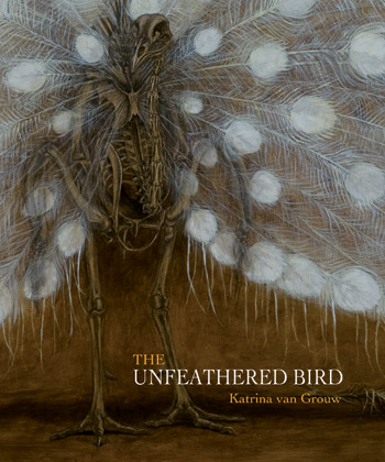
The Unfeathered Bird book by Katrina van Grouw proclaims immediately in its Introduction that it “is not an anatomy of birds.” True– it is far more than that, and it would be a shame if it had just been a dry, technical avian osteology reference book. It is a unique blend of art and science- particularly avian anatomy, evolution, taxonomy, natural history and more. The Unfeathered Bird is written for a general audience; birders/twitchers or just natural history buffs would be ideal targets of its unfettered passion for all things avian. A 12-year-old who is very keen on animals could enjoy it, and it may ignite the flames of ornithological excitement in many young or older readers. I am glad it was not called “The Naked Bird” as that would have caused some serious misconceptions (badum-tish!). The book is dripping with illustrations (at least one every two pages, often more). Almost all of the illustrations (except some paintings in the style of the cover) are in the same brownish sketch style that, like much of the book, evokes a bygone era of dark wooden cabinets and shadowed halls packed with skeletons, with nary an interactive graphics display, animatronic dinosaur or hyperdetailed cladogram in sight. It feels like an homage to the Victorian naturalists’ joy for anatomical detail conveyed through painstakingly detailed woodcuts. And while many still think of feathers as “the defining feature of birds,” enough about feathers already. Seriously. This is a book is about what lies beneath, and how all that non-fluffy stuff is important for birds’ lives, too.
(image-intense post; all can be clicked to embiggritate)
Katrina with peacock feather headdress? (back cover pic and rear view of same skeleton)
Katrina with front cover framed pic and the peacock skeleton that went with it.
The Introduction continues to explain that the book is truly about how the external anatomy of birds is linked to the bony anatomy, which might remind astute readers of modern approaches like the extant phylogenetic bracket. The rest of the book uses both skeletal and unfeathered, quasi-myological illustrations to get this point across vividly. The explanatory text is written at a basic enough level for the average reader and is just the right length, with interesting anecdotes and natural history facts that even the expert reader will find interesting or even inspirational (e.g. possibly a goldmine for research ideas). First there is a 26 page “Basic” section with an introduction to avian osteology, with bountiful sketches to illustrate key organs and text explaining how it all fits together in the fully accoutered bird. The decision to use classical Linnean taxonomy (defunct or re-arranged taxa from the Systema Naturae like Accipitres, Picae, Anseres, Grallae, Gallinae and Passeres; which are the six “Specific” chapters in the second section of the book) was a good one- it enhances the classical feel of the tome and gives the author a great opportunity to discuss convergent evolution and how that misled past ornithologists.
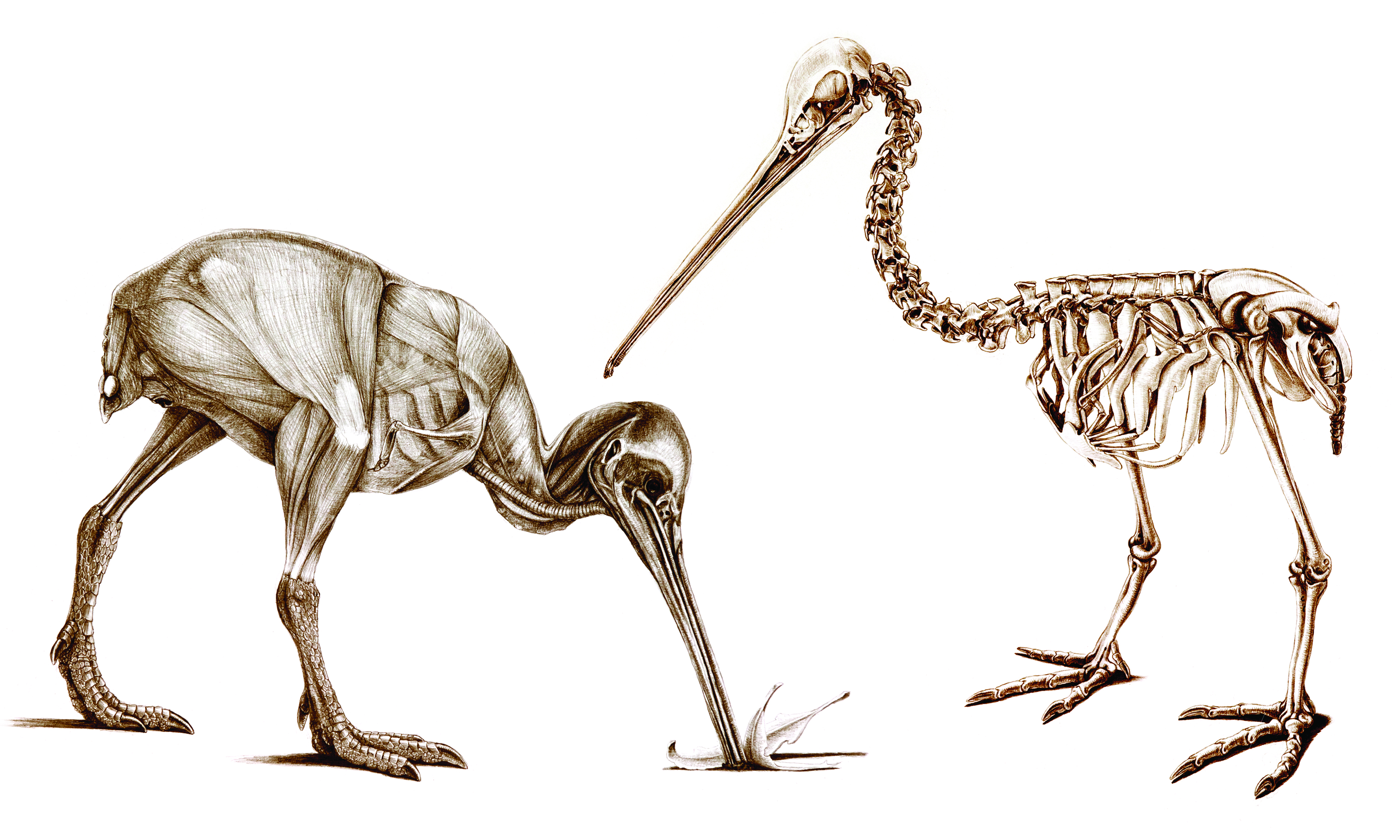
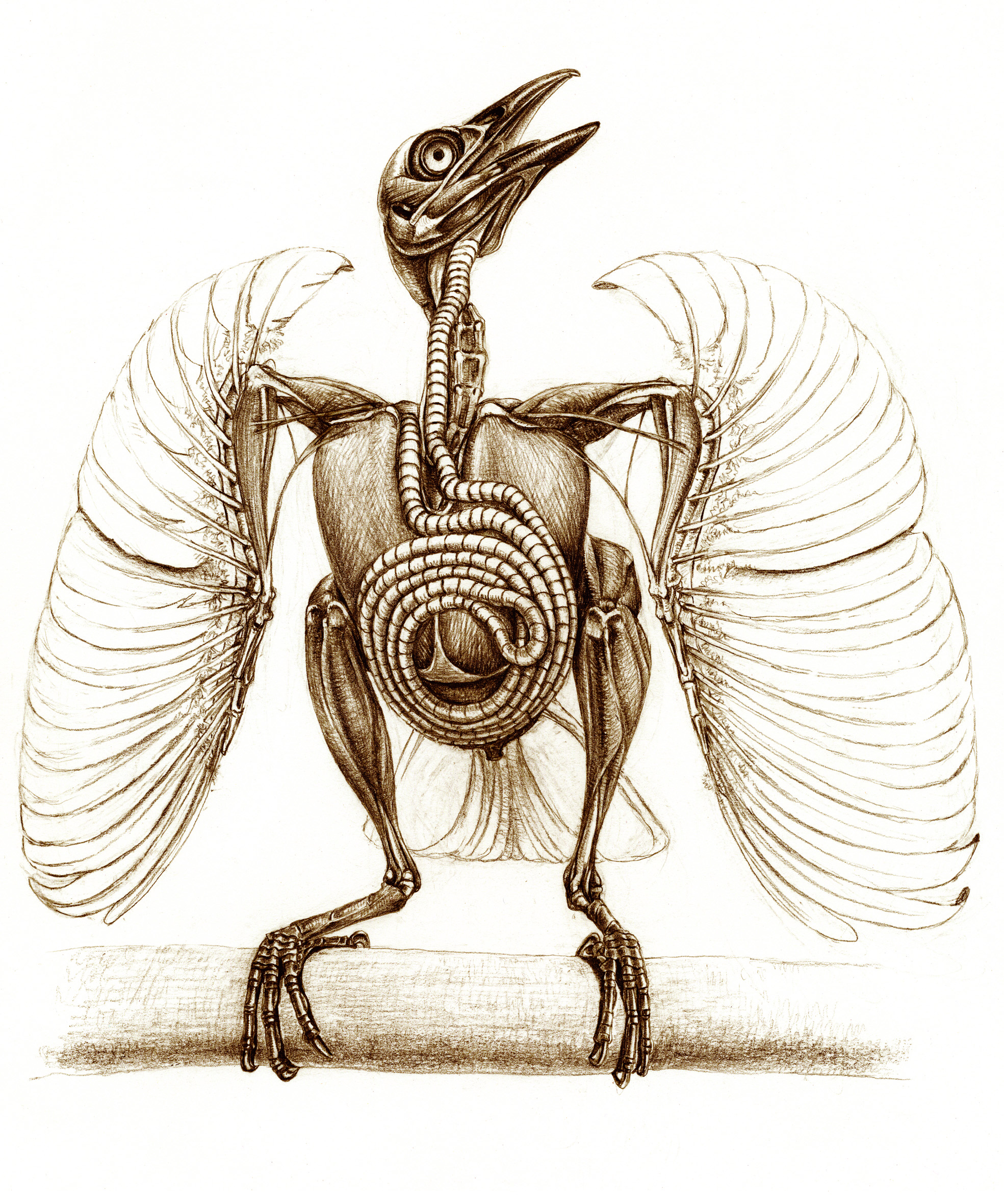
But for me, the book is most pleasurable for the visualizations and the passion for all things birdy that weaves through them and the accompanying text. The removal of feathers, or even all soft tissues, from bird bodies (posed in naturalistic behaviours) that van Grouw renders in her illustrations shows birds in a new light, emphasizing the strangeness and diversity that lie beneath. The author begins the book with a touching Acknowledgments section in which her husband Hein van Grouw, curator of birds at the Natural History Museum’s Tring collection, features very prominently, making it clear that the book was a team operation and comes from the heart after a 25-year journey. This gives the book a special warmth that is preserved throughout the remainder- although the illustrations are of flayed bodies or boiled / beetle-macerated skeletons, the tone is nothing less than an earnest love for birds of all kinds, and a zest for portraying those feelings to the reader in sketches and prose. It is a joyous celebration, not a somber litany, of the wonder of birds that can be gleaned from dead bodies. There is so much powerful, awesome imagery stuffed into those pages that it is hard to summarize. I’ll let five of my favourite images from the book (more are in her gallery and her book’s Facebook page; but even these are just the tip of the icebird) help get this across (used with permission of the author):
Naked kiwi in action.
The unscaled bird: guineafowl feet.
Deplumed sparrowhawk with dove trophy, exalting in its triumph.
Budgerigar has made a friend? Or came to grips with its own mortality?
Trumpet Manucode’s WTF anatomy! Spiraling tracheal coil made me gasp in awe when I saw this image in the book.
Now I’ll depart from this post just being a book review. I went to the Tring collection to do some research, and arranged my trip so I’d also get to see the debut of a Tring special exhibit featuring The Unfeathered Bird, and also to meet Katrina as well as Hein van Grouw. The placement of the exhibit at Tring is apropos, because Katrina was a curator at the museum until a few years ago and Hein still is. But the inspiration for the work and the specimens used (with a few exceptions, including from other museums) are Katrina’s. She (with Hein’s help) procured bodies of birds to dissect, macerate and sketch for the book over its 25 year fledging period, noting in the Acknowledgments that “no birds were harmed” to do this– do read those acknowledgments, as there are some amusing tales there of how she obtained some specimens.
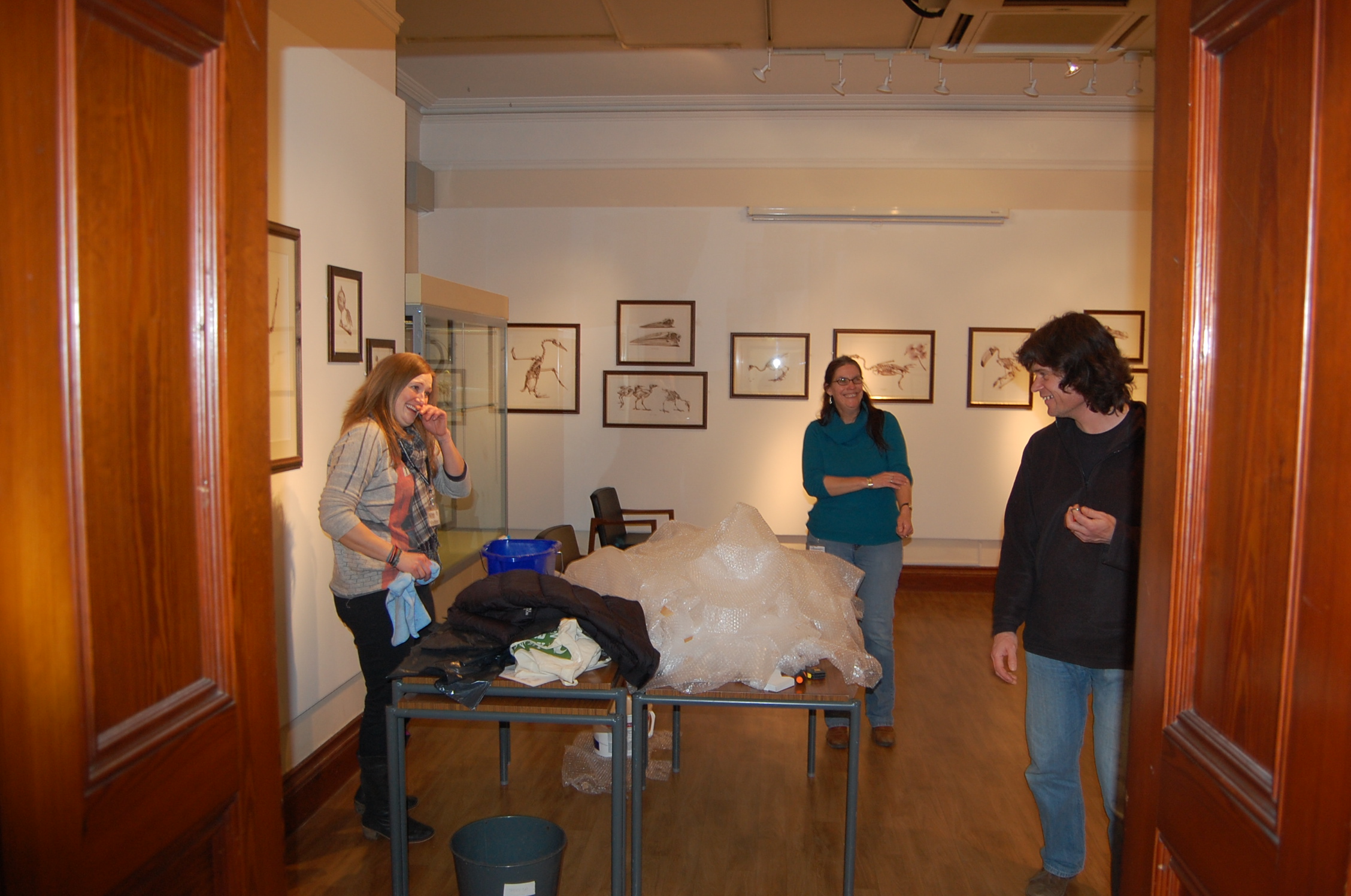
I was fortunate to be able to take some photos of the exhibit while they set it up, and grabbed some candid images of Katrina and colleagues during that process. The following images show off the exhibit, which is all in one clean, bright, simply adorned room in the Tring that lets Katrina’s framed sketches be the focus. Here are some examples:
Poster advert for the book in the Tring collections.
Tring exhibit setup, with Katrina, husband Hein, and helper finishing it up.
Tring exhibit now ready.
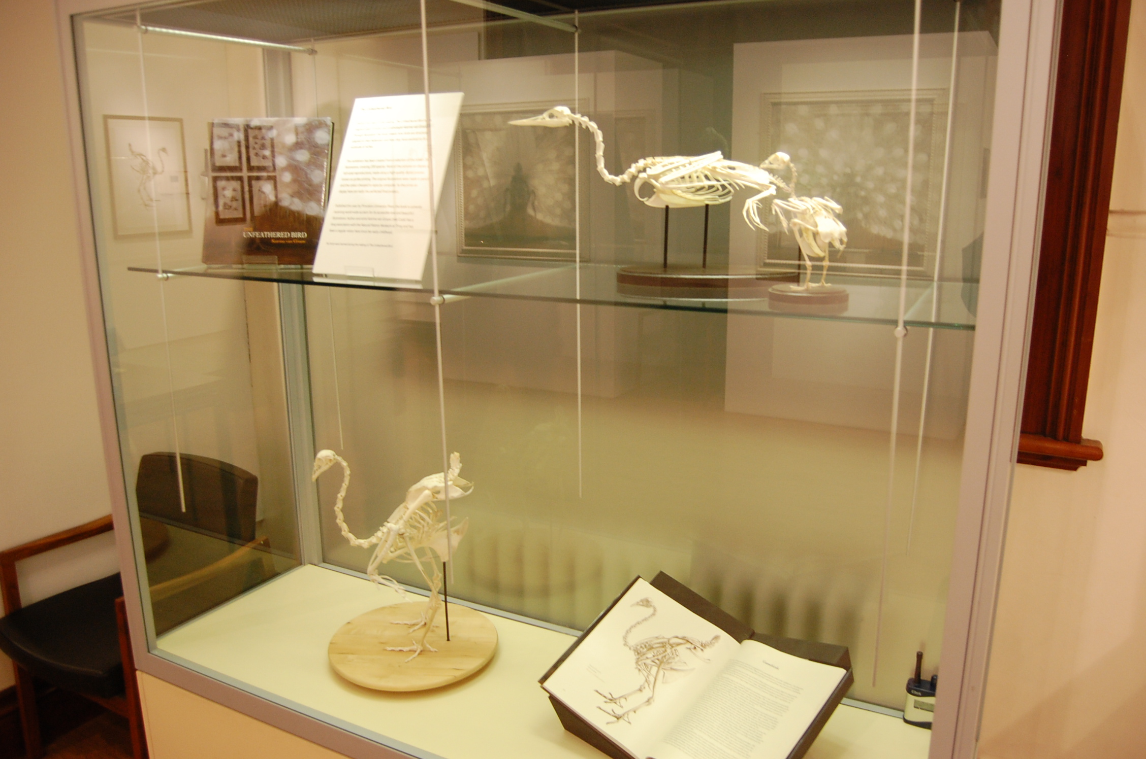
Tring exhibit case.
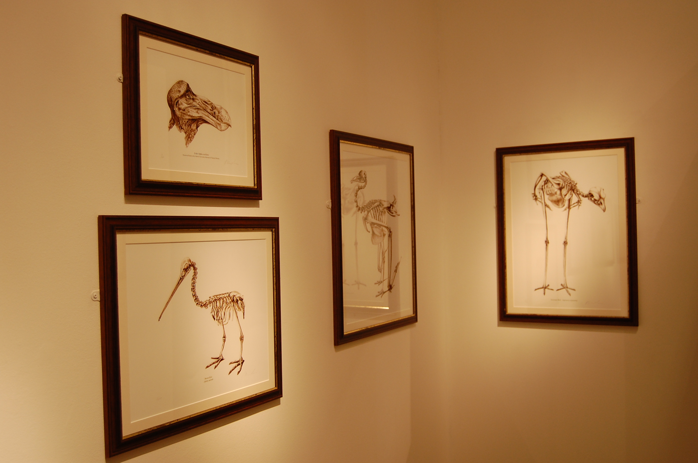
Framed sketches at Tring exhibit.
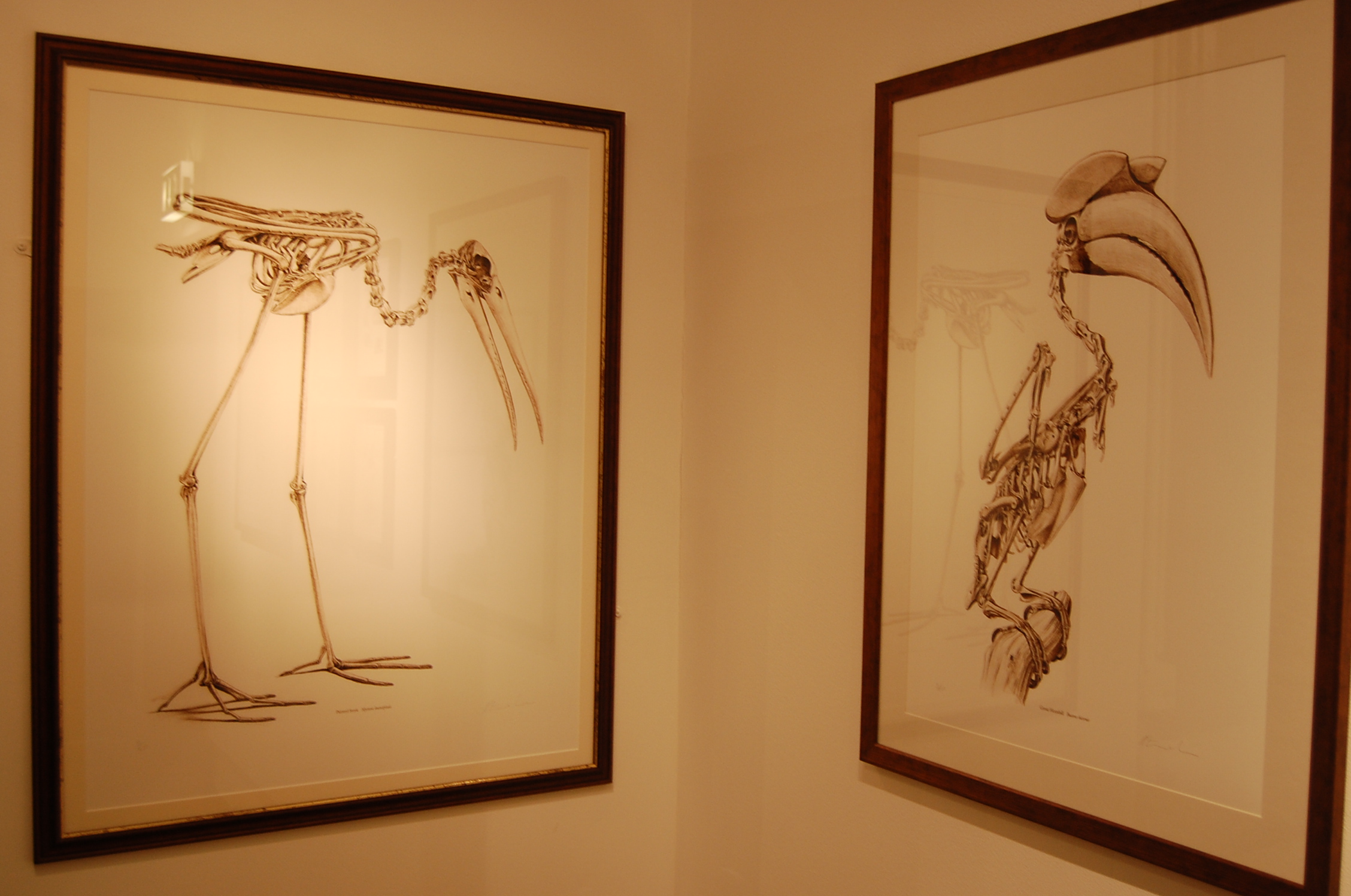
More framed sketches at Tring exhibit.
The exhibit is fun for people who are already Unfeathered Bird fans, and a good way of drawing in new ones. The book is a precious thing that any fan of birds, especially scientists, really needs to have a hard copy of. While it claims not to be an anatomy text, its illustrations provide ample opportunities to use it for that purpose. But really the point of owning all 287-plus pages is to bask in the warmth of true, pure appreciation for classic ornithology, which I found infectious. It is a book by and for bird lovers, but also for those that find the interface of art and science to be fascinating.
I confess I used to hate birds. I found them annoying and boring; all that flitting and twitting and pretentious feathers. “Get over yourselves, already, and calm down too!” was my reaction to them. When I started grad school, I had an open disdain for birds, even moreso than for mammals (OK, except cats). I was a “herp” fan through and through, for most of my life (childhood spent catching anoles in Florida, or stalking frogs in Ohio; during visits to my grandparents). What won me over was studying birds (and eventually mammals, too) as a young scientist, and learning how incredible they are– not just as endpoints in the story of theropod dinosaur evolution, as my thesis focused on, but as amazing animals with spectacular form-function relationships. The Unfeathered Bird is saturated with that amazement, so we’re birds of an unfeather.
Framed sketch of dodo head at Tring exhibit.
Entirely unfeathered Indian peafowl in matching views.
Painted Stork and Great Hornbill sketches.
Red junglefowl, wild ancestor of domestic chickens (and the book ends with several such breeds illustrated).
Katrina told me that she is already deep into writing the next book, whose subject I won’t spoil for you here but maybe we will be lucky enough to have her appear in the Comments and plug it? 🙂 (Her website does say “It was Hein’s stroke of genius to include domestic birds and they’ve provided the inspiration for my next project.” so the cat is out of the bag and amongst the pigeons!) It is great to hear that the book has done quite well sales-wise and critically, such as ~#67 on the Amazon sales list at one point– I hope this paves the way for more such books not only from Katrina, but from others engaged in lateral thinking (and still others) on the boundaries of science-art.
Today I’m shimmying down your interwebz with a late delivery. I’ve promised before to show how we clean up our nasty gooey skeletons to preserve them for future research to use. This is the intended final destination of all critters that are tenants of my freezers– the freezer is just a lovely holiday home, but bony heaven is the end result. I’ve accumulated a little museum of the bones of exotic animals I’ve studied, using these cleaned specimens. Here is how I do that preservation– there are four basic steps, and I’ll show them in four photos.
Stomach-Churning Rating: 8/10; first just dry bones, but then some gooey bones and by the end we ratchet it up to bloody organs.
Step 1) We get the deceased animal from various zoos and other EU sources, CT/MRI scan it, and dissect it. That’s what most of this blog focuses on, so I won’t show that. But I will show the end result, and then how I get to that:
Those are some elephant and rhino bones, some of which you saw on the 2nd day of Freezermas. Elephant bones are super greasy; it’s almost impossible to get rid of that brown grease visible in this photo (upper LH side) without making the bones brittle and over-bleached. The bones of the whiter white rhino on the right show what I’m usually aiming for. How do I get this done? Well, here’s an example for an elephant shank:
I take the elephant shank and make soup. (above) An Asian elephant’s patella, tibia and fibula were dissected, frozen for many years (queued up for cleaning; much freezer burn occurred on this specimen— it was jerky-fied), and then thawed. I put large specimens in this Rose cooker unit, which is a big ham cooker with a heater unit at the bottom. My baby, a Rapidaire MKV 5-ham unit is shown; oooh, ahhh!
The Rose cooker is filled up with tap water and been set it at around 60-90C. Then I let it cook away! A brothy soup develops, and sometimes it smells rather nice (my favourite aroma is giraffe leg). Sometimes… it’s not so nice. We check it every few hours to top up the water and remove stray tissue, and then change the water every day or so.
An elephant shank like this will take 2-3 days of cooking, longer if only switched on during work hours. The key thing is not to let it cook dry, which happened once with a faulty Rose cooker that did not do its normal auto-shutoff when the water ran low… showing up to work to encounter some fire trucks and unhappy college Health & Safety rep is not a good way to start your day, trust me!
This step is only slightly different for smaller (<30cm) specimens. Rather than the Rose cooker, we use the lovingly named “Croc Crock”, which isn’t visually impressive but you can see it here. As the name indicates, we’ve mainly used it for small crocodiles, and it is a crock pot. (a helpful thing is to add some detergent to the water for these small specimens; then bleaching isn’t so necessary)
Step 2) Then I empty out the water through the bottom spout, do the very nasty job of cleaning out the fat and other tissue that has accumulated (think 20 gallons of goo), hose off the bone, and set it in a ~10% bleach solution for at least a day, or up to a week or so for an elephant bone. Once it’s cleared up, I leave it out to dry (for big elephant bones, copious amounts of grease may be emerging for a few weeks). And then…
Step 3) I varnish the dry bones with a clear varnish, and let them dry. Here is how that elephant shank turned out. Pretty good! Finally, they get to join their friends:
Step 4) The prepared bones are labelled, given a number/name that I file in a world class comprehensive electronic database (cough, get on that John, cough!), and they become part of my humble mini-museum, shown above. Voila! The chef’s job is finished. Let science be served!
Happy Freezermas! Sing it: “On the fourth day of Freezermas, this blo-og gave to me: one tibiotarsus, two Darwin pictures, three muscle layers, a-a-and four steps of bone cookery!” ♪♫
Oh it’s Valentine’s day, so, err, have a heart today. Have four, actually!
…a daily picture of anatomy! And today it is two pictures, tra-la-lee!
Welcome back to Freezermas! And HAPPY DARWIN DAY! Last year our whole lab got involved in DD2012, but this blog was just a twinkling in my keyboard back then. This year it was a more mellow, somber occasion for DD2013. But Heinrich Mallison of the dinosaurpalaeo blog took part, and took photos (all credits go to him), and the result kicked ass and took names. Bring it on!
Here is Darwin amidst a selection of greatest hits from my bone collection; post-freezer denizens. How many can you identify? Have a go in the comments below. A few should be quite familiar to blog followers… More about these bones later this week. Incidentally, Darwin is standing on a Kistler forceplate. So biomechanics afficionados can geek out about this, too.
And here I am hamming it up again. Give it a rest, John! But ’tis merely a humble offering to The Master. I’m sure he’d appreciate it. Any guesses what it is?
Happy Freezermas! Sing it: “On the second day of Freezermas, this blo-og gave to me: one tibiotarsus, a-and two silly pictures with Chucky D!”