Maybe it’s uncool to talk about heroes in science these days, because everyone is poised on others’ shoulders, but “Neill” (Robert McNeill) Alexander is undeniably a hero to many researchers in biomechanics and other strands of biology. Our lab probably wouldn’t exist without his pervasive influence- he has personally inspired many researchers to dive into biomechanics, and he has raised the profile of this field and championed its importance and principles like no other one individual. Often it feels like we’re just refining answers to questions he already answered. His influence extends not only to comparative biomechanics and not only around his UK home, but also –via his many, many books on biology, anatomy and related areas, in addition to his research, editorial work and public engagement with science– to much of the life sciences worldwide.
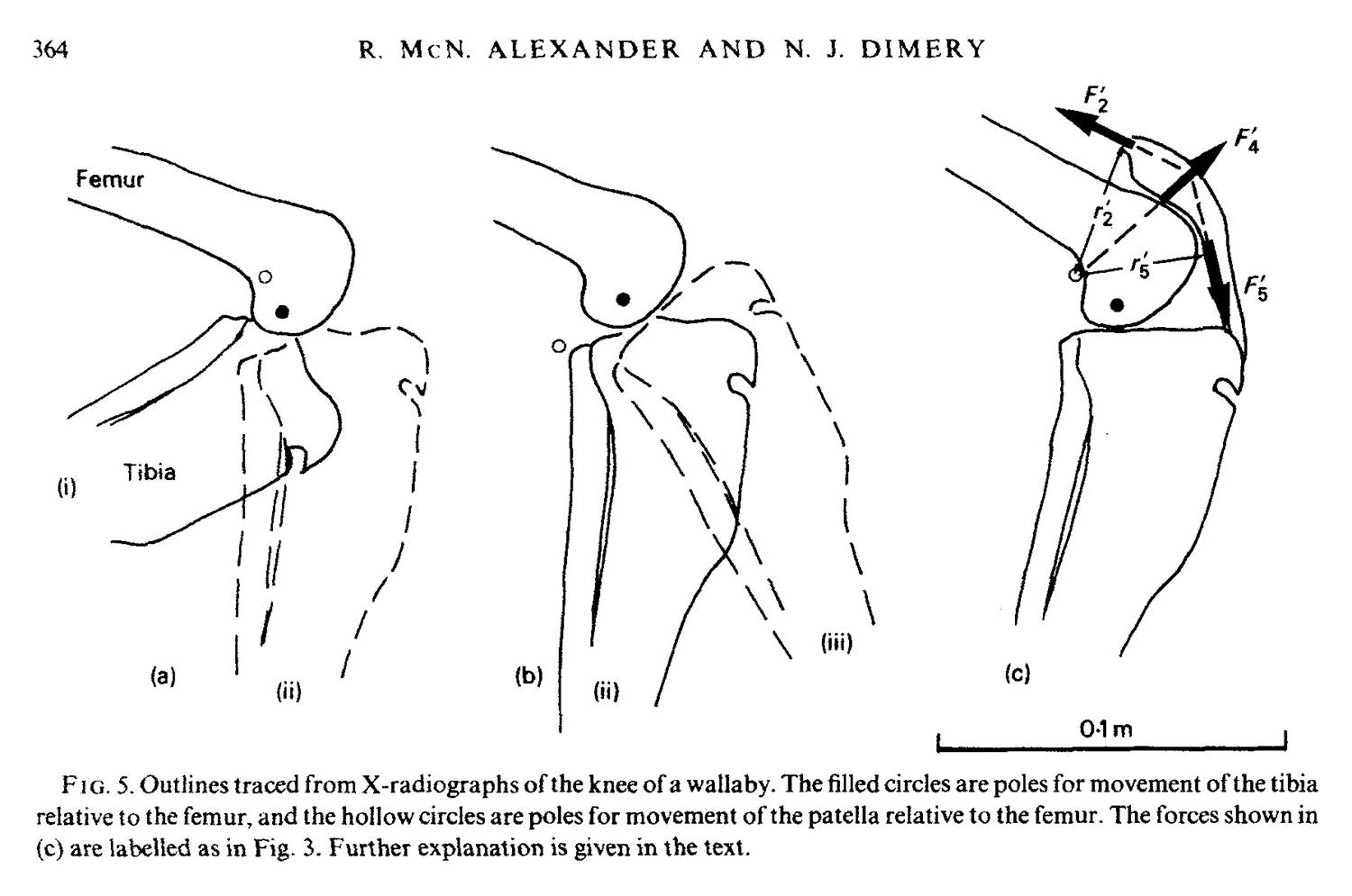
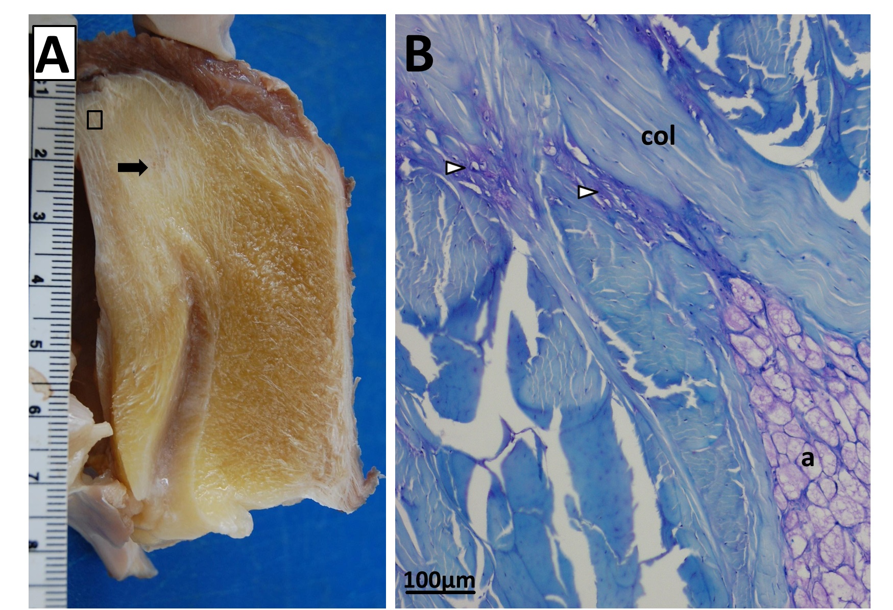
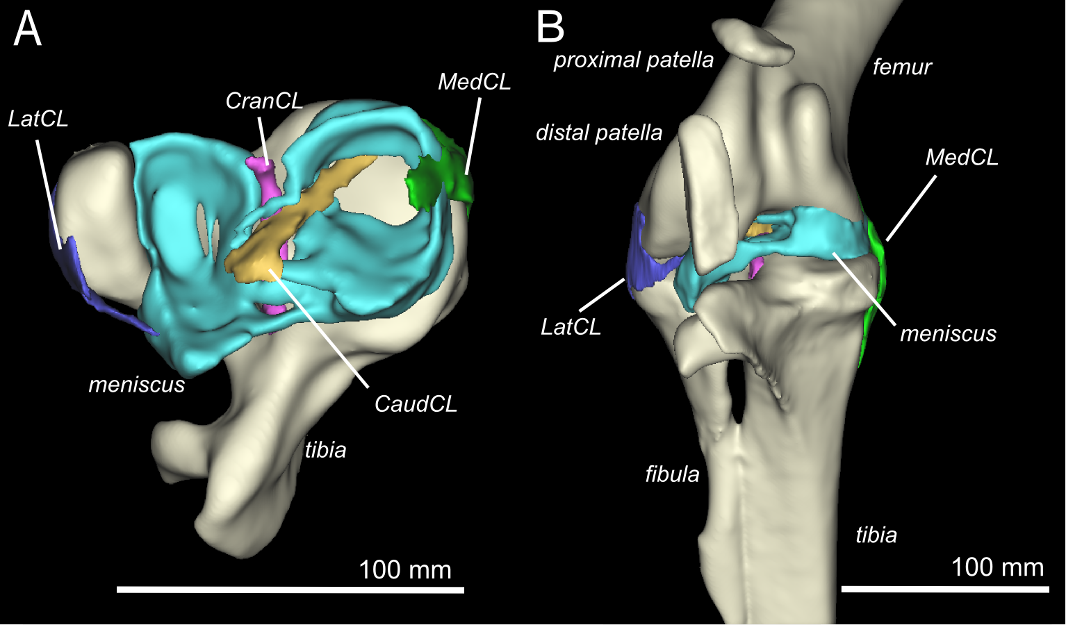
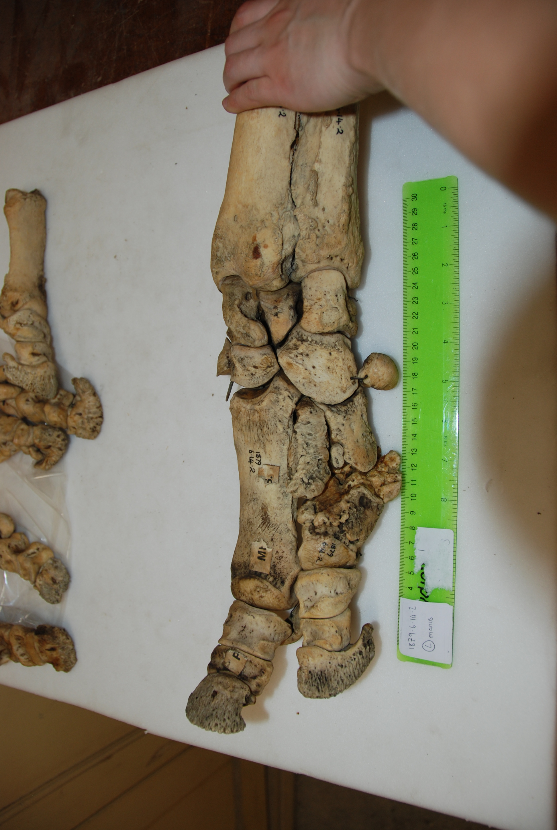
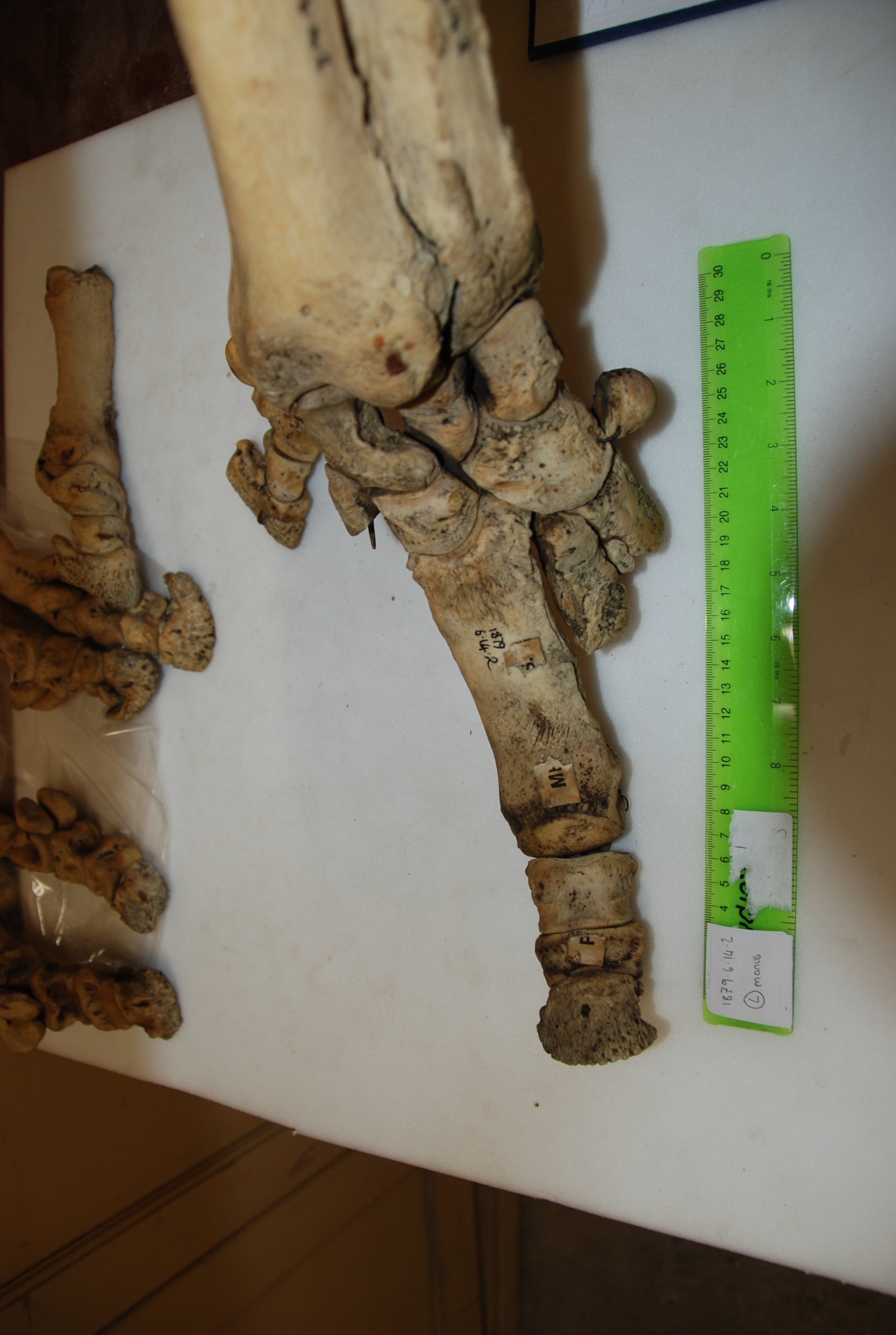
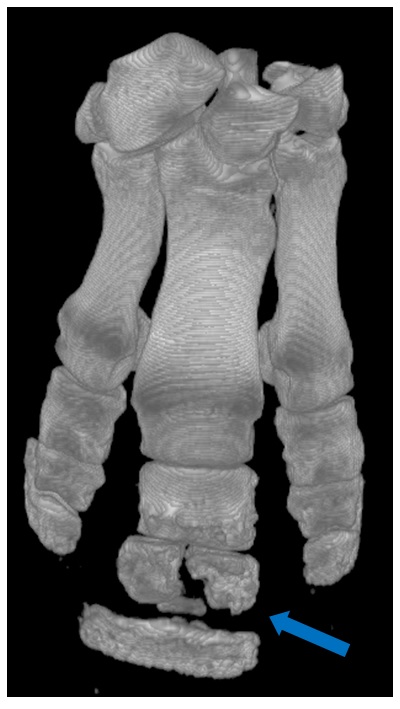
What does a kneecap (patella) do? Alexander and Dimery 1985, they knew. 30 years later, my team is still trying to figure that out!
Sure, one could (and with great humility I’m sure Alexander would) mention others like Galileo and Marey and Muybridge and Fenn and Gray and Manter who came before him and did have a profound impact on the field. Alexander can, regardless, easily be mentioned in the same breath as luminaries of muscle physiology such as AV Hill and even Andrew + Julian Huxley. But I think many would agree that Alexander, despite coming later to the field, had a singular impact on this young field of comparative biomechanics. That impact began in the 1970s, when Dick Taylor and colleagues in comparative physiology were also exploding onto the scene with work at the Concord Field Station at Harvard University, and together biomechanics research there, in the UK, elsewhere in Europe and the world truly hit its stride, with momentum continuing today. I’m trying to think of some women who played a major role in the early history of biomechanics but it was characteristically a woefully male-dominated field. That balance has shifted from the 1970s to today, and my generation would cite luminaries such as Mimi Koehl as key influences. There are many female or non-white-male biomechanics researchers today that are stars in the field, so there seems to have been progress in diversifying this discipline’s population.
Hence, honouring Alexander’s impact on science, today our college gave Neill an honorary doctorate of science (DSc). Last year, I also helped organize a symposium at the Society for Vertebrate Paleontology’s conference in Berlin that honoured his impact specifically on palaeontology, too- compare his book “The Dynamics of Dinosaurs and Other Extinct Giants” to current work and you’ll see what fuelled much of that ongoing work, and how far/not far we’ve come since ~1989. Even 10 years later, his “Principles of Animal Locomotion“, with Biewener’s “Animal Locomotion“, remains one of the best books about our field (locomotion-wise; Vogel’s Comparative Biomechanics more broadly) , and his educational CD “How Animals Move“, if you can get it and make it work on your computer, is uniquely wonderful, with games and videos and tutorials that still would hold up well as compelling introductions to animal biomechanics. Indeed, I’ve counted at least 20 books penned by Alexander, including “Bones: The Unity of Form and Function” (under-appreciated, with gorgeous photos of skeletal morphology!).

1970s Alexander, with a sauropod leg.
And then there are the papers. I have no idea how many papers Neill has written –again and again I come across papers of his that I’ve never seen before. I tried to find out from the Leeds website how many papers he has, but they’re equally dumbfounded. I did manage to count 38 publications in Nature, starting in 1963 with “Frontal Foramina and Tripodes of the Characin Crenuchus,” and 6 in Science. So I think we can be safe in assuming that he has written everything that could be written in biomechanics, and we’re just playing catchup to his unique genius.
Seriously though, Alexander has some awesome publications stemming back over 50 years. I’m a big fan of his early work on land animals, such as with Calow in 1973 on “A mechanical analysis of a hind leg of a frog” and his paper “The mechanics of jumping by a dog” in 1974, which did groundbreaking integrations of quantitative anatomy and biomechanics. These papers kickstarted what today is the study of muscle architecture, which our lab (including my team) has published extensively on, for example. They also pioneered the integration of these anatomical data with simple theoretical models of locomotor mechanics, likewise enabling many researchers like me to ride on Alexander’s coattails. Indeed, while biomechanics often tends to veer into the abstract “assume a spherical horse”, away from anatomy and real organisms, Alexander managed to keep a focus on how anatomy and behaviour are related in whole animals, via biomechanics. As an anatomist as well as a biomechanist, I applaud that.
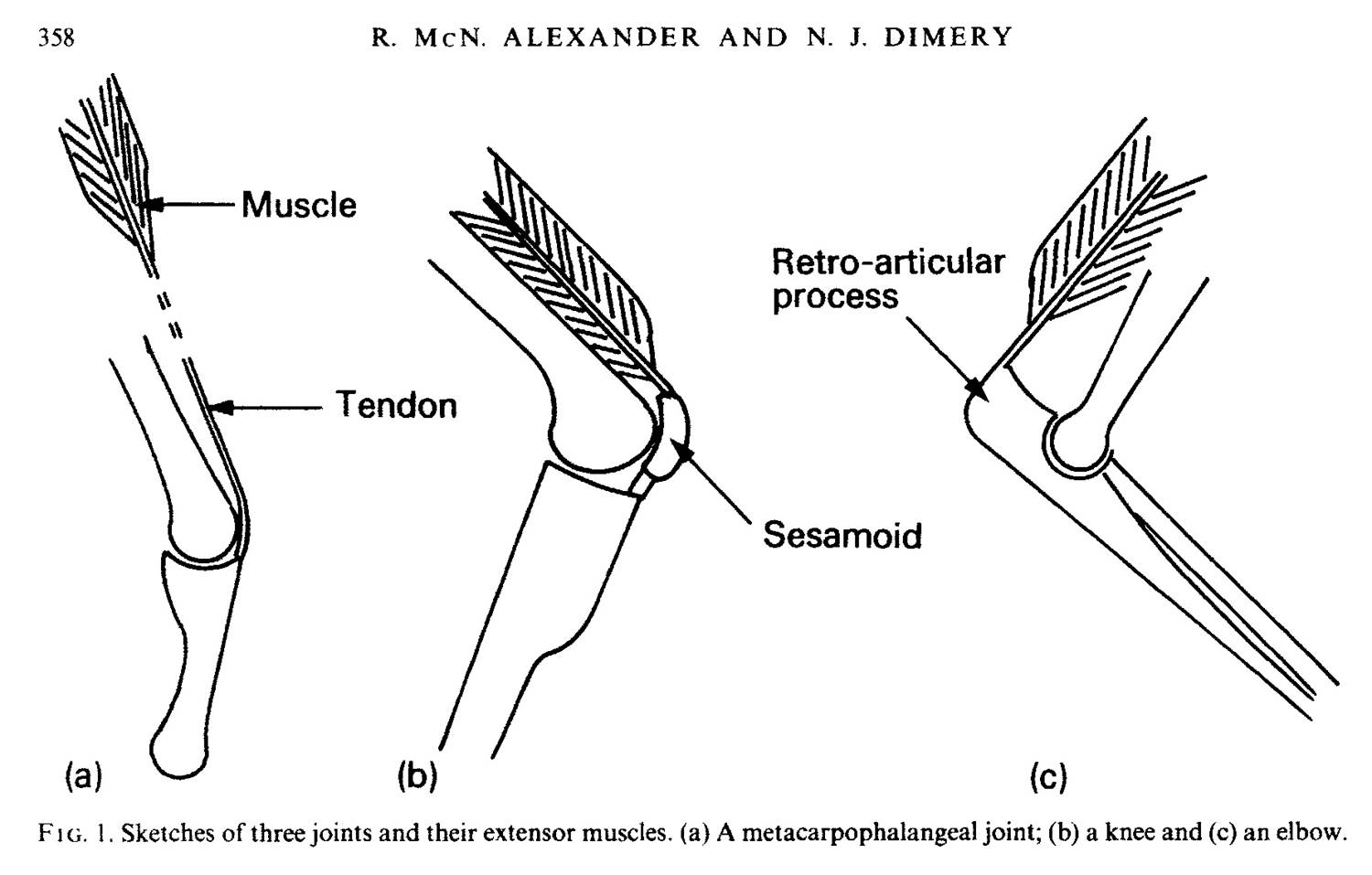
How do muscles work around joints? Alexander and Dimery 1985 figured out some of the key principles.
Alexander has researched areas as diverse as how fish swim, how dinosaurs ran, how elastic mechanisms make animal movement more efficient, how to model the form and function of animals (see his book “Optima for Animals” for optimization approaches he disseminated, typifying his elegant style of making complex maths seem simple and simple maths impressively powerful) and how animals walk and run, often as sole author. In these and other areas he has codified fundamental principles that help us understand how much in common many species have due to inescapable biomechanical constraints such as gravity, and how these principles can inspire robotic design or improvements in human/animal care such as prosthetics. Neill has also been a passionate science communicator, advising numerous documentaries on television.
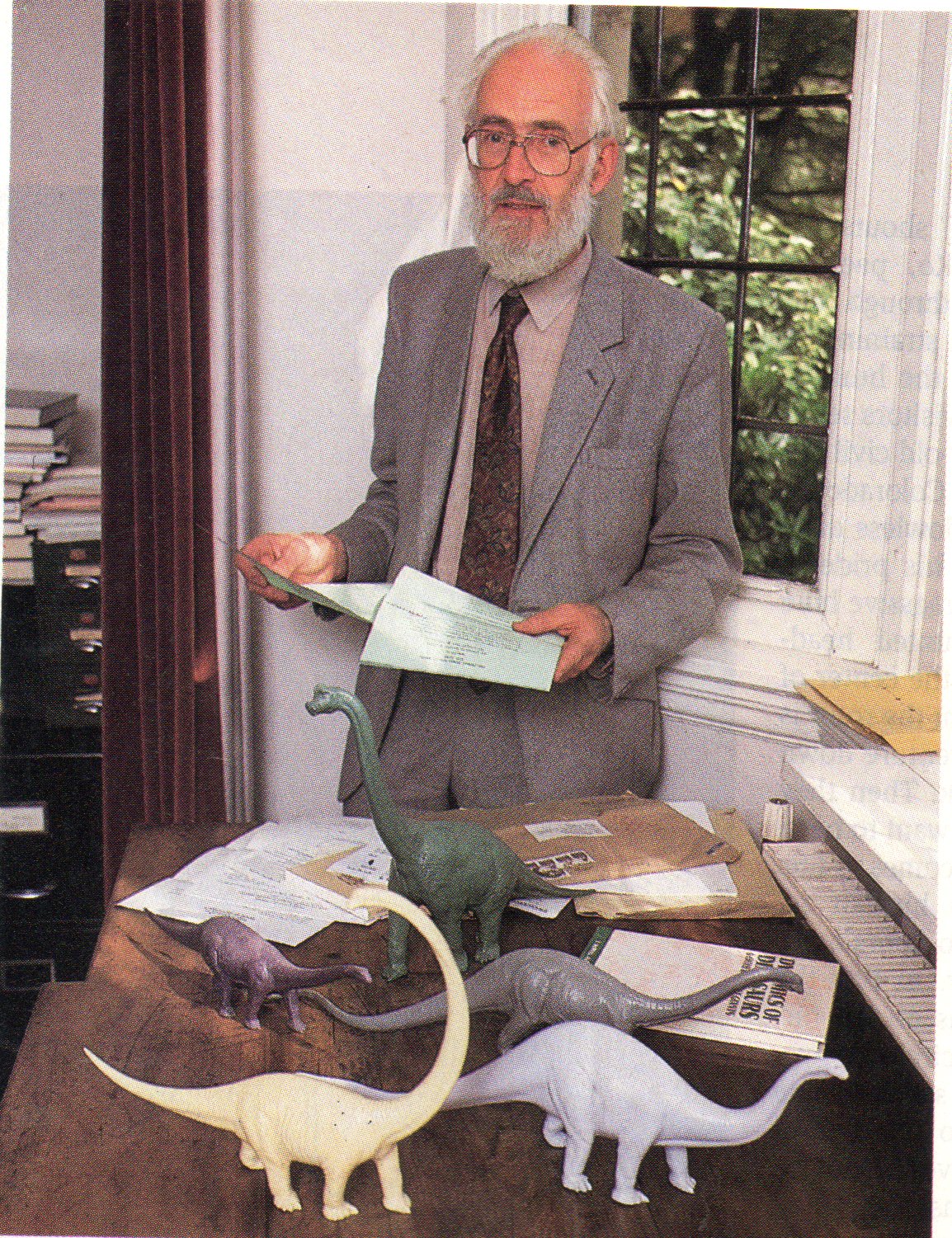
~1990s Alexander, with model dinosaurs used to estimate mass and centre of mass.
Alexander’s “Dynamics of Dinosaurs” book, one of my favourites in my whole collection, is remarkably accessible in its communication of complex quantitative methods and data, which arguably has enhanced its impact on palaeontologists. Alexander’s other influences on palaeobiology include highly regarded reviews of jaw/feeding mechanics in fossil vertebrates (influencing the future application of finite element analysis to palaeontology), considerations of digestion and other aspects of metabolism, analysis of vertebral joint mechanics, and much more. Additionally, he conducted pioneering analyses of allometric (size-related) scaling patterns in extant (and extinct; e.g. the moa) animals that continue to be cited today as valuable datasets with influential conclusions, by a wide array of studies including palaeontology—arguably, he helped compel palaeontologists to contribute more new data on extant animals via studies like these.
Neill Alexander did his MSc and PhD at Cambridge, followed by a DSc at the University of Wales, a Lecturer post at Bangor University and finally settling at the University of Leeds in 1969, where he remained until his retirement in 1999, although he maintains a Visiting Professorship there. I had the great pleasure of visiting him at his home in Leeds in 2014; a memory I will treasure forever, as I had the chance to chat 1-on-1 with him for some hours. He has been Secretary of the Zoological Society of London throughout most of the 1990s, President of the Society for Experimental Biology and International Society of Vertebrate Morphologists, long championing the fertile association of biomechanics with zoology, evolutionary biology and anatomy. More recently, he was a main editor of Proceedings of the Royal Society B for six years.
Many people I’ve spoken to about Neill before have stories of how he asked a single simple question at their talk, poster or peer review stage of publication, and how much that excited them to have attracted his sincere interest in their research. They tend to also speak of how that question cut to the core of their research and gave them a facepalm moment where they thought “why didn’t I think of that?”, but how he also asked that question in a nice way that didn’t disembowel them. I think that those recalling such experiences with Neill would agree that he is a professorial Professor: a model of senior mentorship in terms of how he can advise colleagues in a supportive, constructive and warmly authoritative, scholarly way. For a fairly recent example of his uniquely introspective and concise, see the little treasure “Hopes and Fears for Biomechanics”, a ~2005 lecture you can find here. I really like the “Fears” part. I share those fears- and maybe embody them at times…
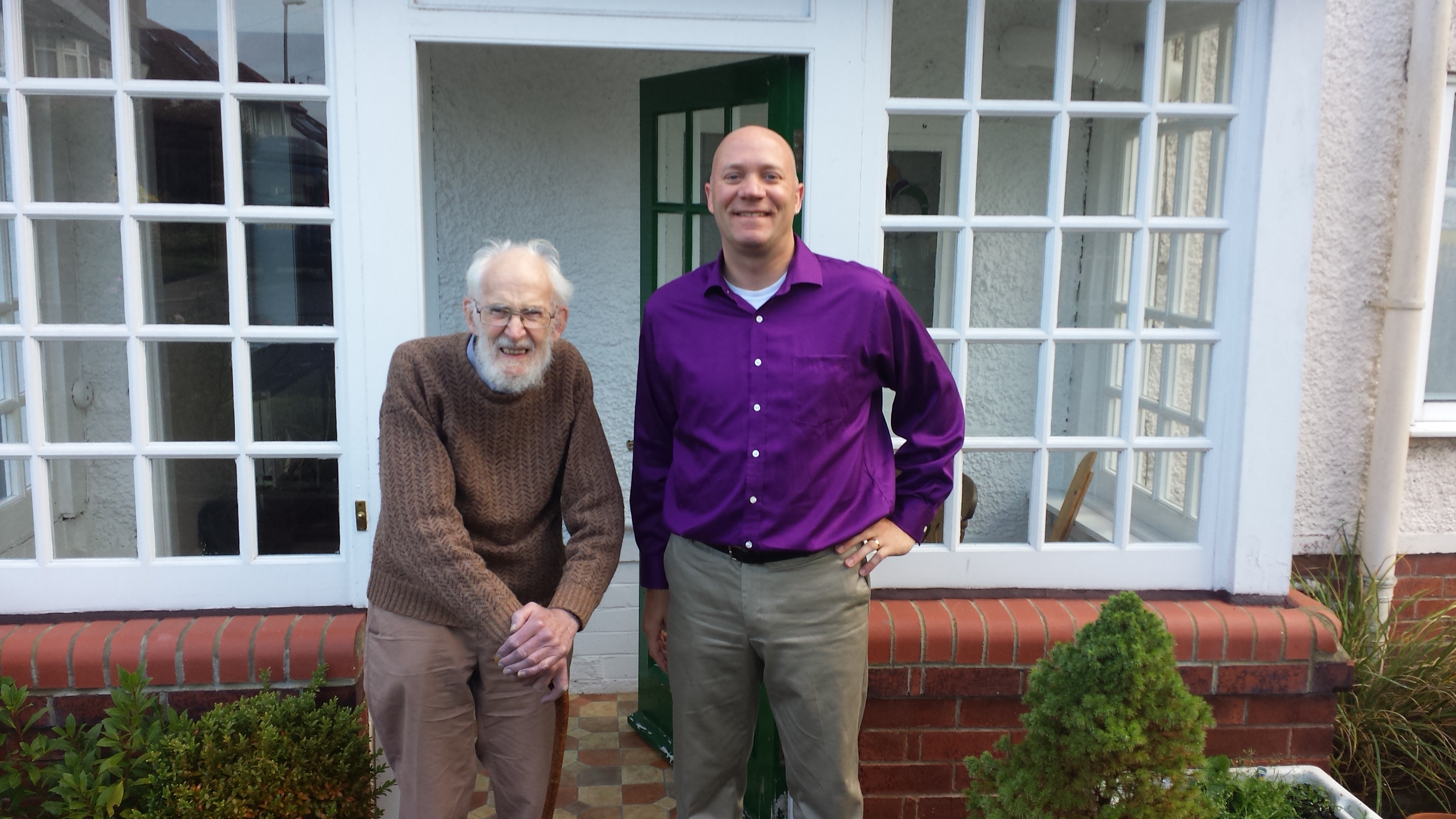
My visit with RMcNeill Alexander in 2014.
Perhaps I have gushed enough, but I could go on! Professor RMcNeill Alexander, to summarise the prodigious extent of his research, is to biomechanics as Darwin is to biology as a whole. One could make a strong case for him being one of the most influential modern biologists. He is recognised for this by his status as a Fellow of the Royal Society (since 1987), and a CBE award, among many other accolades, accreditations and awards. And, if you’ve met him, you know that he is a gentle, humble, naturally curious and enthusiastic chap who instils a feeling of awe nonetheless, and still loves to talk about science and keeps abreast of developments in the field. And as the RVC is honouring Neill today, it is timely for me to honour him in this blog post. There can never be another giant in biomechanics like Alexander, and we should be thankful for the broad scientific shoulders upon which we are now, as a field, poised.
I hope others will chime in with comments below to share their own stories.