I love doing sciencey road trips with my team when I can. Last week, we got a treat: four of us got a behind-the-scenes tour of the fairly new Crocodiles of the World facility near Oxford; just over 90 minutes west of our lab, nestled in the pictureseque Cotswolds region. We were not disappointed, so you get to share in the joy! In photo-blog format. Pics can be clicked to emcrocken.
In the midst of an unpreposessing industrial estate lies: AWESOME!
If you want to bone up on your croc species, go here and here. I won’t go into details. This is an eye candy post!
Reasonably accurate description that caught my eye. My scientific interest in crocodiles starts here, and with their anatomy/relationship with dinosaurs, but I’ve loved crocs since I was an infant (one of my first words, as I may have written here before, was “dock-a-dile”, for my favourite stuffed animal at the time [R.I.P.]).
Siamese crocodiles. The large male is “Hugo.” They were apart when we entered, then got snuggly later, as I’ve often seen this species do. Heavily endangered (<300 in the wild?), so any breeding is a good thing!
The above photo brings me to one of my general points. Crocodiles of the World seems genuinely to be a centre that is breeding crocodiles for conservation purposes (and for education, entertainment and other zoo-like stuff). Essentially every crocodile enclosure had a mated pair, and several were breeding. Such as…
Yes, that is a Dwarf African crocodile, Osteolaemus, and indeed it is a female on her nest-mound. Which means…
Eggs of said Osteolaemus.
And babies of said Osteolaemus! As if the adults aren’t cute enough with their short snouts and doglike size/appearance! These guys have striking yellow colouration, too. I’d never seen it in person before.
That’s not all!
Male American Alligator “Albert” warming up. Smaller female partner “Daisy” lives in same enclosure. Plenty of babies from these guys, too! Daisy comes when called by name, and Albert is learning to do so.
~1 meter long juvenile Nile crocodiles, bred at the facility.
But then crocodile morphological diversity (colours, textures) and behaviour is just too cool not to focus on a bit, so here are some highlights from our visit!
Endearing shot of a crocodylian I seldom get to see anywhere: Paleosuchus trigonatus, the Schneider’s Dwarf Caiman. Spiny armoured hide and quite terrestrial; poorly known in many ways. Some more info is here (note its tortured taxonomy)
Black caiman, Melanosuchusniger, showing some interest in us.
Cuban crocodiles (Crocodylus rhombifer; pound for pound the most badass croc in my experience; badassitude that this photo captures nicely) cooling off by exposing the well-vascularized soft tissues of the mouth region.
But it’s not just crocs there, either, and some of the highlights were non-croc surprises and memorable encounters:
A surprisingly friendly and tame Water monitor (14 yrs old; does kids parties). Note person for scale. Was about 2 meters long, 20 kg or so.
Business end of nice Water monitor, with tongue engaged.
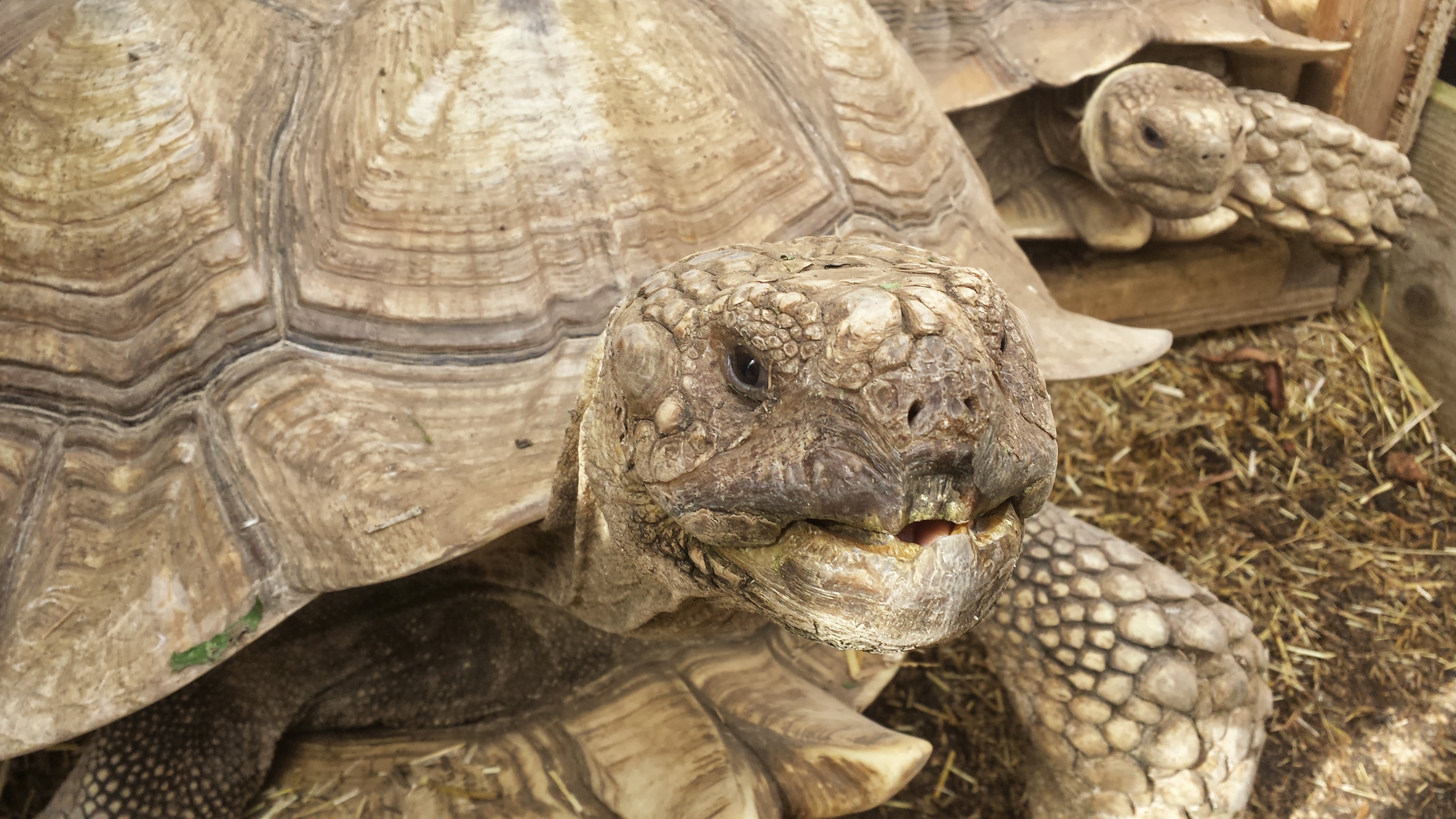
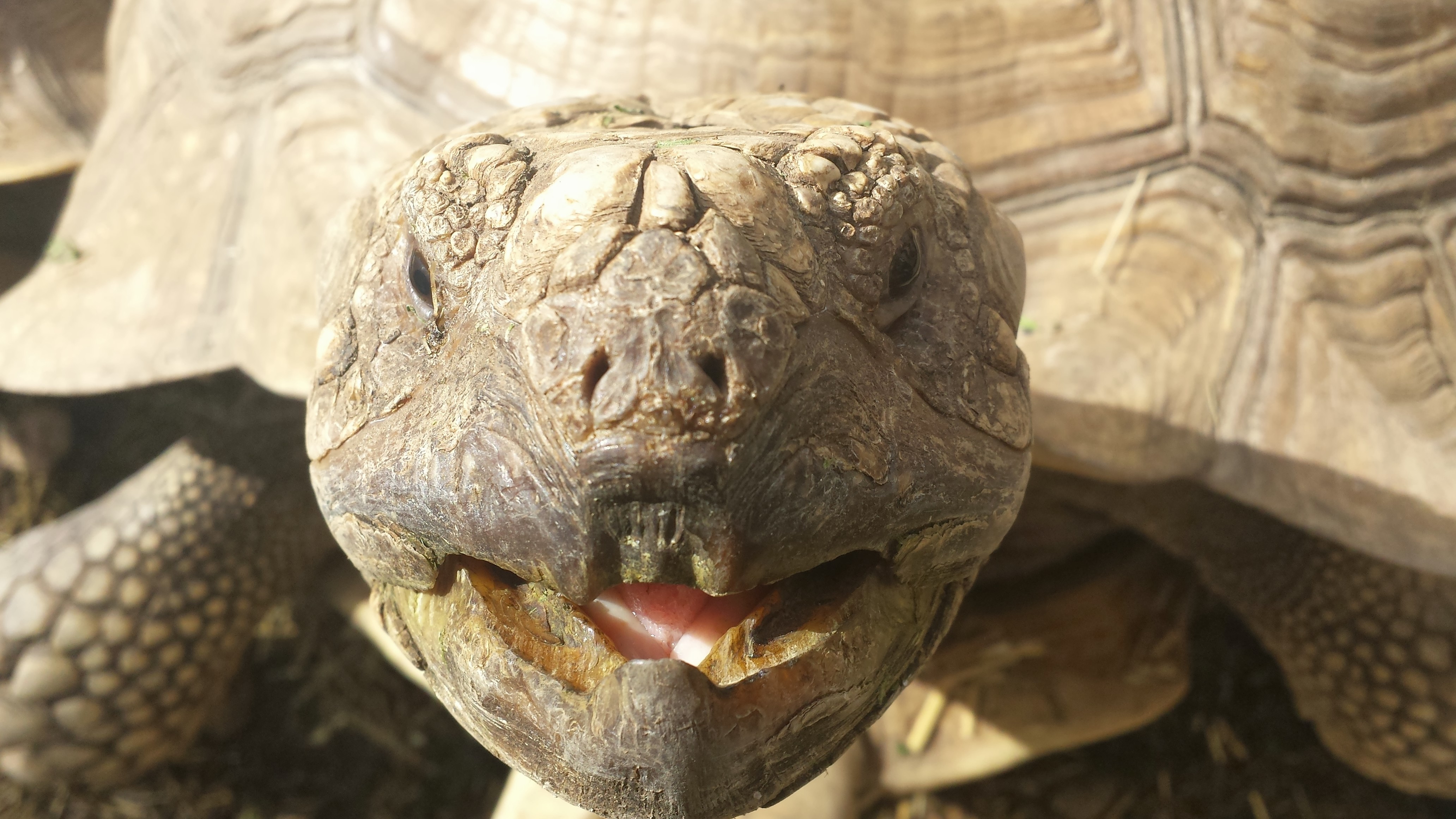
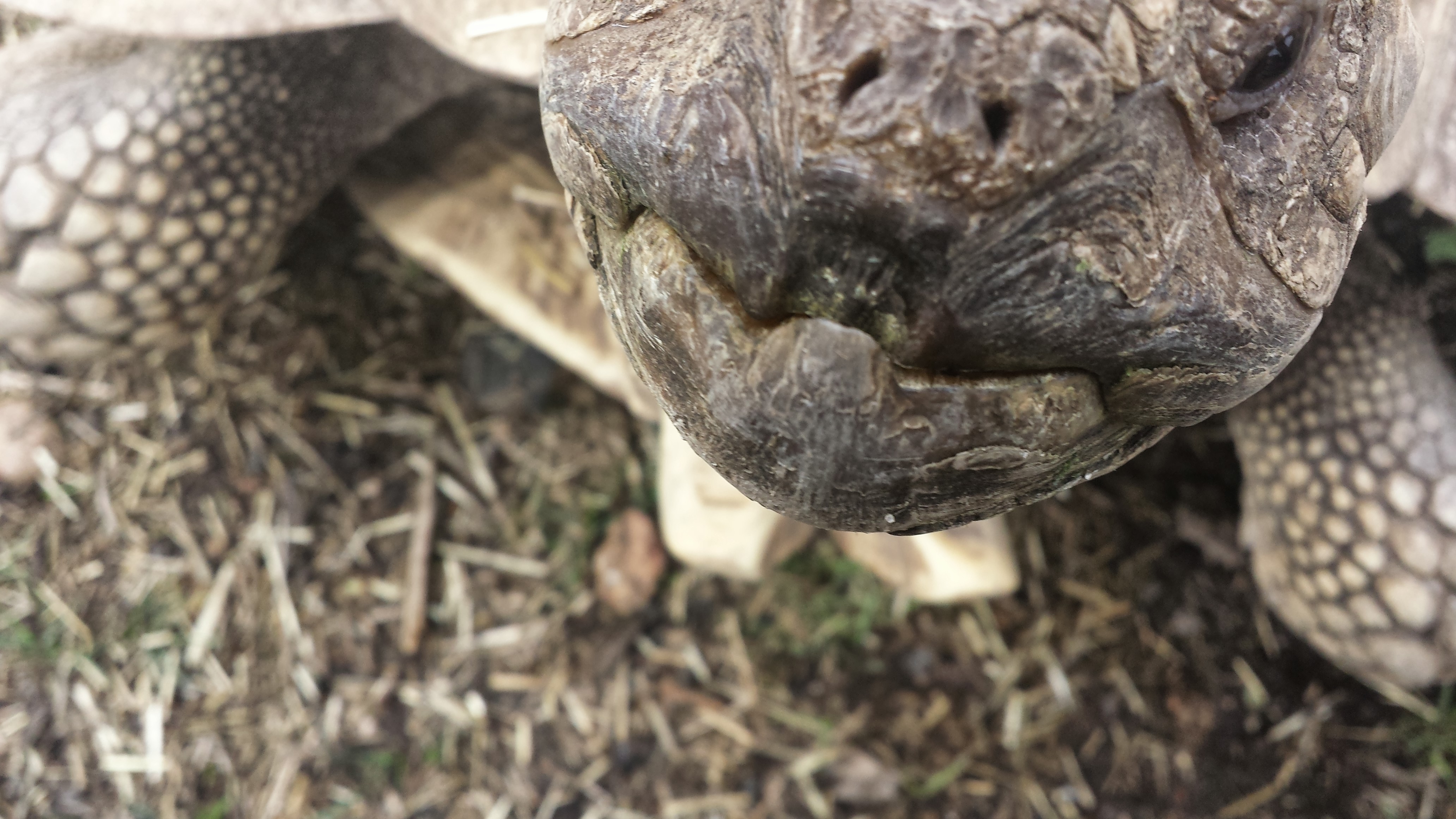
And we got a nice farewell from an African spur-thigh tortoise (Geochelone sulcata) with an oral fixation (action sequence thereof):
Chowmp!
If someone visits this facility and leaves without being converted to a croc-lover, they must be from a different planet than me. It is a celebration of crocodiles; the owner, Shaun Foggett, is the real deal. He sold his home and quit his job as a carpenter to care for crocodiles, and it seems to be a great success– about to get greater, as they have plans to move to a new, bigger, proper site! They are seeking funding, so if you can contribute go here.
Right then… UK residents and visitors: you need to go here! Badly! Get off the blog and go now. If it is a Saturday/Sunday (the cramped industrial estate location only allows the public then).
Otherwise just stew and imagine how much fun you could be having checking out crocodiles. I cruelly posted this on a Tuesday to ensure thorough marination of any croc-geeks.
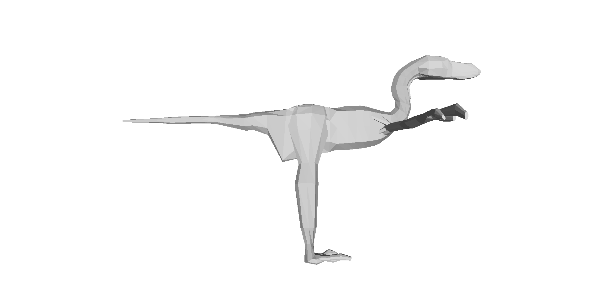
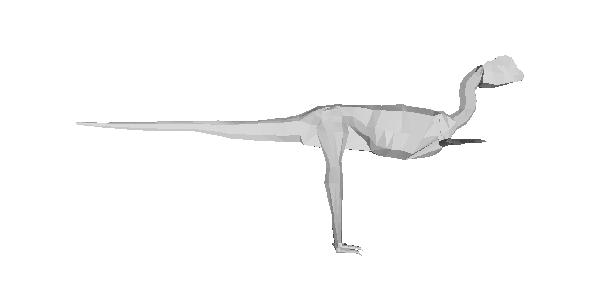
Our 3D computer models of a basal dinosaur and bird, showing methods and key differences in body shape. The numbers at the bottom are museum specimen numbers.
At about the moment I’m posting this, our Nature paper (our more formal page here, and the actual article here) embargo is ending, drawing a 14+ year gestation to a close. The paper is about how dinosaur 3D body shape changed during their evolution, and how that relates to changes in hindlimb posture from early dinosaurs/archosaurs to birds; “morpho-functional evolution” sums up the topic. We used the 3D whole-body computational modelling that I, Allen and Bates (among others) have developed to estimate evolutionary changes in body dimensions, rather than focusing on single specimens or (as in our last study) tyrannosaur ontogeny. We’ve strongly supported the notion (dating back to Gatesy’s seminal 1990 Paleobiology paper) that the centre of mass of dinosaurs shifted forwards during their evolution, and that this shift gradually led to the more crouched (flexed) hind leg posture that characterizes living birds. Here is a movie from our paper showing how we did the modelling:
And here is a summary of our 17 computer models of archosaur bodies, shown as one walks along the tips of the phylogeny shown in the video (the models are not considered to be ancestral to one another; we used a common computer algorithm called squared-change parsimony to estimate ancestral state changes of body dimensions between the 16 numbered nodes of the tree):
But we’ve done much more than just put numbers on conventional wisdom.
We’ve shown, to our own surprise, that the shift of the centre of mass was largely driven by evolutionary enlargements of the forelimbs (and the head and neck, and hindlimbs, to a less strong degree), not the tail as everyone including ourselves has assumed for almost 25 years. And the timing of this shift occurred inside the theropod dinosaur group that is called Maniraptora (or Maniraptoriformes, a slightly larger group), so the change began in animals that were not flying, but not long before flight evolved (depending on whom you ask, what taxonomy they favour and what evidence one accepts, either the smaller clade Eumaniraptora/Paraves or the bird clade Aves/Avialae).
Now, if you don’t like the cliche “rewriting the textbooks”, do have a look through texts on dinosaur/early avian palaeobiology and you probably will find a discussion of how the tail shortened, the centre of mass moved forwards as a consequence, the caudofemoral musculature diminished, and theropod dinosaurs (including birds) became more crouched as a result. We did that to confirm for ourselves that it’s a pretty well-accepted idea. Our study supports a large proportion of that idea’s reasoning, but modifies the emphasis to be on the forelimbs more than the tail for centre of mass effects, so the story gets more complex. The inference about caudofemoral muscles still seems quite sound, however, as is the general trend of increased limb crouching, but our paper approximates the timing of those changes.
Figure 3 from our paper, showing how the centre of mass moved forwards (up the y-axis) as one moves toward living birds (node 16); the funny dip at the end is an anomaly we discuss in the paper.
A final implication of our study is that, because the forelimbs’ size influenced the centre of mass position, and thus influenced the ways the hindlimbs functioned, the forelimbs and hindlimbs are more coupled (via their effects on the centre of mass) than anyone has typically considered. Thus bipedalism and flight in theropods still have some functional coupling– although this is a matter of degree and not black/white, so by no means should we do away with helpful concepts like locomotor modules.
And in addition to doing science that we feel is good, we’ve gone the extra mile and presented all our data (yes, 17 dinosaurs’ worth of 3D whole body graphics!) and the critical software tools needed to replicate our analysis, in the Dryad database (link now working!), which should have now gone live with the paper! It was my first time using that database and it was incredibly easy (about 1 hour of work once we had all the final analysis’s files properly organized)– I strongly recommend others to try it out.
That’s my usual general summary of the paper, but that’s not what this blog article is about. I’ll provide my usual set of links to media coverage of the paper below, too. But the focus here is on the story behind the paper, to put a more personal spin on what it means to me (and my coauthors too). I’ll take a historical approach to explain how the paper evolved.
Embarassing picture of me before I became a scientist. Hardee’s fast food restaurant cashier, my first “real job”, from ~1999- no, wait, more like 1986. The 1980s-style feathered (and non-receding) hair gives it away!
Rewind to 1995. I started my PhD at Berkeley. I planned to use biomechanical methods and evidence to reconstruct how Tyrannosaurus rex moved, and started by synthesizing evidence on the anatomy and evolution of the hindlimb musculature in the whole archosaur group, with a focus on the lineage leading to Tyrannosaurus and to living birds. As my PhD project evolved, I became more interested and experienced in using 3D computational tools in biomechanics, which was my ultimate aim for T. rex.
In 1999, Don Henderson published his mathematical slicing approach to compute 3D body dimensions in extinct animals, which was a huge leap for the field forward beyond statistical estimates or physical toy models, because it represented dinosaurs-as-dinosaurs (not extrapolated reptiles/mammals/whatever) and gave you much more information than just body mass, with a lot of potential to do sensitivity analysis.
I struggled to upgrade my computer skills over the intervening years. I was developing the idea to reconstruct not only the biomechanics of T. rex, but also the evolutionary changes of biomechanics along the whole archosaur lineage to birds– because with a series of models of different species and a working phylogeny, you could do that. To me this was far more interesting than the morphology or function of any one taxon, BUT required you to be able to assess the latter. So Tyrannosaurus became a “case study” for me in how to reconstruct form and function in extinct animals, because it was interesting in its own right (mainly because of its giant size and bipedalism). (Much later, in 2007, I finally finished a collaboration to develop our own software package to do this 3D modelling, with Victor Ng-Thow-Hing and F. Clay Anderson at Honda and Stanford)
Me and a Mystery Scientist (then an undergrad; now a very successful palaeontologist!), measuring up a successful Cretaceous hypercarnivore at the UCMP; from my PhD days at Berkeley, ~2000 or so.
In all this research, I was inspired by not only my thesis committee and others at Berkeley, but also to a HUGE degree by Steve Gatesy, a very influential mentor and role model at Brown University. I owe a lot to him, and in a sense this paper is an homage to his trailblazing research; particularly his 1990 Paleobiology paper.
In 2001, I got the NSF bioinformatics postdoc I badly wanted, to go to the Neuromuscular Biomechanics lab at Stanford and learn the very latest 3D computational methods in biomechanics from Prof. Scott Delp’s team. This was a pivotal moment in my career; I became partly-an-engineer from that experience, and published some papers that I still look back fondly upon. Those papers, and many since (focused on validating and testing the accuracy/reliability of computer models of dinosaurs), set the stage for the present paper, which is one of the ones I’ve dreamed to do since the 1990s. So you may understand my excitement here…
Stanford’s Neuromuscular Biomechanics Lab, just before I left in 2003.
But the new paper is a team effort, and was driven by a very talented and fun then-PhD-student, now postdoc, Dr Vivian Allen. Viv’s PhD (2005-2009ish) was essentially intended to do all the things in biomechanics/evolution that I had run out of time/expertise to do in my PhD and postdoc, in regards to the evolution of dinosaur (especially theropod) locomotor biomechanics. And as I’d hoped, Viv put his own unique spin on the project, proving himself far better than me at writing software code and working with 3D graphics and biomechanical models. He’s now everything that I had hoped I’d become by the end of my postdoc, but didn’t really achieve, and more than that, too. So huge credit goes to Viv for this paper; it would never have happened without him.
We also got Karl Bates, another proven biomechanics/modelling expert, to contribute diverse ideas and data. Furthermore, Zhiheng Li (now at UT-Austin doing a PhD with Dr Julia Clarke) brought some awesome fossil birds (Pengornisand Yixianornis) from the IVPP in Beijing in order to microCT scan them in London. Zhiheng thus earned coauthorship on the paper — and I give big thanks to the Royal Society for funding this as an International Joint Project, with Dr Zhonghe Zhou at the IVPP.
That’s the team and the background, and I’ve already given you the punchlines for the paper; these are the primitive and the derived states of the paper. The rest of this post is about what happened behind the scenes. No huge drama or anything, but hard, cautious work and perseverance.
Me shortly after I moved to the RVC; video still frame from a dinosaur exhibit (c. 2004) I was featured in. Embarassingly goofy pic, but I like the blurb at the bottom. It’s all about the evolutionary polarity, baby!
The paper of course got started during Viv’s PhD thesis; it was one of his chapters. However, back then it was just a focus on how the centre of mass changed, and the results for those simple patterns weren’t very different from those we present in the paper. We did spot, as our Nature supplementary information notes, a strange trend in early theropods (like Dilophosaurus; to a lesser degree Coelophysis too) related to some unusual traits (e.g. a long torso) and suggested that there was a forward shift of centre of mass in these animals, but that wasn’t strongly upheld as we began to write the Nature paper.
On the urging of the PhD exam committee (and later the paper reviewers, too), Viv looked at the contributions of segment (i.e. head, neck, trunk, limbs, tail) mass and centre of mass to the overall whole body centre of mass. And I’m glad he did, since that uncovered the trend we did not expect to find: that the forelimb masses were far more important for moving the centre of mass forwards than the mass (or centre of mass) of the tail was– in other words, the statistical correlation of forelimb mass and centre of mass was strong, whereas changes of tail size didn’t correlate with the centre of mass nearly as much. We scrutinized those results quite carefully, even finding a very annoying bug in the 3D graphics files that required a major re-analysis during peer review (delaying the paper by ~6 months).
The paper was submitted to Nature first, passing a presubmission inquiry to check if the editor felt it fit the journal well enough. We had 3 anonymous peer reviewers; 1 gave extensive, detailed comments in the 3 rounds of review and was very fair and constructive, 1 gave helpful comments on writing style and other aspects of presentation as well as elements of the science, and 1 wasn’t that impressed by the paper’s novelty but wanted lots more species added, to investigate changes within different lineages of maniraptorans (e.g. therizinosaurs, oviraptorosaurs). That third reviewer only reviewed the paper for the first round (AFAIK), so I guess we won them over or else the editor overruled their concerns. We argued that 17 taxa were probably good enough to get the general evolutionary trends that we were after, and that number was ~16 more species than any prior studies had really done.
Above: CT scan reconstruction of the early extinct bird Yixianornis in slab conformation, and then Below: 3D skeletal reconstruction by Julia Molnar, missing just the final head (I find this very funny; Daffy Duck-esque) which we scaled to the fossil’s dimensions from the better data in our Archaeopteryx images. There is also the concern, which the reviewers didn’t focus on but I could see other colleagues worrying about, that some of the specimens we used were either composites, sculpted, or otherwise not based on 100% complete, perfectly intact specimens. The latter are hard to come by for a diversity of extinct animals, especially in the maniraptoran/early bird group. We discussed some of these problems in our 3D Tyrannosaurus paper. The early dinosauromorph Marasuchus that we used was a cast/sculpted NHMUK specimen based on original material, as was our Coelophysis, Microraptor and Archaeopteryx; and our Carnegie ??Caenagnathus??Anzu (now published) specimen was based more on measurements from 1 specimen than from direct scans, and there were a few other issues with our other specimens, all detailed in our paper’s Supplementary Information.
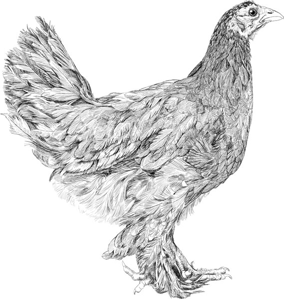
But our intuition, based on a lot of time spent with these models and the analysis of their data, is that these anatomical imperfections matter far, far less than the statistical methods that we employed– because we add a lot of flesh (like real animals have!) outside of the skeleton in our method, the precise morphology of the skeleton doesn’t matter much. It’s not like you need the kind of quality of anatomical detail that you need to do systematic analyses or osteological descriptive papers. The broad dimensions can matter, but those tend to be covered by the (overly, we suspect) broad error bars that our study had (see graph above). Hence while anyone could quibble ad infinitum about the accuracy of our skeletal data, I doubt it’s that bad– and it’s still a huge leap beyond previous studies, which did not present quantitative data, did not do comparative studies of multiple species, or did not construct models based on actual 3D skeletons as opposed to artists’ 2D shrinkwrapped reconstructions (the “Greg Paul method”). We also did directly measure the bodies of two extant archosaurs in our paper: a freshwater crocodile and a junglefowl (CT scan of the latter is reconstructed below in 3D).
One thing we still need to do, in future studies, is to look more carefully inside of the bird clade (Aves/Avialae) to see what’s going on there, especially as one moves closer to the crown group (modern birds). We represented modern birds with simply 1 bird: the “wild-type chicken” Red junglefowl, which isn’t drastically different in body shape from other basal modern birds such as a tinamou. Our paper was not about how diversity of body shape and centre of mass evolved within modern birds. But inspecting trends within Palaeognathae would be super interesting, because a lot of locomotor, size and body shape changes evolved therein; ostriches are probably a very, very poor proxy for the size and shape of the most recent common ancestor of all extant birds, for example, even though they seem to be fairly basal within that whole lineage. And, naturally, our study opens up opportunities for anyone to add feathers to our models and investigate aerodynamics, or to apply our methods to other dinosaur/vertebrate/metazoan groups. If the funding gods are kind to us, later this year we will be looking more closely, in particular, at the base of Archosauria and what was happening to locomotor mechanics in Triassic archosaurs…
Clickum to embiggum:
Australian freshwater crocodile, Crocodylus johnstoni; we CT scanned it in 3 pieces while visiting the Witmer lab in Ohio.
A Red junglefowl cockerel, spotted in Lampang, Thailand during one of my elephant gait research excursions there. Svelte, muscular and fast as hell. This photo is here to remind me to TAKE BLOODY PICTURES OF MY ACTUAL RESEARCH SPECIMENS SO I CAN SHOW THEM!
I’d bore you with the statistical intricacies of the paper, but that’s not very fun and it’s not the style of this blog, which is not called “What’s in John’s Software Code?”. Viv really worked his butt off to get the stats right, and we did many rounds of revisions and checking together, in addition to consultations with statistics experts. So I feel we did a good job. See the paper if that kind of thing floats your boat. Someone could find a flaw or alternative method, and if that changed our major conclusions that would be a bummer– but that’s science. We took the plunge and put all of our data online, as noted above, so anyone can do that, and that optimizes the reproducibility of science.
What I hope people do, in particular, is to use the 3D graphics of our paper’s 17 specimen-based archosaur bodies for other things– new and original research, video games, animations, whatever. It has been very satisfying to finally, from fairly early in the paper-writing process onwards, present all of the complex data in an analysis like this so someone else can use it. My past modelling papers have not done this, but I aim to backtrack and bring them up to snuff like this. We couldn’t publish open access in Nature, but we achieved reasonably open data at least, and to me that’s as important. I am really excited at a personal level, and intrigued from a professional standpoint, to see how our data and tools get used. We’ll be posting refinements of our (Matlab software-based) tools, which we’re still finding ways to enhance, as we proceed with future research.
Above: Two of the 17 archosaur 3D models (the skinny “mininal” models; shrinkwrapped for your protection) that you can download and examine and do stuff with! Dilophosaurus on the left; Velociraptor on the right. Maybe you can use these to make a Jurassic Park 4 film that is better, or at least more scientifically accurate, than Hollywood’s version! 😉 Just download free software like Meshlab, drop the OBJ files in and go!
Now, to bring the story full circle, the paper is out at last! A 4 year journey from Viv’s PhD thesis to the journal, and for me a ~14 year journey from my mind’s eye to realization. Phew! The real fun begins now, as we see how the paper is received! I hope you like it, and if you work in this area I hope you like the big dataset that comes with it, too. Perhaps more than any other paper I’ve written, because of the long voyage this paper has taken, it has a special place in my heart. I’m proud of it and the work our team did together to produce it. Now it is also yours. And all 3200ish words of this lengthy blog post are, as well!
Last but not least, enjoy the wonderful digital painting that Luis Rey did for this paper (another of my team’s many failed attempts to get on the cover of a journal!); he has now blogged about it, too!
Dinosaur posture and body shape evolving up the evolutionary tree, with example taxa depicted. By Luis Rey.
News stories about this paper will be added below as they come out, featuring our favourites:
Synopsis: Decent coverage, but negligible coverage in the general press; just science-specialist media, more or less. I think the story was judged to be too complex/esoteric for the general public. You’d think dinosaurs, evolution, computers plus physics would be an “easy sell” but it was not, and I don’t think we made any big errors “selling” it. Interesting– I continue to learn more about how unpredictable the media can be.
Regardless, the paper has had a great response from scientist colleagues/science afficionados, which was the target audience anyway. I’m very pleased with it, too– it’s one of my team’s best papers in my ~18 year career.
Today I’m doing something a bit unusual for this blog, but which very comfortably fits within its theme. Enough talking about my papers and media appearances and such. Too much self-indulgence, I feel. I want to talk about someone else. And then I will get back to the usual business of this blog: sharing the joy of cool anatomy, with a Mystery Dissection/Image post that is long overdue. Yet first, I wish to share the joy of knowing a cool anatomist– and artist.
One of my great shames as a scientist is that I never cultivated some decent artistic skills that I had as a young boy. And now as an anatomist I feel that my work often suffers from a lack of artistic talent (e.g. the image below, which still makes me hang my head in shame). In addition to scientific know-how, anatomy, when done best, demands the eyes and the hands of an artist. I might have the eyes but I definitely lack the hands. I envy people that have both; Julia Molnar from my team is but one example. And for me, encountering them is always a special delight. What follows is my personal perspective on one of the shining stars in the field.
Right ischium (hip bone; pelvis/synsacrum) of an adult ostrich in side view, showing some muscle origins and stuff, with a 1cm scale bar. Cringe. My boring, amateurish, pixelly line drawing from a paper on pelvic evolution. I hate it and wish I’d done better.
Mieke Roth is a scientific illustrator from the Netherlands, with a Masters degree from the prestigious and hallowed halls of Wageningen University. Her homepage has the tagline “Complex processes beautifully revealed”. This is a wonderfully succinct and eloquent way to describe the magic that she is able to conjure with her skills in scientific illustration. Her “Ultimate Croc Anatomy” project will be the greatest weaving of that sorcery yet. That project is described and will be documented on this page, and has an Indiegogo crowdfunding page here. Mieke describes its goals best:
“I will meticulously dissect a Nile crocodile and document it. I will share the dissection in text, illustrations and video via my website. I will process the data I gathered and each time build a new part of the digital crocodile. From there I will adapt the model for illustrations, books, animations and apps.”
I first became a happy victim of the artistic spells that Mieke casts when I saw her blog entry “How to make an octopus,” which you absolutely must read if you have not already or else you’ll be firmly spanked and sent away from this blog with only lumps of coal in your freezer for Freezermas (what’s Freezermas? Find out soon, and in the meantime be nice– or else!). In her post, Mieke didn’t just look up a picture of an octopus somewhere and redraw it in an abstract, schematic, flat and deceptively simplified way. She went and did her own hands-on research by dissecting an octopus, and then described the steps in converting those observations into a brilliantly novel set of digital illustrations that really brought octopus anatomy to life.
Octopus image by Mieke Roth
Part of what first mesmerized me is that this whole investigative and creative process was lovingly documented on her blog. In doing this, Mieke played the roles of both scientist and artist, by displaying the mundane-but-wonderful labour she did to come up with her final, gorgeous results, and the passion, dedication, scholarship and originality that make her stand head and shoulders above so many gifted scientific or medical illustrators. This thrilled me at both visceral and intellectual levels. It literally gave me chills to witness how good the final product was. As I write this and look back on that blog again, months later, I still feel the magic.
So then I started browsing around her homepage and became punch-drunk from repeated blows of amazement—again and again and again the quality and novelty and thoroughness leaped off the computer screen. Images of nature that I thought I knew well, such as the growth of a chick into a chicken(really great blog here documenting this with sketches), were conveyed in a way that made me see them as if for the first time, with joy and wonder. In the space of a day, I became a huge Mieke Roth fan.
Young chicken sketched by Mieke Roth
And now Mieke is taking it not just a notch, but a huge step, to spend a year documenting the anatomy of the Nile crocodile—how cool is that!?! As an expert in the postcranial anatomy of the Crocodylia, I can confidently state that the available scientific literature on the subject is patchy in coverage and often poor in quality by modern standards. There are big flashes of excellence here and there, such as Larry Witmer’s and Chris Brochu’s teams’ very thorough work on head and neck anatomy, or Colleen Farmer’s and Emma Schachner’s studies of lung morphology. But then I glance at some scholarly books (to avoid offending, I will not cite them here) that are supposed to be key references on the complete anatomy of Crocodylia and I frankly am left cold. While there is some superb work from the 1800s (Gadow, Fürbringer and others come immediately to mind), it is in the flat, often colourless style which the printing technology of that age imposed upon those great masters of anatomy. And while there is superb work by Romer, a tome on the Chinese alligator and a few others, again they tend to be limited to line drawings or spotty coverage of various anatomical systems. We need a visionary with both the scientific and artistic skill to make a subject that could seem dry or arcane become miraculous and accessible. I think you can guess whom I have in mind.
I can think of no one better suited to the ambitious goals and demands of the Ultimate Croc Anatomy project than Mieke Roth. She combines the attention to anatomical detail of a classical 19th century anatomist with the technical wizardry of a modern digital artist. She will have a supportive team of experts to ensure the content is exceptional and up-to-date. I’ll be one of them (and the crocs will come from my freezer), because of my enthusiasm for the project, and I’ve been helping to recruit others. So I urge you not just to join in her crowdfunding effort to carry out a very worthy and exciting project that the world can share, but also to share the joy I had in discovering her work by browsing her online collection of awesomeness. I predict that many of you will feel the power of her spell and become Mieke Roth fans, too, if you are not fortunate enough to be one yet.
A quick report on an exciting event for my team, from this week: We got a box! A big one! With 10 frozen crocodiles.
Stomach-Churning Rating: 5 out of 10. Just 1 picture with some blood.
These come from a breeding centre in southern France, and died of natural causes. Here is a little, icy box of five Crocodylus moreletii, a species that has featured here before:
And five young Nile crocodiles (remember WCROC?), one of which seems to have had an uncomfortable encounter with a larger relative:
I stumbled across some old pics, which I thought I’d lost, from the filming/preparations of 4 episodes of Inside Nature’s Giants (Jan-Feb 2009) at the RVC. They form a nice accompaniment to my previous post reflecting on my experience with the show, and the timing is great because I’m about to head to Raleigh, NC to talk about this research at the Society for Vertebrate Paleontology conference.
Stomach-Churning Rating: 4 at first (just a dead animal; and a rather clean one at that), then about halfway through the dissections start and it edges up to a 7 or so.
These pictures are sadly some of the few I have of the whole, intact body of a gorgeous adult Nile crocodile (Crocodylus niloticus) that the Windfall Films team managed to get to the RVC from La Ferme Aux Crocodiles in Pierrelatte, France. (I have scores of pics of the dissected limbs, shown further below) As the title indicates, it was a nice big croc. And as you’d expect, CT scanning and then dissecting it was no tiny feat, and makes a fun story. Story time, then, after an introductory pic!
Dr Samuel Martin, vet from La Ferme Aux Crocodiles, brought the crocodile (and some smaller specimens) over to our Hawkshead campus in late January 2009, and we quickly moved to run the specimen through our CT scanner to preserve some details of its anatomy (example shown at the end of this post) and for potential usage in the show. As the photos below illustrate, this was hard work for several people.
And then, as we were finishing the last CT scans of the specimen, our ageing medical scanner stopped working. And could not be resuscitated. R.I.P., Picker PQ5000 (buy one or two here!). The crocodile, “WCROC” as my team came to designate it, had claimed its final victim. It took about a year for us to get a new one, and that year sucked. It made me appreciate how lucky we are to have a CT scanner just across the parking lot from my office!
Anyway, the day of filming I was hoping to make it in to watch my colleague and friend Dr Greg Erickson help lead the dissection team, but a wicked blizzard blew up, and as I was starting the 31 mile drive south from my home to the RVC I realized, from the queue of cars that seemed to be 31 miles long (and train lines shut down), that this was going to be a snow day. So I turned around and came home. Another victory for WCROC!
The filming proceeded despite heavy snow delaying many of the key players’ arrivals. I got filmed a day or two later for a little section of the show on the limbs and locomotion of crocodiles but sadly this got cut from the main ING show (but did air in the National Geographic version “Raw Anatomy“, in the USA at least).
The limbs had been left largely intact, although some of the dissectors who didn’t know croc anatomy very well had slashed through parts of the pelvis and, in eagerness to reach key parts to demonstrate in the show, some major muscles got shredded. This is no big surprise; crocodiles have a lot of bones all over the place: in their skin (scutes; bony armour), in their bellies (the belly ribs called gastralia), and almost everywhere else, so some brute force is required to get to the gooey bits. Apparently there had been 6 or so people dissecting at once and things got a little carried away. The curse of WCROC continues?
Oh well; that’s just how documentaries go sometimes, especially with a pioneering show like this and the intensely compressed timescales of filming (time is ££!). There can be pulses of chaos. And the show turned out GREAT! (alternative link if latter does not work outside UK)
Let’s have more photos tell the story of the scanning, which also shows off this beautiful animal’s external anatomy:
Anyway, things turned out fine overall for our research. A week or so later (maybe longer; I forget if the specimen was frozen and thawed out for us) we came in to start dissections. We were really excited to measure the limb muscles of such a big crocodile, for comparison to a growth series (babies to adults) of alligators that my former PhD student (now postdoc; Dr.) Vivian Allen had dissected back in 2008. Here he is with a masked co-dissector, displaying their joy for the task at hand:
And let’s not leave out the exhuberance of visiting research fellow Dr. Shin-Ichi Fujiwara! He wanted to inspect the forelimbs for his ongoing studies of limb posture, joint cartilages and locomotor mechanics.
The remaining images show progressive stages of dissection of WCROC, starting from the pectoral (fore-) limbs with a view of the belly (and the giant jaw-closing muscles visible on the left side of image):
Isolated right forelimb, with coracoid (part of shoulder girdle) sticking through:
Assorted forelimb/upper arm (brachial) muscles:
And the triceps (elbow-straightening) muscles; not that big in such a big animal:
…and on to the pelvic limbs and the huge tail:
With a closer look at the HUGE thigh muscle, the famed M. caudofemoralis longus:
And then an isolated right hindlimb:
Thigh muscles, with which I have a peculiar fascination that stems from my PhD research:
And last, the great, paddle-like hind foot!
What a great experience that was! We have fond memories of WCROC, a great documentary from Windfall Films, some nice data– and a lovely skeleton. Perhaps the curse of WCROC is not so bad. Nothing can go wrong now!
Soon Mieke Roth, scientific illustrator from the Netherlands, is coming here to do a similar dissection on more Nile crocodiles at the RVC. As with the octopus she wrote about in September, she will make a 3D model, but with much more detail and with an emphasis on accuracy and accessibility. The end products will be really cool; think of the visible body, 3d models that can be used in teaching, animations, a book and lots more but also a “how did she do that?” blog. To finance this project (that probably will take a year or more) she will use crowd funding. In several weeks there will be more info on how to participate in her fantastic endeavour. For now, see her video with the initial pitch for “Nile Crocodile 2.0“!
OK Londoners, and Olympics visitors, and anatomy (or just science/biology) buffs, and those not lucky enough to see other versions of the animal Body Worlds show. You have a mission. And that mission is to go see “Animal Inside Out”, a special (£9 for adults is well worth it!) exhbit at the Natural History Museum, open until September 16. This blog will self destruct, very messily, by turning itself inside out in 5 seconds… Boom.
Anatomy to me is beautiful even when it’s “ugly” (messy, wet, mucosal, intestinal, asymmetrical, unlike human, whatever), and that’s a major theme of this blog. Hence I am embarrassed that I hadn’t yet gone to see this Body Worlds spinoff exhibit until now, but can begin to shake off that shame by means of an almost exclusively effusive gushing of blood love for said exhibit. Wow, wow, wow! I went in with no particular expectations, having seen some pictures and knowing some of what to expect, and having other things on my mind. I came out very pleased; the NHM exhibits folks and von Hagens’s crew have created an inspirational spectacle that could do wonders for anatomical sciences and natural history. More about that at the end.
(Warning: possibility of spoilers, but the exhibit is so visual that I don’t think my descriptions can spoil it)
The entrance
No photos are allowed as usual, so all I have to show you is the entrance and some anatomy pics I’ve interspersed from my team’s research to lighten up the text. I suppose I could have asked for special permission to take photos for review usage but this was a very impromptu visit, and with ~4 months of showing left I may well be back again.
Weighing a hippo; spot on at 1600 kg!
There is a brief panel on homology and why it is the major concept underlying comparative anatomy (and a key part of evolution, co-opted from the not-so-evolutionary ideas of Sir Richard Owen, whom the NHM rightly mentions here). Another panel rightly brings up the issue of ethics, which has plagued Body Worlds before. It comforts the visitors that animals were not slaughtered just for this display and that the NHM applied its strict collections criteria to them. Convincing enough for me, and absolutely necessary to bring up early on.
The entry hall then presents you with about five cephalopods (labelled “squid” and “octopus”—a gripe is that species names/details are not given for most specimens on show) prominently occupying the view. The cephalopods, like basically everything else, are plastinated (by a now US-patented set of procedures, I learned from the exhibit book detailed later). They are stunningly frozen in lifelike poses or with gaping cuts to show their interior anatomy, although there was very little explanation here about cephalopod biology and anatomy (about 1 smallish panel). No mention of Cthulhu. Damn. He’d approve of the Grand Guignol scenery.
Toward the back of the first corridor of specimens and cases, there is a stunning scarlet haze outlining the body of a “shark” (species not given) with its huge liver lying below it. The haze, a technique used repeatedly throughout the exhibit, is some kind of corrosion cast of the circulatory system, I gather. A bunch of cross/longitudinal sections of cephalopods, crocodiles, fish, horse hooves and other animals decorate blank spaces on the walls, some with labels showing basic features and some just hung like paintings. Fair enough, but a missed opportunity for a bit more educational content here.
Gratuitious Melanosuchus (black caiman) shot.
A smallish whole shark confronts you as you turn the corner from the crimson chondrichthyan; again of unknown classification. One would think a museum exhibit would care about classification beyond “shark,” but oh well, I am banging the same drum here too much and missing the point, that the exhibit is really a visual, visceral expose rather than a deep prose-driven intellectual dissection. On one of the shark panels it is noted that sharks have red and white kinds of muscle used for slower and faster swimming, but not clarified that this is a very widespread vertebrate (chordate?) feature. This forms my second gripe, that a truly evolutionary approach, such as that taken by dozens of the museum’s research staff as their major paradigm of phylogenetic systematics, could have helped the public grasp the evolutionary, hierarchical nature of homology and depart with accurate information about what features characterize groups at which levels. I’m not asking for cladograms laid out on the floor as at the American Museum of Natural History, although maybe that could work, but the exhibit tended to fall back on an outmoded “this animal has this feature, and that animal has that feature, and these are cool adaptations” shopping list approach rather than a modern comparative approach. Granted, almost all museum exhibits fall into this trap, for various reasons and some of them justified. But with a spare word or phrase here or there, this could have been done better without drowning the visitors in that dreaded sea of bloodprose.
Passing the sharks, we come to one of several thematic sections about body systems, this first one on the skeleton (later, brain/nerves, circulation, muscles, etc.). A few small skeletal specimens of the type that are seen throughout the museum are presented, with a scallop reminding us that skeletons can come in many types among multicellular organisms. There is a horse skull and a stark white whole skeleton of a young-ish ostrich, which was very nicely mounted. However, I was caught off guard by the pelvis, which lacked the curved, ventral “boot” like connection of the pubic bones that ostriches have—presumably explained by its juvenile status although I wasn’t 100% sure it was even an ostrich pelvis. OK, I am having a serious pelvis-nerd moment here; forgive me as my PhD was on this stuff.
Ostrich in the midst of disassembling.
BUT, once again the small interpretive panel had a moment of Fail. The ostrich was explained to have two toes, in contrast to normal birds which have “five”. HUH? Birds have three main toes and variably also a fourth, inner (first) toe called the hallux, used for perching and other activities including walking. None have a fifth toe; indeed their dinosaurian forebears lost that feature some 230ish million years ago. Just an embryonic vestige of the base of the fifth toe is visible in bird embryos today. Furthermore, the panel said that two toes in ostriches can grip the ground more strongly than more toes in other birds. I know of no evidence that shows this, and suspect that the contrary might be true. The standard explanation for toe reduction in ostriches is that it is a lightening feature characteristic of “cursorial” (long-legged, sometimes fleet/efficient) animals, to make swinging the long legs easier. These errors really should have been caught by involving experts in polishing the scientific content of the exhibit.
But I don’t want this post to grumble too much; wrong message. There was so much to celebrate in this exhibit, which was felt impressively spacious and full of cool specimens! Visitors pass some plastinated whole sheep and goats, with panels nicely explaining that goats and sheep look quite similar on the inside and are evolutionary relatives. Having “four stomachs” (technically, a four-chambered stomach; not four distinct organs that were duplicated) is attributed as a sheep trait, then being a ruminant is said to be a goat trait; this might get a little confusing for non—anatomists (both are ruminants and have similar stomachs).
I learned that goats have an extra tail muscle that allows them to swing up/down as well as side-to-side. Hey, I teach veterinary anatomy and I don’t know that!? I must tuck my tail between my legs in shame, but I am no goat so I do not think I can (do satyrs count?). But I wasn’t so sure that goats, as described, were the first animals to be domesticated—I thought that was dogs? Ahh, Wikipedia says dogs, then sheep, then pigs, then goats? I’m outside my expertise here, I admit, and resorting to Wikipedia out of ignorant desperation. Anyway, here, another instance of coulda-been-more-phylogenetically-specific presented itself: the forelimb of goats was said to be connected to the thorax by muscles and ligaments, not a joint, but this is a feature common to most Mammalia. Although audience attentions might be wandering at this point, waiting for the next big spectacle (goats and sheep are not a big crowd draw, even plastinated), some more care as to what was written would be good. Some reindeer and horses and other animals join in the fun later on. Good, but mostly ‘filler’ (wise to put these in the middle of the exhibit, after sharks/cephalopods and before climax) unless you’re a big fan of fairly familiar ungulates with fairly homogeneous postcrania. OK, my bias is showing…
Gratuitious image of emu curled up for CT scan.
Next along the path, a longitudinal section of a whole ostrich caught my attention. Wow again! I had no idea that one could make a section like this of such a large animal, all in one plastic sheet like a giant microscope slide! I stared at this for a while, wondering how both legs could be fit in a ~1cm thick panel, and gave up trying to understand the technology. Von Hagens, you got me there; I’m stumped. Were multiple sections glued together somehow to produce a pseudo-2D slice from many thin 3D sections? I could not tell, and felt humbled and deeply impressed by the technical skill shown in the exhibits so far…
And then the punches kept coming, one-two-three! The exhibit approaches its climax with a crescendo of great specimens in the final hall. First, another maroon marvel. A whole ostrich, standing with wings askew, showing off its entire circulatory system (plus a few wing plumes for aesthetics) from head to toes! Gorgeous, technically brilliant, and well worth at least a 5 minute walk around (you can stroll around many of the displays in 360 degrees- very good move!). A plastinated whole ostrich stands next to it, and for a muscular anatomy geek like me, it was nirvana. However, in a churlish moment I had to look away from a panel explaining that an ostrich is “too heavy to fly” (I admit some younger visitors may need reminding of this). But then I looked into the big open space of this main hall, and the climax was before me. I think I’d had my climax a few times since this, but wow this was enormous in so many ways. All the ways. Mind-blowingly, vastly, geektastically kewl.
Gratuitious rhinoceros leg.
Across from the two posed ostriches and flanked by numerous smaller specimens, the elephant and giraffe stand frozen in vigil. There is also a lovingly detailed dissection of a huge male gorilla by the back wall and exit, with a panel reminding us that gorillas are (among) “our closest relatives.” The giraffe is precariously poised on one front toe-tip, in mid-gallop. What a great pose! There is the requisite explanation of how they solve the blood pressure problem in their neck (e.g. arterial valves), but also the statement, news to me, that they are the only animals able to ruminate while running. Who figured that out and how? I really want to know! Must be hard to check. (or was walking intended? Are my notes wrong?) Across from the full-fleshed plastinated giraffe (which I could see with my eyes closed after all our dissections from a month ago), there was another visually arresting and technically monumental giraffe on exhibit: one represented completely by small, reddish cross-sectional slices, from head to toes in a standing pose. That took me a while to absorb, it was so lovely, almost like a hanging mobile of morphological splendour.
There is a panel about genes and variation and inheritance. It is brief. (and it belongs there) Thank you. Let’s celebrate anatomy for anatomy’s sake for once!
“But John,” you might say, “What about the elephant? No love for the elephant? The star of the show?”
Zoinks! I want one! Stoic and triumphant (except against death and plastination), the Asian elephant is the centrepiece of the collection. (The book explains it was “Samba” from Neunkirchen Zoo, Germany, dead of some circulatory problem in 2005 and the first one plastinated, plus the inspiration for the animal show). I was speechless and paralyzed for a moment. I didn’t even know how to start looking at the partly-exploded-to-show-its-insides elephant. I actually avoided it for a while, looking closely at the other specimens, and building up anticipation, before stepping up and taking a long, intense look at this tall drink of water.
Go see the elephant. If you know basic anatomy, look at its leg muscles. Check out the huge triceps, still attached to the elbow; I like to say it is the size of a graduate student. Same for the analogous superficial gluteal and somewhat-fused biceps femoris muscles on the rear end, around the thigh/knee joint. Huge! I’ve never been able to view a standing dissected elephant, so this really impressed me more than a table full of giant muscle slabs like I normally deal with. And best of all, for me, the “false sixth toes”; the prepollex and prehallux; are visible in all four feet (but not noted anywhere, even in the book; too bad, these things were widely known by anatomists before my work on them). So much to marvel at here. It is an anatomical treasure. I wish I had a 3D image of it to use for anatomical studies- it was so easy to identify every single muscle group (except for a few missing around the shoulder/neck), even in the distal limbs. Hmm, photogrammetry might be possible (nugget of idea begins to crawl around John’s brainlike a Zimmerian parasite)…
Behold, the triceps muscle of an elephant!
Behind that gorgeous elephant, don’t miss the wall mountings of two cross-sectional slices: through the head/neck of a moderate-sized elephant (How!?!?) and distal leg (no predigits but good features). And definitely don’t miss the stool (non-fecal, furniture form). I almost did. A wooden stool is shaped like a newborn elephant and a cross-section of the body is adhered on top of it. I assume you cannot sit there, and I am very glad that it was not, as I first imagined, an actual plastinated baby elephant turned into a stool. That would be bad taste.
The exhibit is in very good taste, without exception, and although I am gore-desensitized to say the least, it is not gory in my view. The plastination process preserves the reality and even some of the colour faithfully, but renders it just unreal enough (past uncanny valley territory?) that it should not be very disturbing to most viewers.
You can’t leave with your own photographs, but you can be schnookered into buying the exhibit book (£12.99) and a couple of packages of nice colour postcards (£4 for six; excellent quality images and cardstock IMO). The book and postcards show many of the exhibit specimens but not all, and include some others that are not on exhibit. I was saddened that the bear was left out—very cool image of that in the book. I’ve only skimmed the book a bit. I was annoyed by a few mistruths about elephants (25mph running speed, “have no ankle joints, which is one of the reasons why elephants cannot jump”, the bones “do not contain any marrow”—wrong, 15mph and there are ankles, they just are not very flexible (but not immobile either); also the bones do contain marrow (how could a large vertebrate survive entirely without it???) but just not as much of it per unit volume, due to lots of spongy bone). But I am still very happy with the 139 pages chock fulla pretty images, which is all I really wanted. Indeed, the book is a great pictorial anatomical reference- some of the species such as elephants and giraffe lack a really good anatomical resource in the modern, or any, literature! The exhibit shop also sells some good anatomy texts, mostly on humans but I recommend “Animal Anatomy for Artists” very strongly; I use that regularly in my own work.
So, £29.99 of schnookering later (haha, poor victimized me!), I emerged and reflected more on what I’d seen. I’m still a bit giddy about it all. I like the minimalism in most aspects- black backgrounds, minimal signage (but just enough to make it educational—when they got the facts right), focus kept on the specimens. Well done there. The spectacle of the specimens I’ve raved plenty about- it is not at all disappointing. It is AWESOME in every sense. I feel I easily got £9 of value from the ticket, and would (probably will!) pay it again. It is a profound experience to see the rich anatomical detail exposed, and be able to circumnavigate the specimens to absorb multiple perspectives. If you know some anatomy, you’ll be doubly rewarded at least, and if you bring your own phylogenetic perspective that can be trebled.
Baby white rhinoceros. Sad infant mortality.
What makes me happiest after my visit is realizing that we are in an anatomical renaissance for science and public interest therein. Exhibits like this and documentaries like “Inside Nature’s Giants” have tapped a public interest and curiosity in the wonders of basic anatomy. Anatomy is at the core of so many biological sciences and is so immediately accessible to people, because we all have anatomy. Anatomy is at the crossroads of art and science; it is visual, variable and complex, yet concrete, objective and easy to relate to. “Animal Inside Out” is a spectacular blend of art and science. They nail the artistic aspect, and the science is done reasonably well (despite my few gripes)—the exhibit’s science speaks for itself, in a way, although many visitors will need a nudge to grasp that.
I’d like to make a call for a permanent exhibit of the likes of “Animal Inside Out” in the UK. We deserve this! Museum exhibits could use something new, other than lame, quickly broken digital pushbuttons and bland skeletons devoid of soft tissue context (although the latter can be sufficient, e.g. at the Paris NMNH). That’s what makes “Animal Inside Out” (and Body Worlds) such a hit- as Hagens is quoted on the book dustcover, animal anatomy that goes beyond digitized abstractions and dusty bones is able “to sharpen our sense of the extraordinary by looking at the self-evident.” I could not say it better myself. This exhibit is extraordinary; that is self-evident after even a peek. It is a loving tribute to how fantastic the totality of animal structure is. Go! Enjoy. Absorb. Gape. Stare. Thrill. Revel. Think. Question. IT’S BEAUTIFUL.
Impressive hippo mouth says “Farewell for now.”
Edit: @samjamespearson on Twitter has kindly posted some photos (for free NHM/AIO publicity) of the exhibits and here are the links, now that they’re out there– SPOILERS! And thanks, Sam! I don’t think these really spoil the intense visual experience of actually being there and walking around the specimens, not at all.
Party time! Let the media onslaught begin! We’ve published a paper in Nature on the limb motions of Ichthyostega (and by implication, some other stem tetrapods). Since we did use some crocodile specimens from Freezersaurus (see below) in this study, I figured WIJF could cover it to help celebrate this auspicious event. Briefly. Particularly since we already did a quasi-blog on it, which is here:
However I want to feature our rockin’ cool animations we did for the paper, to squeeze every last possible drop of science communicationy goodness out of them. So here they are in all their digital glory. Huge credit to Dr. Stephanie Pierce, the brilliant, hardworking postdoc who spearheaded the work including these videos! Dr. Jenny Clack is our coauthor on this study and the sage of Ichthyostega and its relatives- her website is here. Also, a big hurrah for our goddess of artsy science, Julia Molnar, who helped with the videos and other images. Enjoy!
The computer model
The forelimb model
The hindlimb model
We used some of my Nile crocodile collection to do a validation analysis of our joint range of motion (ROM) methods, detailed in the Supplementary info of the paper, which I encourage anyone interested to read since it has loads more interesting stuff and cool pics. We found that a bone-based ROM will always give you a greater ROM than an intact fleshy limb-based ROM. In other words, muscles and ligaments (and articular cartilage, etc.). have a net effect of reducing how far a joint can move. This is not shocking but few studies have ever truly quantitatively checked this with empirical data from whole animals. It is an important consideration for all vert paleo types. Here is a pic of one of the crocodiles from the study, with (A) and without muscles (B; ligaments only):
I’ll close with Julia Molnar’s jaw-droppingly awesome flesh reconstruction from our model. Why Nature wouldn’t use this as a cover pic, I’ll never understand, but I LOVE it! When I first saw it enter my email inbox and then opened it to behold its glory, my squeal of geeky joy was deafening.
(edit: Aha! Fellow Berkeley alum Nick Pyenson’s group made the Nature cover, for their kickass study of rorqual whale anatomy, including a “new” organ! Well, we don’t feel so bad then. Great science– and a win for anatomy!!!)
There’s no better way to kick things off after a holiday than with a celebration of the Inside Nature’s Giants series, which I had a small part in early on, including these shots I took during the time they spent filming at the RVC >3 years ago (!?!?); most of these animals spent multiple holidays inside The Freezers:
Elephant arriving…
Elephant revealed
Private moment with elephant
Stunning emergence of The Guts
So you are impressed by the guts too, ehh? It was pretty amazing to watch it happen. The tension was intense- the animal had been dead for a while and was rather bloated. So cutting it open was a task gingerly taken…
Bloated elephant
RVC dissector Richard Prior stuck a scalpel in the upper abdomen when the time was right… the piercing whistle and the sulphuric odour silenced the crowd watching… and then quickly out came the guts.
Everyone was pretty amazed by the scale.
The guts just went on and on…
Not a 1-person job by any means.
Spreading them out to see the whole GI tract.
I waited patiently and watched the show filming; what a great, professional crew. Then I got to take the legs away for our research.
But not just elephants, no sirree! The Windfall Films/ING team filmed giraffe, crocodile and big cats episodes (4 total) at the RVC too; a crazy period of a few weeks (including a major blizzard that hit us during the croc filming) in 2009. Some of the stars follow:
…and I’m rather fond of that tiger’s neck– check out the hyoids (roaring/tongue apparatus in throat; bottom of movie)!
…and here is the adult Nile crocodile’s head after scanning
…and another view of that big Nile croc, just because I like how this reconstruction turned out
…and here’s one of the small (~1m long, 10kg) juvenile Nile crocodiles from the show, with a pilot CT scan showing the skeleton nicely- and possibly a last meal or stomach stone on the left side of the abdomen (bright white blob; I need to check this now that we’ve dissected it)
Foetal giraffe; stillborn; from the show, in process of dissection in our lab to measure its limb anatomy. Trust me, it looked –and smelled– better on the inside than it did from the outside. Eew.
How most of the specimens from the first 4 episodes ended up after all dissection was done (part of my/RVC’s collection of skeletons). Sadly, I did not get great photos of the 3.7m Nile crocodile or the two giraffes before they were reduced to bits, but I do have the skeletons and CT scans.
Giving a tour (including The Freezers) to A Certain Esteemed Visitor.
(Another) Gratuitous shot with one of the sweet old Red Kangaroos at Alma Park Zoo near Brisbane, Australia. Experiments on hopping we did there will be briefly featured in the new Inside Nature’s Giants show on Channel 4, 16 April @2000- details at http://t.co/SkjsMeVC.
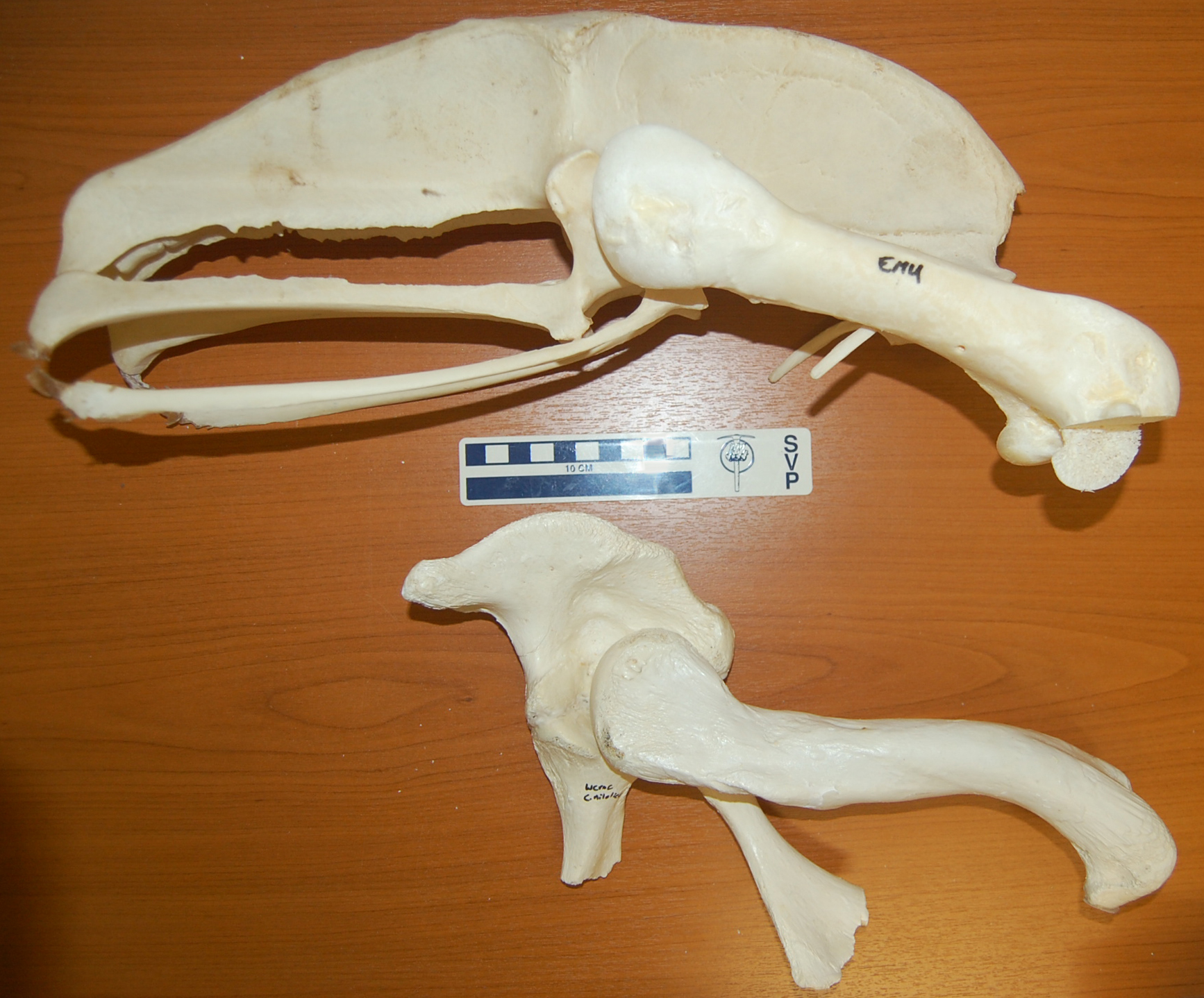
I’m off soon for a sunny break on the beaches of Morocco, but as an Easter gift to you, my (admirable, sagely, few, beautiful) readers, here is an image of two specimens, formerly from my freezers, to consider:
Both are the right pelvis (hip bones; crocodile pelvis is a bit broken toward the bottom) and femur (thigh bones) of living archosaurs– a 27kg emu above, and a 278kg nile crocodile below. The head would be toward the right side of the picture. A tenfold difference in mass between the bird and the crocodile, and yet some of the dimensions are so similar in both of them (femur length etc.), or so vastly different in the bipedal runner vs. the quadrupedal not-so-fast-runner (much bigger pelvis for leg muscle attachments in the former).
This image says it all. It is why I study the evolution of locomotion in land animals. It is why I am so fascinated by the transition from vaguely crocodile-like early archosaurs to modern birds by way of earlier dinosaurs. Anatomy, size, mechanics, behaviour, phylogeny… the photo captures all the facets of why I am so enraptured by research in this field.
It also might evoke thoughts of what features are expected in a terrestrial vs. aquatic animal, and thoughts of how some numbskulls still think big dinosaurs lived in the water (no I will not link to the execrable story from BBC today that I am thinking of!)…
I hope you appreciate it, too. Have a freezer-burn-free holiday period, folks!
Birds and crocodiles are part of the spectacularly diverse group of animals called the Archosauria, or archosaurs if you’re on casual terms with them. Other (extinct) archosaurs include the dinosaurs (non-avian), pterosaurs, and sundry wondrous other beasts like aetosaurs and phytosaurs. Archosaurs have, and presumably their common ancestor had, many specialized features of their anatomy that are related to metabolism and locomotion. That’s a big reason why, as a scientist, I love them.
Yet the bird lineage evolved its own extreme specializations, whereas in some (but not all!) ways crocodilians stayed closer to the ancestral state. Here is a great example of one of the major categories of differences between living crocs and birds: the proportions of the respiratory system, from freezer specimens I’ve CT scanned with my former PhD student Vivian Allen, which were part of a paper we published in Anatomical Record back in 2009. We scanned the thawed specimens with and without the lungs inflated (croc results not shown for inflated state). This was easy; we just stuck a syringe into the windpipe and then tied it off once we had pressurized the lungs. [I’m now working with Colleen Farmer and Emma Schachner on using these specimens to learn more about the surprisingly “bird-like” features of croc lungs despite the smaller total volume of the airways; more about that another day… we can do MUCH better than these images!]
Here, the airways are coloured blue/purple and the flesh has been made transparent yellow, while the skeleton is orange. The relatively massive size of the airways is evident in birds, especially the air sacs (side pockets of the lungs/other air passages), whether they are relaxed or inflated. The lungs (purple) aren’t that differently sized in the two animals.
Australian Freshwater crocodile from CT scan:
Junglefowl (“ancestral wild chicken”) from CT scan; relaxed airways:
Junglefowl from CT scan; inflated airways:
(note that the light blue region is the expanded air sacs; the lung in purple hardly changes because it is fairly rigid in birds)


")
")
")
")
")
")
")
")


")
")


