Feeds:
Posts
Comments

Posts Tagged ‘dem bones’

…a daily picture of anatomy!

Welcome to Freezermas! In the dead of winter, the WIJF blog jumps down your internet to deliver mind-warming science, and images, and evolution! To celebrate Charles Darwin’s birthday (204th = tomorrow Feb 12, 2013), I’m bringing you one Anatomy Vignette each day this week (we’ll see if I can manage the weekend or not)! Let’s do this!

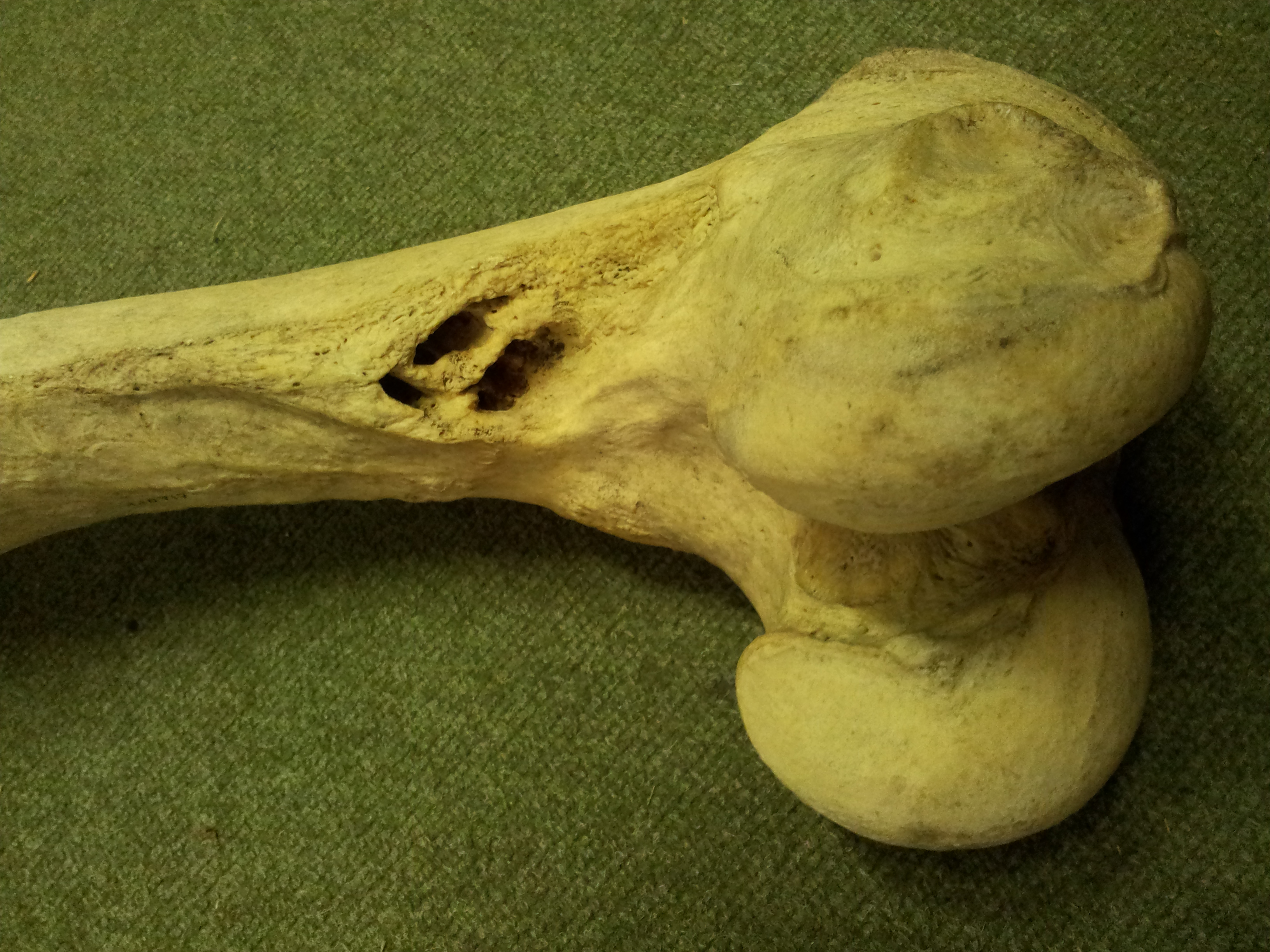
Stomach-Churning Rating: 2/10; just bones; one picture of them, and then a lot of discussion of muscle anatomy but no pictures of it.

Hutch02-Fig4

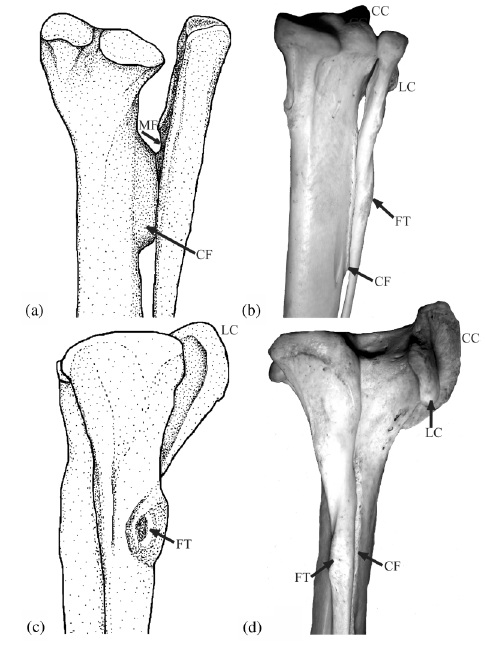
The above image comes from one of my old, somewhat obscure anatomy papers (link to pdf here), from 2003. It’s possibly the first figure I made, entirely by myself, that I’m sort of proud of. It doesn’t totally suck compared with some of my other attempts. I did the stippled line drawing on the left, and on the right is one of my first usages of a digital photo in a paper (digital cameras were finally up to the task around that time; I used my new Nikon Coolpix 900, if memory serves). It was a greatly improved figure over what I’d submitted for this paper originally, which was a rushed, half-baked manuscript for a SICB conference symposium on tendons. I’ll never forget one of the peer reviews of the manuscript, which said something like “the text of this paper is a joy to behold, but the figures are a horror.” They were right, and luckily the images in the paper I submitted changed a lot in revision. (I’m still embarrassed by the incident, though!)

Anyway, the picture is of  the lower hind limb of two theropod dinosaurs: (a,c) an adult Tyrannosaurus rex, and (b,d) a wild turkey (Meleagris) from my personal collections of dissected-then-skeletonized animals (this turkey became a biomechanical model in a 2004 paper of mine, too!).  In both cases we’re looking at a right hind limb; in (a) and (b) from a caudal/posterior/rear view, and in (c) and (d) from a lateral/side-on/profile view.

If you’re having trouble visualizing these bones in the real animal, check this T. rex skeleton in rear and side views and try to find these bones. You can do it! You might also want to look back at my paroxysmic outburst of love for knee joint anatomy.

The thicker long bone is the tibia (your main shank bone; or in a lamb shank, chicken drumstick, etc); the thinner outer bone is the fibula. Together with some smaller bones, for brevity we can call them the tibiotarsus — but only in theropod dinosaurs, or you will anger the freezer gods.

The labels show some cool anatomical features, as follows:

CC” the cranial cnemial crest of the tibia (a projection of bone unique to the knees of birds);

CF” the crista fibularis; or fibular crest; of the tibia (more about this below);

FT” the fibular tubercle (insertion of the big hamstring/biceps muscle M. iliofibularis);

LC” the lateral cnemial crest of the tibia (a big arching swath of bone that both birds and non-avian theropods like Tyrannosaurus have; the CC is just pasted on top of this in birds); and

MF” which denotes a muscle fossa (depression) on the inner surface of the upper end of the fibula, which presumably housed a muscle (M. popliteus) binding the fibula to the tibia in earlier dinosaurs, but is vestigial in birds.

The CF, or fibular crest, is a feature that only theropod dinosaurs, among reptiles, develop like this. It evolved early in their history and thus was passed on to birds with other ancient features like hollow bones and bipedalism. It binds the fibula closely appressed to the tibia, making those bones act more like a single functional unit –and sometimes they even fuse together. The CF also transmits forces from the whopping big M. iliofibularis muscle’s insertion (the FT label) across the puny fibula onto the robust tibia. The MF once held a muscle that also helped keep those two bones together, but additionally it could have contracted to move them relative to each other a little bit, as in other living animals (many mammals and reptiles have a big M. popliteus and/or M. interosse[o]us). So these features all have a common functional, anatomical and evolutionary (and developmental; different story for evo-devo fans) relationship. By binding the fibula and tibia together, these structures helped early bipeds (the first theropods and kin) support themselves on one leg at a time during standing and moving, and also helped begin to reduce the limbs to lighten them for easier, faster swinging. So we can think of these features as specializations that helped theropod dinosaurs, and ultimately birds, get established as bipedal animals.

The CC and LC have a similar story to tell; for one, they are muscle attachments, again mainly for thigh muscles. And again, the LC dates back to early theropods (and some other dinosaurs had a version of it; usually smaller). These crests serve mainly as insertions for the “quadriceps” (in human/mammal terms) or triceps (in reptile/bird terms) muscle group’s major tendon, spanning from the pelvis/femur across the thigh and knee to this region. In birds, we call this structure of insertion the patellar tendon or (less appropriately) ligament. But dinosaurs had no patella, ever, so the triceps femoris tendon would be the proper technical term. Regardless, that crest (LC, and later LC too) helped the attached muscles to straighten the knee joint or support body weight during standing/moving, by giving them better leverage. So it would have been important for early bipeds, too, like the CF, MF and other features above. Your cnemial crest (tibial tuberosity) is pathetic by comparison. Don’t even look at it. Droop your knees in primate shame!

Bumps and squiggles on bones might seem puny details just for anatomists to study and describe in long, tedious monographs, but each is part of the great story of evolution, and each has a story to tell that fits into that story. Back in Darwin’s day, some of the world’s greatest scientists of the age (Richard Owen and Thomas Huxley being but two spectacular examples) pored over these seemingly innocuous features, and so they became part of nascent evolutionary theory even then. This week, I’ll be celebrating a lot of those details, which I still feel are important today, and the stories they help to tell.

Happy Freezermas! Sing it: “On the first day of Freezermas, this blo-og gave to me: a tibiotarsus with a CF and FT!”

Read Full Post »

I’m not sure if this is a new tradition at this blog or not (probably not), but hey let’s give it a name: an Anatomy Vignette. Just something curious I notice during my research that deserves more than just a tweet. I borrowed some bones from the University of Cambridge Museum of Zoology (whom I love, because they have great exhibits and are very research-friendly) to CT scan for some projects. I noticed this:

femur-path

And I thought “Ouch! That’s nasty, dude.” (the holes in the bone just above the knee joint– these should just be a roughened area where the adductor muscles and other leg muscles attach)

So I was interested to see the CT scan images to find out how these possibly osteomyelitic lesions continued into the bone. They’re really pervasive, continuing into the marrow cavity quite far up the femur, as this shows (good CT-viewing practice to match up what you are seeing in the photo above with this movie):

I would be surprised if this was not the reason this animal died (presumably being euthanased at a UK zoo). There would have been extensive infection and pain resulting from this bony disease. How did it originate? Who knows. Maybe the animal strained a muscle and bacteria got inside, or maybe there was a fall or other injury. Hard to tell.

Oh, and also note the lack of a true marrow cavity in hippos, which is true for all the long bones. The “cavity” is filled in with cancellous bone. Same with rhinos, elephants, and many other species… science doesn’t entirely know why but this feature surely does help support the body on land, and grants at least some extra negative buoyancy in water; at a cost of some extra weight to lug around, of course.

And so ends this Anatomy Vignette.

Read Full Post »

To kick off the New Year just right, our tetrapod team has a new paper in Nature, following up on last year’s Ichthyostega not-so-good-at-walking study (also see here). Yet this paper has a more anatomically descriptive — and also an “evo-devo” — twist to it. For brevity, I’ll let our press release tell the story, since I think it does a good job of it (like I always preach scientists should do, we worked with our PR company to write this together, so we’re happy with how the press release came out). In a nutshell, our study used some very fancy synchotron radiation techniques to image the 3D anatomy of the backbone in early land vertebrates. Our findings surprised even us, and ended up turning around palaeontology/comparative anatomy’s view of how the backbone evolved, giving us a new glimpse into our inner tetrapod.

Stick around for the videos at the end, which are the first four supplementary movies from the paper and are rather pretty (there are two more, for imaging/segmenting afficionados, but they are not as pretty or interesting for most of this blog’s readership). The final figure (Figure 1 from our paper) gives some extra visual context.

The paper is:

Pierce, S.E., Ahlberg, P.E., Hutchinson, J.R., Molnar, J.L., Sanchez, S., Tafforeau, P., Clack, J.A. 2013. Vertebral architecture in the earliest stem tetrapods. Nature, published online [here].

I should note that I’m just 3rd author, so I deserve only modest credit. But I helped. Even though no freezers were involved, or harmed, in the process.

Ichy_vertebrae_final_sm-01

Above image: Julia Molnar‘s illustration of Ichthyostega showing anatomical changes of its spine from front to back, with neural arch/spine in pink, twin pleurocentra in yellow, and intercentrum in green. These four parts, three kinds of bones, made up the backbone of the first land vertebrates. These parts evolved in different ways in later animals, but formed one main bone in all living lineages of vertebrates.

RVC PRESS RELEASE:

Scientists reassemble the backbone of life using a particle accelerator

Research published today (Sunday 13 January 2013) in the journal Nature documents, for the first time, the intricate three-dimensional structure of the backbone in the earliest four-legged animals (tetrapods).

The international team of scientists, led by Dr Stephanie E. Pierce from The Royal Veterinary College and Professor Jennifer A. Clack from the University of Cambridge, bombarded 360 million year old early tetrapod fossils with high energy synchrotron radiation. The resulting high resolution X-ray images allowed the researchers to reconstruct the backbones of the extinct animals in exceptional detail.

The backbone, also known as the spine or vertebral column, is a bony structure found in all tetrapods, along with other vertebrates such as fish. It is formed from many elements or vertebrae all connected in a row – from head to tail. Unlike the backbone of living tetrapods (e.g. humans), in which each vertebra is composed of only one bone, early tetrapods had vertebrae made up of multiple parts.

Lead author Dr Pierce says: “For more than 100 years, early tetrapods were thought to have vertebrae composed of three sets of bones – one bone in front, one on top, and a pair behind. But, by peering inside the fossils using synchrotron X-rays we have discovered that this traditional view literally got it back-to-front.”

For the analysis, the European Synchrotron Radiation Facility (ESRF) in France, where the three fossil fragments were scanned with X-rays, used a new protocol to reveal tiny details of the fossil bones buried deep inside the rock matrix.

Using this new technology, the team of scientists discovered that what was thought to be the first bone – known as the intercentrum – is actually the last in the series. And, although this might seem like a trivial oversight, this re-arrangement in vertebral structure has over-arching ramifications for the functional evolution of the tetrapod backbone. (see here for a now out-of-date image from Wikipedia)

Dr. Pierce explains: “By understanding how each of the bones fit together we can begin to explore the mobility of the spine and test how it may have transferred forces between the limbs during the early stages of land movement”.

But, the findings didn’t end there. One of the animals – known as Ichthyostega – was also found to have an assortment of hitherto unknown skeletal features including a string of bones extending down the middle of its chest.

Professor Clack says: “These chest bones turned out to be the earliest evolutionary attempt to produce a bony sternum.  Such a structure would have strengthened the ribcage of Ichthyostega, permitting it to support its body weight on its chest while moving about on land.”

This unexpected discovery supports recent work done by the same authors that showed Ichthyostega probably moved by dragging itself across flat ground using synchronous ‘crutching’ motions of its front legs – much like that of a mudskipper or seal.

Dr Pierce adds: “The results of this study force us to re-write the textbook on backbone evolution in the earliest limbed animals.”

The next step, the researchers say, is to understand how the backbone aided locomotion in these early tetrapods using sophisticated biomechanical analysis.

The study was funded by the Natural Environment Research Council.

Additional support was provided by the European Research Council and the ESRF, of which the Science and Technology Facilities Council (STFC) is the UK shareholder.

MOVIES:

These are rotating images of the anatomy, colour-coded, of the four species of early tetrapod that we examined for this study. Each shows the same basic pattern of having a “reverse rhachitomous” (pleurocentra in the front, intercentrum in the back; trying to think of a mullet joke…) anatomy. This is opposite the pattern that essentially all studies since famed evolutionary biologist/palaeontologist Edward Drinker Cope coined the term “rhachitomous” in 1878 have portrayed these and related animals as having. And this realization forces a re-examination of how the backbone structures first evolved in tetrapods and which parts (intercentra? pleurocentra? And where?) formed the spines of later animals.

For once, as authors we all felt that this finding really deserved the painfully hackneyed “rewrite the textbooks” label. It changes a lot of what we thought we knew about this classic evolutionary transition of anatomy. Check a vertebrate palaeontology/comparative anatomy textbook and you’ll likely find rhachitomous vertebrae and/or changes of pleurocentra vs. intercentra told in a way that we now are pretty sure is wrong.

You can also see the “sternebrae” (sternal elements; parts of the sternum that evolved independently in later land animals) in the first movie.  This, to my knowledge, is by far the oldest such evidence. I know of ossified sternal plates in Early Permian mesosaurs like Stereosternum, but nothing earlier although perhaps in some synapsid I don’t know, or a basal diapsid of some kind? Chime in in the comments if you know of something I missed. Regardless, the sternebrae in Ichthyostega have nothing to do directly with those convergently evolved in lissamphibians, lepidosaurs, synapsids and archosaurs, although there may be some parallel developmental mechanisms involved and at least similar dermal tissues recruited into ossification patterns. Even so, these sternebrae are further evidence of how that taxon, at least, was beginning to make forays onto land, as they’d have helped it to support its belly on land and breathe.

The segmented PPC-SRµCT of Ichthyostega stensioi MGUH VP 6115 spinning in yaw and roll.

The segmented PPC-SRµCT of Ichthyostega eigili MGUH VP 29017a spinning in yaw and roll.

The segmented PPC-SRµCT of Acanthostega gunnari MGUH f.n. 1227 spinning in yaw.

The segmented µCT of Pederpes finneyae GLAHMS 100815 spinning in yaw.

FIGURE:

Figure 1_Pierce et al

Above: (a,b) How we used to think the vertebrae were composed in early tetrapods like Ichthyostega. (c) How we found that Ichthyostega‘s posterior thoracic vertebrae actually tend to look. (d) Ichthyostega‘s anterior lumbar vertebral morphology. (e) Acanthostega according to Coates’s important description. (f) Our revision of the anatomy of Acanthostega (anterior dorsal). (g) Our new interpretation of Pederpes‘s morphology, from a posterior dorsal. Focus on the yellow vs. green elements. In a,b and e they are in different positions (reversed) compared with our new versions in c,d,f,g.

To put the above figure and movies into broader context, check this Wikipedia image. We think the red/pink bones (pleurocentra) are in the wrong place relative to the blue ones (intercentrum); the ones currently there in this image actually belong to the vertebral unit behind that one, so the pleurocentra should be moved to the front (left end) of each unit. But also look down toward the bottom of the figure. Some of those vertebrae may need to have their blue/pink bits re-examined and interpreted, too. Is it turtles intercentra all the way down?

There you have it! Welcome to your new, revised, irradiated, reverse-rhachitomous inner tetrapod’s vertebrae. Propagation phase-contrast X-ray synchrotron microtomography FTW!!!!

Science media articles arising from this study–

I like to keep track of media stories covering our research, using this blog, so here are some of the stories about this paper. It’s funny… this was one of the most broadly important papers I’ve ever been on, but the coverage was relatively scant. It was too technical. We knew that would be a problem, and really had a hard time putting into words why the study was so surprising even to us! Most writers wanted the “how did the animals move?” angle, which was not what the study was about. I still feel that this angle was not even needed; the study (and again I take minimal credit for it) is exciting without it. To comparative anatomy and evo-devo specialists, anyway. Well, that’s science for you; sometimes it is just too hard to explain its value to the outside world, even when you feel its importance in your very spine… And the press coverage was not terrible by any means; no sour grapes from me. Regardless, we’re glad it has been well received by specialist researcher colleagues we’ve spoken to, and that matters a lot.

NERC’s Planet Earth (nice story from our funder)- “Scientists had fossil backbone backwards”

BBC online (the only story aside from NERC’s that did more than read the press release) “Tetrapod anatomy: Backbone back-to-front in early animals”

Discovery News online– “First Land Animals Shuffled Like Seals” (good, but is sort of mixing up our this study, our 2012 one and Ahlberg et al’s 2005 seal-analogue study; latter two were more about movement. As often happens, a lot of other media stories basically copied this one’s headline/angle.)

Discover 80beats– “Paleontologists Use 3-D Models to Rewrite Evolution” (also in “top stories”)

Popsci– “Particle Accelerator Reveals That First Land Animals Walked Like Seals”

Daily FMail (nice pics)- “Astonishing 3D images reveal the first four-legged land animals in amazing detail – and overturn a century of research” (wins longest headline award)

Red Orbit– “Study Reveals First Ever Images Of Early Tetrapod Backbone And How It Helped In Land Evolution”

Examiner.com– “X-ray study rewrites tetrapod backbone evolution (Photos)”

Everything Dinosaur– “Ichthyostega Gets a Re-Think”

Business Standard– “Scientists recreate earliest quadraped’s backbone” (Proofread, editors! Quadruped.)

Geekosystem– “Early Land-Dwelling Animals Moved About Like Seals, Probably Didn’t Balance Balls on Their Noses” (scores some pts for humour)

…and the PR-copying, non-spellchecking fail of the week award goes to… Physorg! “Scientists reassemble the backbone of life with a particle acceleratorynchrotron [sic] X-rays”

Warming up the acceleratorynchrotron for our next study… 🙂

Read Full Post »

A vignette from research I’m engaged in with a couple of different projects follows. Below is a photo I took of two humeri (upper arm bones; humerus is singular).

One is from a Black Rhinoceros; Diceros bicornis (modern; specimen #H.6481 from the University Museum of Zoology, Cambridge), which was collected in 1873 in Bogos, Abyssinia by zoologist ?Edward? Gerrard.

The other, larger one is from a giant long-necked and (presumably) hornless rhinocerotoid; Paraceratherium [AKA Indricotherium, Baluchitherium] (extinct of course; specimen #NHMUK PV M 12251 from The Natural History Museum, London); which was collected in 1911 in the Siwalik Hills of India by palaeontologist Forster Cooper. My photo is shown with kind permission of the Natural History Museum, London.

For an idea of scale, the smaller one is 39 cm (just over a foot) long, so about the same length as your humerus, give or take a bit. It comes from an animal that probably weighed around one tonne (1000 kg; 2200 lbs) or so. Look back at the picture, and pause to reflect on the scale. This is one of the largest living land animals right here, and despite that size it is quite an athlete (watch the classic John Wayne chasing-animals-around-Africa film Hatari! if you want elegant proof, or browse Youtube videos of boisterous rhinos).

But any living rhino pales in comparison to the giant Oligocene form, whose humerus is twice the length (~80 cm; almost as long as your entire leg, probably) and quite a bit more robust. The best estimates of mass for such an animal are up to 15-20 tonnes, on a par with the largest mammoths and other elephant relatives. That’s like a ten-rhino rhino! Sure, they all pale somewhat in scale against the largest sauropods (or whales, which cheat by living in water). Yet for my money (warning: subjective value judgement ahead!) a rhinoceros is cooler than any sauropod at the same size, and sauropods are extinct so we have less left to study. (I’m being deliberately provocative for my sauropod researcher friends, but in a loving way)

The scale, and often cramped conditions, make it hard getting a good photo of a Paraceratherium skeleton or reconstruction, but here’s one I took at Tokyo’s Museum of Nature and Science.

Now, of course if you know me, you know I am thinking about how such giant land animals moved. Authors such as Gregory Paul and Per Christiansen have made arguments based on real data, both qualitative anatomy and quantitative bone dimension measurements, that even giant rhinos like Paraceratherium could trot and gallop much like living rhinos do, despite their giant size. They have inferred from the limb joint structure that these giant rhinos were more crouched, were less columnar (vertical-limbed) than living elephants are (although I’ve shown with my team that this characterization of elephants is quite misleading; they get quite un-columnar, rather crouched, as they attain faster speeds). If Paul and Christiansen were correct, it would be remarkable. I can’t definitively show either way, just yet. But I want to see how well this argument holds up with other data and methods, so I’ve been planning to test this idea for a long time. We’ll see how it goes.

Anyway, that was my brief tale of two scales. On one hand we have living “giants” in the form of the five currently remaining species of rhinoceroses, which are quite extraordinary in many ways, albeit in big trouble. On the other hand we have amazing, mysterious uber-giants like Paraceratherium, two or more times the size in linear dimensions and an order of magnitude greater in weight. Both are certainly giants by any measure of size in land animals.

But was the bigger rhino living in a rather different world, even more dominated by gravity than its smaller relative is today? (No, gravity was no different! It was only 30 or so million years ago; relatively recent!) Or did they live in relatively similar worlds of just being “bloody huge and devastatingly powerful, thank you very much”? I find that question really exciting and wondrous to ponder. What do you think?

Read Full Post »

More mysterious morphology for you

I hope that you like it too

But there is a trick

The bone here is thick

And the beast might be rude, it’s true!

(What is it and what from? Answers must be in limerick form to count. Pilot scans explained in this post.)

This post is dedicated in memory of the late, great Professor Farish Jenkins, Jr; one of the best anatomists and functional morphologists ever. Excellent retrospectives here and here and here.

Aaaaaand here is the current scoreboard, as promised last time; starting from this post onwards–

RULES: 5 pts for correct, spot-on and FIRST right answer, 4 pts for very close or second, 3 pts for partly right or third in line with right answer, 2 pts for a good try, 1 pt consolation prize for just trying, or for a good joke!

If you post as “anonymous” name then it all goes into the same tomb of the unknown anatomist.

If you change your answer, you lose ~1 pt. Answers posted via Twitter, Facebook, email or whatever do not count! No appeals. I am a frigid dictator. 🙂

(more…)

Read Full Post »

A quick and easy Mystery Dissection post for you today– these objects are left over from a dissection we did awhile ago. What are they, and (for extra points) from what species (be as precise as possible)?

Speed round. Let’s see how many correct answers we can get in the next 24 hours!

Stomach-Churning Factor: 1. They won’t bite.

Difficulty: small image, oblique angle, object on the left side of the image is in the way (and not related to this post).

This will lead into a full-length blog post, hopefully to come sometime late this week, after Halloween. And there should be a Halloween bonus post this year!

Go for it!

Read Full Post »

Hi folks, as my birthday present to you, and big thanks for racking up 70,000 blog views in 7 months (and my 50th post!), here is a new installment of Mystery CT Slice!

This time with a pilot (or scout) scan of an odd object. A pilot/scout scan is a quick, low exposure scan used to plan a series of CT slices, which shows a a larger area that is then narrowed down to focus just on the object of interest and a bit of buffer room for those slices. It generally isn’t used for much else, but sometimes can make a neat picture. As you can see here, the pilot scan area was excessively massive relative to the object. The two odd objects below the primary object of interest are scanning phantoms, used to calibrate density from Hounsfield units to actual real-world density (one is water at 1 g/cm3; the other is “cortical bone” at 1.69 g/cm3). Ignore them.

But what is this object and from what taxon? Be as specific as you can, but pinning it down to genus/species level will be bloody hard!

Stomach Churning Rating: 1/10; it doesn’t get much tamer than a pilot scan.

Difficulty Level: small image, hazy, not a lot of diagnostic traits visible, 1 main element.

Read Full Post »

Well, I happened to take some more photos of a few of my favourite skeletons/bones from our Anatomy Museum during my last jaunt over to our Camden campus, and figured I might as well share them. So here you go! (Right now it’s so hot I wish I was a skeleton! Gigantothermy is not always so great) First, two views of our seated polar bear, which I learned this week is a celebrity— a model for the kickass armoured polar bears in the film The Golden Compass:


Then, also out in our cafe area you’ll find some nice smaller specimens in addition to our elephant. Such as:

A decent mount of a three-toed sloth is above; and below I’ll share several skulls including a second hippo (male? quite different morphology from the other one I showed):

And another charismatic megafauna, a ?black? rhinoceros (shown previously as a mounted skeleton in our old hall):

And a small gharial (Gavialis) skull:

Which can be nicely juxtaposed with a more robust Caiman (or our earlier Alligator):

And then a small wallaby:

Let’s go back inside. I have a few more friends for you to meet. Such as our chimp next to a Lucy skeleton (both casts), briefly glimpsed in my first post:

And a really, really gnarly-faced bulldog! Shudder.

During my brief perusal of the exhibits the other day, I realized I had never shared our nice knee joint dissection in my post on those specimens, nor had I included it in my knotations about knee joints. This is particularly egregious as I am now doing a year-long fellowship/sabbatical to study knee joints, in particular the patella (kneecap) of birds. Here, a dog, with helpful labels of the anatomy around the stifle:

And that’s all folks! I’m preparing a particularly wacky post for later, which will include lots of whimsical anatomy, so stay tuned and keep coooooooool!

Read Full Post »

Well, here we are at the end of our virtual tour of the RVC’s little-anatomy-museum-that-could. Soon we will return to our regularly scheduled programme of dismembered cadavers and frozen body segments. I know you’ve missed them.

This post has a Stomach-Churning Rating (SCR) of 2/10, unless either penis bones or the backs of knees really gross you out (then maybe 4/10 at worst, although there is a link hidden in the text that might really freak you out).

OK, here we go. You might remember this chap, the famed racehorse Foxhunter:

And the museum features sundry other domestic-type species and their close kin, such as this sheep (a Soay Ram):

And this skeleton that is probably also a sheep, unless it is a tricky goat (notice framed photo of Bodyworlds exhibit on wall):

As well as his barnyard buddy, a quite robust pig:

But I want to focus more here on the surprises that the museum’s collection of skeletons has to offer, like this tiger poised below the pig:

Along with a wide assortment of smaller mammals such as this kinkajou coatimundi (note: label is wrong; see this Twitter chat we had that solved this mystery):

And a rhesus monkey (our primate skeletal collection is not shabby at all!):

And a whole shelf of ossified dog (and other carnivore) penises, oh joy! (os baculum; the “penis bone” of various Primata, Rodentia, Insectivora, Carnivora and Chiroptera– the infamous mnemonic P.R.I.C.C. of comparative anatomy):

But, great as penises may be and much as the internet may love them, there is much more to see here. Carry on reading, or incur the wrath of the giant fossil walrus baculum [oosik]! (this, Freezerinos, is called an argument ad baculum, I have just discovered; had to share!)

As I mentioned way back in my first post, what really sold me on the museum when I first viewed it back in 2003 was the megafauna! Here’s a photo I dug up of how the rhino used to appear in the museum’s old settings:

And the hippo was close by:

My off-with-their heads bias rears its ugly cranial appendage here, but I’ve already shown you its skull, so rest easy craniophiles; you had your day of glory. It’s time to kneel before Zod, or in this case kneel before my collection of animal knee photos! I hate to remind you of the trauma, but I did promise this with the mystery emu knee dissection, so suppress your PTSD [knee TSD?] and come along quietly now… Let an elephant knee soothe your tortured soul (from here on, all knees are left knees, in caudal/rear view):

I’m going to continue on without providing longwinded interpretations. I’ll leave you to draw your own, and just enjoy the diversity of knee anatomy, with some surprises toward the end. Descending in the size scale from multi-tonnes to semi-tonnes, first, a rhino:

And then our hippo:

The RVC does not have a mounted giraffe skeleton but I can show you the knee from our dissections, now nice and tidy (note the absence of a fibula, reduced to a small tarsal (ankle) bone in many artiodactyls– but not pigs or hippos as you can see above; what consequences this change has for knee joint mechanics is entirely unknown! However, the fibula has such a small/nonexistent connection to the femur/knee joint in many large mammals that the consequences may be negligible; who knows.):

Now, a horse of course:

One reason I find knees so fascinating scientifically is that they are mechanically so complex (and often over-simplified as simple hinges) and yet so fragile (knee injuries are common in many species; poorly adapted humans in particular! To wit…). But I also love knees because they feature prominently in discussions of form and function in extinct animals; in particular, dinosaurs. Let’s not forget Scrotum humanum, either…

Consider a few representative dinosaur images (mostly from the AMNH; not RVC!) here, starting with my old pal Tyrannosaurus (sorry, dislocated knee in this mount; ouch!):

Now, a ceratopsian (probably Triceratops??), I think; photo-labelling fail! Could easily be some sort of hadrosaur or something, though:

For what its worth, sans massive cartilages, a sauropod knee to round out the holy trinity of Dinosauria:

Consider here how the fibula plays a greater role in the dinosaurian knee joint (as in birds, too, to some degree), compared with the mammals above. But also consider the whopping amount of soft tissues missing here, as made evident in the emu post. Daunting indeed.

We kneed to know more about knees before we can knavigate their aknatomy and make knew iknfereknces about their fukntiokn! And so I’m coming back full circle to my earlier anatomical studies this year to look more closely at knees in a variety of species; more about that later.

And that’s the end. I hope you liked the kneet photos and knurture the kneed to come back for my knext post, which will kontain more knormal spellikng.

Read Full Post »

And so we return to the series of posts on non-frozen, but still anatomically awesome, specimens from the RVC’s Anatomy Museum. Refer to posts on dissections, skulls and the introduction if you missed the last three.

Today is for the birds. Feel free to cry fowl if you feel this post is a poultry sum of images. Oh I could go on with lame puns, but I am merciful…

We’ll start with what is presumably not a Norwegian Blue; presumably neither resting and certainly bereft of beautiful plumage, but nonetheless a remarkable bird and great fodder for a wide range of silly jokes:

Which provides us with a segue into our series of nicely mounted skeletons of domestic poultry, first with the super-sized American variety termed Meleagris:

And then with the less titanic but still impressive, fast-growing, large-breasted Gallus:

Which is a reminder of the non-defunct poultry that the RVC maintains, including a sporadic series of chickens that our lab hosts for our research (blog to come soon!), first shown in the fluffy 2 week old stage:

And what a difference 2 weeks makes!

But back to the museum. Perhaps in sympathy for the plight of broiler chickens, a local raptor hangs, wings akimbo, to display various external features:

Plodding along, and missing the cranial end of the skeleton in this photo (in John’s typical photography/research style; d’oh!) is a nice big Maribou stork:

Keeping the birds company is a fellow archosaur, which reminds me of WIJF’s previous post on pelvic differences in archosaurs; here an Alligator:

Nearby there is an ostrich pelvis for a similar comparison as in the latter post. And not far from that is a nice view of an ostrich foot, along with other birds’ feet in a display on perching/pedal adaptations:

For a really stunning image of an ostrich foot, check out this plastinated specimen (more pics like it here). We really like ostriches, so we also have an ostrich head and neck:

These ratite displays remind me of our emu flock that we are maintaining (not at the museum!), which is 13 strong at the moment and very cute at ~8 weeks of age (intriguingly, a similar ~3kg body mass to a 6-week old broiler chicken! But much leggier.):

If you happen to visit the Anatomy Museum to peruse plastinated poultry or oggle other oddities, save time for a stroll to the nearby Grant Museum of Zoology; one of London’s greatest natural history treasures (edit- see recent TetZoo blog post on this) — and one that is drenched in history. Great flightless bird exhibits, too, such as this kiwi:

Or a stunning assortment of dodo bones:

Or, coming full circle, an emu (or so I think… naughty John’s headless-photo bias at play again!).

And the emu will escort you to the exit. Thanks for visiting! We’re nearing the end of this series, so I hope you have enjoyed it.

Read Full Post »

« Newer Posts - Older Posts »