I’m not sure if this is a new tradition at this blog or not (probably not), but hey let’s give it a name: an Anatomy Vignette. Just something curious I notice during my research that deserves more than just a tweet. I borrowed some bones from the University of Cambridge Museum of Zoology (whom I love, because they have great exhibits and are very research-friendly) to CT scan for some projects. I noticed this:
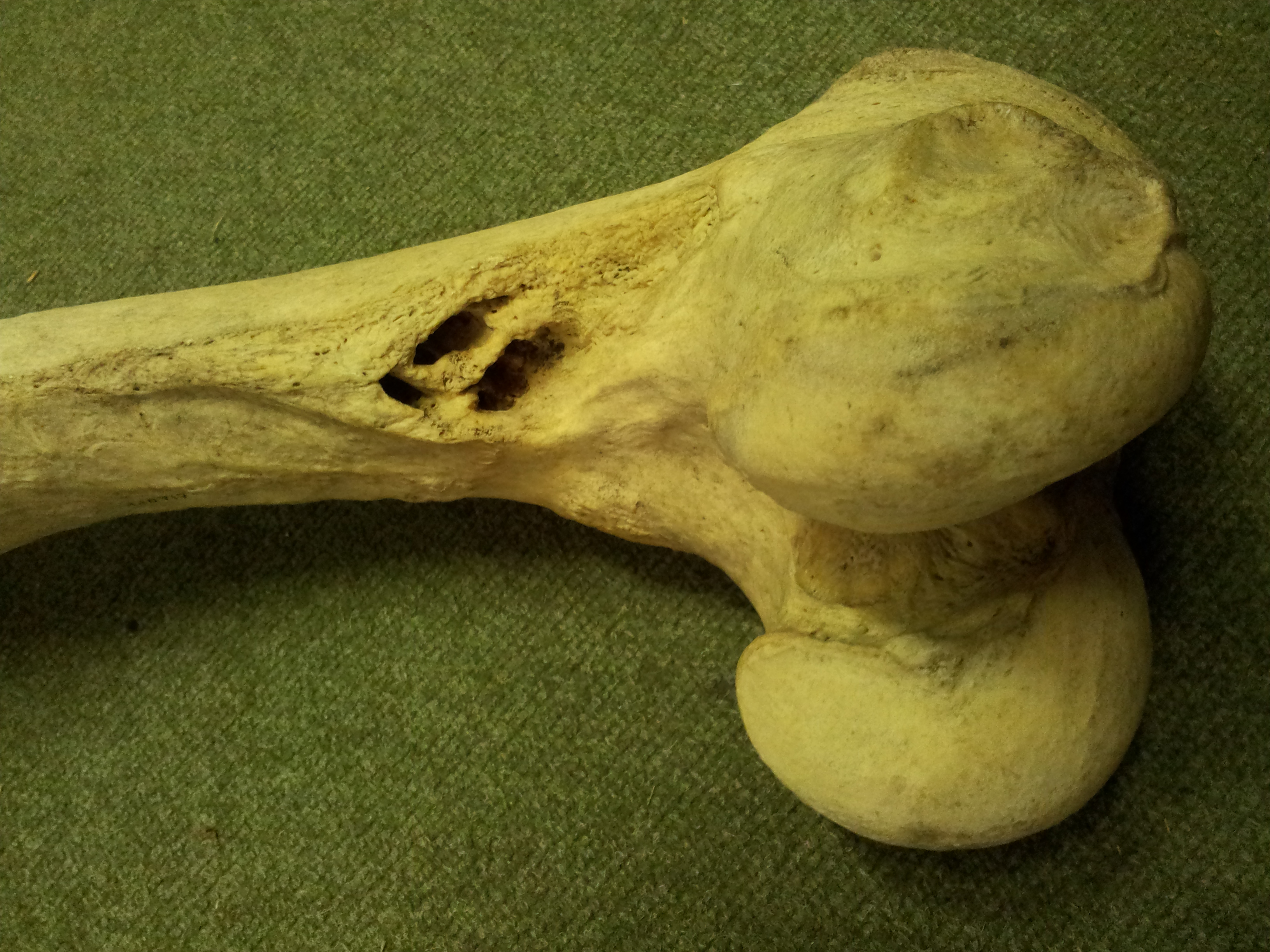
And I thought “Ouch! That’s nasty, dude.” (the holes in the bone just above the knee joint– these should just be a roughened area where the adductor muscles and other leg muscles attach)
So I was interested to see the CT scan images to find out how these possibly osteomyelitic lesions continued into the bone. They’re really pervasive, continuing into the marrow cavity quite far up the femur, as this shows (good CT-viewing practice to match up what you are seeing in the photo above with this movie):
I would be surprised if this was not the reason this animal died (presumably being euthanased at a UK zoo). There would have been extensive infection and pain resulting from this bony disease. How did it originate? Who knows. Maybe the animal strained a muscle and bacteria got inside, or maybe there was a fall or other injury. Hard to tell.
Oh, and also note the lack of a true marrow cavity in hippos, which is true for all the long bones. The “cavity” is filled in with cancellous bone. Same with rhinos, elephants, and many other species… science doesn’t entirely know why but this feature surely does help support the body on land, and grants at least some extra negative buoyancy in water; at a cost of some extra weight to lug around, of course.
And so ends this Anatomy Vignette.


Awesome stack. . .I got distracted by the rockin’ density phantoms, though. Out of curiosity, what did you use for those?
These are Gammex, Inc. commercial phantoms; 1.67 and 1 g/cm^3 (bone and water). We have a nice set covering from pretty low to high density (20 or so phantoms) but usually just 2 (plus air =0) are enough.
Cool! Thanks for the info.
Great stuff, John, thanks for posting. Nice to see a CT stack posted, they are so good to look at.
And I didn’t know about the pachyderms having no marrow cavity..wow. Kinda makes sense from a biomechanical perspective, though?
Keep them coming!
[…] « A WIJF Anatomy Vignette: A Hippopotamus Femur With Funky Pathology […]
Haziroglu described a Supratrochlear Foramen in the Humerus of Cattle but after examination of thousands of humerus I never can see a similar finding.
http://onlinelibrary.wiley.com/doi/10.1111/j.1439-0264.1990.tb00893.x/abstract
Hmm could be breed variation, or pathology, or ???? With just 3 cattle I don’t know how they can be sure.
and we observed similar foramina in bovine skull !!