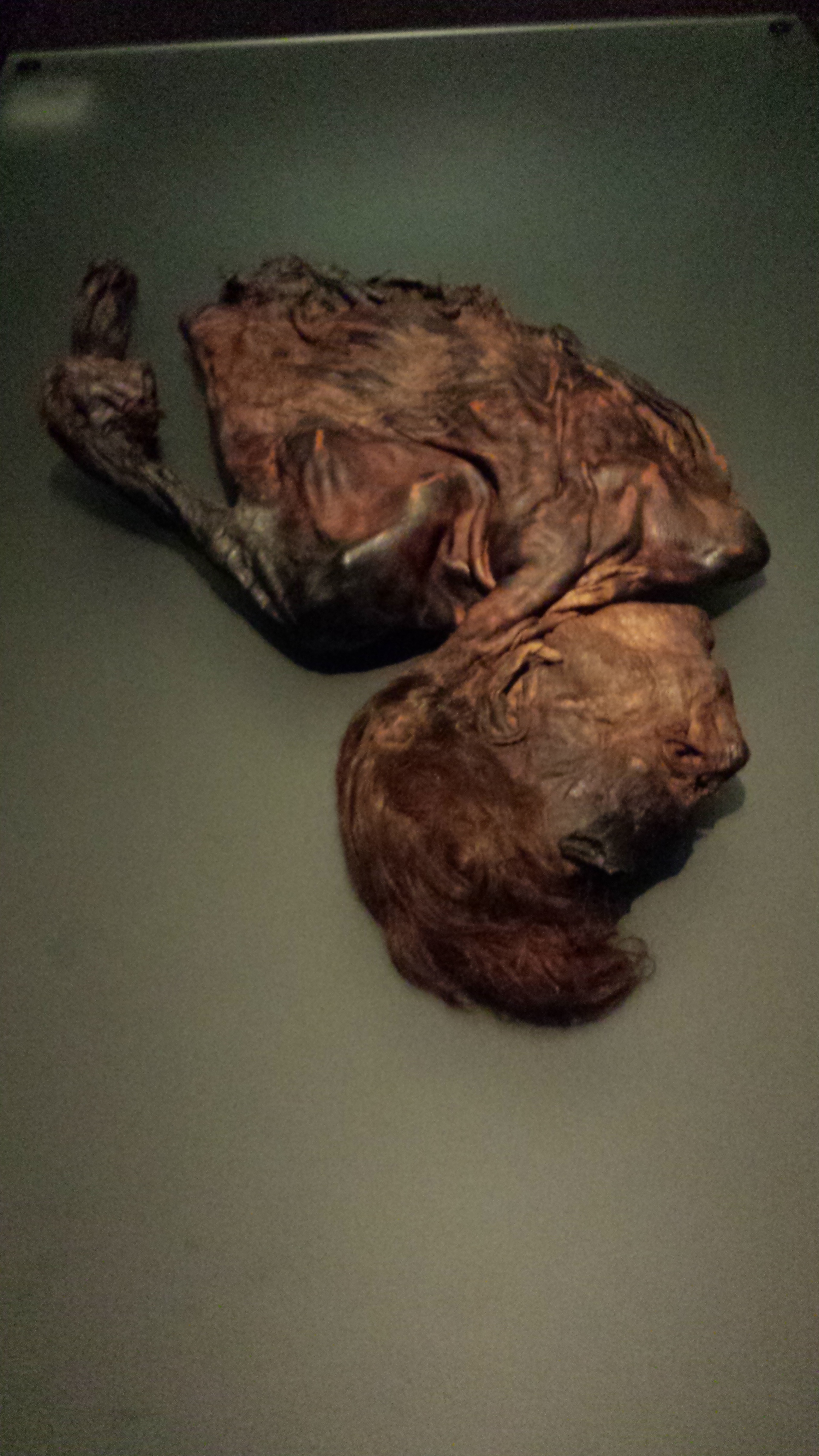
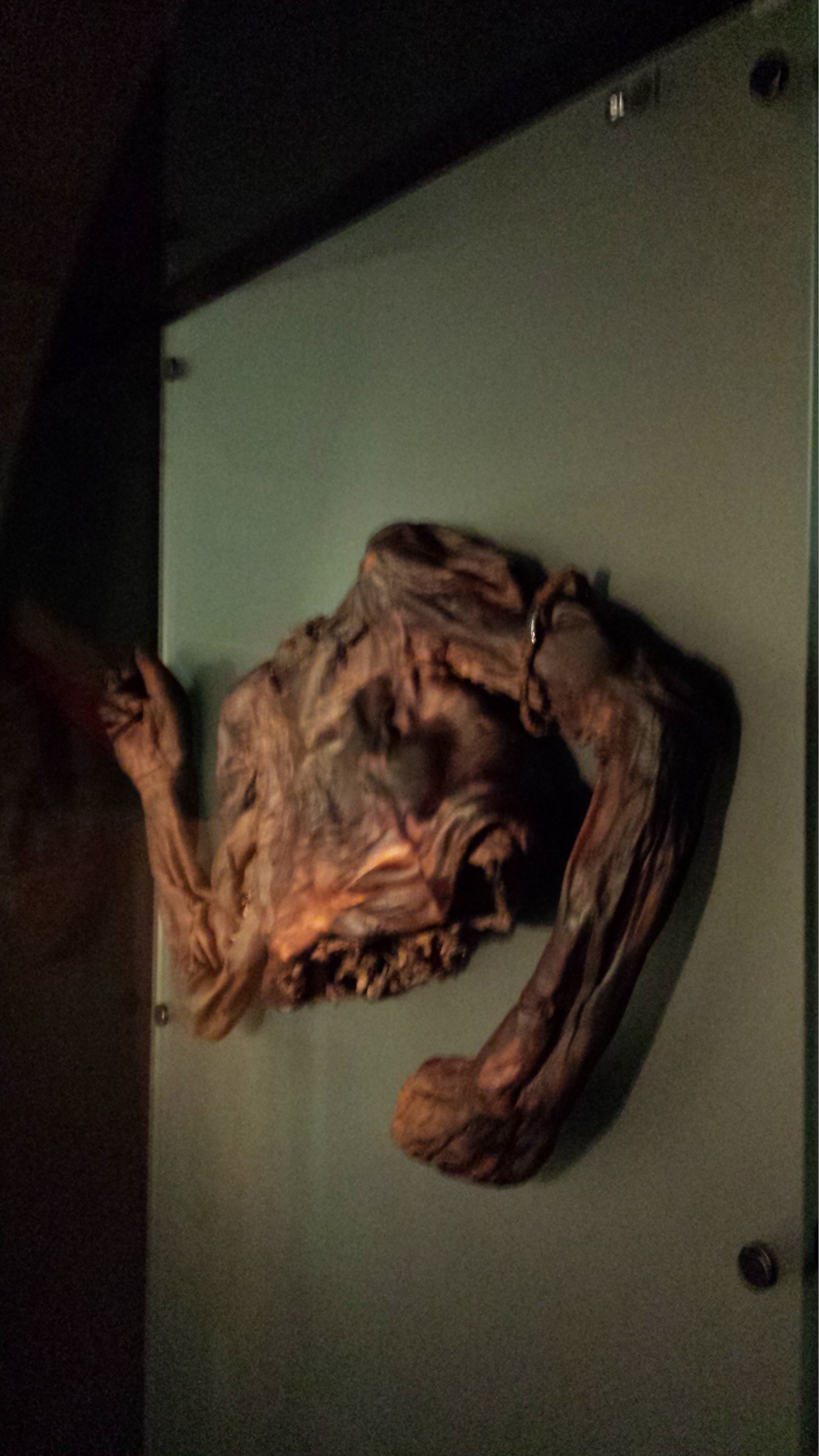
Jason Anderson, vertebrate paleontologist and anatomist at the University of Calgary (Canada), shared these two intriguing photos with me, and agreed for me to share them with you. Yay, thanks Jason! Good timing for a badly needed Mystery Dissection post:
What are they (species ID) *AND* from what region of the body?
(they are the same region, same specimen, same animal)
RULE: Your answer must be in the form of a winter holiday song (at least four lines)!
If you’ve been following some of my recent tweets, I’ve been tweeting about the “joys” of increased academic paperwork around holiday-time; e.g. this one:
I love doing sciencey road trips with my team when I can. Last week, we got a treat: four of us got a behind-the-scenes tour of the fairly new Crocodiles of the World facility near Oxford; just over 90 minutes west of our lab, nestled in the pictureseque Cotswolds region. We were not disappointed, so you get to share in the joy! In photo-blog format. Pics can be clicked to emcrocken.
In the midst of an unpreposessing industrial estate lies: AWESOME!
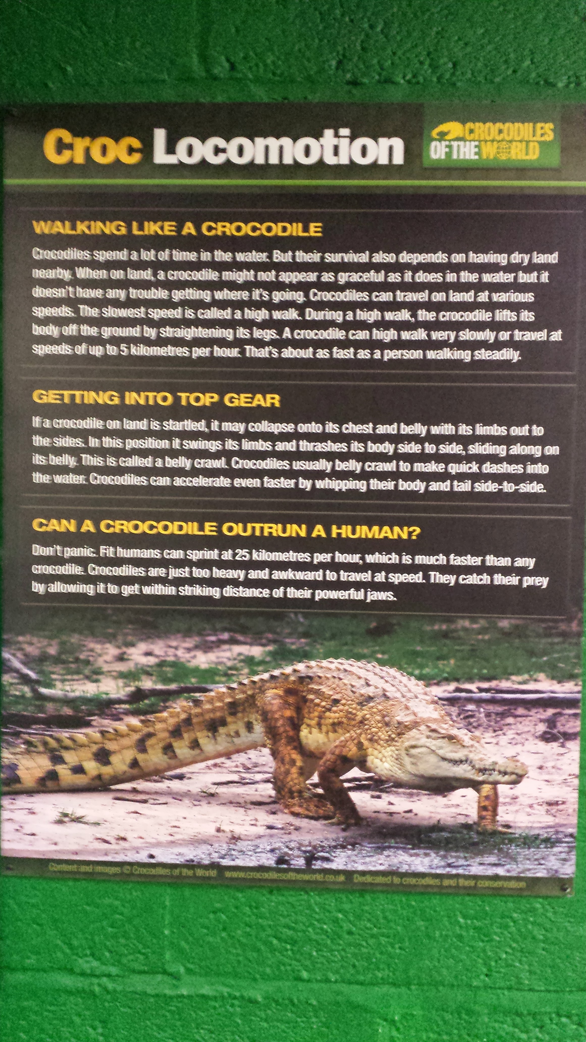
If you want to bone up on your croc species, go here and here. I won’t go into details. This is an eye candy post!
Reasonably accurate description that caught my eye. My scientific interest in crocodiles starts here, and with their anatomy/relationship with dinosaurs, but I’ve loved crocs since I was an infant (one of my first words, as I may have written here before, was “dock-a-dile”, for my favourite stuffed animal at the time [R.I.P.]).
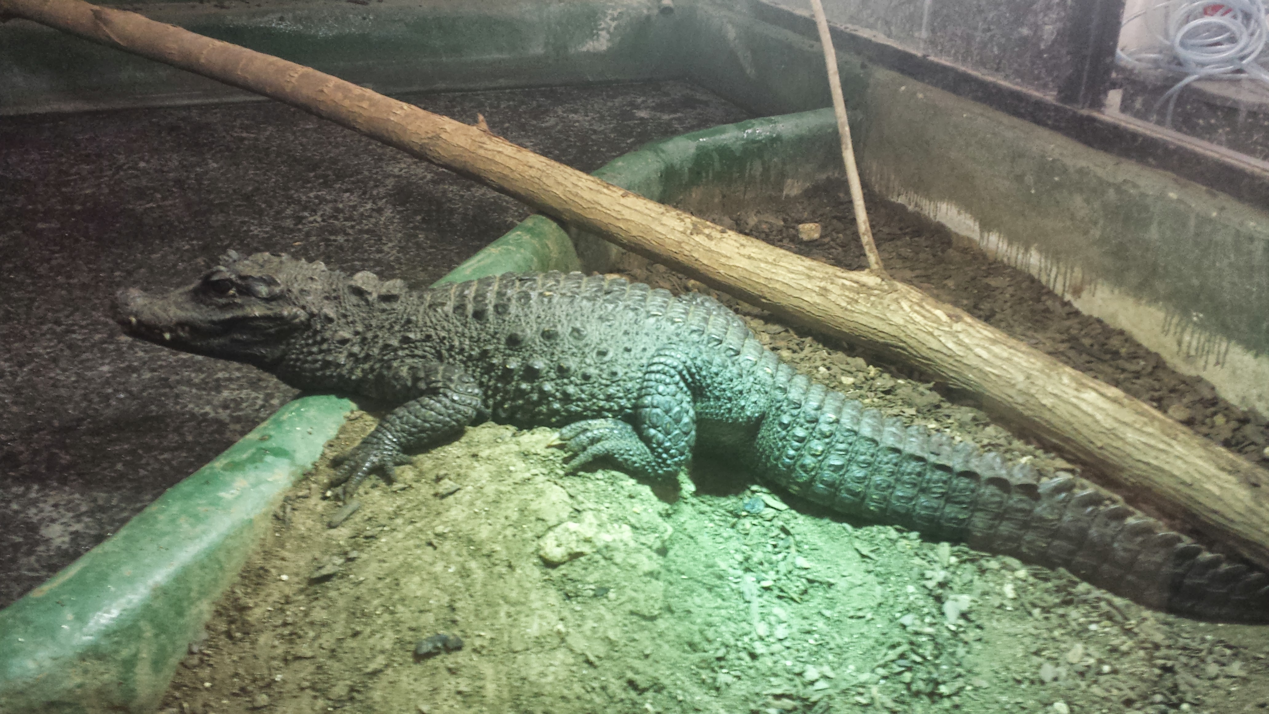
Siamese crocodiles. The large male is “Hugo.” They were apart when we entered, then got snuggly later, as I’ve often seen this species do. Heavily endangered (<300 in the wild?), so any breeding is a good thing!
The above photo brings me to one of my general points. Crocodiles of the World seems genuinely to be a centre that is breeding crocodiles for conservation purposes (and for education, entertainment and other zoo-like stuff). Essentially every crocodile enclosure had a mated pair, and several were breeding. Such as…
Yes, that is a Dwarf African crocodile, Osteolaemus, and indeed it is a female on her nest-mound. Which means…
Eggs of said Osteolaemus.
And babies of said Osteolaemus! As if the adults aren’t cute enough with their short snouts and doglike size/appearance! These guys have striking yellow colouration, too. I’d never seen it in person before.
That’s not all!
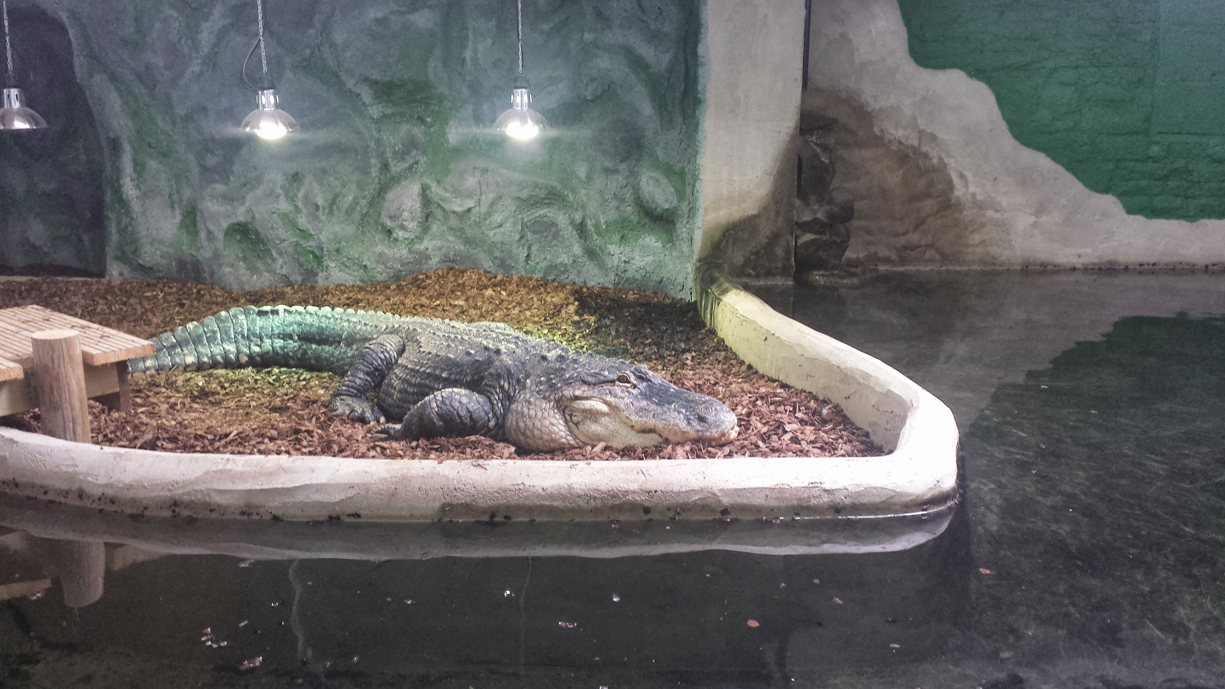
Male American Alligator “Albert” warming up. Smaller female partner “Daisy” lives in same enclosure. Plenty of babies from these guys, too! Daisy comes when called by name, and Albert is learning to do so.
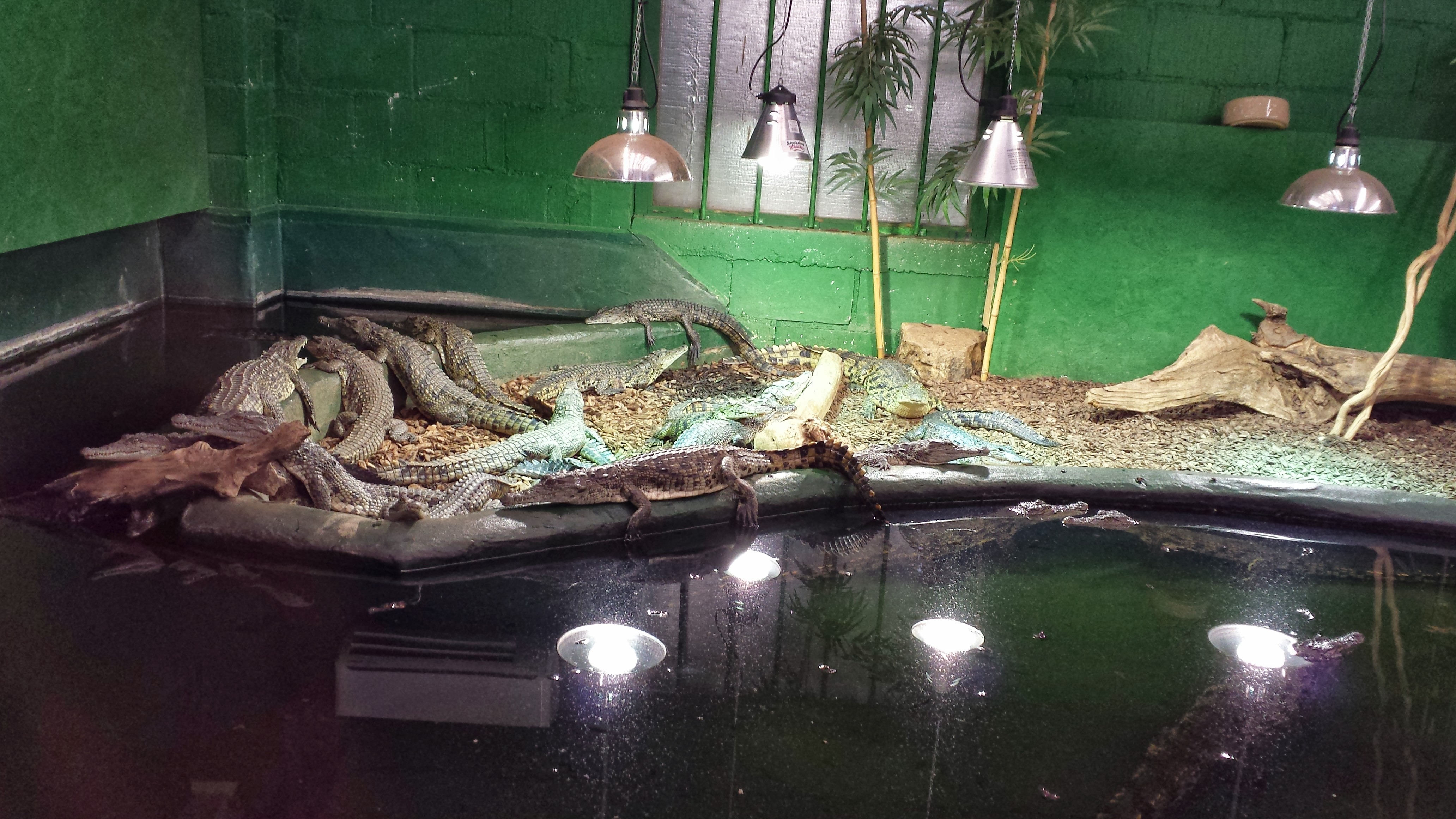
~1 meter long juvenile Nile crocodiles, bred at the facility.
But then crocodile morphological diversity (colours, textures) and behaviour is just too cool not to focus on a bit, so here are some highlights from our visit!
Endearing shot of a crocodylian I seldom get to see anywhere: Paleosuchus trigonatus, the Schneider’s Dwarf Caiman. Spiny armoured hide and quite terrestrial; poorly known in many ways. Some more info is here (note its tortured taxonomy)
Black caiman, Melanosuchusniger, showing some interest in us.
Cuban crocodiles (Crocodylus rhombifer; pound for pound the most badass croc in my experience; badassitude that this photo captures nicely) cooling off by exposing the well-vascularized soft tissues of the mouth region.
But it’s not just crocs there, either, and some of the highlights were non-croc surprises and memorable encounters:
A surprisingly friendly and tame Water monitor (14 yrs old; does kids parties). Note person for scale. Was about 2 meters long, 20 kg or so.
Business end of nice Water monitor, with tongue engaged.
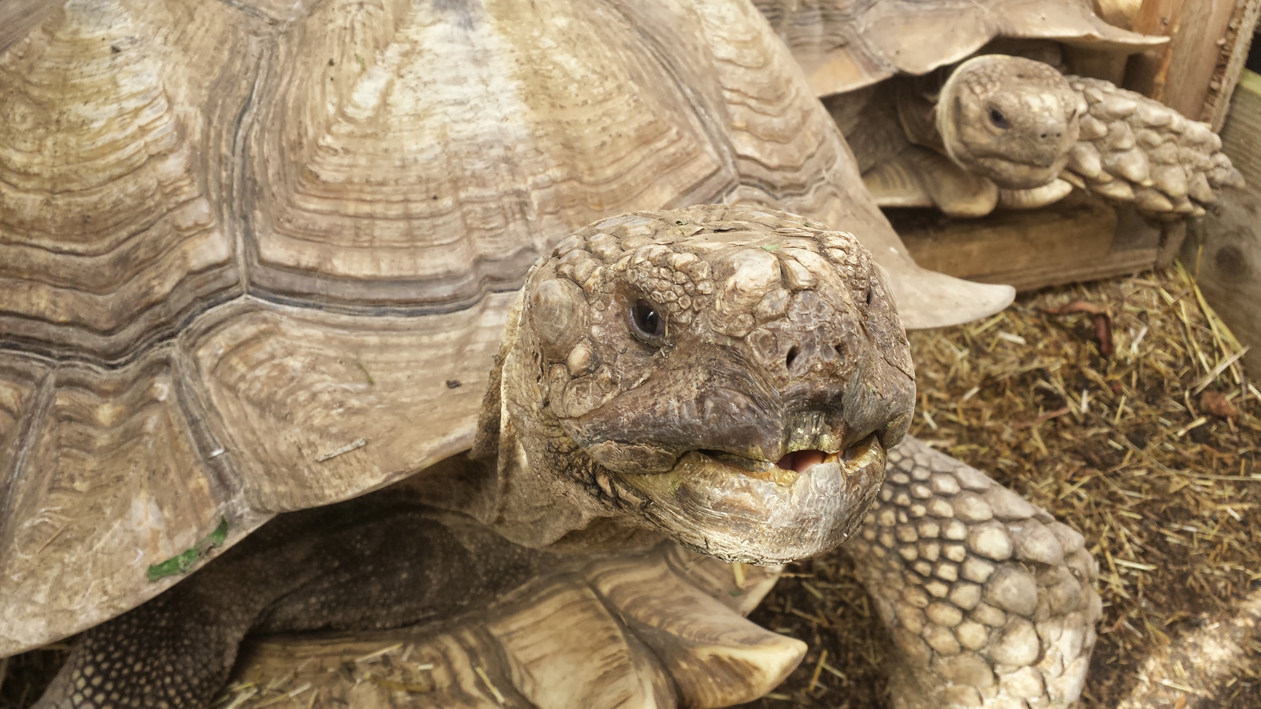
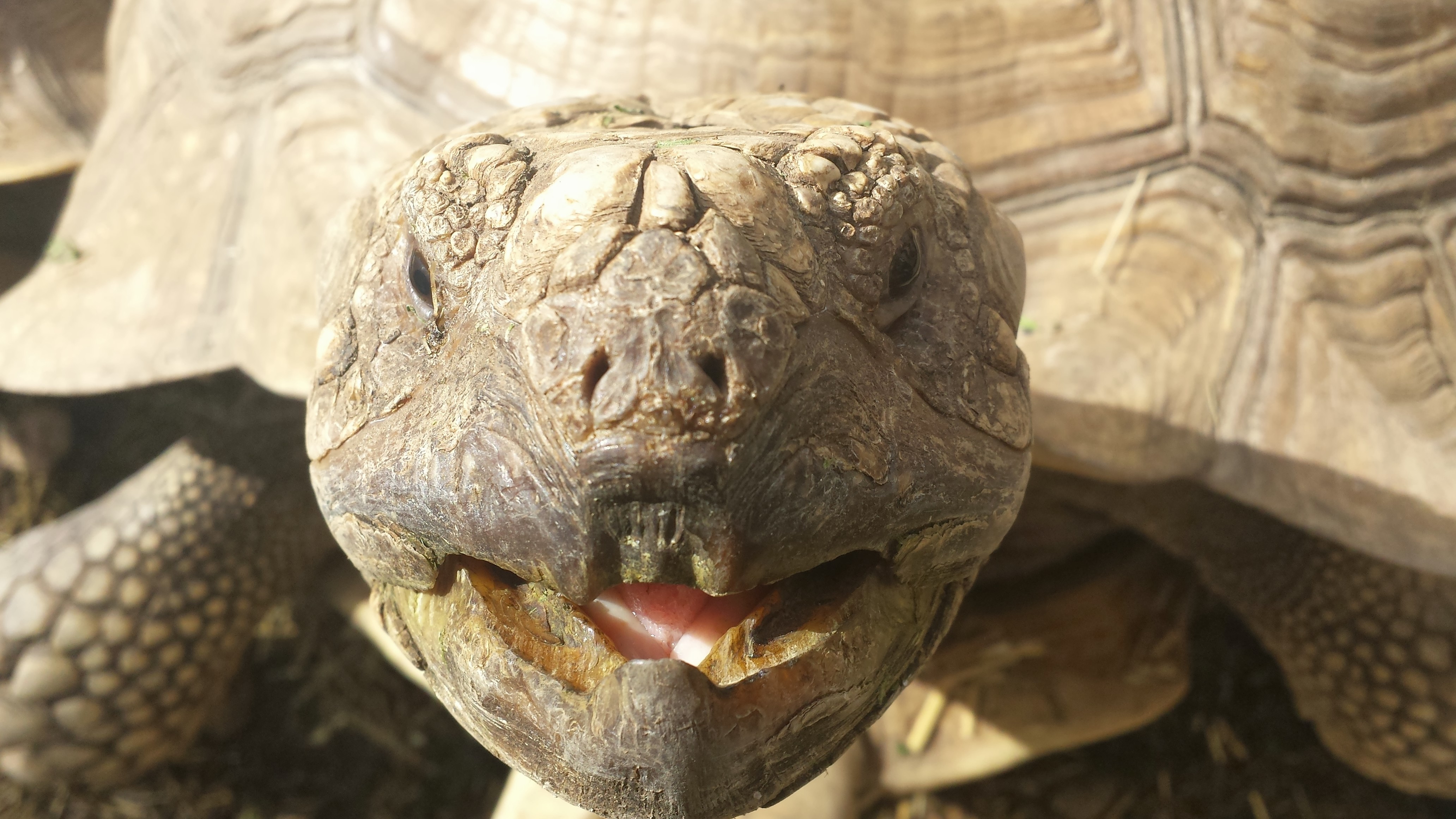
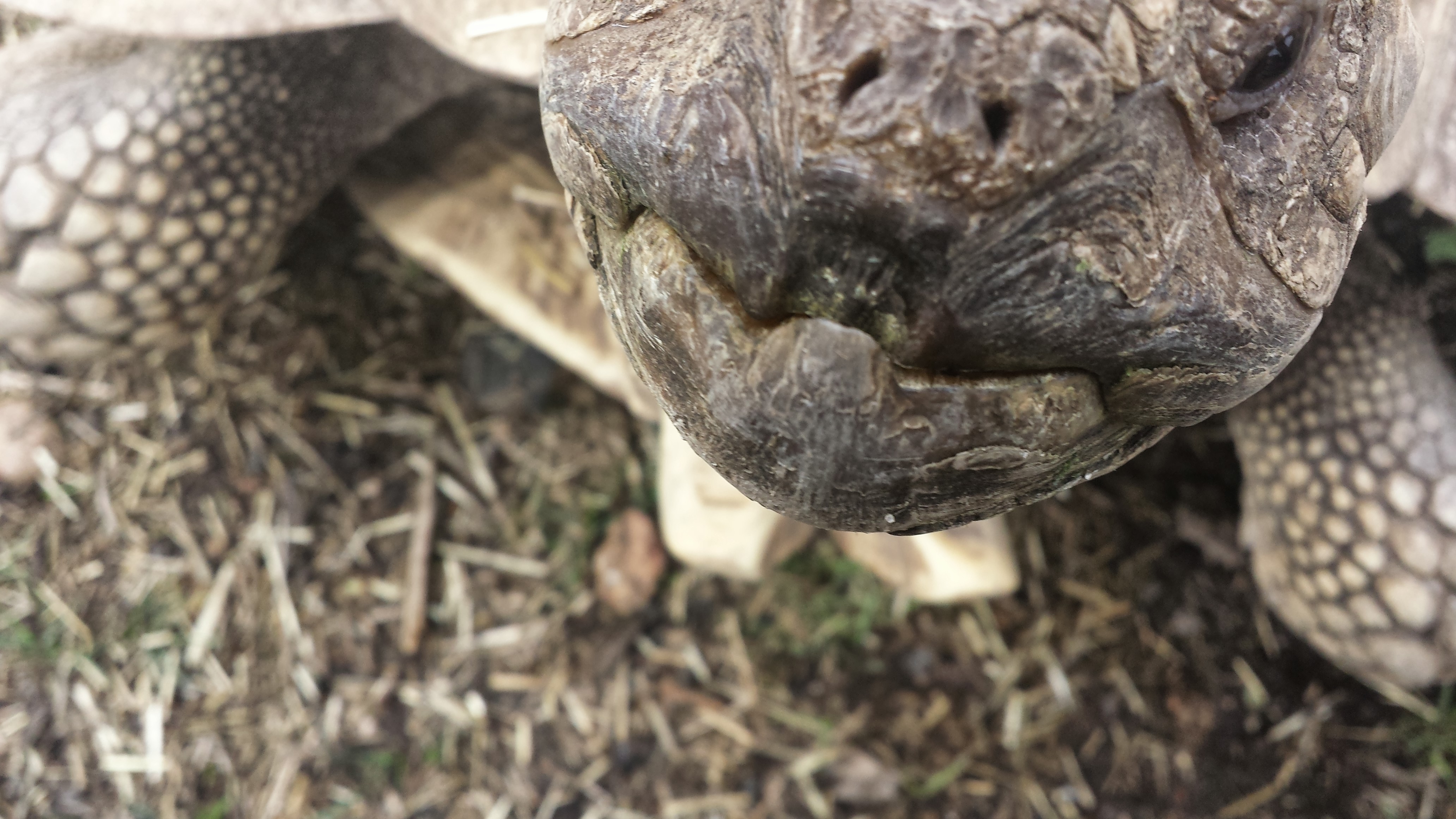
And we got a nice farewell from an African spur-thigh tortoise (Geochelone sulcata) with an oral fixation (action sequence thereof):
Chowmp!
If someone visits this facility and leaves without being converted to a croc-lover, they must be from a different planet than me. It is a celebration of crocodiles; the owner, Shaun Foggett, is the real deal. He sold his home and quit his job as a carpenter to care for crocodiles, and it seems to be a great success– about to get greater, as they have plans to move to a new, bigger, proper site! They are seeking funding, so if you can contribute go here.
Right then… UK residents and visitors: you need to go here! Badly! Get off the blog and go now. If it is a Saturday/Sunday (the cramped industrial estate location only allows the public then).
Otherwise just stew and imagine how much fun you could be having checking out crocodiles. I cruelly posted this on a Tuesday to ensure thorough marination of any croc-geeks.
My team had a new technician arrive, Kyle Chadwick from Uni. Virginia, and NSF Postdoctoral Research Fellow, Dr. Ashley Heers (see here for an example of new stuff she’s starting here at the RVC!), started working with me at the RVC, and then these guys showed up…
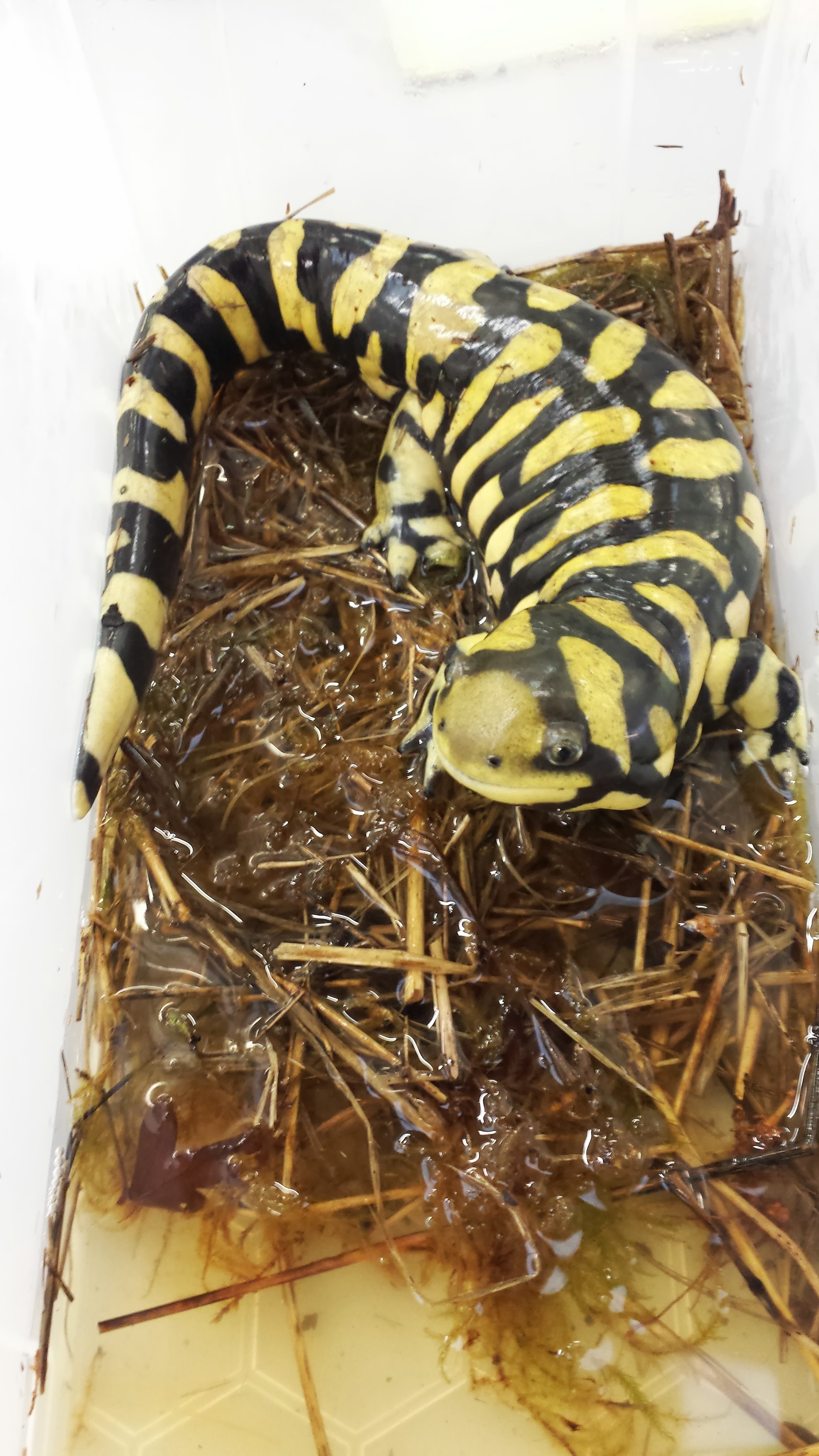
First a tiger salamander (Ambystoma) paid a visit, for filming an episode of the Windfall Films/PBS documentary “Your Inner Fish” (a la the famous book):
So cute! Tiger salamander, soon to be a TV celebrity.
And that gorgeous salamander was a star performer in strutting his stuff for the camera to demonstrate the locomotion of modern tetrapods, including some lovely slo-mo footage from our lab cameras:
(if that’s too slow for you, try the normal-speed footage. I’ll admit, salamanders don’t really need slo-mo video for normal walking, but I like it)
So cool!
But then we got a special package… with three frozen fire salamanders (Salamandra salamandra) from colleagues in Germany!
Three new occupants of the freezers, for planning our studies of salamander locomotion
This marks the start of an exciting new period in my team’s work in the lab. I’ve always liked salamanders and newts, and we’ve scanned and modelled plenty (e.g. this old post), but now we’re going to work with live fire salamanders (a first for me)! We are using the dead ones to plan the new studies with the live ones– these new studies will involve lots of high speed videos and force platform analysis (as shown above), in conjunction with XROMM (biplanar fluoroscopy/3D skeletal motion analysis) and other techniques including computer simulations. We got initial approval this week to work with these salamanders, and found a reputable source this week too, so it was definitely Salamander Week in my group!
This research all will feed into our upcoming studies of extinct tetrapods: we’re using salamanders to figure out how salamanders move and what limits their speed and gait, and then we’re using the same sorts of computer tools to try to estimate how extinct tetrapods may have moved and how locomotion evolved, in much more specific detail than our prior work had done, which was mainly about using 3D reconstructions of anatomy to show what those animals could not do. More about the project here.
Watch this space for more scampering salamanders!
UPDATE: And here’s one! Not quite scampering, but…
Setting up our two fluoroscopes for a test run of our gait studies– but with one of the deceased salamanders. Gotta get good images before any live animal work begins!
An example of the kind of footage we’re aiming for (single 2D fluoroscope view from Nadja Schilling’s team’s research; see XROMM website for more details on the methodology)
UPDATE 2:
I did a CT scan with a normal medical grade CT scanner at the highest resolution we can manage (0.625 mm slices). Check out the results below, which amuse me:
Looks like a toy; too crude resolution. But we can see major structures, and we can very nicely see the “microchip” (which looks HUGE) that was placed in this animal’s back when in captivity, and then another structure is visible near the pelvis which might be another chip or else remains of some food, pathology, or a really odd pelvis– I am not totally sure!
So this is why we tend to use microCT, which can go down to as low as ~5 micron resolution, to get 3D anatomy of animals this small. It’s no surprise to me, but it is fun to see how far we could push our normal CT machine. The results aren’t horrid but wouldn’t have much scientific value for us. They did confirm for us that this specimen is heavily ossified, so the faint images of bone that we are getting in our x-ray fluoroscopes (above) are due to something going wrong with our camera system, not the animal’s immature skeleton. Stay tuned for more updates as the science happens!
UPDATE 3:
20 wonderful adult Fire Salamanders have joined our team and are relaxing over the coming week before we start taking them for walks. Here is one exploring its new home:
UPDATE 4:
August 11-15, 2014 we are in Jena, Germany using their fancy biplanar radiography system (“x-ray video”) to study our salamanders, at last! Follow the tweets starting here, for more information as it happened! https://twitter.com/JohnRHutchinson/status/500187568416518144
and this video of “Jabba” the corpulent salamander walking-
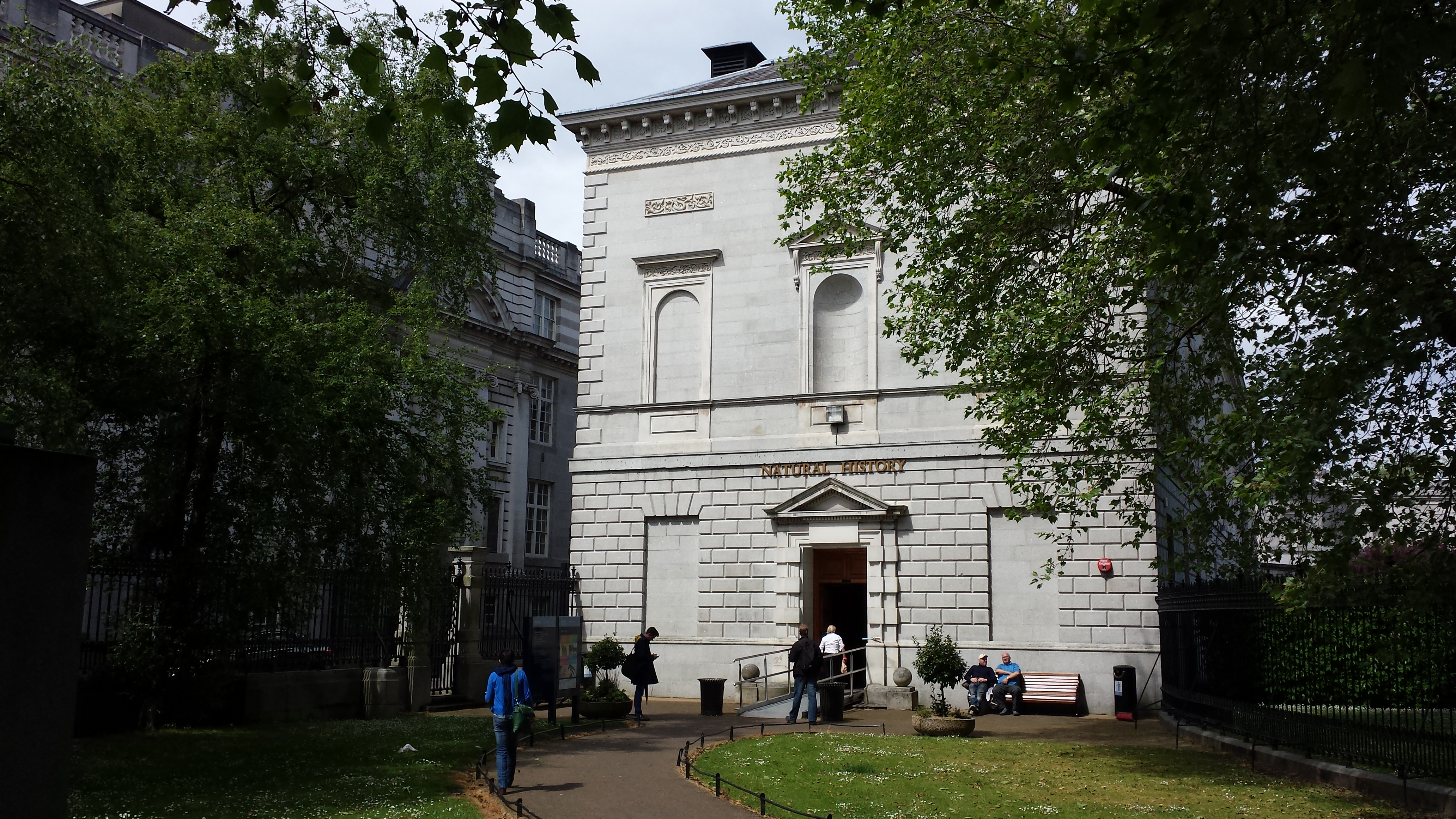
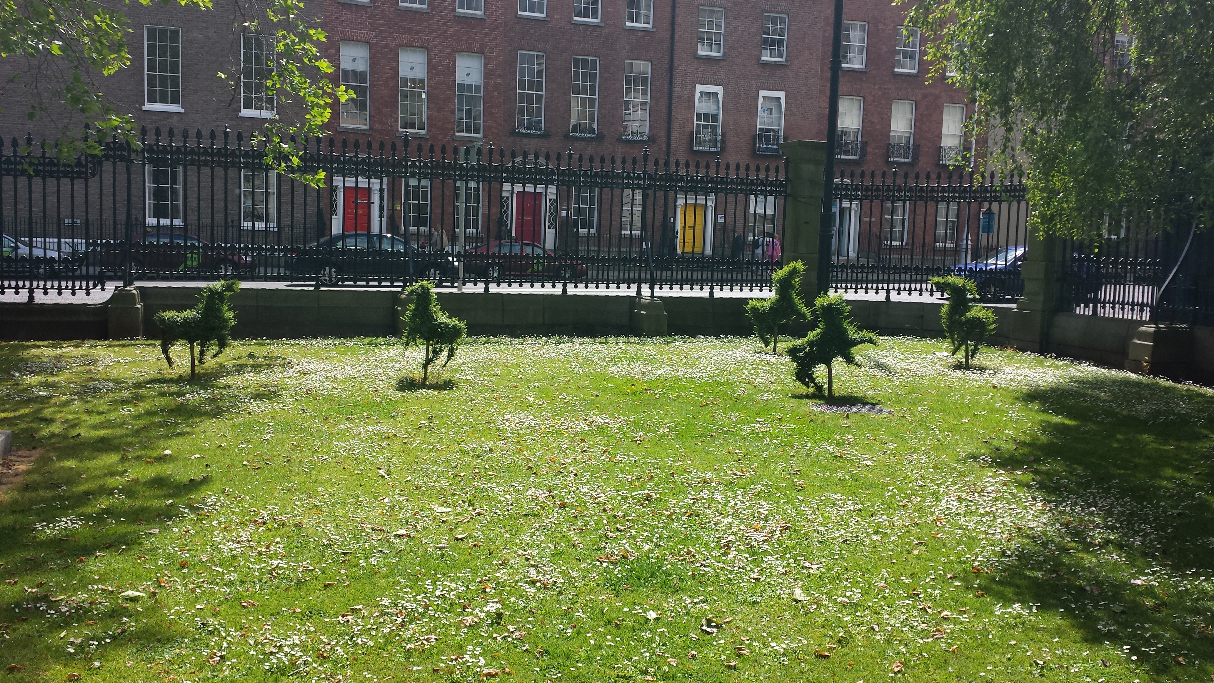
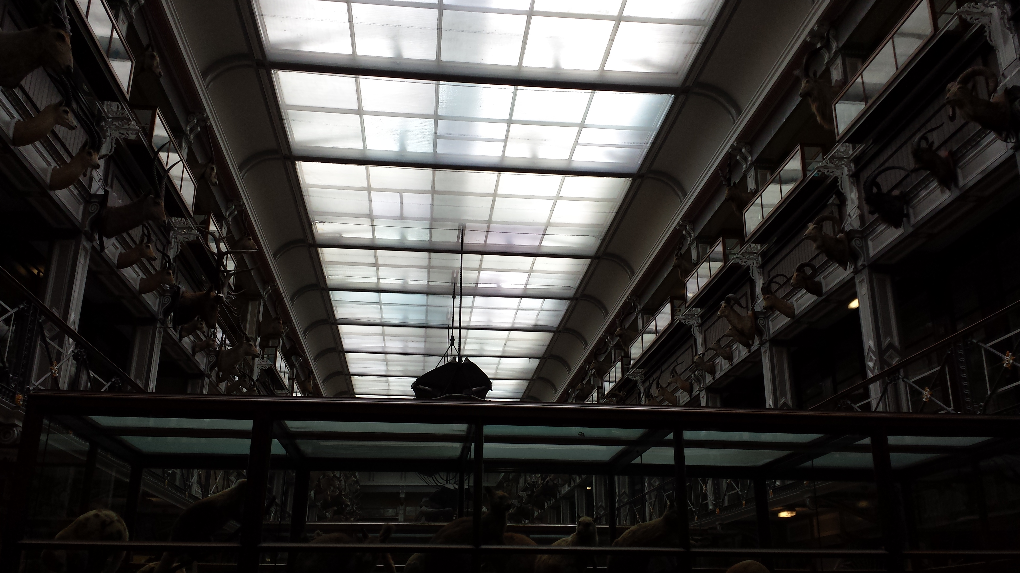
A photo blog post for ya here! I went to Dublin on a ~28 hour tour, for a PhD viva (now-Dr Xia Wang; bird feather/flight evolution thesis) earlier this month. And I made a beeline for the local natural history museum (National Museum of Ireland, Natural History building) when I had free time. So here are the results!
Stomach-Churning Rating: Tame; about a 1/10 for most, but I am going to break my rule about showing human bodies near the end. Just a warning. The bog bodies were too awesome not to share. So that might be 4/10-8/10 depending on your proclivities. They are dry and not juicy or bloody, and don’t look as human as you’d expect.
Simple Natural History museum entrance area.
Adorable frolicking topiaries outside the NHM.
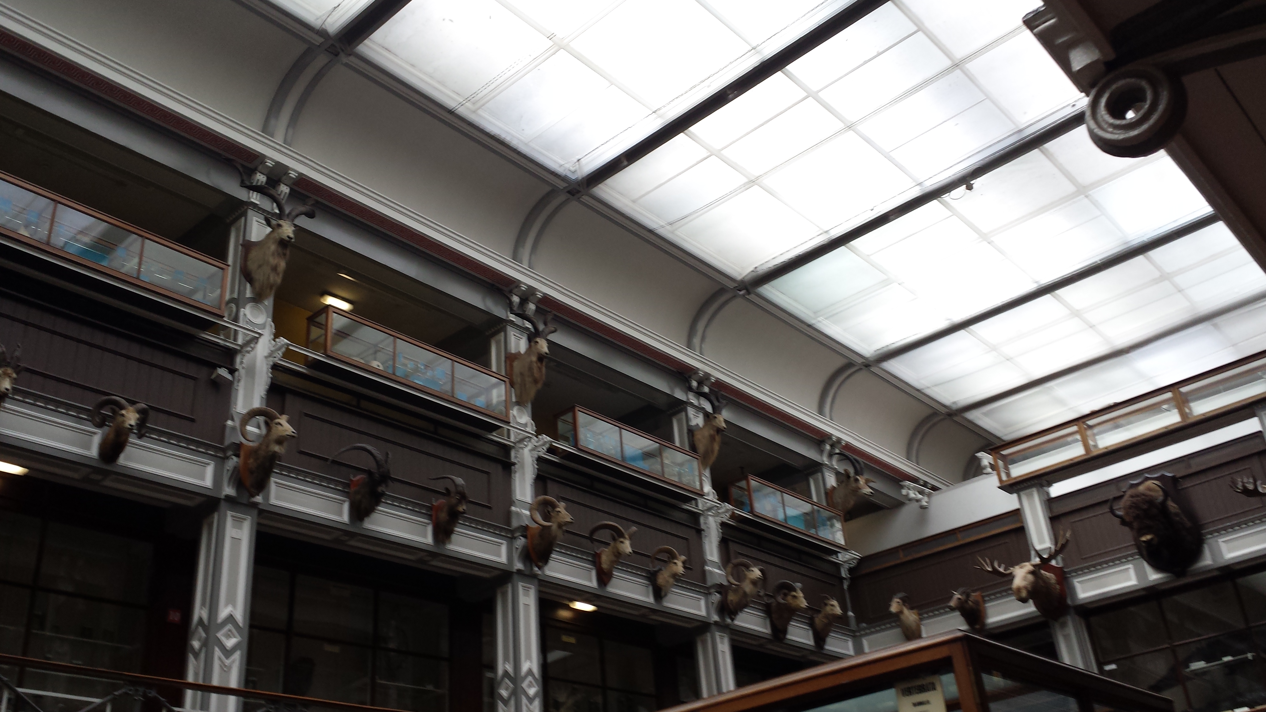
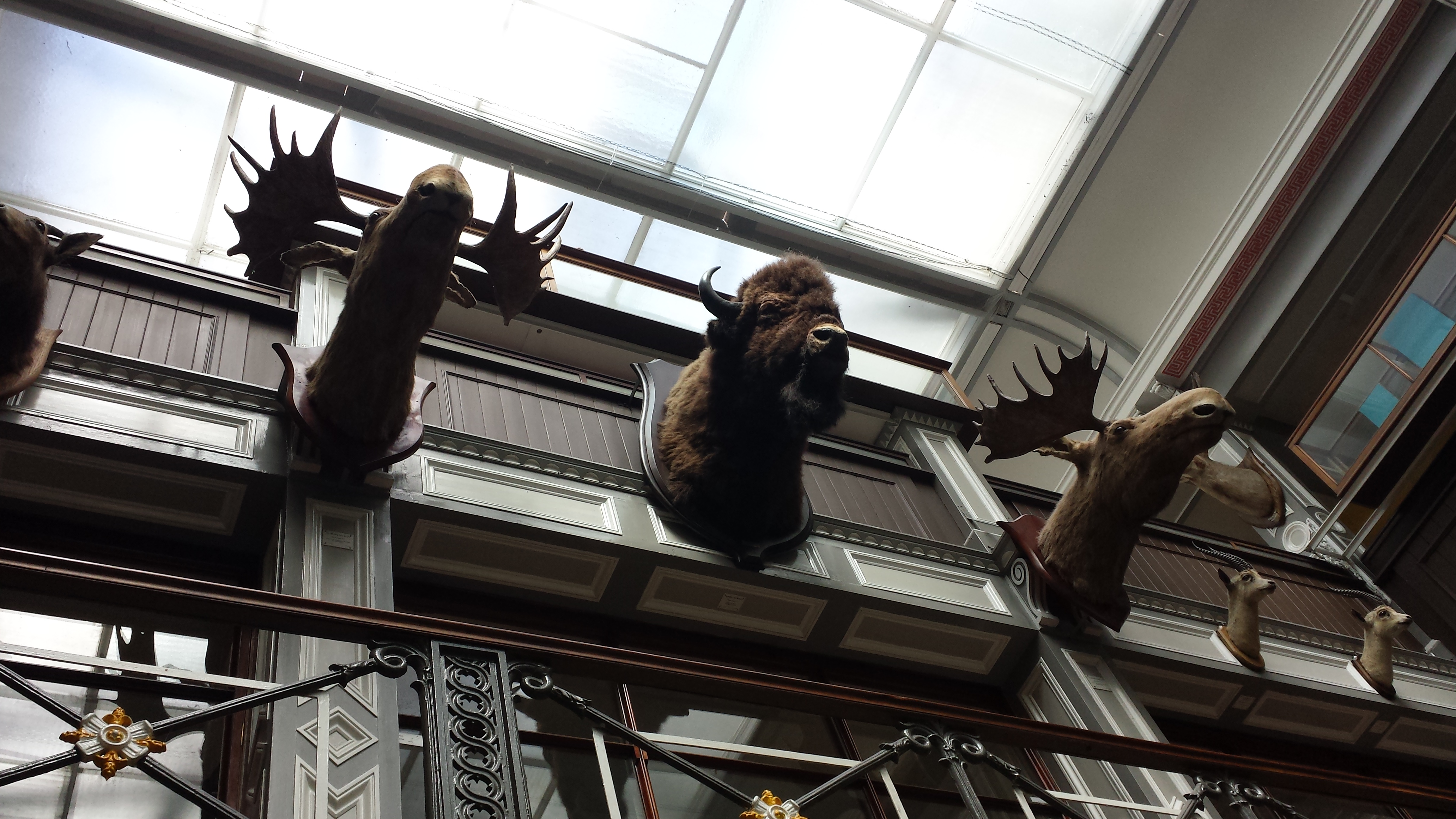
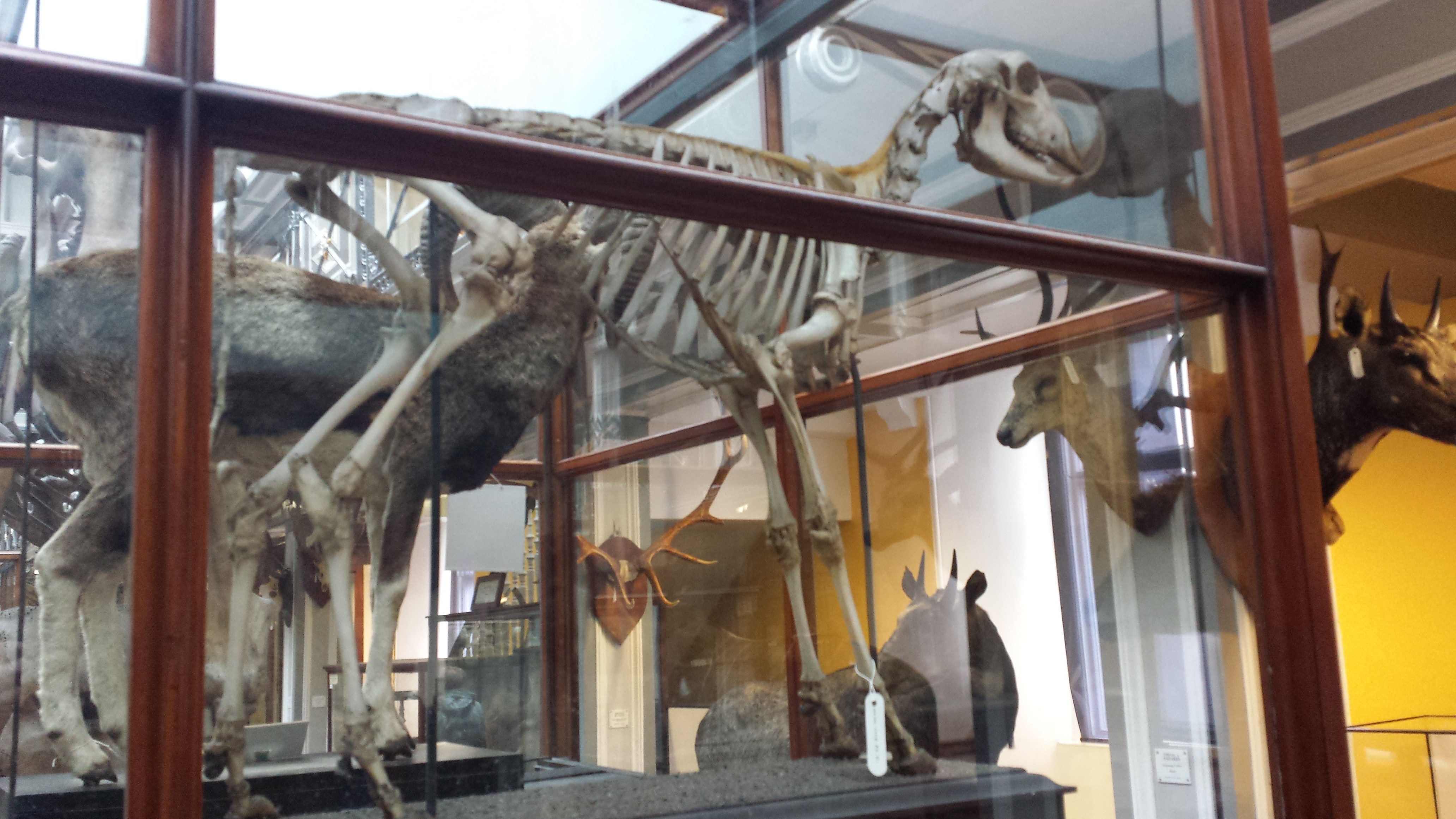
Inside, it was a classical Victorian-style, dark wood-panelled museum stuffed with stuffed specimens. It could use major refurbishment, but I do love old-fashioned exhibits. Get on with it and show us the animals; minimize interpretive signage and NO FUCKING INTERACTIVE COMPUTER PANELS! So by those criteria, I liked it. Some shots of the halls: And on to the specimens!
Giant European deer (“Irish elk”). I looked at these and thought, “why don’t we see female deer without antlers ever? then noticed one standing next to these (you can barely see it in back); too bad my photo is crappy.
Superb mounted skeleton of giraffe (stuffed skin was standing near it).
A sheep-y or a goat-y beastie; I dunno but it shows off a nice example of the nuchal ligament (supports the head/neck).
Yarr, narwhals be internet gold!
Giant blown glass models of lice!
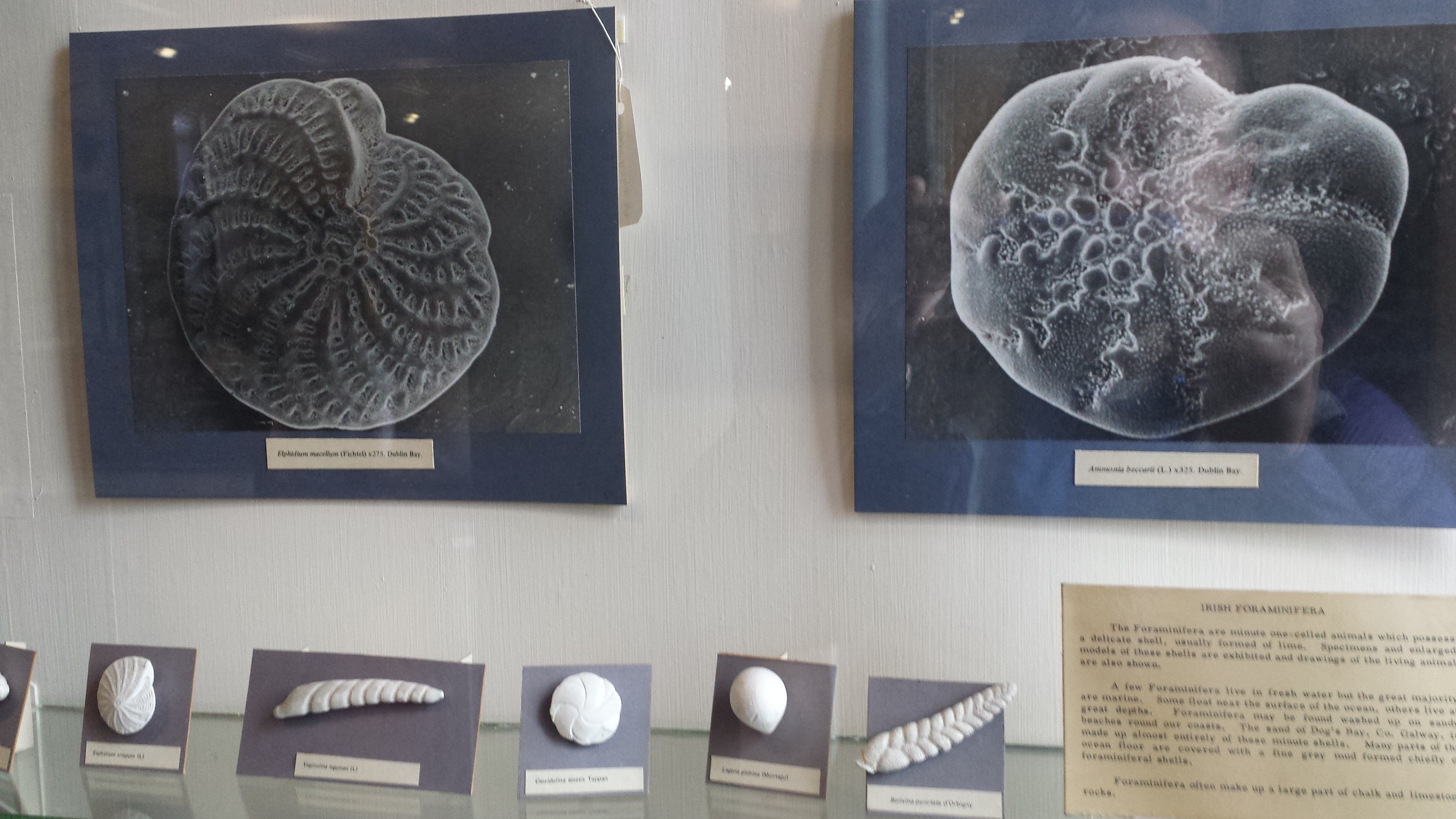
Who doesn’t like a good giant foramanifera image/model?
“That’s one bigass skate,” I murmured to myself.
“That’s one bigass halibut,” I quipped.
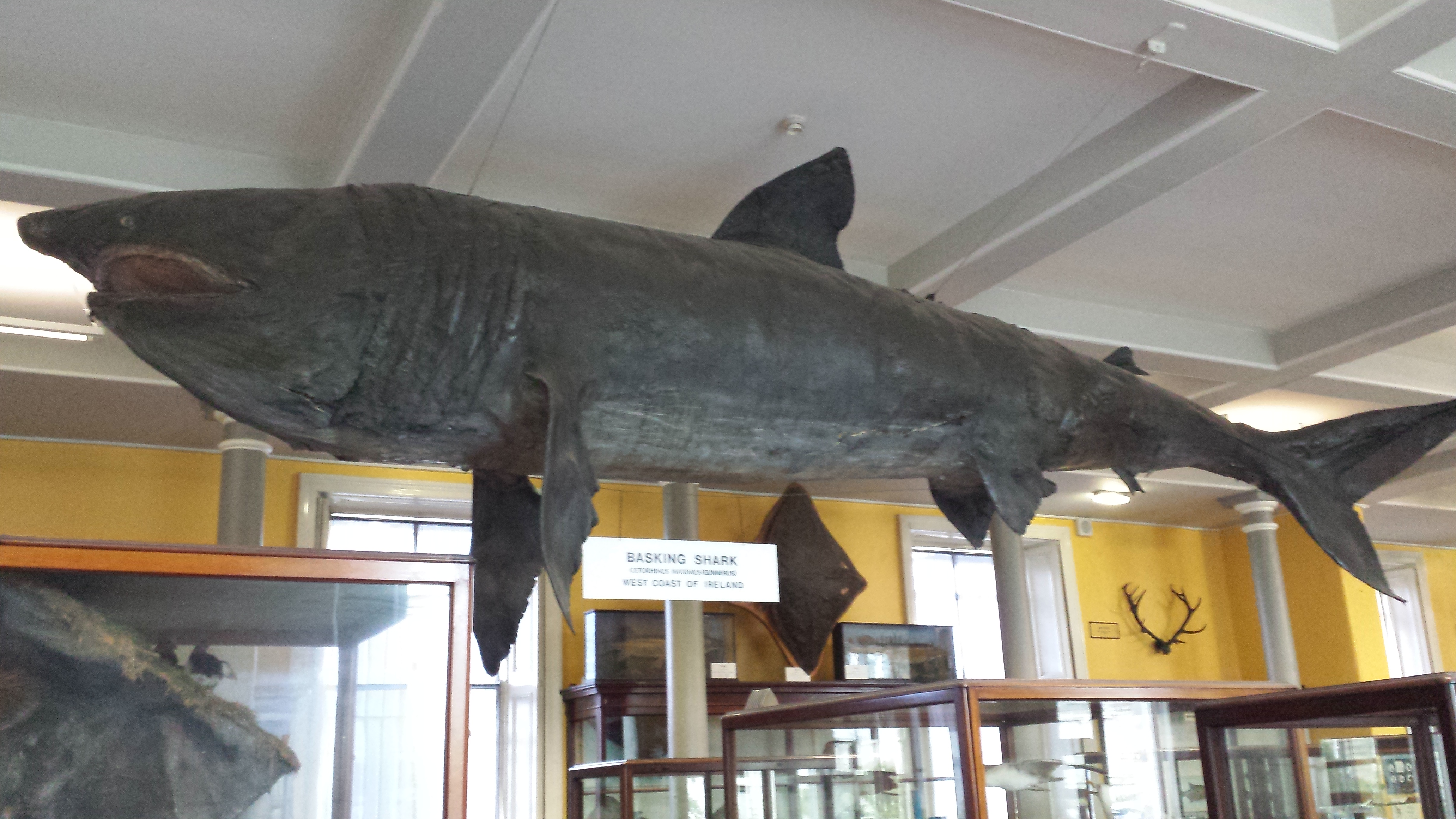
Tatty basking shark in entry hall.
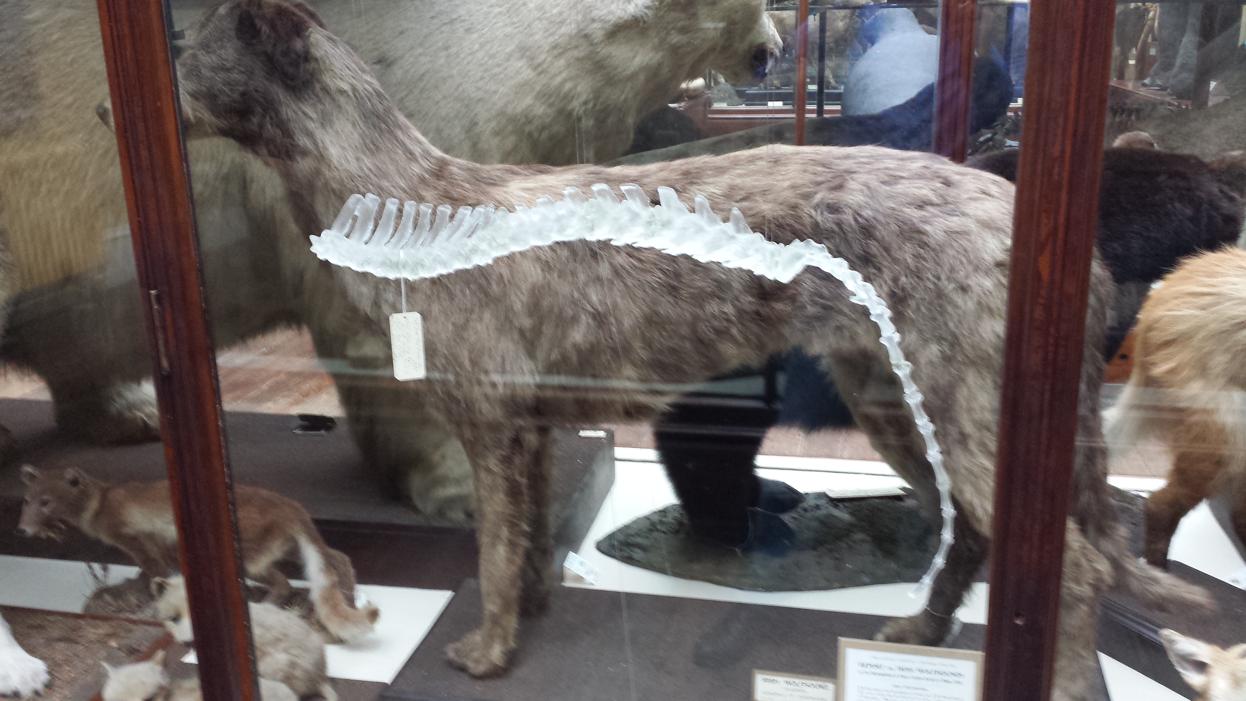
Irish wolfhound, with a glass sculpture of its spine hanging near it, for some reason.
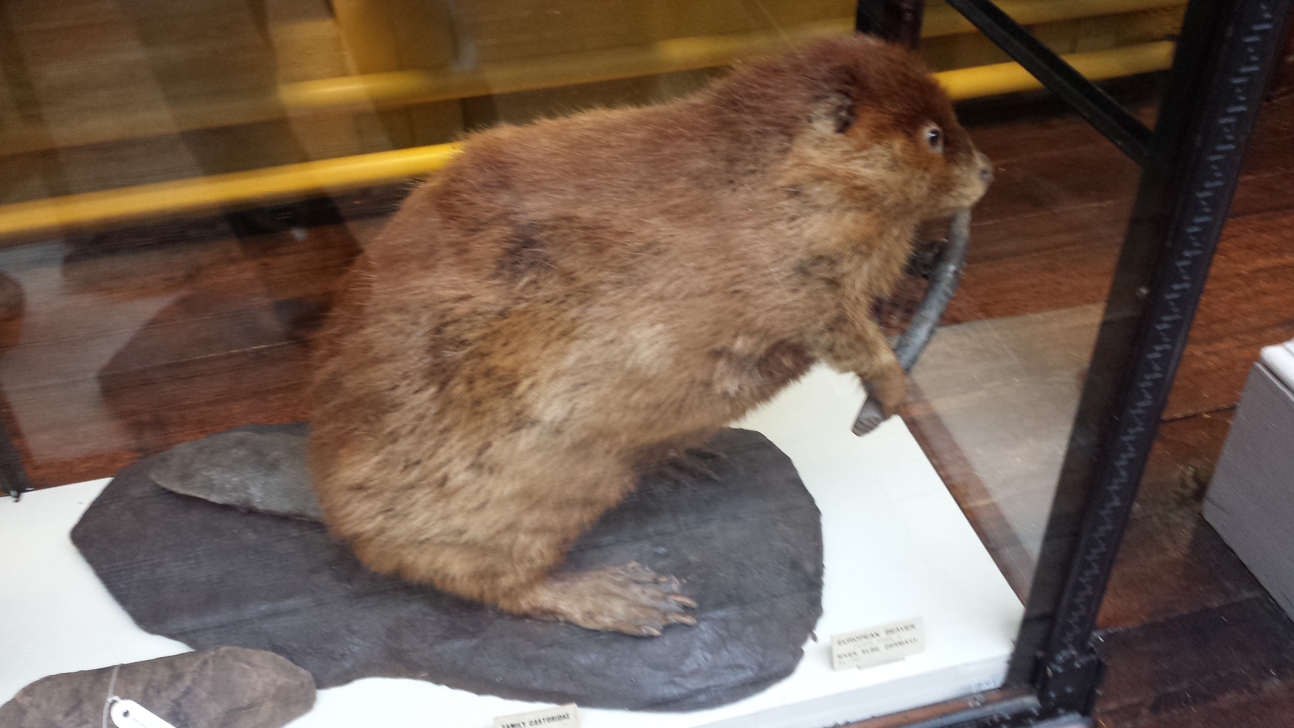
Stand back everyone! That beaver has a club!
Skull of a pilot whale/dolphin.
Nice anteater skeleton and skin.
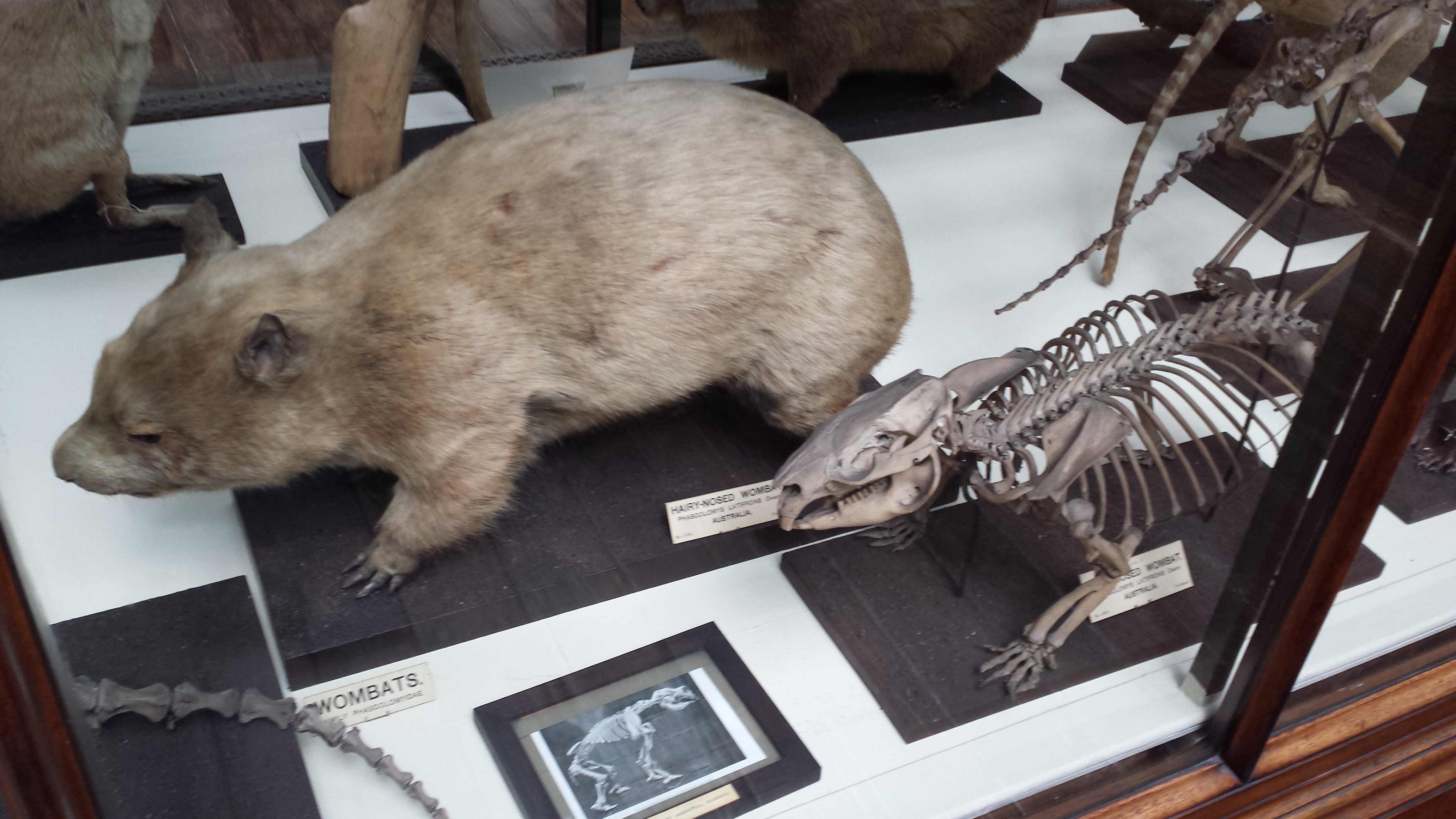
Nice wombat skeleton and skin.
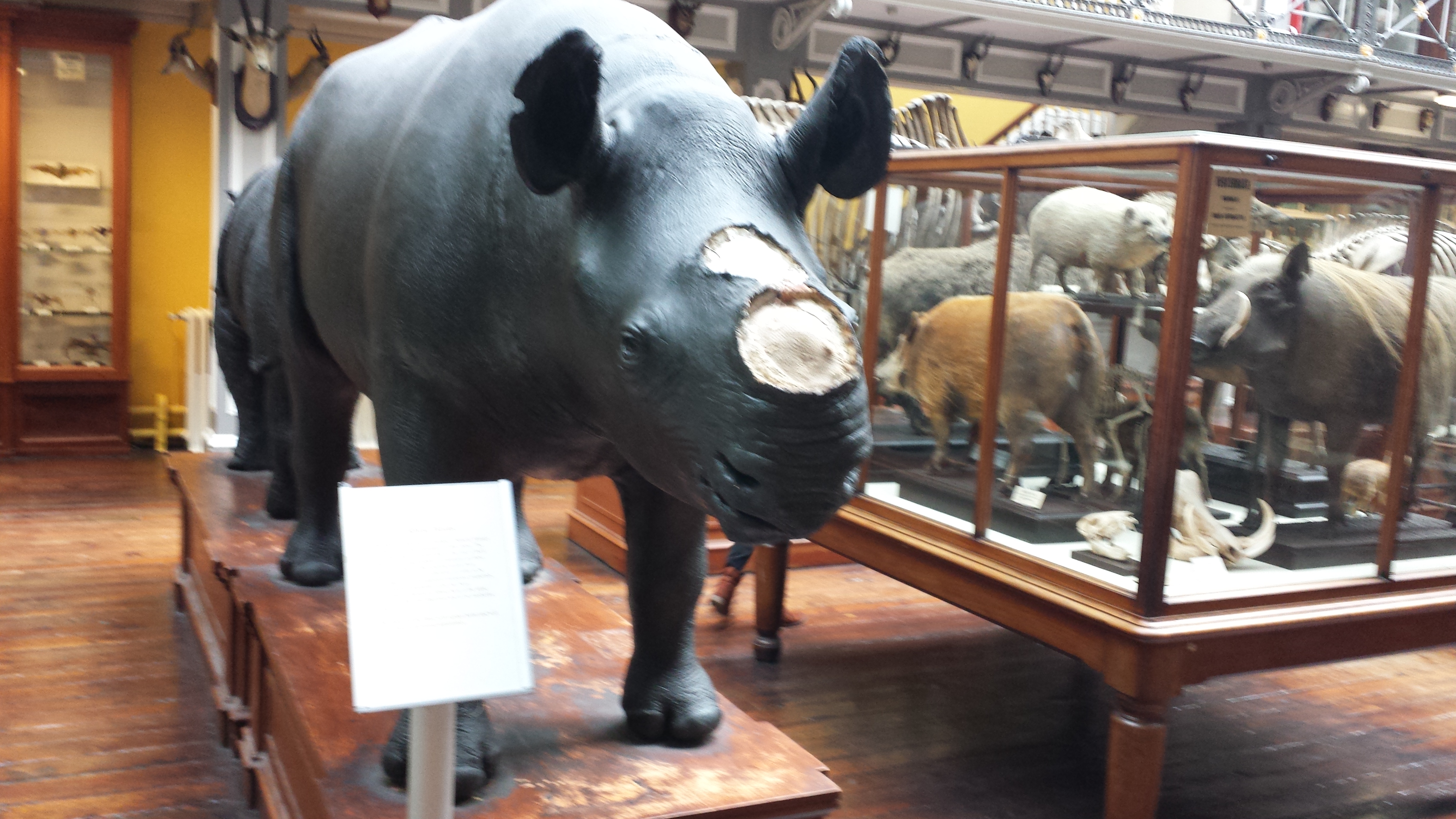
Sad display of a stuffed rhino with the horn removed, and signage explaining the problem of thefts of those horns from museum specimens of rhinos worldwide.
But then the stuffed animals started to get to me. Or maybe it was the hangover. Anyway, I saw this…
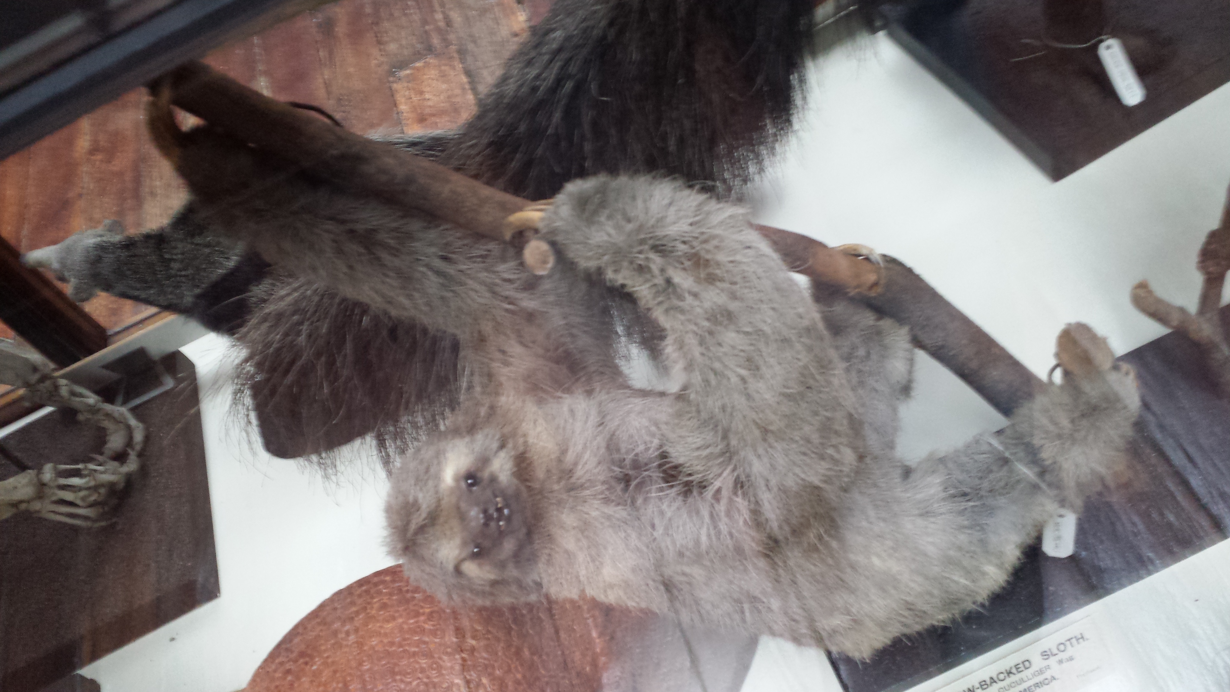
A proboscis monkey mother who seemed to be saying “Hey kid, you want this yummy fruit? Tough shit. I’m going to hold it over here, out of reach.” with a disturbing grimace. That got me thinking about facial expressions in stuffed museum specimens of mammals more, and I couldn’t help but anthropomorphize as I toured the rest of the collection, journeying deeper into surreality as I progressed. What follows could thus be employed as a study of the Tim-Burton-eseque grimaces of stuffed sloths. Click to emslothen.
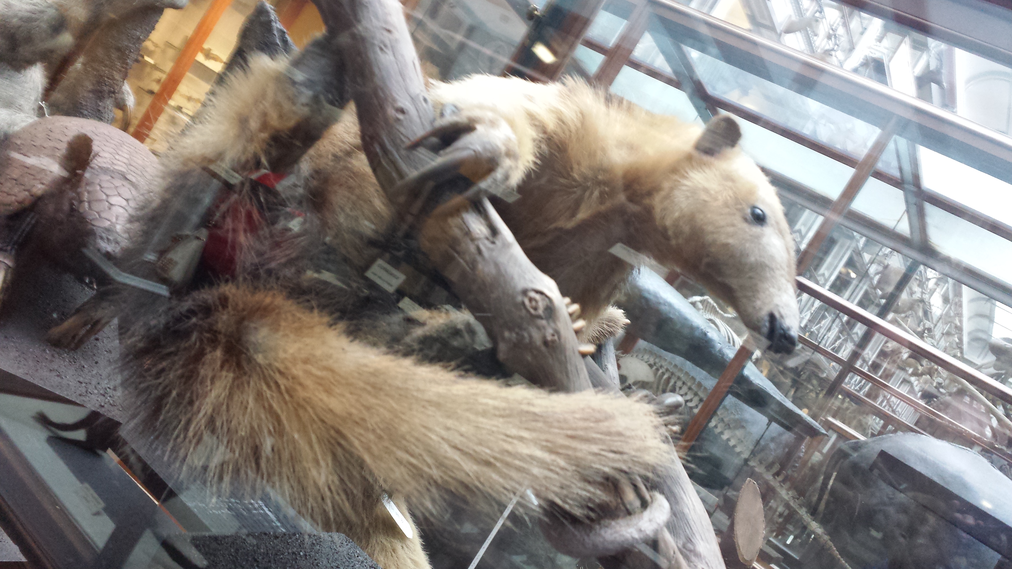
Tree anteater has a go at the awkward expression game.
This completed my tour of the museum; there were 2 more floors of specimens but they were closed for, sigh, say it with me… health and safety reasons. Balconies from which toddlers or pensioners or drunken undergrads could accidentally catapult themselves to their messy demise upon the throngs of zoological specimens below. But the National Museum’s Archaeology collection was just around the block, so off I went, following whispered tales of bog bodies. There will be a nice, calm, pretty photo, then the bodies, so if peaty ~300 BCE cadavers are not your cup of boggy tea, you can depart this tour now and lose no respect.
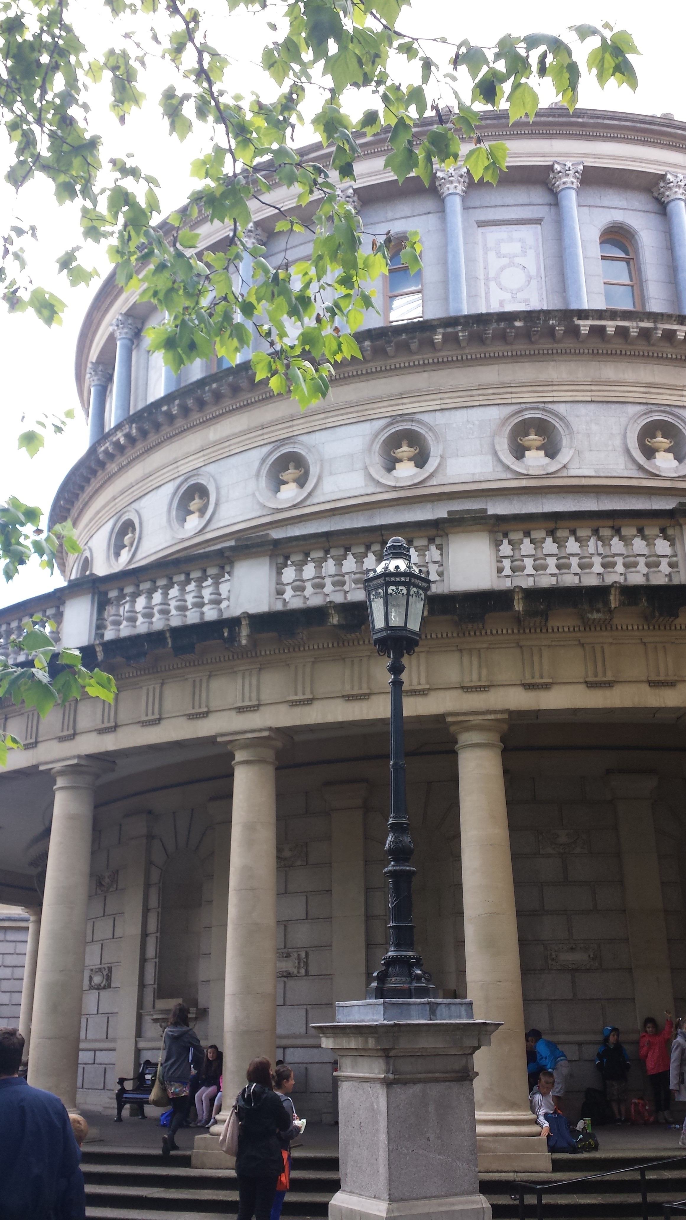
Impressive entrance to the National Museum’s Archaeology building.
The bog bodies exhibit is called “Kingship and Sacrifice“. It is packed with cylindrical chambers that conceal, and present in a tomb-like enclosed setting, the partial bodies of people that were killed and then tossed in peat bogs as honoraria for the ascension of a new king. The peaty chemistry has preserved them for ~2300 years, but in a dessicated, contorted state. The preservation has imparted a mottled colouration and wrinkled texture not far off from a Twix chocolate bar’s. Researchers have studied the bejesus out of these bodies (including 3D medical imaging techniques) and found remarkable details including not just wounds and likely causes of death (axes, strangling, slit throats etc) but also clothing, diet, health and more.
Here they are; click to (wait for it)… emboggen:
Did you find the Celtic armband on one of them?
Finally (actually this happened first; my post is going back in time), I visited UCD’s zoology building for the PhD viva and saw a few cool specimens there, as follows:
Giant deer in UCD zoology building foyer, with a lovely Pleistocene landscape painted on the wall behind it.
Sika deer in an awkward posture (what is it supposed to be doing?) in Univ Coll Dublin zoology building’s foyer.
The pose of this ?baboon?mandrill struck me as very peculiar and menacing- reminiscent of a vampire bat’s pose.
A whole lotta chicken skeletons in a UCD teaching lab.
After the viva we went out for some nice Chinese food and passed some Dublin landmarks like this:
Trinity College entrance, I think.Former Irish Parliament; now the Bank of Ireland.
And we wandered into a very posh Irish pub called the Bank (on College Green), which displayed this interesting specimen, as well as some other features shown below:
Replica of illuminated 9th Century gospel manuscript “The Book of Kells”, with gorgeous Celtic art.
Vaults near toilets in the Bank pub. Almost as cool as having giant freezers down there.
Nice glass ceiling of the Bank pub.
And Irish pub means one big, delicious thing to me, which I will finish with here– much as I finished that night off:
Just a quickie here! I’m finishing a little sabbatical at Brown University and had a bit of downtime, then ran across this confusing image that seems to have loveable, sometimes-superhero Sesame Street character Grover in it, and also poses a tough but solveable Mystery CT Slice post! So go for it! Can you find Grover? (no points for that) and can you tell us (1) what the image is of (animal/species, region of anatomy, identifiable bits), and (2) what the heck is wrong with this image and why?
Stomach-Churning Rating: 1/10 unless you have bad childhood memories associated with Grover.
This is the mystery image below, not the Grover image above! You cheeky monkey.
No rhyming in your answers or you lose 10000000 points! Grover is grumpy today and hates rhymes. He had a bit too much Hefeweissen and polka music last night. Pity the poor creature.
I have a rant to do, and an anatomy vignette or two, but before I do, here is a puzzleroo: It’s a reconstructed CT scan. I’ve digitally cut off the head to be tricksy. Come on man, I ain’t just whistlin’ Dixie! What is this beastie? Not hard in the leasty.
(your answer needs neither rhyme nor Shakespearean meter, but do take the time and provide the Latin binomen for reala– don’t just call it Peter or Sheila!)
To kick off the New Year just right, our tetrapod team has a new paper in Nature, following up on last year’s Ichthyostega not-so-good-at-walking study (also see here). Yet this paper has a more anatomically descriptive — and also an “evo-devo” — twist to it. For brevity, I’ll let our press release tell the story, since I think it does a good job of it (like I always preach scientists should do, we worked with our PR company to write this together, so we’re happy with how the press release came out). In a nutshell, our study used some very fancy synchotron radiation techniques to image the 3D anatomy of the backbone in early land vertebrates. Our findings surprised even us, and ended up turning around palaeontology/comparative anatomy’s view of how the backbone evolved, giving us a new glimpse into our inner tetrapod.
Stick around for the videos at the end, which are the first four supplementary movies from the paper and are rather pretty (there are two more, for imaging/segmenting afficionados, but they are not as pretty or interesting for most of this blog’s readership). The final figure (Figure 1 from our paper) gives some extra visual context.
The paper is:
Pierce, S.E., Ahlberg, P.E., Hutchinson, J.R., Molnar, J.L., Sanchez, S., Tafforeau, P., Clack, J.A. 2013. Vertebral architecture in the earliest stem tetrapods. Nature, published online [here].
I should note that I’m just 3rd author, so I deserve only modest credit. But I helped. Even though no freezers were involved, or harmed, in the process.
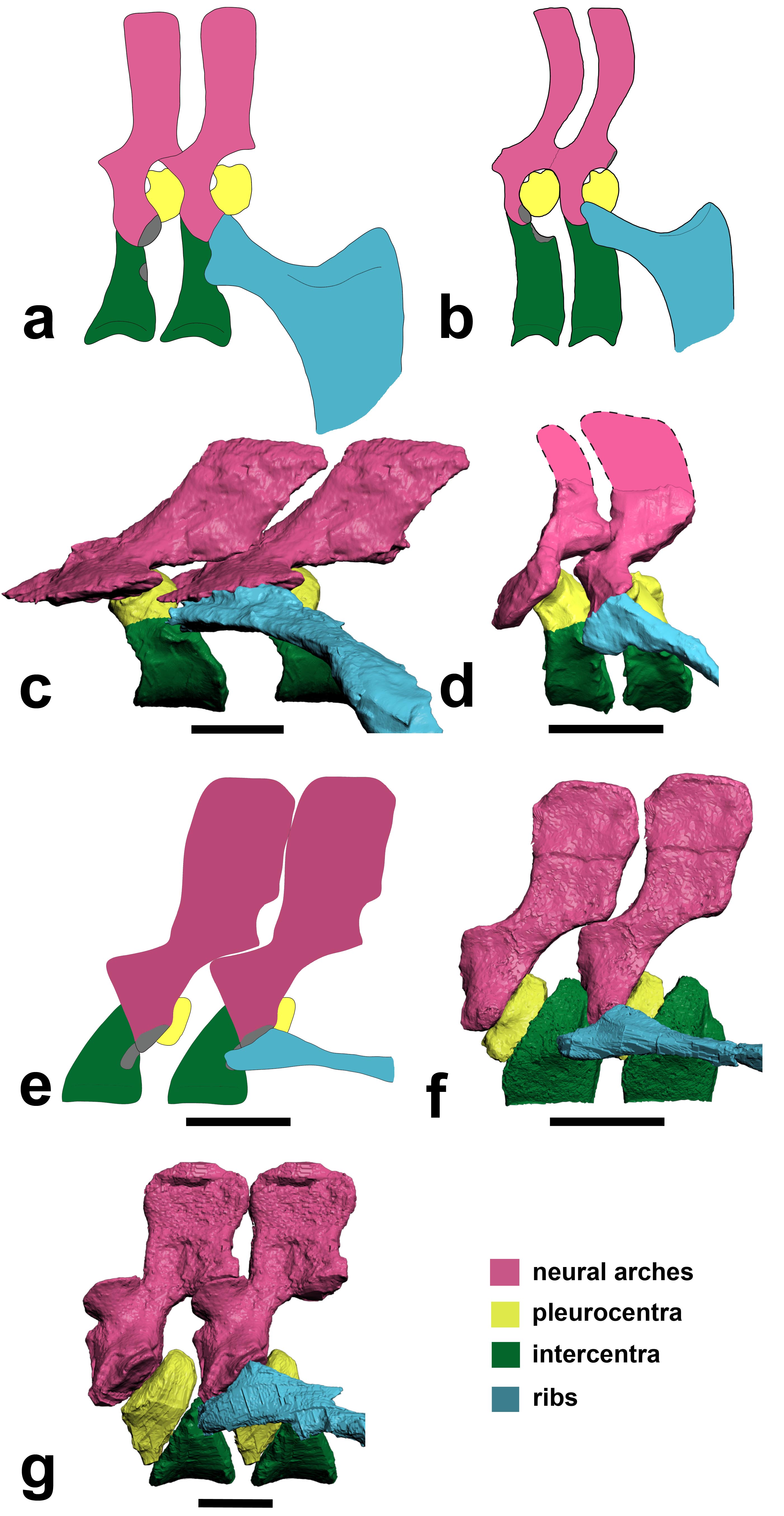
Above image: Julia Molnar‘s illustration of Ichthyostega showing anatomical changes of its spine from front to back, with neural arch/spine in pink, twin pleurocentra in yellow, and intercentrum in green. These four parts, three kinds of bones, made up the backbone of the first land vertebrates. These parts evolved in different ways in later animals, but formed one main bone in all living lineages of vertebrates.
RVC PRESS RELEASE:
Scientists reassemble the backbone of life using a particle accelerator
Research published today (Sunday 13 January 2013) in the journal Nature documents, for the first time, the intricate three-dimensional structure of the backbone in the earliest four-legged animals (tetrapods).
The backbone, also known as the spine or vertebral column, is a bony structure found in all tetrapods, along with other vertebrates such as fish. It is formed from many elements or vertebrae all connected in a row – from head to tail. Unlike the backbone of living tetrapods (e.g. humans), in which each vertebra is composed of only one bone, early tetrapods had vertebrae made up of multiple parts.
Lead author Dr Pierce says: “For more than 100 years, early tetrapods were thought to have vertebrae composed of three sets of bones – one bone in front, one on top, and a pair behind. But, by peering inside the fossils using synchrotron X-rays we have discovered that this traditional view literally got it back-to-front.”
For the analysis, the European Synchrotron Radiation Facility (ESRF) in France, where the three fossil fragments were scanned with X-rays, used a new protocol to reveal tiny details of the fossil bones buried deep inside the rock matrix.
Using this new technology, the team of scientists discovered that what was thought to be the first bone – known as the intercentrum – is actually the last in the series. And, although this might seem like a trivial oversight, this re-arrangement in vertebral structure has over-arching ramifications for the functional evolution of the tetrapod backbone.(see here for a now out-of-date image from Wikipedia)
Dr. Pierce explains: “By understanding how each of the bones fit together we can begin to explore the mobility of the spine and test how it may have transferred forces between the limbs during the early stages of land movement”.
But, the findings didn’t end there. One of the animals – known as Ichthyostega – was also found to have an assortment of hitherto unknown skeletal features including a string of bones extending down the middle of its chest.
Professor Clack says: “These chest bones turned out to be the earliest evolutionary attempt to produce a bony sternum. Such a structure would have strengthened the ribcage of Ichthyostega, permitting it to support its body weight on its chest while moving about on land.”
This unexpected discovery supports recent work done by the same authors that showed Ichthyostega probably moved by dragging itself across flat ground using synchronous ‘crutching’ motions of its front legs – much like that of a mudskipper or seal.
Dr Pierce adds: “The results of this study force us to re-write the textbook on backbone evolution in the earliest limbed animals.”
The next step, the researchers say, is to understand how the backbone aided locomotion in these early tetrapods using sophisticated biomechanical analysis.
These are rotating images of the anatomy, colour-coded, of the four species of early tetrapod that we examined for this study. Each shows the same basic pattern of having a “reverse rhachitomous” (pleurocentra in the front, intercentrum in the back; trying to think of a mullet joke…) anatomy. This is opposite the pattern that essentially all studies since famed evolutionary biologist/palaeontologist Edward Drinker Cope coined the term “rhachitomous” in 1878 have portrayed these and related animals as having. And this realization forces a re-examination of how the backbone structures first evolved in tetrapods and which parts (intercentra? pleurocentra? And where?) formed the spines of later animals.
For once, as authors we all felt that this finding really deserved the painfully hackneyed “rewrite the textbooks” label. It changes a lot of what we thought we knew about this classic evolutionary transition of anatomy. Check a vertebrate palaeontology/comparative anatomy textbook and you’ll likely find rhachitomous vertebrae and/or changes of pleurocentra vs. intercentra told in a way that we now are pretty sure is wrong.
You can also see the “sternebrae” (sternal elements; parts of the sternum that evolved independently in later land animals) in the first movie. This, to my knowledge, is by far the oldest such evidence. I know of ossified sternal plates in Early Permian mesosaurs like Stereosternum, but nothing earlier although perhaps in some synapsid I don’t know, or a basal diapsid of some kind? Chime in in the comments if you know of something I missed. Regardless, the sternebrae in Ichthyostega have nothing to do directly with those convergently evolved in lissamphibians, lepidosaurs, synapsids and archosaurs, although there may be some parallel developmental mechanisms involved and at least similar dermal tissues recruited into ossification patterns. Even so, these sternebrae are further evidence of how that taxon, at least, was beginning to make forays onto land, as they’d have helped it to support its belly on land and breathe.
The segmented PPC-SRµCT of Ichthyostegastensioi MGUH VP 6115 spinning in yaw and roll.
The segmented PPC-SRµCT of Ichthyostegaeigili MGUH VP 29017a spinning in yaw and roll.
The segmented PPC-SRµCT of Acanthostegagunnari MGUH f.n. 1227 spinning in yaw.
The segmented µCT of Pederpes finneyae GLAHMS 100815 spinning in yaw.
FIGURE:
Above: (a,b) How we used to think the vertebrae were composed in early tetrapods like Ichthyostega. (c) How we found that Ichthyostega‘s posterior thoracic vertebrae actually tend to look. (d) Ichthyostega‘s anterior lumbar vertebral morphology. (e) Acanthostega according to Coates’s important description. (f) Our revision of the anatomy of Acanthostega(anterior dorsal). (g) Our new interpretation of Pederpes‘s morphology, from a posterior dorsal. Focus on the yellow vs. green elements. In a,b and e they are in different positions (reversed) compared with our new versions in c,d,f,g.
To put the above figure and movies into broader context, check this Wikipedia image. We think the red/pink bones (pleurocentra) are in the wrong place relative to the blue ones (intercentrum); the ones currently there in this image actually belong to the vertebral unit behind that one, so the pleurocentra should be moved to the front (left end) of each unit. But also look down toward the bottom of the figure. Some of those vertebrae may need to have their blue/pink bits re-examined and interpreted, too. Is it turtles intercentra all the way down?
There you have it! Welcome to your new, revised, irradiated, reverse-rhachitomous inner tetrapod’s vertebrae. Propagation phase-contrast X-ray synchrotron microtomography FTW!!!!
Science media articles arising from this study–
I like to keep track of media stories covering our research, using this blog, so here are some of the stories about this paper. It’s funny… this was one of the most broadly important papers I’ve ever been on, but the coverage was relatively scant. It was too technical. We knew that would be a problem, and really had a hard time putting into words why the study was so surprising even to us! Most writers wanted the “how did the animals move?” angle, which was not what the study was about. I still feel that this angle was not even needed; the study (and again I take minimal credit for it) is exciting without it. To comparative anatomy and evo-devo specialists, anyway. Well, that’s science for you; sometimes it is just too hard to explain its value to the outside world, even when you feel its importance in your very spine… And the press coverage was not terrible by any means; no sour grapes from me. Regardless, we’re glad it has been well received by specialist researcher colleagues we’ve spoken to, and that matters a lot.
NERC’s Planet Earth (nice story from our funder)- “Scientists had fossil backbone backwards”
BBC online (the only story aside from NERC’s that did more than read the press release) “Tetrapod anatomy: Backbone back-to-front in early animals”
Discovery News online– “First Land Animals Shuffled Like Seals” (good, but is sort of mixing up our this study, our 2012 one and Ahlberg et al’s 2005 seal-analogue study; latter two were more about movement. As often happens, a lot of other media stories basically copied this one’s headline/angle.)
Discover 80beats– “Paleontologists Use 3-D Models to Rewrite Evolution” (also in “top stories”)
Popsci– “Particle Accelerator Reveals That First Land Animals Walked Like Seals”
Daily FMail (nice pics)- “Astonishing 3D images reveal the first four-legged land animals in amazing detail – and overturn a century of research” (wins longest headline award)
Red Orbit– “Study Reveals First Ever Images Of Early Tetrapod Backbone And How It Helped In Land Evolution”
Examiner.com– “X-ray study rewrites tetrapod backbone evolution (Photos)”
Business Standard– “Scientists recreate earliest quadraped’s backbone” (Proofread, editors! Quadruped.)
Geekosystem– “Early Land-Dwelling Animals Moved About Like Seals, Probably Didn’t Balance Balls on Their Noses” (scores some pts for humour)
…and the PR-copying, non-spellchecking fail of the week award goes to… Physorg! “Scientists reassemble the backbone of life with a particle acceleratorynchrotron [sic] X-rays”
Warming up the acceleratorynchrotron for our next study… 🙂
Hi folks, as my birthday present to you, and big thanks for racking up 70,000 blog views in 7 months (and my 50th post!), here is a new installment of Mystery CT Slice!
This time with a pilot (or scout) scan of an odd object. A pilot/scout scan is a quick, low exposure scan used to plan a series of CT slices, which shows a a larger area that is then narrowed down to focus just on the object of interest and a bit of buffer room for those slices. It generally isn’t used for much else, but sometimes can make a neat picture. As you can see here, the pilot scan area was excessively massive relative to the object. The two odd objects below the primary object of interest are scanning phantoms, used to calibrate density from Hounsfield units to actual real-world density (one is water at 1 g/cm3; the other is “cortical bone” at 1.69 g/cm3). Ignore them.
But what is this object and from what taxon? Be as specific as you can, but pinning it down to genus/species level will be bloody hard!
Stomach Churning Rating: 1/10; it doesn’t get much tamer than a pilot scan.
Difficulty Level: small image, hazy, not a lot of diagnostic traits visible, 1 main element.
Party time! Let the media onslaught begin! We’ve published a paper in Nature on the limb motions of Ichthyostega (and by implication, some other stem tetrapods). Since we did use some crocodile specimens from Freezersaurus (see below) in this study, I figured WIJF could cover it to help celebrate this auspicious event. Briefly. Particularly since we already did a quasi-blog on it, which is here:
However I want to feature our rockin’ cool animations we did for the paper, to squeeze every last possible drop of science communicationy goodness out of them. So here they are in all their digital glory. Huge credit to Dr. Stephanie Pierce, the brilliant, hardworking postdoc who spearheaded the work including these videos! Dr. Jenny Clack is our coauthor on this study and the sage of Ichthyostega and its relatives- her website is here. Also, a big hurrah for our goddess of artsy science, Julia Molnar, who helped with the videos and other images. Enjoy!
The computer model
The forelimb model
The hindlimb model
We used some of my Nile crocodile collection to do a validation analysis of our joint range of motion (ROM) methods, detailed in the Supplementary info of the paper, which I encourage anyone interested to read since it has loads more interesting stuff and cool pics. We found that a bone-based ROM will always give you a greater ROM than an intact fleshy limb-based ROM. In other words, muscles and ligaments (and articular cartilage, etc.). have a net effect of reducing how far a joint can move. This is not shocking but few studies have ever truly quantitatively checked this with empirical data from whole animals. It is an important consideration for all vert paleo types. Here is a pic of one of the crocodiles from the study, with (A) and without muscles (B; ligaments only):
I’ll close with Julia Molnar’s jaw-droppingly awesome flesh reconstruction from our model. Why Nature wouldn’t use this as a cover pic, I’ll never understand, but I LOVE it! When I first saw it enter my email inbox and then opened it to behold its glory, my squeal of geeky joy was deafening.
(edit: Aha! Fellow Berkeley alum Nick Pyenson’s group made the Nature cover, for their kickass study of rorqual whale anatomy, including a “new” organ! Well, we don’t feel so bad then. Great science– and a win for anatomy!!!)