Feeds:
Posts
Comments

Posts Tagged ‘ratite’

And I post my blog and stare
Into x-rays of an ostrich
I’ve always known that radiographs never lie
People always say “that’s cool”
To see x-rays of an ostrich
So keen to know what
Lies behind the skin

(evolved from “Eyes of A Stranger” by Queensrÿche, from the epic masterpiece of Operation: Mindcrime (1988). One of my favourite albums of all time, and a fantastic concept album too. The band was operating at their peak. Tight! Drug addict Nikki gets brainwashed by the evil Dr. X and made to assassinate a nun, Sister Mary, who was a prostitute, and then there’s like a revolution or something, and things get all screwed up and no one ends up happy – or alive. All the while, Geoff Tate is singing his guts out. Anyway, I got to see them play the whole album live in 1990 in Madison, WI, for the filming of Operation: Livecrime, which was like a Mecca moment for me back then. Look for me (pre-bald years) in about the 6th row. )

What does that album have to do with the number 2 (two days left in Freezermas)? Hmm… Track 2 is the instrumental Anarchy-X, and today’s post is about X-rays as well as that funky ostrich (2 legs good! 2 toes good, too!) again, so I’m satisfied, and by this point you’re probably just oggling the mind-blowing images below anyway, so fuck it!

Stomach-Churning Rating: 2/10; just X-rays.

Tech/MRes Kyle Chadwick, Renate Weller and the equine imaging team at the RVC took these x-rays of our birdie for us and for an artist who is doing a big x-ray animal art show (more news on this soon!)– thanks to all of them for some truly awesome images! I could stare at the intricate details in these images for hours– go ahead, do it. Click to emostrichinate them (this post needs to be viewed on nice big screen), and oggle away…

Head and neck.

Head and neck.

Another view of the same.

Another view of the same. The highly flexible esophagus and trachea can be seen going diagonally across the neck; twisting from ventral to dorsal. It’s floppy, so it can do that.

Neck near the head; tapering.

Neck near the head; tapering.

Middle of neck. Check out the rings of the trachea!

Middle of neck. Check out the rings of the trachea!

Base of neck and shoulder

Base of neck and shoulder.

Shoulder and chest. Hard to image; thick and dense (still was frozen).

Shoulder and chest. Hard to image; thick and dense (still was frozen), hence the whiteout toward the left side of the image.

Check out that wing!!

Check out that wing!!

Ankle- note the big calloused pad that ostriches rest on (right side of image).

Ankle- note the big calloused pad that ostriches rest on (right side of image).

That two-toed foot... but did you know that normally the missing 2nd toe is still there as a fibrous remnant on the 3rd toe?

That two-toed foot… but did you know that normally the missing 2nd toe is still there as a fibrous remnant on the 3rd toe?

Tomorrow: the final day of Freezermas. What will it be?

Read Full Post »

Freezermas continues! Today we have a treat for you. Lots of detailed anatomy! This post comes from my team’s dissections of an ostrich last week (~3-7 February 2014), which I’ve been tweeting about as part of a larger project called the Open Ostrich.

However, before I go further, it’s as important as ever to note this:

Stomach-Churning Rating: 9/10: bloody pictures of a dissection of a large ostrich follow. Head to toes, it gets messy. Just be glad it wasn’t rotten; I was glad. Not Safe For Lunch!

If the introductory picture below gets the butterflies a-fluttering in your tummy, turn back now! It gets messier. There are tamer pics in my earlier Naked Ostriches post (still, a rating of 6/10 or so for stomach-churning-ness there).

All photo credits  (used with permission) on this post go to palaeoartist Bob Nicholls (please check out his website!), who got to attend and get hands-on experience in extant dinosaur anatomy with my team and Writtle College lecturer Nieky VanVeggel (more from Nieky soon)!

Research Fellow Jeff Rankin, myself and technician/MRes student Kyle Chadwick get to work.

Research Fellow Jeff Rankin, myself and technician/MRes student Kyle Chadwick get to work, removing a wing.

This is a male ostrich, 71.3 kg in body mass, that had gone lame in one foot last summer and, for welfare reasons, we had to put down for a local farmer, then we got the body to study. We took advantage of a bad situation; the animal was better off being humanely put down.

The number for today is 6; six posts left in Freezermas. But I had no idea I’d have a hard time finding a song involving 6, from a concept album. Yet 6 three times over is Slayer’s numerus operandi, and so… The concept album for today is Slayer’s  1986 thematic opus “Reign in Blood” (a pivotal album for speed/death metal). The most appropriate track here is the plodding, pounding, brooding, then savagely furious “Postmortem“, which leads (literally and figuratively, in thunderous fashion) to the madness of the title track, after Tom Araya barks the final verse:

“The waves of blood are rushing near, pounding at the walls of lies

Turning off my sanity, reaching back into my mind

Non-rising body from the grave showing new reality

What I am, what I want, I’m only after death”

I’m not going to try to reword those morbid lyrics into something humorous and fitting the ostrich theme of this post. I’ll stick with a serious tone for now. I like to take these opportunities to provoke thought about the duality of a situation like this. It’s grim stuff; dark and bloody and saturated with our own inner fears of mortality and our disgust at what normally is politely concealed behind the integumentary system’s viscoelastic walls of keratin and collagen.

But it’s also profoundly beautiful stuff– anatomy, even in a gory state like this, has a mesmerizing impact: how intricately the varied parts fit together with each other and with their roles in their environment, or even the richness of hues and multifarous patterns that pervade the dissected form, or the surprising variations within an individual that tell you stories about its life, health or growth. Every dissection is a new journey for an anatomist.

OK I’ve given you enough time to gird yourself; into the Open Ostrich we go! The remainder is a photo-blog exploration of ostrich gross anatomy, from our detailed postmortem.

(more…)

Read Full Post »

A photo blog post for ya here! I went to Dublin on a ~28 hour tour, for a PhD viva (now-Dr Xia Wang; bird feather/flight evolution thesis) earlier this month. And I made a beeline for the local natural history museum (National Museum of Ireland, Natural History building) when I had free time. So here are the results!

Stomach-Churning Rating: Tame; about a 1/10 for most, but I am going to break my rule about showing human bodies near the end. Just a warning. The bog bodies were too awesome not to share. So that might be 4/10-8/10 depending on your proclivities. They are dry and not juicy or bloody, and don’t look as human as you’d expect.

Simple Natural History museum entrance area.

Simple Natural History museum entrance area.

Adorable frolicking topiaries outside the NHM.

Adorable frolicking topiaries outside the NHM.

Inside, it was a classical Victorian-style, dark wood-panelled museum stuffed with stuffed specimens. It could use major refurbishment, but I do love old-fashioned exhibits. Get on with it and show us the animals; minimize interpretive signage and NO FUCKING INTERACTIVE COMPUTER PANELS! So by those criteria, I liked it. Some shots of the halls: hall2 hall1 hall3 hall4 hall5 hall6 And on to the specimens!

Giant European deer ("Irish elk"). I looked at these and thought, "why don't we see female deer without antlers ever? then noticed one standing next to these; photo was crappy though. :(

Giant European deer (“Irish elk”). I looked at these and thought, “why don’t we see female deer without antlers ever? then noticed one standing next to these (you can barely see it in back); too bad my photo is crappy.

Superb mounted skeleton of giraffe (stuffed skin was standing near it).

Superb mounted skeleton of giraffe (stuffed skin was standing near it).

A sheep or a goat-y thingy; I dunno but it shows off a nice example of the nuchal ligament (supports the head/neck).

A sheep-y or a goat-y beastie; I dunno but it shows off a nice example of the nuchal ligament (supports the head/neck).

Yarr, narwhals be internet gold!

Yarr, narwhals be internet gold!

Giant blown glass models of lice!

Giant blown glass models of lice!

Who doesn't like a good giant foramanifera image/models? Not me.

Who doesn’t like a good giant foramanifera image/model?

"That's one bigass skate," I murmured to myself.

“That’s one bigass skate,” I murmured to myself.

"That's one bigass halibut," I quipped.

“That’s one bigass halibut,” I quipped.

Tatty basking shark in entry hall.

Tatty basking shark in entry hall.

Irish wolfhound, with a glass sculpture of its spine hanging near it, for some reason.

Irish wolfhound, with a glass sculpture of its spine hanging near it, for some reason.

Stand back folks! The beaver has a club!

Stand back everyone! That beaver has a club!

Skull of a pilot whale/dolphin.

Skull of a pilot whale/dolphin.

Nice anteater skeleton and skin.

Nice anteater skeleton and skin.

Nice anteater skeleton and skin.

Nice wombat skeleton and skin.

Sad display of a stuffed rhino with the horn removed, and signage explaining the problem of thefts of those horns from museum specimens of rhinos worldwide.

Sad display of a stuffed rhino with the horn removed, and signage explaining the problem of thefts of those horns from museum specimens of rhinos worldwide.

But then the stuffed animals started to get to me. Or maybe it was the hangover. Anyway, I saw this…
creepy proboscis (1) creepy proboscis (2)

A proboscis monkey mother who seemed to be saying “Hey kid, you want this yummy fruit? Tough shit. I’m going to hold it over here, out of reach.” with a disturbing grimace. That got me thinking about facial expressions in stuffed museum specimens of mammals more, and I couldn’t help but anthropomorphize as I toured the rest of the collection, journeying deeper into surreality as I progressed. What follows could thus be employed as a study of the Tim-Burton-eseque grimaces of stuffed sloths. Click to emslothen.

sloths (1) sloths (5)sloths (4) sloths (3) sloths (2)

Tree anteater has a go at the awkward expression game.

Tree anteater has a go at the awkward expression game.


This completed my tour of the museum; there were 2 more floors of specimens but they were closed for, sigh, say it with me… health and safety reasons. Balconies from which toddlers or pensioners or drunken undergrads could accidentally catapult themselves to their messy demise upon the throngs of zoological specimens below. But the National Museum’s Archaeology collection was just around the block, so off I went, following whispered tales of bog bodies. There will be a nice, calm, pretty photo, then the bodies, so if peaty ~300 BCE cadavers are not your cup of boggy tea, you can depart this tour now and lose no respect.

Impressive entrance to the National Museum's Archaeology building.

Impressive entrance to the National Museum’s Archaeology building.

The bog bodies exhibit is called “Kingship and Sacrifice“. It is packed with cylindrical chambers that conceal, and present in a tomb-like enclosed setting, the partial bodies of people that were killed and then tossed in peat bogs as honoraria for the ascension of a new king. The peaty chemistry has preserved them for ~2300 years, but in a dessicated, contorted state. The preservation has imparted a mottled colouration and wrinkled texture not far off from a Twix chocolate bar’s. Researchers have studied the bejesus out of these bodies (including 3D medical imaging techniques) and found remarkable details including not just wounds and likely causes of death (axes, strangling, slit throats etc) but also clothing, diet, health and more.

Here they are; click to (wait for it)… emboggen:

BogBodies (1) BogBodies (2) BogBodies (3) BogBodies (4) BogBodies (5) BogBodies (6)

Did you find the Celtic armband on one of them?

Finally (actually this happened first; my post is going back in time), I visited UCD’s zoology building for the PhD viva and saw a few cool specimens there, as follows:

Giant deer in UCD zoology building foyer.

Giant deer in UCD zoology building foyer, with a lovely Pleistocene landscape painted on the wall behind it.

Sika deer in awkward posture in Univ Coll Dublin zoology building's foyer.

Sika deer in an awkward posture (what is it supposed to be doing?) in Univ Coll Dublin zoology building’s foyer.

The pose of this ?baboon? struck me as very peculiar, and menacing- reminiscent of a vampire bat's pose, to me.

The pose of this ?baboon?mandrill struck me as very peculiar and menacing- reminiscent of a vampire bat’s pose.

A whole lotta chicken skeletons in a UCD teaching lab.

A whole lotta chicken skeletons in a UCD teaching lab.

After the viva we went out for some nice Chinese food and passed some Dublin landmarks like this:

Trinity College entrance, I think.

Trinity College entrance, I think.Former Irish Parliament; now the Bank of Ireland.

And we wandered into a very posh Irish pub called the Bank (on College Green), which displayed this interesting specimen, as well as some other features shown below:

Replica of illuminated old Gaelic manuscript.

Replica of illuminated 9th Century gospel manuscript “The Book of Kells”, with gorgeous Celtic art.

Vaults near toilets in the Bank pub.

Vaults near toilets in the Bank pub. Almost as cool as having giant freezers down there.

Nice glass ceiling of the Bank pub.

Nice glass ceiling of the Bank pub.

And Irish pub means one big, delicious thing to me, which I will finish with here– much as I finished that night off:

Ahhh...

Ahhh… ice cold.

Read Full Post »

Less words, more pictures in this post, and I’ll get the one lame cake joke out of the way early. I’ve nearly finished my research blitz through the postcranial material of the NHM-Tring’s osteological collection and have made some pit-stops for cake skulls now and then when I see one that pleases me. Now I shall present a survey of some of the species I’ve examined. I’ll proceed up from the base of the crown clade of living birds (Neornithes/Aves; the most recent common ancestor of living birds and all its descendants) and first take a tour of Palaeognathae; the ratites and kin; then move another step up into the Neognathae, first featuring the lineage featuring the ground fowl (Galliformes) and then the waterfowl (Anseriformes). If all this taxonomy and phylogeny is a bit much, check out this page for a brush-up on the bushy branches of bird biodiversity.

First, lots of bones of our cast of currasows, chachalacas, cassowaries and other kooky characters. And then, perhaps, a stop to the excessive alliteration. Finally, I will finish with some examples of species oddity (hat tip to Chris Hadfield).

Stomach-Churning Rating: 2/10- some bony pathologies but still just dry bones. Minimal cake jokes, and no filthy swearing this time.


BRING ON THE BONES:

My photographs are shown with kind permission from the Natural History Museum, London.

Exploded skull of an ostrich/ This takes skill.

Exploded skull of an ostrich, Struthio camelus. This kind of careful preparation takes crazy skill, and creates a thing of rare beauty.

Neat skull of a cassowary, Casuarius casuarius.

Imposing skull of a cassowary, Casuarius casuarius, with a rather worn head casque.

Mummified Owen's Little Spotted kiwi, Apteryx owenii.

Mummified Owen’s Little Spotted Kiwi, Apteryx owenii. The feathers were still soft and fluffy, but I would not call this specimen cuddly.

Dorsal view of the back/hips of the Great Spotted kiwi, Apteryx haasti.

Dorsal view of the back/hips of the Great Spotted Kiwi, Apteryx haasti. I like this photo and am not sure why. The symmetry and shading pleases me, I guess.

Front view of the back/hips of the Great Spotted kiwi, Apteryx haasti.

Front view of the back/hips of the Great Spotted Kiwi, Apteryx haasti, watching over my laptop and watching me while I write this blog on my laptop… so meta(ornithine)!

Wing of a kiwi, showing the fragile bones and feather attachments.

Wing of a kiwi, showing the fragile bones and feather attachments. “Apteryx” = “no wings”… well not quite. Click to emkiwi(?) so you can identify the individual bones, from the humerus right down to the fingers! I love this specimen.

The left leg (in front view) of the elephant-bird, Aepyornis maximus, from Madagascar, with a small moa nearby in left side view.

The titanic left leg (in front view) of the Elephant Bird, Aepyornis maximus, from Madagascar, with a small moa nearby in left side view. There’s so much awesomeness about elephant birds I don’t know where to start, but this is one good place to do so.

Mummified Unulated tinamou, Crypturellus undulatus.

The smaller end of the palaeognath scale: a mummified Undulated Tinamou, Crypturellus undulatus. Somehow the head got stuck into the abdominal cavity underneath the sternum, so this tinamou almost had its head up its arse. A tinamou with head in its proper position looks and sounds like this (video).

And now we take a left turn into the Galloanseres, most basal branch of the neognath birds, to see some of the neglected, strange early branches off from the “main line” that led to the modern diversity of ducks, geeses and swans (Anatinae, Anserinae).

Screamers (Anhimidae) are to Anseriformes as megapodes (see below; brush turkeys) are to Galliformes. By that I mean that both screamers and megapodes are very early branches off the main line of their respective lineages’ evolution, and both are quite strange when seen in that context… an unfair one, frankly; over-focused on the most familiar, “modern” or most speciose group. More about this issue further below.

This was my first hands-on experience with screamer anatomy; I was familiar from reading Tetrapod Zoology and other material about them. Check out the sound that gives them their name here! I’m now a big fan- they have so many strange features: oddly chunky but often very light bones, big feet with long toes, and then these switchblade-wrists, which would make Batman jealous:

Crested screamer, Chauna torquata, showing the wicked spur on the carpometacarpus.

Crested Screamer, Chauna torquata, showing the wicked spur (and smaller one) on the carpometacarpus.

Horned screamer, Anhima cornuta; similar carpometacarpal spur as in Chauna.

Horned Screamer, Anhima cornuta; similar carpometacarpal spurs as in Chauna.

Torso of a screamer seen in top view. Nice narrow body.

Torso of a screamer seen in top view. Nice narrow body, and no uncinate processes (spur-like bony struts that cross the ribs and act as levers for the muscles that move the ribcage during breathing)

The long, gracile, clawed toes of a screamer.

The long, gracile, clawed toes of a screamer. Those toes, especially as they belong to an animal called a screamer, are spooky for me. Note also: very little toe-webbing for a “waterfowl.”

Not to be outdone, on the Galliformes side of Galloanserae, we have some funky headgear in the Maleo (a megapode bird/Megapodiidae; a very basal branch of “brush turkeys” and kin) and curassows (part of the Cracidae; odd South American birds whose males make booming sounds, presumably using their head-casques as resonating chambers?):

Skull of a male maleo, Macrocephalon maleo.

Skull of a male Maleo, Macrocephalon maleo. AR Wallace famously pursued it, and here is its funky call.

Australian brush-turkeys, Alectura lathami i, at the Alma Park Zoo near Brisbane, Australia; they run wild there. Here they are doing what they are best known for: making a mound-like nest.

Australian brush-turkeys, Alectura lathami, at the Alma Park Zoo near Brisbane, Australia; they run wild there. Here they are doing what they are best known for: making a mound-like nest. We were doing kangaroo biomechanics experiments and they were everywhere. I was in awe to see such exotic (to me) birds; locals seemed not so enthused (the birds are loud and make a lot of mess).

Skull of Helmeted curassow, Crax/Pauxi pauxi.

Skull of Helmeted Curassow, Crax/Pauxi pauxi,  showing that resonating chamber. Along with this boom-boom-room, the male uses a piece of food that he holds to draw in the female; if she takes it, then it’s sexy time.

Foot of a Russian Black Grouse, Tetrao tetrix (nothing to do with a certain videogame), with and without flesh.

Foot of a Siberian Black Grouse, Tetrao tetrix (nothing to do with a certain videogame), with and without flesh. Regard the broad, feathered feet, well insulated and with plenty of surface area for prancing around in the snow or moorlands. Tetrao engage in a cool display pattern called lekking, in which the males group together and show off to watching females.

A theme in the section above that is not to be missed is that there is some amazing disparity of anatomical forms in these basal lineages of poultry-relatives. Don’t dismiss the Galloanserae as just boring food-birds! Heaps of not-so-well-studied species exist here, surely with a treasure trove of cool neontological and evolutionary questions waiting for the right person to ask! Darwin’s chickens may get their share of neglect, but that pales in comparison to how little we understand about many basal Galloanserae.

What a lot of people think of as a “ground fowl” or galliform way of life is more of a way of life somewhat typical of the Phasanidae- chickens, pheasants and their familiar kin. Megapodes, curassows, guans, grouse and other Galliformes do not necessarily do things in the “typical” ground fowl way, much as the earlier branches of the Anseriformes don’t always look/act like “proper water fowl” (i.e. Anatidae). The phenomenon at play here is one of the great bugaboos in biology: essentialism— the often implicit misconception that variation away from some abstract ideal is negligible, uninteresting or just not conceivable due to mental blinders. When we say something like “the chicken is a fascinating species” we are sliding down the essentialistic slope. There is no “the chicken.” Not really. Oh dear, speaking of slippery slopes, I’d best stop here before I start talking about species concepts. And no one wants that to happen! Anyway, essentialism still pervades a lot of modern scientific thinking, and has its place as a conceptual crutch sometimes. But in biology, essentialism can be very insidious and misleading. It burrows in deep into the scientific mind and can be hard to root out. Unfortunately, it is entrenched in a lot of science education, as it makes things easier to teach if you sweep aside the exceptions to the essentialist “rules” in biology. I catch myself thinking in static, essentialist ways sometimes. The punishment is no cake for a week; so awful. 🙂

And speaking of “normal” or “typical,” morphology is of course often not that way even within a species, age class or gender. Pathology is a great example; by definition it is abnormal. It is a shattering of the “essence” of animals, brought on by some malady.

Next I’ve highlighted some of the amazing pathologies I’ve seen in the Tring skeletons. There have been so many I’ve been unable to keep track of them– some of these birds had the stuffing beaten out of them, and I’m not talking about Thanksgiving turkeys. Some were captive animals, in which the pathology might be blamed on living an inappropriate environment, but some were wild-caught — given the extreme pathologies, it’s a wonder those even survived to be found, but perhaps less a surprise that they were caught.


BONES GONE BONKERS:

View of left knee of a specimen of the Highland guan, Penelopina nigra, showing some nasty osteoarthritis around the whole joint.

View of left knee of a specimen of the Highland Guan, Penelopina nigra, showing some nasty osteoarthritis around the whole joint. Eew.  A happier Guan sounds like this.

Femora and tibiae of the Blue-throated Piping Guan, Aburria cumanensis. Amazing pathology involving the left femur (broken, rehealed) and tibiotarsus (secondary infection?).

Femora and tibiotarsi of the Blue-throated Piping Guan, Aburria cumanensis. Amazing pathology involving the left femur (broken, rehealed) and tibiotarsus (secondary infection?). Interestingly, the non-fractured limb also showed some pathology, perhaps indicating general infection and/or arthritis in reaction to the severe damage to the other leg, or just increased load-bearing on that leg.

Little Chachalaca, Ortalis motmot, showing a broken and rehealed right femur and the tibiotarsus.

Little Chachalaca, Ortalis motmot, showing a broken and rehealed right femur and the tibiotarsus. As in the guan above, this animal was not walking for many weeks; its femur had snapped in two, but somehow melted back together. The tibiotarsus didn’t look too great, either; lumpy and bendy. In better times, the Chachalaca does the cha-cha like this.

These two specimens blew my mind. On the right is a normal Tetrao tetrix (Black grouse); on the left is one hybridized with another (unknown) species.

These two specimens blew my mind. On the left is a normal Tetrao tetrix (Black Grouse); on the right is one hybridized with another (unknown) species.

In the picture above, what amazed me first was the very unusual flattened pelvis/synsacrum of Tetrao, which characteristically is light and wide. But in the hybrid this morphology was completely gone; the pelvis had a more standard “galliform” (read: Phasianid)-like shape, deeper and narrower and more solid in build. I am guessing that the hybrid was a cross with a pheasant like Phasianus itself, whose anatomy would be more like this. Somewhere in here there is a fantastic evo-devo/morphometrics project waiting to happen.

That’s my quick specimen-based tour of “basal birds”. Beyond these two clades of Palaeognathae and Galloanseres, there lies the forebidding territory of Neoaves: much of living avian diversity, and extremely contentious in its phylogenetic relationships. I’m tackling them next for my research on the evolution of the patella/kneecap. But first, I’ll be at the NHM-Tring today for a whirlwind tour through the respectably speciose “normal” Galloanseres clades of Phasianidae and Anserinae+Anatidae, so off I go! (It’s my wife’s birthday celebration, so cake may have to wait for later this time)

So what do you think? What’s your favourite neglected “primitive” bird group (more apropos: early branching avian lineage that may still be very specialized, rare and poorly understood), or cool factoid about palaeognaths and basal neognaths?

No quaggas were harmed during the writing of this post.

No quaggas were harmed during the writing of this post. Polly wanna quagga?

Read Full Post »

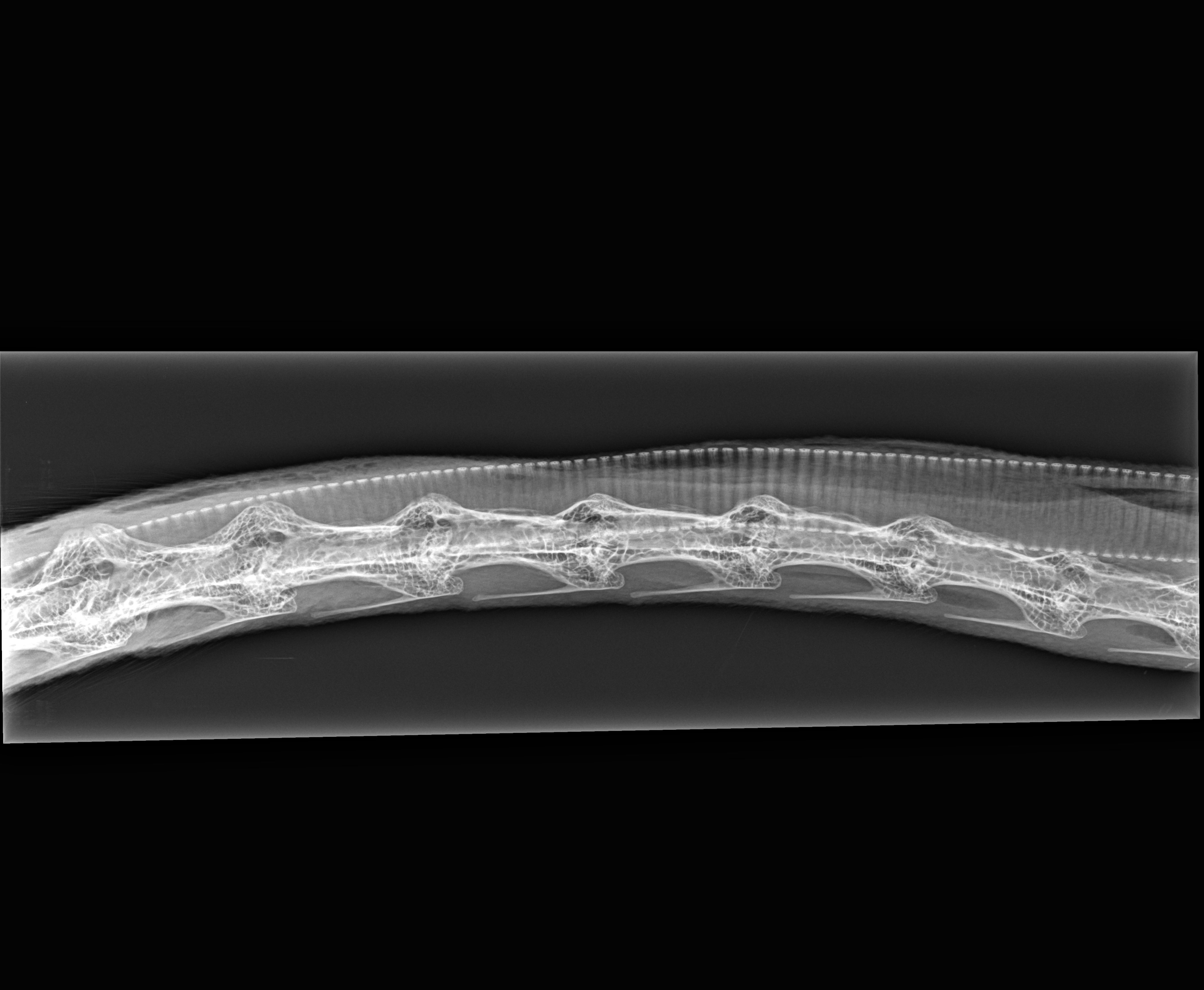
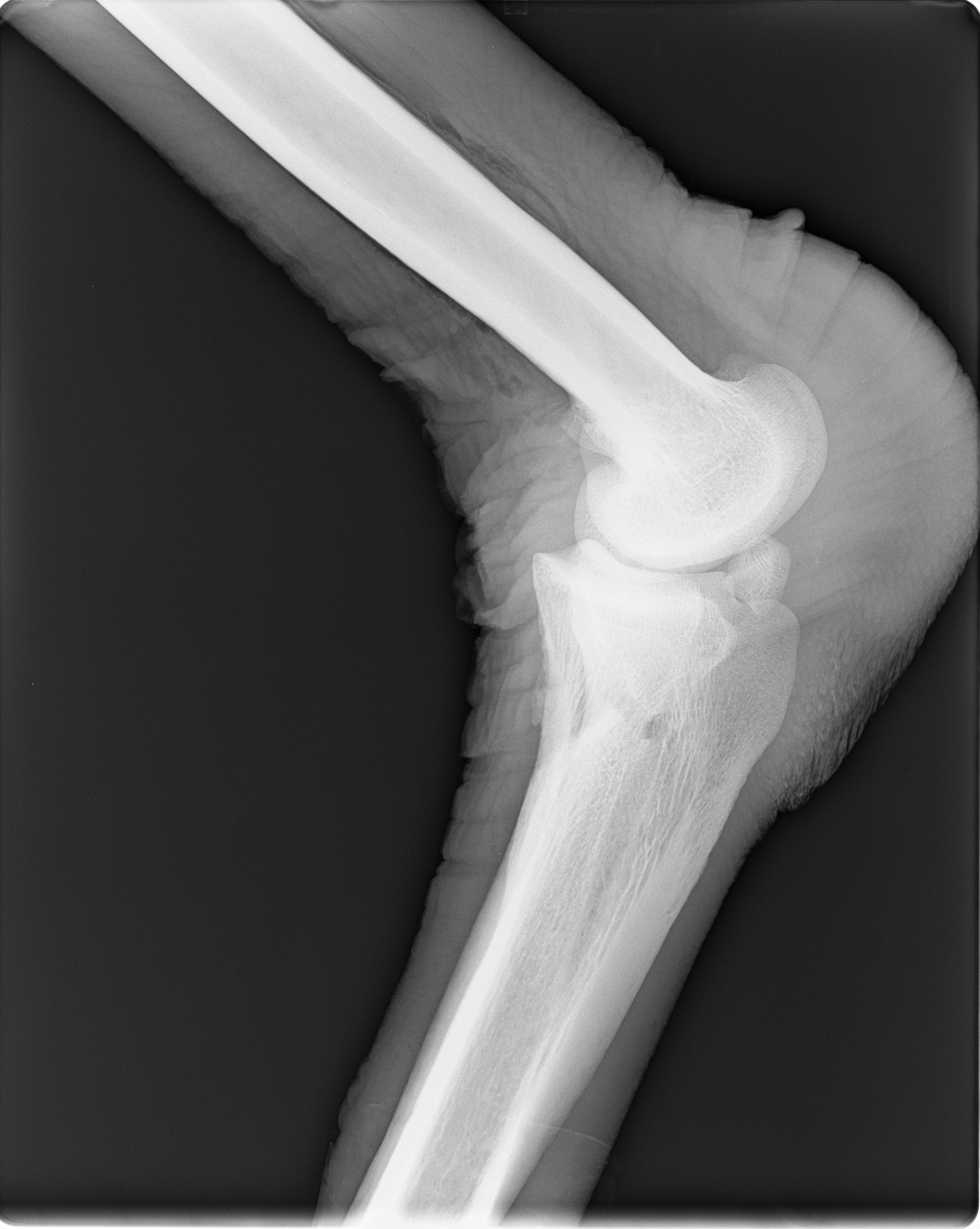
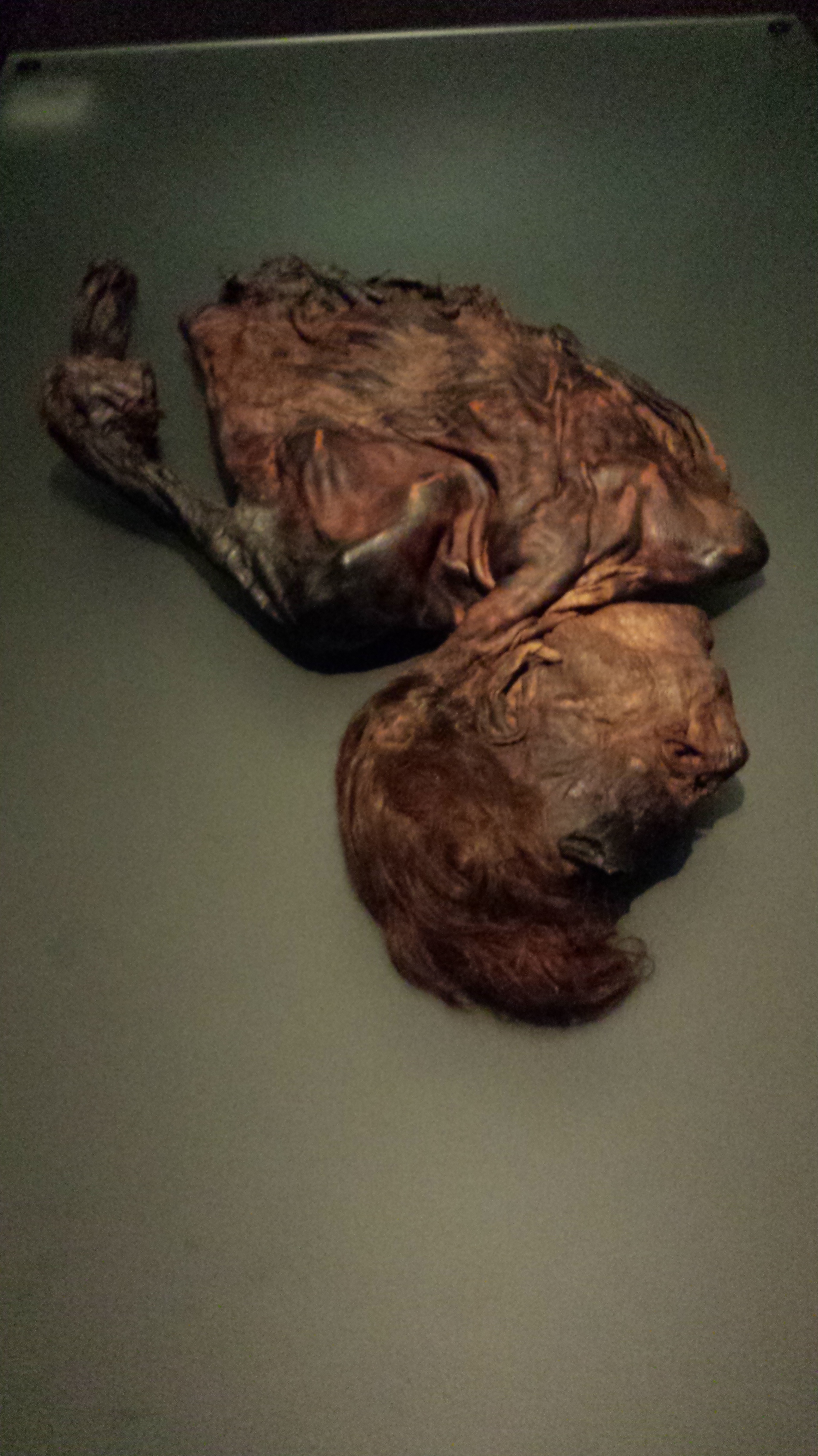
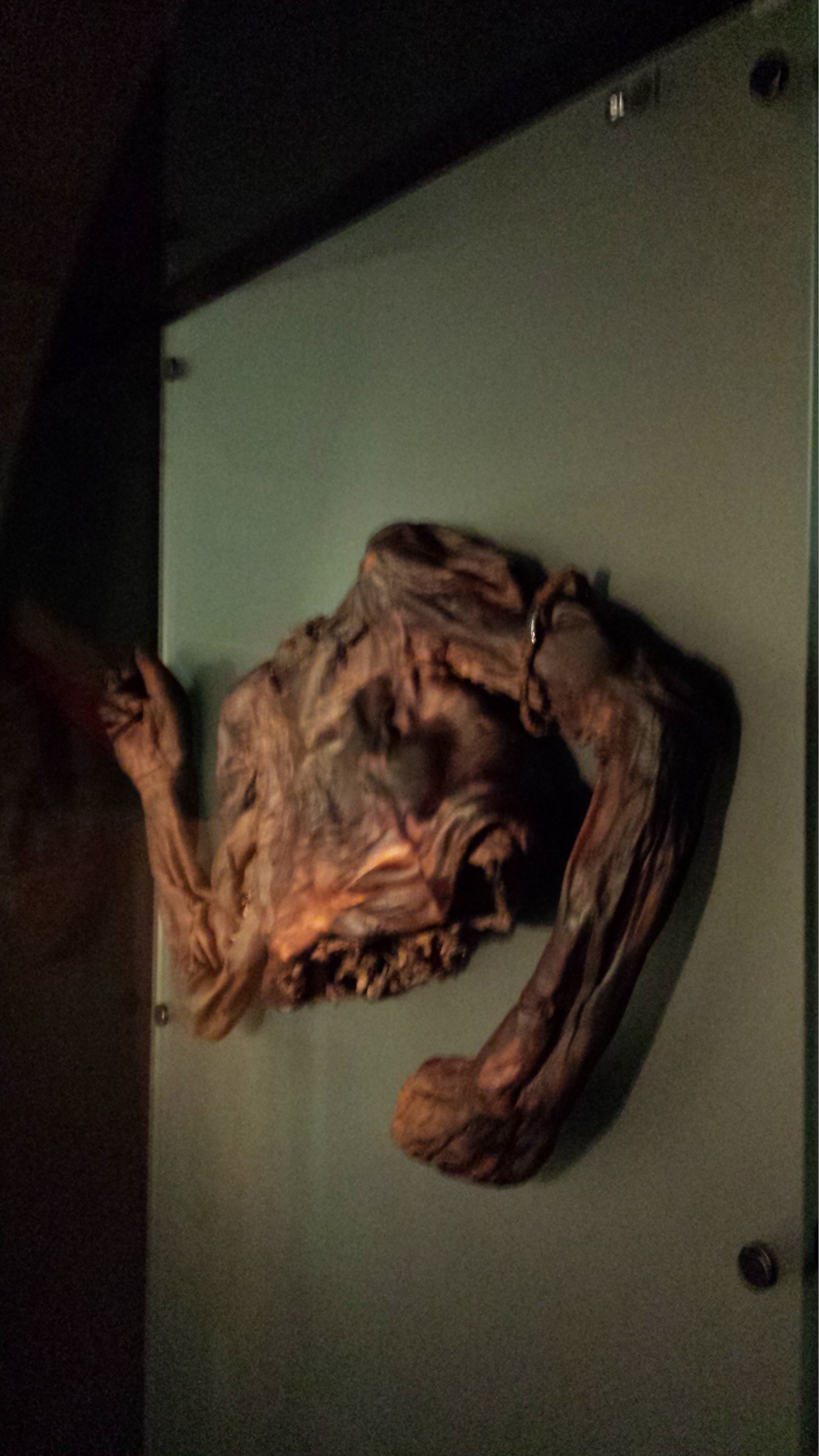
…a daily picture of anatomy! And today it is three pictures, scoob-a-dee!

Welcome back again to Freezermas! 

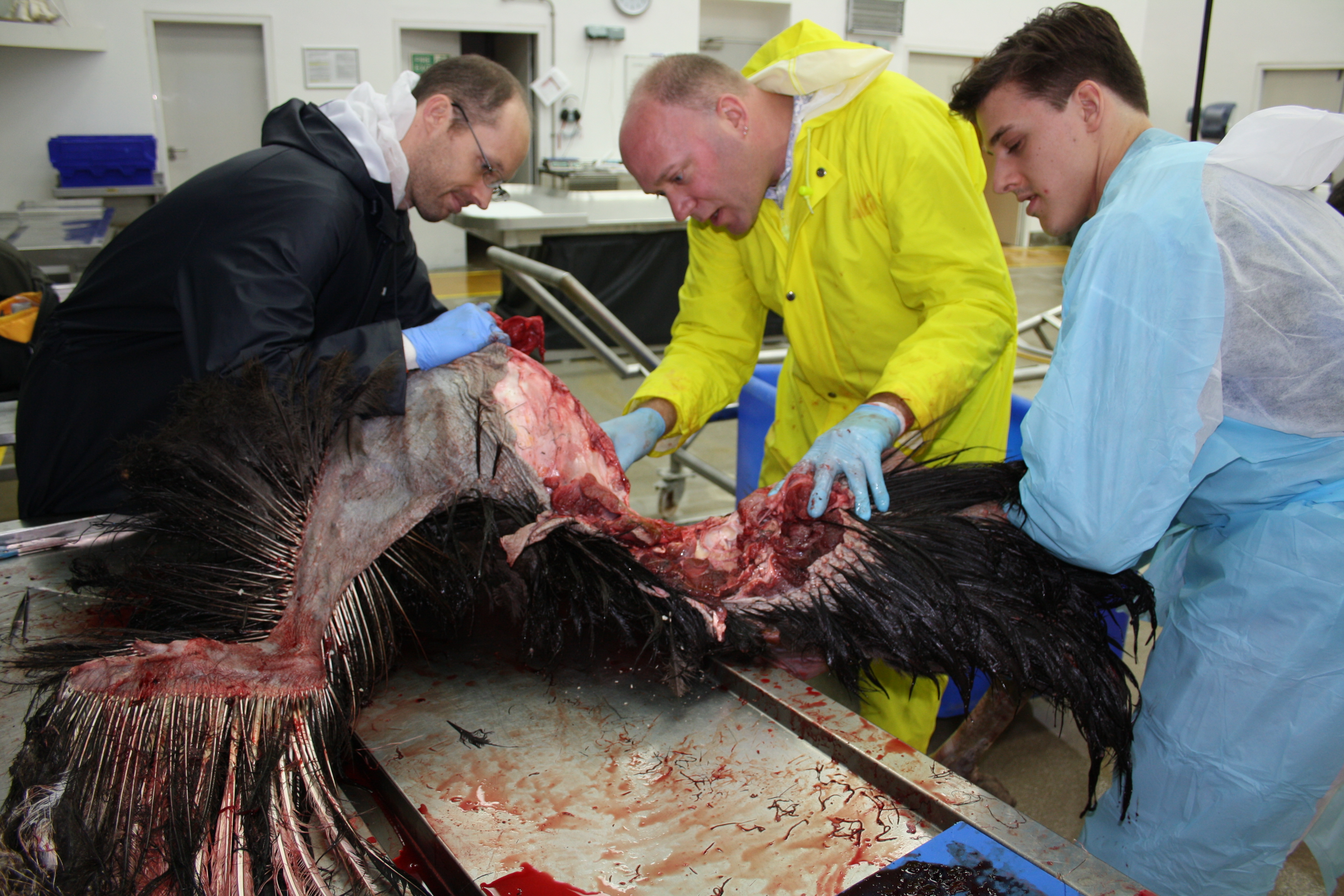
For the previous days of Freezermas we first had 1 picture, then 2, now guess how many we have today? Right, we’ve settled into a groove and have three (plus one silly one). Today is fresh beefy anatomy day! No focus on bones, but on soft tissues– however, once again, I’m representin’ bird legs! And this time, no mystery things to identify; sorry. But if you want to muscle in on some myology, today is the day for you. I will unwrap the thigh of an ostrich and consider the major muscles that power rapid running in this biped, and how they illuminate the evolution of bipedal motion along the line of descent to birds. For more ostrich escapades, see this old post. And we’re off!

Stomach-Churning Rating: 7/10; plenty of fresh, red, meaty meat from ostrich leg muscles.

Ostrich thigh muscles 1

Here you are looking at a right hindlimb of an ostrich, in side/lateral view. To help orient yourself, the hip lies deep in the middle of the image and the knee is the rounded bump near the bottom right corner, with the shank angling sharply back toward the bottom left.

I’ve labelled six muscles in yellow. As usual for sauropsid (bird/reptile) pelvic limb muscles, they have sensible names that reflect their attachments. They don’t have so many silly old mammalian names like pectineus or latissimus, which tell you rather little about the muscles themselves. We can thank 19th century anatomists like two of my anatomist heroes, Hans Gadow and Alfred Romer (who refined Gadow’s earlier work and made it more popular among English-speakers and palaeontologists), for that enlightened nomenclature.

The six muscles seen above are the IC (iliotibialis cranialis), IL (iliotibialis lateralis), “AMB2?” (one of the ambiens muscles– correctly identified; ignore the ?), ITC (iliotrochantericus caudalis), CFP (caudofemoralis pars pelvica) and FCLP (a mouthful to say: flexor cruris lateralis pars pelvica). The ambiens is the one oddly, non-anatomically named muscle, and has nothing to do with helping you sleep (pssst– wake up! Muscles are exciting!), but everything to do with the state of total awesomeness, which is what “ambiens” means. Maybe. Or I am making shit up.

Amazed ostrich

The IC, IL and AMB2 are parts of the triceps femoris group (discussed in my 1st Freeezermas post), or for mammal fans the quadriceps. The IC and AMB are in front of the hip so they flex it (move the thigh forward; protract it); the IL is right around the hip so it can flex or extend the hip (protract/retract the femur); all three of these can extend (straighten) the knee joint to varying degrees. The IC is fairly typical for a bird except for its size, and helps to quickly swing the leg through the air between steps. Some birds have multiple parts of the IL, but ostriches and many others have simplified it to one major mass; regardless, it is a major muscle used to support the weight of the body.

The AMB2 is a remarkable muscle unique to ostriches; it can also be called the dorsal ambiens muscle. Typical birds just have a single head of the AMB sitting on the preacetabular (pubic) tubercle, so in front of and below the hip. It has a crazy tendon that snakes past the knee (in some birds, perforating/grooving the patella) into the lower leg muscles and may be able to even pull on the toes. But ostriches, for some reason, added a second head of this muscle that was shifted way up onto the front of the pelvis (the ilium; dorsal bladelike bone). Crocodilians also have a 2nd ambiens muscle but in a different position, and almost certainly as an example of convergent evolution. The function of the ambiens is mysterious, but this muscle has featured prominently in avian systematics/taxonomy, evolution (invoked as a key muscle used in perching) and more.

These muscles of the triceps femoris group are easily identifiable in crocodiles and other reptiles because they are remarkably similar in their attachments. The main changes these muscles experienced during the evolution of bipedalism, dinosaurs and later birds are simply proportional– they got bigger, with stronger, larger attachments on the pelvis and the front of the knee (the CC/LC, if you remember from Freezermas day 1).

The ITC is a muscle that is very dear to me. I’ve written a lot about it, and I love saying the name “Iliotrochantericus caudalis”- it is musical to me. For mammal fans, think gluteal muscles (medial gluteal in particular). It is a huge, pennate muscle (short and strongly angled muscle fibres in a “sandwich” with a tendinous sheet between the two layers of fibres). It has a short, broad tendon that wraps around the trochanteric crest (a structure on the upper front end of the femur with a history that goes wayyyy back into dinosaurs; long story!) to insert in a scarred depression. The ITC seems to mainly rotate the femur around its long axis to help support the body. I could go on and on about this muscle, which is part of the enigmatic “deep dorsal” thigh muscle group — the homologies of this group among land vertebrates are still controversial and confusing. But I will spare you the on-and-on. Incidentally, the ITC  is the “oyster” in birds that is the best cut of meat. And in ostriches it makes a massive steak.

The CFP also has a cool evolutionary history. It runs from the back of the pelvis to the middle of the femur, closely adjoined to the caudal head of the muscle (CFC), which is more vestigial. In birds the CFP is usually not a large muscle, but in other sauropsids/reptiles it can be fairly hefty, although almost never as hefty as its more famous counterpart the caudofemoralis longus (= CFC in birds). Probably any dinosaur specialist is familiar with its origin and its insertion: respectively, the “brevis fossa” on the back of the ilium; a big shelf of bone; and the fourth trochanter of the femur; a crest of bone that is reduced to a scar/tubercle in birds. Much like its tail-based counterpart, the CFP became progressively reduced closer and closer to birds. This is related to a reduction in the amount of movement of the femur/thigh during locomotion, as birds shortened their tails and shifted their balance forward, as Steve Gatesy showed in a classic 1990 paper. Hopefully there will be more about this subject in a future paper from my team…

The FCLP is another muscle that didn’t change much, except by getting larger as we trace its evolution from early reptiles to birds. It is a “hamstring” muscle that is an important power source during locomotion in birds like the ostrich, because it retracts the lower limb (flexes the knee; hence flexor cruris in its name) as well as the femur/thigh (extends the hip). Your semitendinosus muscle is a good comparison to it. Indeed, these two differently named muscles are homologous– our very distant tetrapod ancestor had the same single muscle, and its descendants didn’t change it that much on our lineage or on the avian/reptile one.

Ostrich thigh muscles 3

I’ve reflected the IL muscle out of the way so we can see the second layer of muscles underneath it. Now we see two more muscles of the thigh, and large ones at that– the FMTL (femorotibialis lateralis) and ILFB (iliofibularis).

The FMTL simply is a part of the triceps femoris group that only comes from the femur and hence only, but due to its large size powerfully, straightens the knee. Unlike the other muscles in this group, it has no action about the hip joint. It is very similar to your vastus lateralis muscle: its fleshy origin dominates the surface of the femur (thigh bone). There are two other parts of that muscle, hidden in this figure, much like our vastus group has multiple parts. Again, this is a muscle that enlarged on the lineage leading to modern birds.

And that evolutionary enlargement applies, too, to the ILFB, whose prominent insertion I discussed on day 1 of Freezermas. This huge “biceps” muscle (it is single-headed unlike in humans, so the name “biceps” does not apply well) is the most powerful of the “hamstring”-type muscles that extend the hip and flex the knee. Therefore it is important for the “knee-driven” locomotion of birds. And hence the ILFB enlarged during avian evolution– which is very evident from changes of both its bony origin on the back of the pelvis/ilium and its insertion on the fibula.

Ostrich thigh muscles 2

Here, for the terminus of today’s trio of struthious tributes and tribulations, I’ve moved the ILFB  out of the way so you can see the various inner/medial layer of thigh muscles. Some of the former muscles are more exposed now, and we can see three new ones: the FCM (flexor cruris medialis), PIFM+(PIF)L (the tongue-twisting puboischiofemoralis medialis et lateralis), and tiny ISF (ischiofemoralis).

The FCM (~mammalian semitendinosus) is merely another, smaller part of the FCLP’s “hamstring” group, and its thin tendon blends with that of the FCLP, so it very much works with that muscle in locomotion, and has a similar evolutionary history.

The PIFM+L are “adductors”, but in birds they don’t really do any adduction (drawing the legs inwards) because they are right behind, rather than below or inside, the hip. They act as hip extensors/retractors of the femur, and probably aid more in holding the femur steady (“postural muscles”) than playing a major role in producing power for locomotion like the ILFB/hamstring group does. In earlier reptiles, they were much more important, for preventing the legs from splaying too far away from the body.

The ISF is usually quite a large muscle in birds, but ostriches and some other ratites have reduced it to a thin slip of muscle– often mistaken for other muscles (indeed, like a few other muscles I’ve described here, modern anatomists still get confused by this muscle– an otherwise superb recent description by Gangl et al., among others, mis-identifies this and some other muscles— an error an upcoming paper from my group will rectify). Normally the ISF sits atop a bone-free window on the outer surface of the pelvis, the ilio-ischiadic fenestra (literally a window in Latin) in birds; in ostriches it has moved more onto the ischium. In contrast, in other sauropsids it lies inside the pelvis, so during its evolution it became more lateral, but the insertion on the upper femur was maintained. It is a weak rotator and extensor of the hip, especially in ostriches in which its role is probably proportionately puny.

And there you have read a healthy chunk of my 2001 PhD thesis, condensed into less jargonious language. You might now know almost half of the key muscles of the avian hind limb. If you made it this far, you are one awesome anatomical enthusiast. If you eat meat, apply this lesson to the next chicken thigh you consume, to consumate this enthusiasm.

A broader point I’d like to make here is that anatomy is best conveyed not only along with the functional narrative (How does anatomy work?) but also the evolutionary tale (Where did anatomy come from and What were the consequences of its changes? Why did it change?). This takes it away from dry memorization of terms and locations, and carries it into the realm of explaining why nature is the way it is, and how every organism’s biology has a richly detailed and complex background. This style portrays nature as much more like that tangled bank that Darwin so enchantingly envisioned. I’ve tried to do that justice here, using this one ostrich whom we affectionately called Twinkletoes, or Twinkie, when we dissected it back in 2002.

Happy Freezermas! Sing it: “On the third day of Freezermas, this blo-og gave to me: one tibiotarsus, two silly pictures, a-and three muscle layers from Twinkie!”

Read Full Post »

And so we return to the series of posts on non-frozen, but still anatomically awesome, specimens from the RVC’s Anatomy Museum. Refer to posts on dissections, skulls and the introduction if you missed the last three.

Today is for the birds. Feel free to cry fowl if you feel this post is a poultry sum of images. Oh I could go on with lame puns, but I am merciful…

We’ll start with what is presumably not a Norwegian Blue; presumably neither resting and certainly bereft of beautiful plumage, but nonetheless a remarkable bird and great fodder for a wide range of silly jokes:

Which provides us with a segue into our series of nicely mounted skeletons of domestic poultry, first with the super-sized American variety termed Meleagris:

And then with the less titanic but still impressive, fast-growing, large-breasted Gallus:

Which is a reminder of the non-defunct poultry that the RVC maintains, including a sporadic series of chickens that our lab hosts for our research (blog to come soon!), first shown in the fluffy 2 week old stage:

And what a difference 2 weeks makes!

But back to the museum. Perhaps in sympathy for the plight of broiler chickens, a local raptor hangs, wings akimbo, to display various external features:

Plodding along, and missing the cranial end of the skeleton in this photo (in John’s typical photography/research style; d’oh!) is a nice big Maribou stork:

Keeping the birds company is a fellow archosaur, which reminds me of WIJF’s previous post on pelvic differences in archosaurs; here an Alligator:

Nearby there is an ostrich pelvis for a similar comparison as in the latter post. And not far from that is a nice view of an ostrich foot, along with other birds’ feet in a display on perching/pedal adaptations:

For a really stunning image of an ostrich foot, check out this plastinated specimen (more pics like it here). We really like ostriches, so we also have an ostrich head and neck:

These ratite displays remind me of our emu flock that we are maintaining (not at the museum!), which is 13 strong at the moment and very cute at ~8 weeks of age (intriguingly, a similar ~3kg body mass to a 6-week old broiler chicken! But much leggier.):

If you happen to visit the Anatomy Museum to peruse plastinated poultry or oggle other oddities, save time for a stroll to the nearby Grant Museum of Zoology; one of London’s greatest natural history treasures (edit- see recent TetZoo blog post on this) — and one that is drenched in history. Great flightless bird exhibits, too, such as this kiwi:

Or a stunning assortment of dodo bones:

Or, coming full circle, an emu (or so I think… naughty John’s headless-photo bias at play again!).

And the emu will escort you to the exit. Thanks for visiting! We’re nearing the end of this series, so I hope you have enjoyed it.

Read Full Post »

Welcome to the first of a series of image galleries with highlights from the RVC‘s Anatomy Museum! Our veterinary school dates back to the 1789 epic dissection of the unbeaten racehorse Eclipse by surgeon Mr Charles Vial de Saint Bels, which led to the college’s founding in 1791 (incidentally, the RVC retains Eclipse’s skeleton to this day, and 80% of living racehorses come from Eclipse’s lineage!).

What, you didn’t know we have an anatomy museum? Well this is another of London’s many hidden museum treasures. It is based at our Camden campus, just a 10min jaunt from King’s Cross or St Pancras stations (or Mornington Crescent tube), in the colourful Camden Town neighborhood. It doesn’t have its own website, yet, and my posts are not intended to play that role, but I want to informally and unofficially celebrate its glory because I think we have a great museum full of wonderful features and people deserve to see them.

For example, when I first interviewed for (what became) my job at the RVC in 2003, one of the first sights at the Camden campus was the original, classic ~Victorian style (dark and gloomy, stained wooden cabinets, room chock full of skeletons) anatomy museum which presented the entrant with a lovely view of this:

Which sadly is my only photo of the skeleton of an Asian elephant that shows it in its original position, crowded next to the skeletons of a white rhino, common hippo, horse and other animals. If you know me and my penchant for giant critters, that was like being shown the Promised Land! Since then, modernity has required us to clear out the dusty Victorian room and rehouse the specimens in more airy, spacious surroundings. Which has worked out pretty well in our case, I think. Here is the elephant now, in the midst of our cafe next to our Anatomy Museum (sadly, the rhino and hippo are mostly now tucked away in storage, and no, there is no rhino horn here for people to steal. Sheesh!):

Much easier to walk around, drink coffee with, etc., and it has gained a second skull (with the skull of a baby also on display nearby). So you might immediately be able to see why I like our museum– any museum with a mounted elephant skeleton rocks, in my opinion. But also, I’m gradually cleaning up my freezer specimens, building a little museum of “my” own that will eventually become an official part of the RVC museum’s collection, so there is a connection to this blog too.

Anyway, here is what a visitor gets as a first impression upon entering our museum:

Namely, a horse who is less famous than Eclipse but still no slouch in his day, Foxhunter the show jumping horse, who won Britain its only gold medal at the Olympics 70 years ago (nice timing)! Then, looking around the museum, you will see:

A cow skeleton to your left, which is no shock at a vet school, but then look more closely, to the right:

A nice tiger skeleton is mounted there, with a pig skeleton atop it, and a hippo skull hanging out nearby (closer view of that in a later post). Through the green doors to the right is our lovely cafe, with the elephant and a few more specimens including a splendid mount of a sitting polar bear (to be shown later). And then, meandering around back to the left through the museum hall you will find:

A nice replica chimp skeleton next to a cast of “Lucy”, the famed Australopithecus early hominin! So there’s some decent evolutionary context in the exhibit, too; not just your standard domestic critters with little broader conceptual unification. But I think some of the museum’s greatest treasures  are the preserved specimens of lovingly dissected animal anatomy demonstrations, such as toward the back of the room:

These were done over past decades, many winning awards for the skill displayed in making them, and it is sad that this skill is becoming more and more rare, with shifts toward less hands-on, more computerized education and training. At least BodyWorlds and Animal Inside Out bucks this trend! It’s fortunate we have museums to show off the skill of preparators and dissectors so the beauty of such specimens can continue to be appreciated. I’ll show some closeups later.

There are plenty of surprises in the RVC’s Anatomy Museum, so if you get a chance and expect to be near our Camden Campus, come take a look sometime. Casual, unheralded visitors are not normally welcome, as the museum is more of an in-house educational resource than a public one. But I am told that scientists could easily get entry to study specimens on prior request, and with plenty of advance notice other members of the general public probably could, too. Mr Andrew Crook (recently awarded an MBE for his efforts using our museum and other facilities to educate local students) is the main contact person but please don’t swamp him with requests. It would be best to contact me first for advice and contact details.

So there’s a little introduction to our Anatomy Museum, and coming posts will show you more of the cool specimens within– stay tuned!

I’ll have our friend the ostrich skull show you the way out–

Read Full Post »

Like other birds, ostriches are fluffy. Too fluffy for some anatomists– so fluffy, it’s hard imagining or estimating what they look like beneath all the feathers. A few years ago, we received an ostrich from a UK farmer. The male bird had been killed by a kick to the neck from another rival, and at the time was supposedly “Britain’s largest ostrich.” As the feathers were valuable to him, the farmer delivered the animal to us whole but plucked. I wanted to dissect it mainly to refresh my memory on ostrich anatomy while developing a biomechanical model of their limbs (see below). Taphonomy expert Jason Moore then buried it for his studies of how bodies decompose.

[Side note: ostriches and other ratites (flightless birds, members of the palaeognath group, whose evolution remains fascinatingly complex) are often brought up as uniquely dinosaur-like. That’s rather misleading; all birds are living dinosaurs, so all birds are descended from an ancestor that was equally ‘dinosaur-like’. What we see of them today is a snapshot that is biased by their recent evolutionary history. During their apparently multiple losses of flight, ratite birds increased in body size and “re-evolved” (or simply enhanced) some traits that were more marked in extinct dinosaurs than in the most recent common ancestor of living birds. Some of those more ‘primitive’ traits may be due to flightlessness, some due to large size, some due to their extreme running specializations; science hasn’t sorted all that out just yet. But the point is, ostriches and other ratites are far from the ancestral form that all birds sprung from, which was probably more like a small, flying tinamou-like animal. Their similarities are due to convergent evolution. And they’re still quite different from something like an “ostrich-mimic” dinosaur- which is a sad misnomer because it’s more that ostriches mimicked (in a naughty teleological sense) ostrich-mimic dinosaurs like Struthiomimus than the other way around; the ornithomimosaurs did it first (Huzzah!). Ratites have just gone back, in some ways but not others (e.g. no long tail or large arms) to a superficially more primitive body form. There have been some wacky ideas to the contrary before, such as the idea that ratites evolved entirely separately from other living birds from different dinosaur stock, but they’re so discredited now by multiple lines of evidence that I won’t glorify them by spending time discussing each. This tangent has gone on too long and must die.]

Anyway, back to the plucked ostrich in question. My first look at it really stunned me. It was a powerful example of just how ‘dinosaurian’ most of the anatomy of living birds is, for reasons noted above. I’d never seen a naked ostrich and now I’ll never look at them the same again. Maybe you won’t, either…

First, some images of the animal once it was brought into our dissection room (which you might recognize from the great Inside Nature’s Giants documentary).

The device near the top of the screen is a digital scale; we were weighing the bird before we cut in…

Close-up view of the hugely muscular legs (each leg is around 25% of the animal’s body weight, and mostly muscle; about 50% more bulky than our legs), and the arms (shown more below).

129 kg weight sans feathers; not bad! That’s about 284 pounds for those folks still mired in the medieval Imperial system of units. 🙂

The swollen, bloody region just below the head (on the left above) is where the mortal blow struck. Ouch!

I love the hands of ratite birds. Yes, those are little claws attached to the three vestigial fingers (thumb/first finger at top, long middle finger, and tiny third finger bound to it). Darren Naish covered some of this in a previous post, and let’s not forget SV-POW’s excellent series of “things to make and do” involving various critters including ostriches.

Ostriches and I go way back. Here I am from my less bald immature postdoctoral days at Stanford University in 2002, dissecting a smaller (female, 65kg) ostrich for some biomechanical modelling (still mostly unpublished; aaargh!).

And yes, I had a third hand back then; later lost during a tragic dissection incident involving a battleaxe and a bottle of tequila. I don’t want to talk about that.

Ostrich packed for transport. Just barely fit in the trunk of my little 1993 Toyota Tercel (R.I.P.)!

Once we complete dissections. we put everything together in some fancy biomechanical computer models (a subject of a future post), resulting in a nice, 3D,  poseable, anatomically-realistic model of the entire limb musculature, shown above. This is a right hindlimb in side view, with the individual muscle paths abstracted as red lines. More about this when it is finally published…

This is just a teaser showing off some of the cool external anatomy of ostriches-in-the-buff, and what we’ve done with the anatomical data we’ve gathered. I’ll do a post later showing what’s inside, which is also pretty amazing. Hope you enjoyed it!

Read Full Post »

« Newer Posts