Today I’m shimmying down your interwebz with a late delivery. I’ve promised before to show how we clean up our nasty gooey skeletons to preserve them for future research to use. This is the intended final destination of all critters that are tenants of my freezers– the freezer is just a lovely holiday home, but bony heaven is the end result. I’ve accumulated a little museum of the bones of exotic animals I’ve studied, using these cleaned specimens. Here is how I do that preservation– there are four basic steps, and I’ll show them in four photos.
Stomach-Churning Rating: 8/10; first just dry bones, but then some gooey bones and by the end we ratchet it up to bloody organs.
Step 1) We get the deceased animal from various zoos and other EU sources, CT/MRI scan it, and dissect it. That’s what most of this blog focuses on, so I won’t show that. But I will show the end result, and then how I get to that:
Those are some elephant and rhino bones, some of which you saw on the 2nd day of Freezermas. Elephant bones are super greasy; it’s almost impossible to get rid of that brown grease visible in this photo (upper LH side) without making the bones brittle and over-bleached. The bones of the whiter white rhino on the right show what I’m usually aiming for. How do I get this done? Well, here’s an example for an elephant shank:
I take the elephant shank and make soup. (above) An Asian elephant’s patella, tibia and fibula were dissected, frozen for many years (queued up for cleaning; much freezer burn occurred on this specimen— it was jerky-fied), and then thawed. I put large specimens in this Rose cooker unit, which is a big ham cooker with a heater unit at the bottom. My baby, a Rapidaire MKV 5-ham unit is shown; oooh, ahhh!
The Rose cooker is filled up with tap water and been set it at around 60-90C. Then I let it cook away! A brothy soup develops, and sometimes it smells rather nice (my favourite aroma is giraffe leg). Sometimes… it’s not so nice. We check it every few hours to top up the water and remove stray tissue, and then change the water every day or so.
An elephant shank like this will take 2-3 days of cooking, longer if only switched on during work hours. The key thing is not to let it cook dry, which happened once with a faulty Rose cooker that did not do its normal auto-shutoff when the water ran low… showing up to work to encounter some fire trucks and unhappy college Health & Safety rep is not a good way to start your day, trust me!
This step is only slightly different for smaller (<30cm) specimens. Rather than the Rose cooker, we use the lovingly named “Croc Crock”, which isn’t visually impressive but you can see it here. As the name indicates, we’ve mainly used it for small crocodiles, and it is a crock pot. (a helpful thing is to add some detergent to the water for these small specimens; then bleaching isn’t so necessary)
Step 2) Then I empty out the water through the bottom spout, do the very nasty job of cleaning out the fat and other tissue that has accumulated (think 20 gallons of goo), hose off the bone, and set it in a ~10% bleach solution for at least a day, or up to a week or so for an elephant bone. Once it’s cleared up, I leave it out to dry (for big elephant bones, copious amounts of grease may be emerging for a few weeks). And then…
Step 3) I varnish the dry bones with a clear varnish, and let them dry. Here is how that elephant shank turned out. Pretty good! Finally, they get to join their friends:
Step 4) The prepared bones are labelled, given a number/name that I file in a world class comprehensive electronic database (cough, get on that John, cough!), and they become part of my humble mini-museum, shown above. Voila! The chef’s job is finished. Let science be served!
Happy Freezermas! Sing it: “On the fourth day of Freezermas, this blo-og gave to me: one tibiotarsus, two Darwin pictures, three muscle layers, a-a-and four steps of bone cookery!” ♪♫
Oh it’s Valentine’s day, so, err, have a heart today. Have four, actually!
For the previous days of Freezermas we first had 1 picture, then 2, now guess how many we have today? Right, we’ve settled into a groove and have three (plus one silly one). Today is fresh beefy anatomy day! No focus on bones, but on soft tissues– however, once again, I’m representin’ bird legs! And this time, no mystery things to identify; sorry. But if you want to muscle in on some myology, today is the day for you. I will unwrap the thigh of an ostrich and consider the major muscles that power rapid running in this biped, and how they illuminate the evolution of bipedal motion along the line of descent to birds. For more ostrich escapades, see this old post. And we’re off!
Stomach-Churning Rating: 7/10; plenty of fresh, red, meaty meat from ostrich leg muscles.
Here you are looking at a right hindlimb of an ostrich, in side/lateral view. To help orient yourself, the hip lies deep in the middle of the image and the knee is the rounded bump near the bottom right corner, with the shank angling sharply back toward the bottom left.
I’ve labelled six muscles in yellow. As usual for sauropsid (bird/reptile) pelvic limb muscles, they have sensible names that reflect their attachments. They don’t have so many silly old mammalian names like pectineus or latissimus, which tell you rather little about the muscles themselves. We can thank 19th century anatomists like two of my anatomist heroes, Hans Gadow and Alfred Romer (who refined Gadow’s earlier work and made it more popular among English-speakers and palaeontologists), for that enlightened nomenclature.
The six muscles seen above are the IC (iliotibialis cranialis), IL (iliotibialis lateralis), “AMB2?” (one of the ambiens muscles– correctly identified; ignore the ?), ITC (iliotrochantericus caudalis), CFP (caudofemoralis pars pelvica) and FCLP (a mouthful to say: flexor cruris lateralis pars pelvica). The ambiens is the one oddly, non-anatomically named muscle, and has nothing to do with helping you sleep (pssst– wake up! Muscles are exciting!), but everything to do with the state of total awesomeness, which is what “ambiens” means. Maybe. Or I am making shit up.
The IC, IL and AMB2 are parts of the triceps femoris group (discussed in my 1st Freeezermas post), or for mammal fans the quadriceps. The IC and AMB are in front of the hip so they flex it (move the thigh forward; protract it); the IL is right around the hip so it can flex or extend the hip (protract/retract the femur); all three of these can extend (straighten) the knee joint to varying degrees. The IC is fairly typical for a bird except for its size, and helps to quickly swing the leg through the air between steps. Some birds have multiple parts of the IL, but ostriches and many others have simplified it to one major mass; regardless, it is a major muscle used to support the weight of the body.
The AMB2 is a remarkable muscle unique to ostriches; it can also be called the dorsal ambiens muscle. Typical birds just have a single head of the AMB sitting on the preacetabular (pubic) tubercle, so in front of and below the hip. It has a crazy tendon that snakes past the knee (in some birds, perforating/grooving the patella) into the lower leg muscles and may be able to even pull on the toes. But ostriches, for some reason, added a second head of this muscle that was shifted way up onto the front of the pelvis (the ilium; dorsal bladelike bone). Crocodilians also have a 2nd ambiens muscle but in a different position, and almost certainly as an example of convergent evolution. The function of the ambiens is mysterious, but this muscle has featured prominently in avian systematics/taxonomy, evolution (invoked as a key muscle used in perching) and more.
These muscles of the triceps femoris group are easily identifiable in crocodiles and other reptiles because they are remarkably similar in their attachments. The main changes these muscles experienced during the evolution of bipedalism, dinosaurs and later birds are simply proportional– they got bigger, with stronger, larger attachments on the pelvis and the front of the knee (the CC/LC, if you remember from Freezermas day 1).
The ITC is a muscle that is very dear to me. I’ve written a lot about it, and I love saying the name “Iliotrochantericus caudalis”- it is musical to me. For mammal fans, think gluteal muscles (medial gluteal in particular). It is a huge, pennate muscle (short and strongly angled muscle fibres in a “sandwich” with a tendinous sheet between the two layers of fibres). It has a short, broad tendon that wraps around the trochanteric crest (a structure on the upper front end of the femur with a history that goes wayyyy back into dinosaurs; long story!) to insert in a scarred depression. The ITC seems to mainly rotate the femur around its long axis to help support the body. I could go on and on about this muscle, which is part of the enigmatic “deep dorsal” thigh muscle group — the homologies of this group among land vertebrates are still controversial and confusing. But I will spare you the on-and-on. Incidentally, the ITC is the “oyster” in birds that is the best cut of meat. And in ostriches it makes a massive steak.
The CFP also has a cool evolutionary history. It runs from the back of the pelvis to the middle of the femur, closely adjoined to the caudal head of the muscle (CFC), which is more vestigial. In birds the CFP is usually not a large muscle, but in other sauropsids/reptiles it can be fairly hefty, although almost never as hefty as its more famous counterpart the caudofemoralis longus (= CFC in birds). Probably any dinosaur specialist is familiar with its origin and its insertion: respectively, the “brevis fossa” on the back of the ilium; a big shelf of bone; and the fourth trochanter of the femur; a crest of bone that is reduced to a scar/tubercle in birds. Much like its tail-based counterpart, the CFP became progressively reduced closer and closer to birds. This is related to a reduction in the amount of movement of the femur/thigh during locomotion, as birds shortened their tails and shifted their balance forward, as Steve Gatesy showed in a classic 1990 paper. Hopefully there will be more about this subject in a future paper from my team…
The FCLP is another muscle that didn’t change much, except by getting larger as we trace its evolution from early reptiles to birds. It is a “hamstring” muscle that is an important power source during locomotion in birds like the ostrich, because it retracts the lower limb (flexes the knee; hence flexor cruris in its name) as well as the femur/thigh (extends the hip). Your semitendinosus muscle is a good comparison to it. Indeed, these two differently named muscles are homologous– our very distant tetrapod ancestor had the same single muscle, and its descendants didn’t change it that much on our lineage or on the avian/reptile one.
I’ve reflected the IL muscle out of the way so we can see the second layer of muscles underneath it. Now we see two more muscles of the thigh, and large ones at that– the FMTL (femorotibialis lateralis) and ILFB (iliofibularis).
The FMTL simply is a part of the triceps femoris group that only comes from the femur and hence only, but due to its large size powerfully, straightens the knee. Unlike the other muscles in this group, it has no action about the hip joint. It is very similar to your vastus lateralis muscle: its fleshy origin dominates the surface of the femur (thigh bone). There are two other parts of that muscle, hidden in this figure, much like our vastus group has multiple parts. Again, this is a muscle that enlarged on the lineage leading to modern birds.
And that evolutionary enlargement applies, too, to the ILFB, whose prominent insertion I discussed on day 1 of Freezermas. This huge “biceps” muscle (it is single-headed unlike in humans, so the name “biceps” does not apply well) is the most powerful of the “hamstring”-type muscles that extend the hip and flex the knee. Therefore it is important for the “knee-driven” locomotion of birds. And hence the ILFB enlarged during avian evolution– which is very evident from changes of both its bony origin on the back of the pelvis/ilium and its insertion on the fibula.
Here, for the terminus of today’s trio of struthious tributes and tribulations, I’ve moved the ILFB out of the way so you can see the various inner/medial layer of thigh muscles. Some of the former muscles are more exposed now, and we can see three new ones: the FCM (flexor cruris medialis), PIFM+(PIF)L (the tongue-twisting puboischiofemoralis medialis et lateralis), and tiny ISF (ischiofemoralis).
The FCM (~mammalian semitendinosus) is merely another, smaller part of the FCLP’s “hamstring” group, and its thin tendon blends with that of the FCLP, so it very much works with that muscle in locomotion, and has a similar evolutionary history.
The PIFM+L are “adductors”, but in birds they don’t really do any adduction (drawing the legs inwards) because they are right behind, rather than below or inside, the hip. They act as hip extensors/retractors of the femur, and probably aid more in holding the femur steady (“postural muscles”) than playing a major role in producing power for locomotion like the ILFB/hamstring group does. In earlier reptiles, they were much more important, for preventing the legs from splaying too far away from the body.
The ISF is usually quite a large muscle in birds, but ostriches and some other ratites have reduced it to a thin slip of muscle– often mistaken for other muscles (indeed, like a few other muscles I’ve described here, modern anatomists still get confused by this muscle– an otherwise superb recent description by Gangl et al., among others, mis-identifies this and some other muscles— an error an upcoming paper from my group will rectify). Normally the ISF sits atop a bone-free window on the outer surface of the pelvis, the ilio-ischiadic fenestra (literally a window in Latin) in birds; in ostriches it has moved more onto the ischium. In contrast, in other sauropsids it lies inside the pelvis, so during its evolution it became more lateral, but the insertion on the upper femur was maintained. It is a weak rotator and extensor of the hip, especially in ostriches in which its role is probably proportionately puny.
And there you have read a healthy chunk of my 2001 PhD thesis, condensed into less jargonious language. You might now know almost half of the key muscles of the avian hind limb. If you made it this far, you are one awesome anatomical enthusiast. If you eat meat, apply this lesson to the next chicken thigh you consume, to consumate this enthusiasm.
A broader point I’d like to make here is that anatomy is best conveyed not only along with the functional narrative (How does anatomy work?) but also the evolutionary tale (Where did anatomy come from and What were the consequences of its changes? Why did it change?). This takes it away from dry memorization of terms and locations, and carries it into the realm of explaining why nature is the way it is, and how every organism’s biology has a richly detailed and complex background. This style portrays nature as much more like that tangled bank that Darwin so enchantingly envisioned. I’ve tried to do that justice here, using this one ostrich whom we affectionately called Twinkletoes, or Twinkie, when we dissected it back in 2002.
Happy Freezermas! Sing it: “On the third day of Freezermas, this blo-og gave to me: one tibiotarsus, two silly pictures, a-and three muscle layers from Twinkie!”
One for the weekend morning crowd here. The early bird gets the… cadaver?
At last I’ve managed to pore thru my photos and find something that works for a Mystery Dissection image, so without further adieu here it is! Answer will come tomorrow (Monday) night.
What is the largest structure evident (i.e. what is the picture mainly featuring) and from what group of organisms (be as specific as you can).
Remember, we have a scoreboard now, and rules for scoring. See here. Regular points for this round– Xmas is over, folks!
To recap, Mark Robinson is in the lead w/14pts, tied w/Filippo, but with Heinrich and RH close behind at 12 pts, followed by the 5-person Gang of Awesomeness at 7 pts.
I have a lot to be thankful for as a scientist, including a great, steady set of blog readers interested in my freezer and its sundry tenants. And now and then I get a fun surprise, like Redditors stumbling across my posts and ramping up my blog views by a factor of 10-20 fold. So this weekend I did (and am still doing at this moment) an “Ask Me Anything” (AMA) on Reddit, by suggestion, and I just crossed 1000 Twitter followers. So I figure I should give some thanks.
And I will give those thanks in a way that I can only do on this blog. With kickass pictures of incredible animal anatomy! Much as I started this blog with giraffes, I will return to them now. And I will let the pictures, with brief captions, tell the tale. These photos are from a dissection our team did quite a few years ago, on an adult giraffe that died suddenly in a local zoo. I forget who snapped these photos– my thanks to them anyway, as I didn’t take them but it was someone from our team.
Stomach-Churning Rating: a 7/10 or even 8/10, depending on your fortitude. Blood, a freshly dead animal, guts, brains, and more. So before we go further, while you brace yourself if need be, a pic to liven things up. Here I am with my cat (taken a few years ago, too), wishing you Happy Holidays — and much fortitude.
Away we go!
Left side of the neck. Purplish-blue vessel toward the bottom/eft is the jugular vein, shown next. Nuchal ligament, shown further below, is toward the top.
The jugular vein, opened to show the valves (little pockets), which prevent blood from flowing back down the neck.
Cross-section of trachea (windpipe). A narrow tube should give less dead space to move in/out with each breath, so it makes sense for such a huge, long-necked animal to have such a thin trachea.
The nuchal ligament, which runs along the spine and helps hold up that long neck.
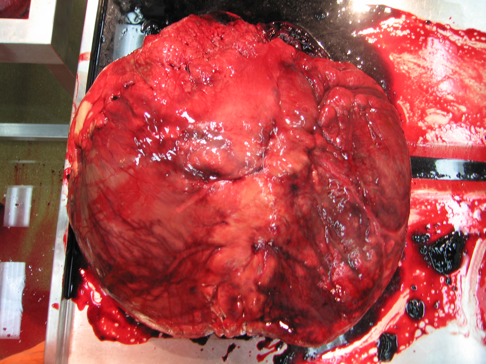
The big heart, needed to pump blood up that long neck to the head. Compare with the elephant and rhino hearts posted here before.
Left shoulder and ribcage, muscles of the triceps peeled back. Shoulder blade (scapula) visible. The neck extends up to the left corner.
Left side of chest, rumen (fermenting tank) showing through behind ribcage. Forelimb has been entirely removed here.
The left cheek’s teeth (molars)– and check out the spines on the inside of the cheek! They are keratinous growths to aid in chewing, food movement, digestion, protection against thorns, etc. These extend into the stomach, too! These amazed me the first time I saw them, in an okapi (giraffe cousin).
The brain, in bottom view. Olfactory nerves leading to the nostrils near the top (whitish), and optic chiasm for the eyes (“X” shape behind the olfactory nerves) are visible, then the medulla oblongata, smallish cerebellum and the spinal cord. For a human brain diagrammed and labelled in similar view, see here.
Like rhinos, elephants and many other large mammals, giraffes (especially in captivity) are vulnerable to foot/hoof pathologies, such as this very skewed/divergent pair of nails on the right front foot. This can lead to them walking very abnormally, getting infections or arthritis and other problems, so it is very serious.
The tapetum lucidum; reflective coating of the eye that can aid in night vision and protect the eye a bit. Gorgeous!
Hey, Americans and others happening to be gobbling down Meleagris gallopavo today– don’t forget to practice your anatomy! Such a great opportunity. Dig in to that carcass and horrify/amaze your family and friends! This pic might help you get started (info below if you want it), and is my WIJF blog wish of happiness to you all, today.
Stomach-Churning Rating: 6 out of 10; a small picture of some fresh turkey leg muscles, but not that bad really.
Click to embiggen.
Wondering what’s shown here?
On the left: an ossified (turned into bone!) tendon, probably part of the M. flexor perforans et perforatus group (a wickedly complex set of muscles that go from the knee region to the toes, and act mainly to flex the knee, extend the ankle and (plantar)flex the toes; i.e curl the toes up). What’s particularly cool is that, towards the top, you can see the divisions where the pennate (angled) fibers of the short, meaty muscle belly sat. If you are eating a turkey drumstick, you will be picking some of these out of the meat, although many turkeys seem to have fewer bony tendons due to human breeding and young age at slaughter.
In the middle, top: a crude experiment where we hung a frozen turkey’s body in a few different orientations to determine its centre of mass, important for biomechanical calculations. Mad science, but simple science.
In the middle, bottom: the right hip joint of a turkey in lateral (side) view, showing a few of the key muscles of the thigh. The ITC is M. (abbreviated Latin for Musculus) iliotrochantericus caudalis. Practice saying that (ill-ee-oh-tro-kan-tare-ick-us caw-dahl-iss) to impress your friends. It sits in a depression in the ilium (top pelvic bone), in front of the hip joint. The ITC is also important for helping birds to support their weight, as Steve Gatesy and I discussed in our 2000 Paleobiology paper. The ITC leaves a lovely crescent-shaped scar on the top of the femur (thigh bone). Show off your culinary skills by noting to your dinner party that this muscle is the best bit of the bird, AKA the “oyster”. (A little tip is here for how to find it; in a chicken but the anatomy is almost the same in a turkey)
The OM is the obturatorius medialis (obb-turr-ahh-tor-ee-us mee-dee-ahl-iss), an antagonist to the ITC, used to swing the leg. It is mostly hidden inside the pelvis so you just see its tendon (dotted line), and especially in turkeys (seriously, they have very nicely visible muscle attachments on their leg bones, for any bird!), a little knobby bit of bone that helps guide the tendon to keep it in its little groove on the femur. Unless you’re very industrious and break open the body cavity to excavate into the pelvis, you won’t be eating this muscle.
The IFE; M. iliofemoralis externus (ill-ee-oh-fem-oh-rahl-iss ex-ter-nuss); arching over the ITC and OM tendons, is a vestigial muscle, often lost in birds, and having little major function but helping a bit to draw the leg away from the body (abduction). Even though it is a puny muscle, it still has a nice little pit for its insertion on the femur. Turkeys are just cool that way. But it’s not much in the way of eating.
And now you know three of the ~40 main muscles of the avian leg, well done!
I love these muscles not only because I did a lot of my PhD (and later) research on them, but also because they leave great scars on bird and other dinosaurian bones that allow us to reconstruct how muscles evolved. I better stop here or I’ll be writing for days… don’t wind me up further! 🙂
On the right: the foot of a turkey in front and back views. Lots of ossified tendons are visible if you squint. Why do birds only have ossified tendons below their knee joints, and why only some muscles in some birds, and not so commonly in most other species of land animals? This is one of those cool mysteries that remain for people doing evolutionary or biomechanics research to sort out.
Hope you enjoyed a quick anatomy tour with our pal Meleagris!
A quick and easy Mystery Dissection post for you today– these objects are left over from a dissection we did awhile ago. What are they, and (for extra points) from what species (be as precise as possible)?
Speed round. Let’s see how many correct answers we can get in the next 24 hours!
Stomach-Churning Factor: 1. They won’t bite.
Difficulty: small image, oblique angle, object on the left side of the image is in the way (and not related to this post).
This will lead into a full-length blog post, hopefully to come sometime late this week, after Halloween. And there should be a Halloween bonus post this year!
I stumbled across some old pics, which I thought I’d lost, from the filming/preparations of 4 episodes of Inside Nature’s Giants (Jan-Feb 2009) at the RVC. They form a nice accompaniment to my previous post reflecting on my experience with the show, and the timing is great because I’m about to head to Raleigh, NC to talk about this research at the Society for Vertebrate Paleontology conference.
Stomach-Churning Rating: 4 at first (just a dead animal; and a rather clean one at that), then about halfway through the dissections start and it edges up to a 7 or so.
These pictures are sadly some of the few I have of the whole, intact body of a gorgeous adult Nile crocodile (Crocodylus niloticus) that the Windfall Films team managed to get to the RVC from La Ferme Aux Crocodiles in Pierrelatte, France. (I have scores of pics of the dissected limbs, shown further below) As the title indicates, it was a nice big croc. And as you’d expect, CT scanning and then dissecting it was no tiny feat, and makes a fun story. Story time, then, after an introductory pic!
Dr Samuel Martin, vet from La Ferme Aux Crocodiles, brought the crocodile (and some smaller specimens) over to our Hawkshead campus in late January 2009, and we quickly moved to run the specimen through our CT scanner to preserve some details of its anatomy (example shown at the end of this post) and for potential usage in the show. As the photos below illustrate, this was hard work for several people.
And then, as we were finishing the last CT scans of the specimen, our ageing medical scanner stopped working. And could not be resuscitated. R.I.P., Picker PQ5000 (buy one or two here!). The crocodile, “WCROC” as my team came to designate it, had claimed its final victim. It took about a year for us to get a new one, and that year sucked. It made me appreciate how lucky we are to have a CT scanner just across the parking lot from my office!
Anyway, the day of filming I was hoping to make it in to watch my colleague and friend Dr Greg Erickson help lead the dissection team, but a wicked blizzard blew up, and as I was starting the 31 mile drive south from my home to the RVC I realized, from the queue of cars that seemed to be 31 miles long (and train lines shut down), that this was going to be a snow day. So I turned around and came home. Another victory for WCROC!
The filming proceeded despite heavy snow delaying many of the key players’ arrivals. I got filmed a day or two later for a little section of the show on the limbs and locomotion of crocodiles but sadly this got cut from the main ING show (but did air in the National Geographic version “Raw Anatomy“, in the USA at least).
The limbs had been left largely intact, although some of the dissectors who didn’t know croc anatomy very well had slashed through parts of the pelvis and, in eagerness to reach key parts to demonstrate in the show, some major muscles got shredded. This is no big surprise; crocodiles have a lot of bones all over the place: in their skin (scutes; bony armour), in their bellies (the belly ribs called gastralia), and almost everywhere else, so some brute force is required to get to the gooey bits. Apparently there had been 6 or so people dissecting at once and things got a little carried away. The curse of WCROC continues?
Oh well; that’s just how documentaries go sometimes, especially with a pioneering show like this and the intensely compressed timescales of filming (time is ££!). There can be pulses of chaos. And the show turned out GREAT! (alternative link if latter does not work outside UK)
Let’s have more photos tell the story of the scanning, which also shows off this beautiful animal’s external anatomy:
Anyway, things turned out fine overall for our research. A week or so later (maybe longer; I forget if the specimen was frozen and thawed out for us) we came in to start dissections. We were really excited to measure the limb muscles of such a big crocodile, for comparison to a growth series (babies to adults) of alligators that my former PhD student (now postdoc; Dr.) Vivian Allen had dissected back in 2008. Here he is with a masked co-dissector, displaying their joy for the task at hand:
And let’s not leave out the exhuberance of visiting research fellow Dr. Shin-Ichi Fujiwara! He wanted to inspect the forelimbs for his ongoing studies of limb posture, joint cartilages and locomotor mechanics.
The remaining images show progressive stages of dissection of WCROC, starting from the pectoral (fore-) limbs with a view of the belly (and the giant jaw-closing muscles visible on the left side of image):
Isolated right forelimb, with coracoid (part of shoulder girdle) sticking through:
Assorted forelimb/upper arm (brachial) muscles:
And the triceps (elbow-straightening) muscles; not that big in such a big animal:
…and on to the pelvic limbs and the huge tail:
With a closer look at the HUGE thigh muscle, the famed M. caudofemoralis longus:
And then an isolated right hindlimb:
Thigh muscles, with which I have a peculiar fascination that stems from my PhD research:
And last, the great, paddle-like hind foot!
What a great experience that was! We have fond memories of WCROC, a great documentary from Windfall Films, some nice data– and a lovely skeleton. Perhaps the curse of WCROC is not so bad. Nothing can go wrong now!
Soon Mieke Roth, scientific illustrator from the Netherlands, is coming here to do a similar dissection on more Nile crocodiles at the RVC. As with the octopus she wrote about in September, she will make a 3D model, but with much more detail and with an emphasis on accuracy and accessibility. The end products will be really cool; think of the visible body, 3d models that can be used in teaching, animations, a book and lots more but also a “how did she do that?” blog. To finance this project (that probably will take a year or more) she will use crowd funding. In several weeks there will be more info on how to participate in her fantastic endeavour. For now, see her video with the initial pitch for “Nile Crocodile 2.0“!
In case you haven’t heard, Saturday, September 22nd, 2012 (today, at this writing) is World Rhino Day! The main websites include here and here. Ivan Kwan has also posted a fantastic blog entry “Rhinos are not prehistoric survivors” for WRD2012- check it out! And if you haven’t seen the WitmerLab’s AWESOME Visible Interactive Rhino site, you really really need to (in fact, quit reading this and go there first; it is soooooo good!).
I’ve written about the global rhino crisis before, and about rhino foot pathologies. The title of today’s post may be “cute”, or at least goofy, but the real situation is as grim as the images I’ll share. I won’t repeat the explanation, but all five living species of rhinoceroses are in serious trouble. There’s a good chance that most or all of them will go extinct quite soon– see the previous links for more information on this. Javan and Sumatran rhinos are dangling the most precariously over the precipice of extinction. My goal in this post is to share the beautiful, complex and exotic anatomy of rhinoceros anatomy and movement, and the joy of contributing new scientific information about poorly understood species.
Stomach-Churning Rating: 7/10— dissections, and there are a couple of pics where the specimens are not so fresh, and there’s big skin, and a huge heart.
Baby white rhinoceros. Will frozen specimens like this be all we have of rhinos someday?
The purpose of today’s rhino post is to share a bit more; especially images; of the work my team has done on rhinoceros gait and limb anatomy; all of it unpublished but hopefully coming soon. We’ve steadily been collecting data since ~2005. Because my previous post went through some of this, I’ll keep it brief and image-focused.
First, a video of one of our amusing encounters with a white rhinoceros, at Woburn Safari Park. In this study, we wanted to measure, for the first time really, the gaits (footfall patterns) that a white rhinoceros uses at different speeds, and how often it uses those different gaits. We attached a GPS unit on a horse surcingle around the rhino’s torso, which measured the animal’s speed once a second. We then observed 5 individuals (1 at a time over various days), following them in my station wagon (estate car) across the safari park. We filmed them with a conventional camcorder to document their gaits, and concentrated on the two periods of the day that they’d normally be active: when released from their overnight barn, and when coming in for the night back to that barn. They got rather excited and frisky some of those times. The GPS belt then kept recording speeds for the rest of the day; unsurprisingly, the rhinos generally did not do much. I have to thank Nick Whiting, rhino handler, for his help making this research happen. I’ve been meaning for too long to finish the final paper… soon, I hope! Enjoy this tense scene of a rhino investigating my car (driven by me and with an undergraduate student filming) then having a nice canter/gallop across the field (accompanied by my jubilant narration).
Like our foot pressure research, we aim that this work provides baseline data useful to caretakers of rhinos; for example, to test if a particular animal is lame. This follows what we’ve successfully done with elephant gaits and feet, translating basic research into more clinical application. But my major scientific interest is in understanding more about what makes any rhinoceros, even a 2-tonne White rhino, so much more athletic than any elephant (even a baby or 2-tonne small adult Asian elephant). As the video shows, they can use a variety of gaits including cantering and galloping, and trotting at slower running speeds. No elephant ever does that, and no one knows precisely why. The leg bones are more robust, but the muscles aren’t that dramatically larger in rhinos.
An Indian rhinoceros forelimb- note the characteristic knobbly hide, unlike the smoother, more elephant-like hide of a White rhinoceros.
Similarly, the anatomical work we do with rhinos is intended to not only be useful science for comparative biologists like me, showing how rhino limbs work and how they differ from those of other animals, but also to aid clinicians in comparing normal vs. pathological anatomy. For conveying that anatomical work, I’m lucky to have been granted permission to use a professional photographer’s pictures of some of my freezers’ rhino specimens– big thanks to James King-Holmes and the Science Photo Library. The watermarked images below belong to them. I ask that you do not use them elsewhere, honouring their license to me for personal usage on this website (and I will only use them here). I’m in all the images, which makes me feel weird putting them up here, but it’s about the rhinos (and freezers), not me. First: the infamous “rhino foot freezer”, featuring some of its denizens:
…and inside we go (and I begin to get frosty and numb-fingered from holding a foot; my smile soon fades):
Taking a rest with the skinned white rhinoceros foot:
And now warming up at the “digital freezer”, our CT scanner, and preparing to scan another rhinoceros foot, which segues nicely out of this image sequence:
Now over to some 3D anatomy– segmented reconstructions of rhinoceros fore (top) and hind (bottom) feet, from CT scans; if you’ve frequented this blog you know the drill. Here, the longest bones are the metacarpals/metatarsals and the upper bones are the carpals/tarsals, then the bones near the botttom are the phalanges, which connect to the hooves (visible in the bottom image):
I’ll wrap up with a series of images of basic limb muscle anatomy from dissections we’ve done of baby and adult Indian and White rhinoceroses. First, here’s what a rhino looks like underneath the skin:
But ahh that skin, that fabled “pachyderm” skin! A rhino’s greatest defense is also a real chore to get through in a dissection. Here, we enlist the help of a crane and hook, hurrying to get down to the muscles of this forelimb before rotting takes over too much (as with other big animals, this is a tough race against time even in chilly England!):
Here is a closer look at that amazing armoured skin; sometimes 10cm or so thick:
Back to the forelimb muscles– stocky and well-defined for this athletic animal:
(late addition) Here are the massive shoulder muscles, such as the serratus and latissimus dorsi (this is a left limb in side view; head is toward the left):
And now a close look at the forearm muscles:
And then over to the hindlimb, here from an adult Indian rhino, whose thigh bone (femur) shows the characteristic giant “third trochanter” (toward the bottom centre of the image), which is an expanded bony attachment for the giant “gluteobiceps” muscle complex that retracts the femur for the power stroke in locomotion. Also, this specimen showed fascinating anatomy that I’d never seen before: the third trochanter has a thin bar of bone that extends up (toward the bottom left in the image) to fuse with the greater trochanter, opposite the head of the femur (upper left corner):
Damn my photography skills, cutting off the edge of that image and instead giving a view of my boots! Anyway, another interesting feature of that femur: the medial (inner) condyle of the femur (knee joint surface) has a pink stripe of worn cartilage. This is indicative of at least a moderate stage of arthritis, shown here (look for the pinkness amidst the shiny, healthy white cartilage on the upper right side). It is an exemplar of serious welfare problems that some captive, and probably some wild as well, rhinos face:
(late addition) Back up the limb, this baby White rhino shows the massive thigh muscles, especially that “gluteobiceps” that attaches to the third trochanter, noted above, and also showing the hamstrings:
Moving down the limb, we encounter the glorious three-toed perissodactyl foot of rhinos, and the robust hooves/nails, which are reasonably healthy in this animal– unlike others I’ve seen:
And the sole of that foot, showing a fairly healthy pad, below. Toward the rear (away from the nails), it culminates in a modest-sized fat pad, or digital cushion, akin to that in elephants but far less well developed and lacking the false “sixth toe” (predigit) (see also CT scan movie of the hindfoot above):
Here’s a view inside that marvelous foot, showing the HUGE digital flexor tendons. These help support the toes against gravity and, in theory, can act to curl them up– although in a rhino’s foot, as in an elephant’s, the toes are more like a single functional hoof, with reduced independence compared to a carnivore or primate:
And that ends our tour of rhinoceros limb anatomy and function. Help spread the word of how precious and threated rhinos are; educate yourself and others! And if you overhear someone talking about using rhino horn for medicine, try to politely educate them on the utter fallacy of this tradition. It is this cruel, greedy, ignorant practice that needs to die; not rhinos. I don’t enjoy receiving dead rhinos, on a personal level, even though the science excites me. I’d rather have many more alive and living good, healthy lives. And my team is trying to do what we can to help others on the “front lines” of rhino conservation make that happen.
For example, Will Fowlds, vet and co-owner of Amakhala Game Reserve, South Africa, recently sent us some images of a white rhino that had been caught in a poacher’s foot snare some years ago. The poor rhino still was having problems healing– we inspected x-ray images and external photos and helped to make an initial diagnosis of osteomyelitis, a nasty infectious, inflammatory foot bone/joint disease. We are following this case to hope that the rhino recovers and contribute help where we can, but the tough job belongs to the keepers/vets on the ground, not to mention the rhinos…
Furthermore, we’ve done foot pressure research covered here, and here is an example of the data we’ve collected (image credit: Dr Olga Panagiotopoulou), showing high pressures on the toes and low pressures on the foot pads:
Big thanks to people on my team that have helped with this and related research: Dr Olga Panagiotopoulou (and Dr Todd Pataky at Shinshu University, Japan), Dr Renate Weller in the VCS Dept at the RVC, Liz Ferrer at Berkeley, and former undergraduate student researchers Sophie Regnault, Richard Harvey, Hinnah Rehman, Richard Sheehan, Kate Jones, Bryony Armson and Suzannah Williams.
A White rhino’s heart, with more images below, all courtesy of William Perez’s Veterinary Anatomy Facebook pages. A mass of around 10kg (22 lbs weight) is not unusual! (Compare with even larger elephant heart)
The Olympics are over! Hooray for everybody! Here I’m taking a different spin on the blog’s Mystery Dissection series: a mysterious method– what is going on here and why? I know someone will get this, but experts might want to hold back and let non-specialists have their moment in the sun first…
Stomach-churning rating: 6/10. (but probably too late!) Be glad this image does not include an olfactory component.
The scene is from ~2005 and took place in our old lab- note the filthy floor. It rarely got much cleaner than that. Before we moved in, this was an abattoir at some point. We used to have experiments going on in this hangar-like area of our small office building, right next to our office rooms. The smell and noise was… unpleasant. Luckily, now we have a separate office building (renovated from this old structure) and a wholly new, giant gait laboratory full of science toys.


")
")
")
")
")
")
")
")


")


