Good morning, Freezerinos! Here is a twin treat for you to puzzle over. Two things, perhaps rather squidlike at first glance, but not cephalopods. There is a conceptual connection between the two images. Can you identify both of these structures? Huge bonus points if you can identify the taxon they belonged to, but stabs at it are encouraged; there are clues in the images…
(labels have been removed to protect the innocent)
With the help of Heinrich Mallison of dinosaurpalaeo and his student Sebastian Marpmann (now having finished the Great Giraffe Deconstruction), I did a quick cleanup and reorganization of the big walk-in freezer we’ve all come to know and love as Freezersaurus. This had to be done because some of the big stuff was becoming a terrible obstacle to cross in order to get anything from inside; cue health-and-safety paperwork nightmares. And yes, the ice penis is now gone. End of an era…
All photos henceforth by Doktor Mallison:
Emerging from Freezersaurus; most big stuff removed (note our giraffe's metacarpus trying to escape, on right side)
The big stuff. Emus, elephant feet, too much giraffe (metacarpus continuing to sidle away), and horse legs.
Carnage remaining inside; needed to be taken out gradually and reorganized, to make more space on floor for intermittent human presence (i.e. walking in).
Elephant feet, mystery giraffe pelvis, oh my!
For ME!?!? Aww shucks, you shouldn't have. Reorganizing the small stuff on shelves. PhD student Mike Pittman makes a guest appearance, delivering crocodile vertebrae.
Sebastian poses with giraffe buddy. We emphasize the Buddy System when dealing with Freezersaurus; she is a treacherous hostess. Note that Sebastian has also cunningly halted the abscondence of the giraffe metacarpus.
Job done! Farewell Freezersaurus! You look mahvelous! Wall of archosaurs on the left; wall of synapsids on the right, and sundry giant mammals in the middle.
And so we finished, and so you’ve now had a very intimate look at Freezersaurus too! Don’t you feel lucky? 🙂
Without further ado, what’s up with this specimen from The Freezers? What is it, what animal etc?
(admin note, 28 Oct 2012: Mystery Dissections 2-3 do not exist, mysteriously. At the time, in the jumbled freezers of my mind, they corresponded to Mystery CT Slice(1) and Mystery CT Slice 2. But we can pretend that MD2-3 are just an eternal mystery of this blog, subject of numerous conspiracy theories that you are welcome to expound upon!)
There’s no better way to kick things off after a holiday than with a celebration of the Inside Nature’s Giants series, which I had a small part in early on, including these shots I took during the time they spent filming at the RVC >3 years ago (!?!?); most of these animals spent multiple holidays inside The Freezers:
Elephant arriving…
Elephant revealed
Private moment with elephant
Stunning emergence of The Guts
So you are impressed by the guts too, ehh? It was pretty amazing to watch it happen. The tension was intense- the animal had been dead for a while and was rather bloated. So cutting it open was a task gingerly taken…
Bloated elephant
RVC dissector Richard Prior stuck a scalpel in the upper abdomen when the time was right… the piercing whistle and the sulphuric odour silenced the crowd watching… and then quickly out came the guts.
Everyone was pretty amazed by the scale.
The guts just went on and on…
Not a 1-person job by any means.
Spreading them out to see the whole GI tract.
I waited patiently and watched the show filming; what a great, professional crew. Then I got to take the legs away for our research.
But not just elephants, no sirree! The Windfall Films/ING team filmed giraffe, crocodile and big cats episodes (4 total) at the RVC too; a crazy period of a few weeks (including a major blizzard that hit us during the croc filming) in 2009. Some of the stars follow:
…and I’m rather fond of that tiger’s neck– check out the hyoids (roaring/tongue apparatus in throat; bottom of movie)!
…and here is the adult Nile crocodile’s head after scanning
…and another view of that big Nile croc, just because I like how this reconstruction turned out
…and here’s one of the small (~1m long, 10kg) juvenile Nile crocodiles from the show, with a pilot CT scan showing the skeleton nicely- and possibly a last meal or stomach stone on the left side of the abdomen (bright white blob; I need to check this now that we’ve dissected it)
Foetal giraffe; stillborn; from the show, in process of dissection in our lab to measure its limb anatomy. Trust me, it looked –and smelled– better on the inside than it did from the outside. Eew.
How most of the specimens from the first 4 episodes ended up after all dissection was done (part of my/RVC’s collection of skeletons). Sadly, I did not get great photos of the 3.7m Nile crocodile or the two giraffes before they were reduced to bits, but I do have the skeletons and CT scans.
Giving a tour (including The Freezers) to A Certain Esteemed Visitor.
(Another) Gratuitous shot with one of the sweet old Red Kangaroos at Alma Park Zoo near Brisbane, Australia. Experiments on hopping we did there will be briefly featured in the new Inside Nature’s Giants show on Channel 4, 16 April @2000- details at http://t.co/SkjsMeVC.
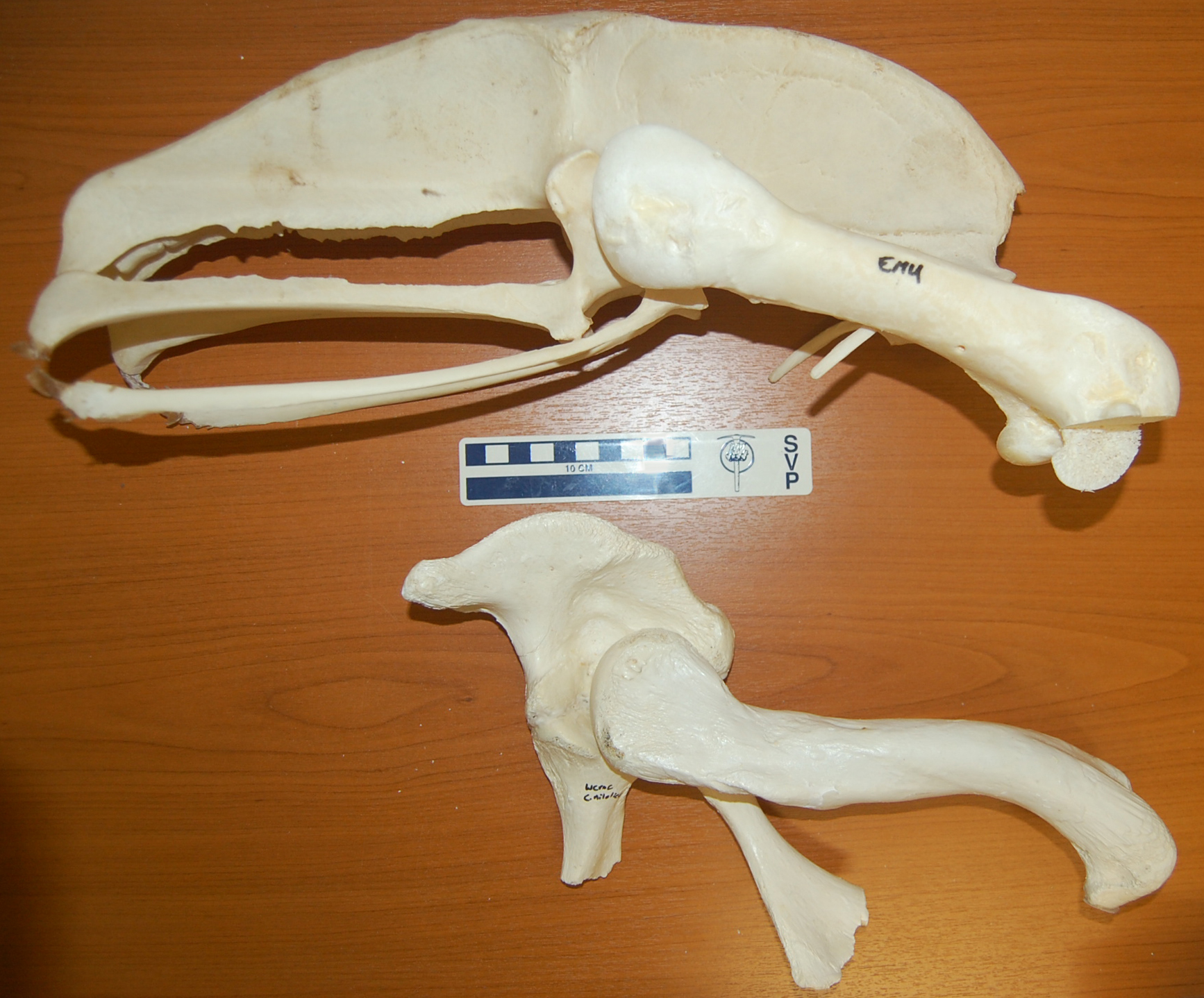
I’m off soon for a sunny break on the beaches of Morocco, but as an Easter gift to you, my (admirable, sagely, few, beautiful) readers, here is an image of two specimens, formerly from my freezers, to consider:
Both are the right pelvis (hip bones; crocodile pelvis is a bit broken toward the bottom) and femur (thigh bones) of living archosaurs– a 27kg emu above, and a 278kg nile crocodile below. The head would be toward the right side of the picture. A tenfold difference in mass between the bird and the crocodile, and yet some of the dimensions are so similar in both of them (femur length etc.), or so vastly different in the bipedal runner vs. the quadrupedal not-so-fast-runner (much bigger pelvis for leg muscle attachments in the former).
This image says it all. It is why I study the evolution of locomotion in land animals. It is why I am so fascinated by the transition from vaguely crocodile-like early archosaurs to modern birds by way of earlier dinosaurs. Anatomy, size, mechanics, behaviour, phylogeny… the photo captures all the facets of why I am so enraptured by research in this field.
It also might evoke thoughts of what features are expected in a terrestrial vs. aquatic animal, and thoughts of how some numbskulls still think big dinosaurs lived in the water (no I will not link to the execrable story from BBC today that I am thinking of!)…
I hope you appreciate it, too. Have a freezer-burn-free holiday period, folks!
Heinrich Mallison’s photo-rific dinosaurpalaeo blog has the first of what might, if the Gods of the Freezers remain kind, be a series of posts on our dissections of some of the verrrrrry same giraffe limbs featured earlier on this blog. Have a brush with greatness- see the giraffe legs in deconstruction! For free! What more fun could you possibly have (legally)?
Here now is the promised blog post, which uses the rhino foot mystery pic as a springboard to address a phenomenon that is a bit better known, partly because it is an even worse situation and involving (arguably) even more charismatic critters: elephants.
A rotating movie of a CT scan reconstruction is a good way to kick this off:
This shows the right hind foot of an Asian elephant that had mild pathology; mostly a roughening of some of the bone surfaces that is called osteitis (proliferative bone growth possibly due to infection or other irritation) and perhaps a mild case of degenerative joint disease such as osteoarthritis. But this is nothing compared to the severe cases we’ve observed in other elephant feet, and indeed may not have anything to do with why this elephant died (I’m not sure; I was given very little medical history for this one).
If you want more elephant anatomy lessons, see the videos from the posting on six-toed elephants. I will proceed assuming some basic familiarity with bones of the feet in animals, although you may be just fine even without that.
About 50% of elephants in captivity die from foot disorders of one kind or another. Elephant keepers spend a huge amount of time and energy taking the best care of elephant feet that they can, but a variety of factors including anatomy, biomechanics, exercise, obesity, ground surface, hygiene, “hoof” care including trimming, nutrition, and much more are part of the very complex causal nexus underlying these disorders. Wild elephants get similar problems, too, but less frequently (e.g. in drought periods, I’m told); there are few solid data on this, however.
Onwards, then! I shall present a cavalcade of horrific examples of the kinds of elephant foot pathology that we have observed in specimens that have come through my freezers at the RVC.
Let’s start with what one of our vets might see on examination of a live elephant at a zoo:
This is an x-ray image of the third (on the left) and fourth (on the right) toes of an elephant’s front foot. The RVC (Dr. Renate Weller and myself) have developed protocols to take such x-rays on live elephants. The anatomy shown here is pretty normal and non-pathological. So with that in mind, check this out; toes four (on the left) and five (on the right), different animal:
Ouch! Digit 4 (“ring finger”) has a proliferation of bone that is characteristic of an animal with osteomyelitis: a flowering of bone in response to infection and painful swelling, probably caused by an abscess on the toe’s sole/nail. This animal was put down because of its unresolvable misery from this disorder. Oddly, we see toe 4 as well as 3 and 5 as the most commonly pathological; toes 1 and 2 seldom are. We’ll be discussing this in a new paper coming out soon; I’ll get back to that another day.
Assuming such conditions don’t resolve, the next place the foot may end up is in The Freezer at the RVC, and then into our CT scanner before we do postmortem dissections and a report on the pathologies so the zoo knows what went wrong. Here’s an example of what we cut off the end of the fourth toe of such an animal:
Just looks like a glob of tissue, right? The joint between two segments of the toe is visible as a pinkish white structure on the right side, with some bleeding on the cartilage where it wore down to the bone surface. But it gets worse. Here is how that same toe bone looked when we cleaned it up (boiling and bleaching away soft tissues):
Here, that same roughened joint surface is visible at the top of the specimen. Two toe bones have become fused together (the bottom one is not visible), encased in a cocoon of lacy, spiny bone. Again, ouch. The next specimen had a different kind of “ouch”- its fifth toe basically shattered:
That toe is almost unrecognizable, having disintegrated rather than proliferated its bony scaffolding. Other specimens may be in less extreme states of pathology but still likely to have been in pain:
The label here says it all; third toe with a cyst where an infection entered the bone.
This one, the end of a third metatarsal, shows degenerative joint disease with a loss of articular cartilage, and holes where abrasion has worn down into the bone and caused bleeding. In contrast, and to give you a breather from the horrors, here is a healthy, younger elephant’s similar joint surface:
Nice white, fresh, shiny cartilage! Ahhh…
But then we dive back into Grand Guignol-level aberrations:
Here we’re looking at the back side of a right hind foot of an elephant, at the level of the ankle joint. The joint capsule surrounding the ankle joint has been cut open in my dissection to expose the terribly pathological, but still somewhat white and shiny, cartilages (middle of the image) which have been abraded (in some regions) but also extended by new bone formation (in other regions) to creep around the back of the ankle. Here, the bone growth was fulfulling a role to limit joint mobility and thereby restrict painful joint motions- the joint was fusing into an ankylosis (no, not an ankylosaur, but same Greek root). Here is a closer look, removing the tibia and fibula that were at the top of the screen in the above image, and looking down onto the ankle joint surface:
You should be able to more clearly see how the cartilage and underlying bone are not forming a smooth edge, as they should on the talus (ankle bone), but rather an irregular, jagged contour (area to the right of the label). This animal would have been visibly lame, to say the least; elephant ankles can’t move much even in normal animals but this one was even less mobile. We’ve had some specimens where the ankle was so fused it was totally immobile and took a saw to separate the two sides of the joint. Oddly, I haven’t seen an ankylosis like that in the wrist, which in normal elephants is as flexible a joint as the ankle is inflexible.
Pathologies like these sadly aren’t uncommon in elephant feet but zoo/park keepers are doing their best to turn the trend around. Zoo conditions generally were a lot worse 50 years ago. The pictures below document this, from museum specimens we’ve studied (among many others) at the University Museum of Zoology at Cambridge and the Natural History Museum in London. See what pathologies you can spot! Some are from wild-shot animals, reinforcing that foot pathologies are not just a zoo thing. (click to embiggen)
Zoo/park conditions are improving now— in the UK for example, elephants are being moved into more safari park-like environs and given more varied surfaces to walk on or even dig (e.g. sand at Chester Zoo). But because elephants live long lives, and foot pathologies sometimes cannot be reversible (or even detectable, sometimes), any pathologies existing now may well still be evident, or even worsen despite the best care, for decades to come. The lag time for fixing the global problem of elephant foot pathologies is not a short one. I won’t get into the controversy over whether elephants should be in zoos/parks or not, but at least for the short to medium term they are, and we need to make the best of that. The images in this post help show why, and perhaps point a way toward how.
I will go into more detail soon on the broader subject that this involves, but am posting this image as a teaser– what’s up with this foot from my freezer?
Other than the obvious dead-ness and non-attached-to-body-ness…
Utterly puerile post ahead. I was just in one of those silly moods… Six-year-old daughter, lately with a strong potty-humour tendency, will do that to you. So with that forewarning in mind…
I was rummaging around in the back of Freezersaurus yesterday and was quite surprised to encounter this:
I am deeply, deeply disturbed. And shocked. And a bit violated. Cover your shame, Freezersaurus! Now, I’ll freely admit, penises are great. Hilarious floppy bits to the common person, and fascinating adaptations to the scientist; e.g., duck penises, alligator penises… I’ll never forget the time my invertebrate zoology teacher showed a video of barnacle penises (immobile animals that need to reproduce by copulation– you do the math). But I digress. I was conveying my disturbed feelings about this blatant ICE PENIS in my freezer. Clearly Freezersaurus was either very happy to see me; perhaps titillated by all my rummaging around; or I need to get out more and get my mind out of the anatomical gutter. C’mon, look closer:
In any other place, it might just be an icicle. But here, under the baleful Eye-of-Sauron-like gaze of Freezersaurus’s fan unit, it can be only one thing. Penisicle. I’m not sure what to do with it now. It was such an awkward moment, I had to back off and leave The Freezer to its privacy. Not sure if I can go back there, especially not alone.
My therapy sessions start Monday. I’ll keep you posted on my progress. In the meantime: My gift to you: another emanation from The Freezer; because the last Mystery Dissection pic was too hard and then too easy after a not-so-subtle nudge…
OK, I had an inspiration for another short image-based blog post. People seemed to enjoy the wallaby mystery photo, so I’ll start a tradition here of mystery images from dissections of “my” specimens. That could even be an easy quasi-weekly thing to do.
What’s this animal? Binomial required. One guess per person please, unless you all get stumped. Bonus myology-nerd points to those who can name all the labelled muscles without checking literature (I’ve used these abbreviations a lot in my papers!).
Too hard I think! Extra hint sneak peek as per comments; same specimen — hmm what’s this, a jaw or something else? Bad image (these things aren’t easy to come by for photo ops), but quite distinctive…


")
")