Feeds:
Posts
Comments

Archive for 2013

Models of a basal dinosaur and bird, showing methods and key differences in body shape.

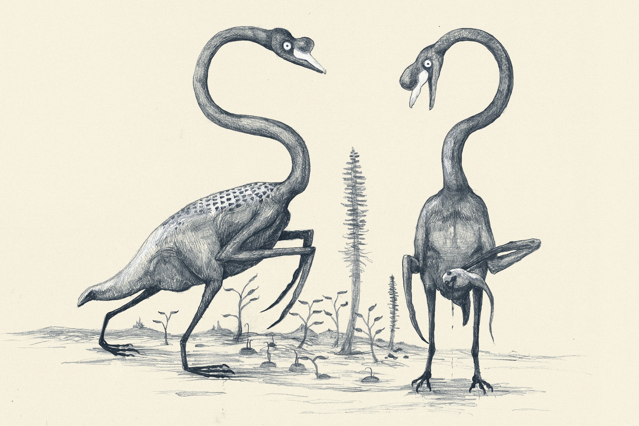
Our 3D computer models of a basal dinosaur and bird, showing methods and key differences in body shape. The numbers at the bottom are museum specimen numbers.

At about the moment I’m posting this, our Nature paper (our more formal page here, and the actual article here) embargo is ending, drawing a 14+ year gestation to a close. The paper is about how dinosaur 3D body shape changed during their evolution, and how that relates to changes in hindlimb posture from early dinosaurs/archosaurs to birds; “morpho-functional evolution” sums up the topic. We used the 3D whole-body computational modelling that I, Allen and Bates (among others) have developed to estimate evolutionary changes in body dimensions, rather than focusing on single specimens or (as in our last study) tyrannosaur ontogeny. We’ve strongly supported the notion (dating back to Gatesy’s seminal 1990 Paleobiology paper) that the centre of mass of dinosaurs shifted forwards during their evolution, and that this shift gradually led to the more crouched (flexed) hind leg posture that characterizes living birds. Here is a movie from our paper showing how we did the modelling:

And here is a summary of our 17 computer models of archosaur bodies, shown as one walks along the tips of the phylogeny shown in the video (the models are not considered to be ancestral to one another; we used a common computer algorithm called squared-change parsimony to estimate ancestral state changes of body dimensions between the 16 numbered nodes of the tree):

But we’ve done much more than just put numbers on conventional wisdom.

We’ve shown, to our own surprise, that the shift of the centre of mass was largely driven by evolutionary enlargements of the forelimbs (and the head and neck, and hindlimbs, to a less strong degree), not the tail as everyone including ourselves has assumed for almost 25 years. And the timing of this shift occurred inside the theropod dinosaur group that is called Maniraptora (or Maniraptoriformes, a slightly larger group), so the change began in animals that were not flying, but not long before flight evolved (depending on whom you ask, what taxonomy they favour and what evidence one accepts, either the smaller clade Eumaniraptora/Paraves or the bird clade Aves/Avialae).

Now, if you don’t like the cliche “rewriting the textbooks”, do have a look through texts on dinosaur/early avian palaeobiology and you probably will find a discussion of how the tail shortened, the centre of mass moved forwards as a consequence, the caudofemoral musculature diminished, and theropod dinosaurs (including birds) became more crouched as a result. We did that to confirm for ourselves that it’s a pretty well-accepted idea. Our study supports a large proportion of that idea’s reasoning, but modifies the emphasis to be on the forelimbs more than the tail for centre of mass effects, so the story gets more complex. The inference about caudofemoral muscles still seems quite sound, however, as is the general trend of increased limb crouching, but our paper approximates the timing of those changes.

Figure 3 from our paper, showing how the centre of mass moved forwards (up the y-axis) as one moves toward living birds (node 16); the funny dip at the end is an anomaly we discuss in the paper.

Figure 3 from our paper, showing how the centre of mass moved forwards (up the y-axis) as one moves toward living birds (node 16); the funny dip at the end is an anomaly we discuss in the paper.

A final implication of our study is that, because the forelimbs’ size influenced the centre of mass position, and thus influenced the ways the hindlimbs functioned, the forelimbs and hindlimbs are more coupled (via their effects on the centre of mass) than anyone has typically considered. Thus bipedalism and flight in theropods still have some functional coupling– although this is a matter of degree and not black/white, so by no means should we do away with helpful concepts like locomotor modules.

And in addition to doing science that we feel is good, we’ve gone the extra mile and presented all our data (yes, 17 dinosaurs’ worth of 3D whole body graphics!) and the critical software tools needed to replicate our analysis, in the Dryad database (link now working!), which should have now gone live with the paper! It was my first time using that database and it was incredibly easy (about 1 hour of work once we had all the final analysis’s files properly organized)– I strongly recommend others to try it out.

That’s my usual general summary of the paper, but that’s not what this blog article is about. I’ll provide my usual set of links to media coverage of the paper below, too. But the focus here is on the story behind the paper, to put a more personal spin on what it means to me (and my coauthors too). I’ll take a historical approach to explain how the paper evolved.


This paper’s story, with bits from the story of my life:

Embarassing picture of me before I became a scientist. Hardee's fast food restaurant cashier, my first "real job."

Embarassing picture of me before I became a scientist. Hardee’s fast food restaurant cashier, my first “real job”, from ~1999- no, wait, more like 1986. The 1980s-style feathered (and non-receding) hair gives it away!

Rewind to 1995. I started my PhD at Berkeley. I planned to use biomechanical methods and evidence to reconstruct how Tyrannosaurus rex moved, and started by synthesizing evidence on the anatomy and evolution of the hindlimb musculature in the whole archosaur group, with a focus on the lineage leading to Tyrannosaurus and to living birds. As my PhD project evolved, I became more interested and experienced in using 3D computational tools in biomechanics, which was my ultimate aim for T. rex.

In 1999, Don Henderson published his mathematical slicing approach to compute 3D body dimensions in extinct animals, which was a huge leap for the field forward beyond statistical estimates or physical toy models, because it represented dinosaurs-as-dinosaurs (not extrapolated reptiles/mammals/whatever) and gave you much more information than just body mass, with a lot of potential to do sensitivity analysis.

I struggled to upgrade my computer skills over the intervening years. I was developing the idea to reconstruct not only the biomechanics of T. rex, but also the evolutionary changes of biomechanics along the whole archosaur lineage to birds– because with a series of models of different species and a working phylogeny, you could do that. To me this was far more interesting than the morphology or function of any one taxon, BUT required you to be able to assess the latter. So Tyrannosaurus became a “case study” for me in how to reconstruct form and function in extinct animals, because it was interesting in its own right (mainly because of its giant size and bipedalism). (Much later, in 2007, I finally finished a collaboration to develop our own software package to do this 3D modelling, with Victor Ng-Thow-Hing and F. Clay Anderson at Honda and Stanford)

Me and a Mystery Scientist (then an undergrad; now a successful palaeontologist), measuring up a successful Cretaceous hypercarnivore at the UCMP; from my PhD days at Berkeley, ~2000 or so.

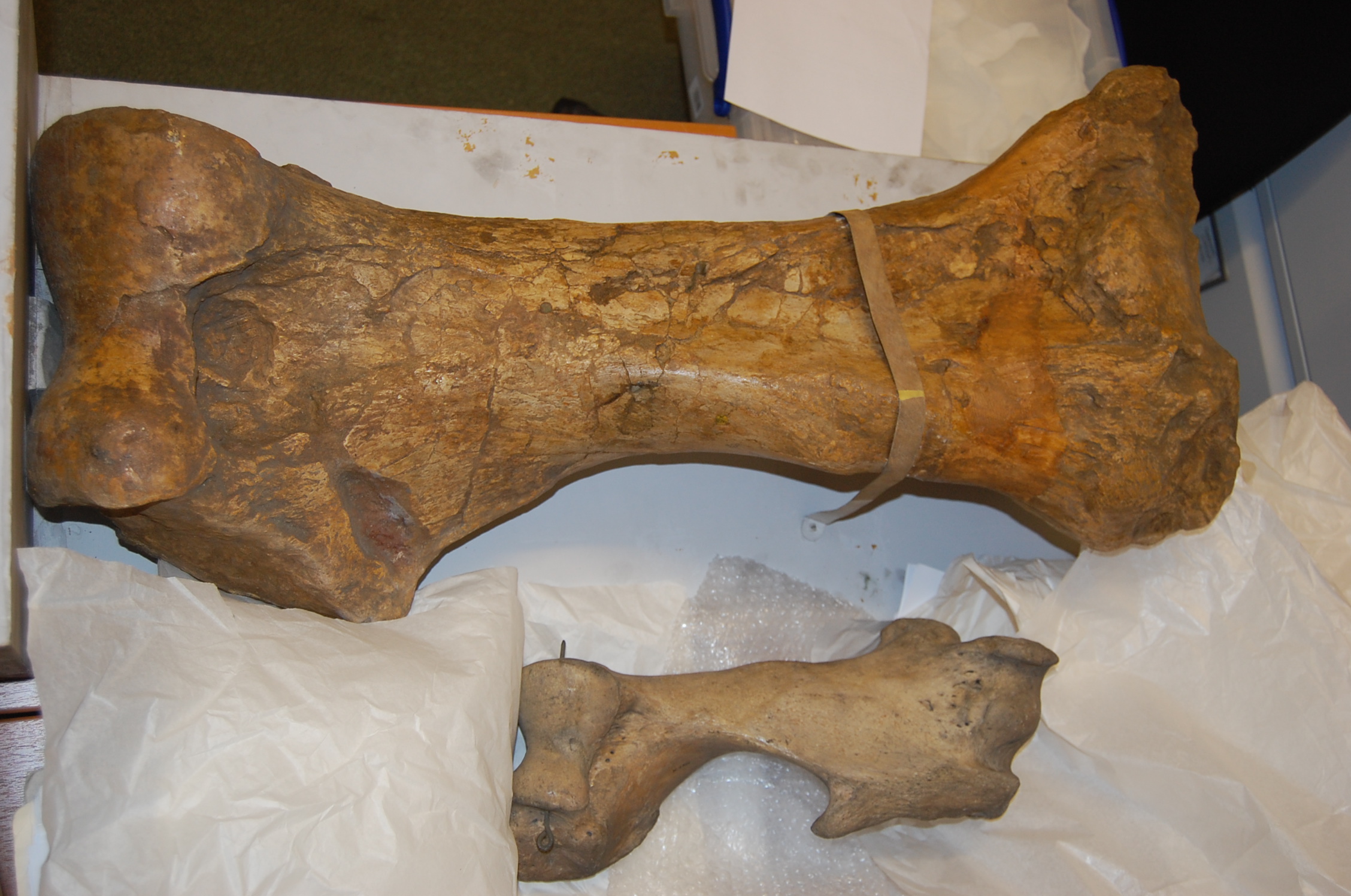
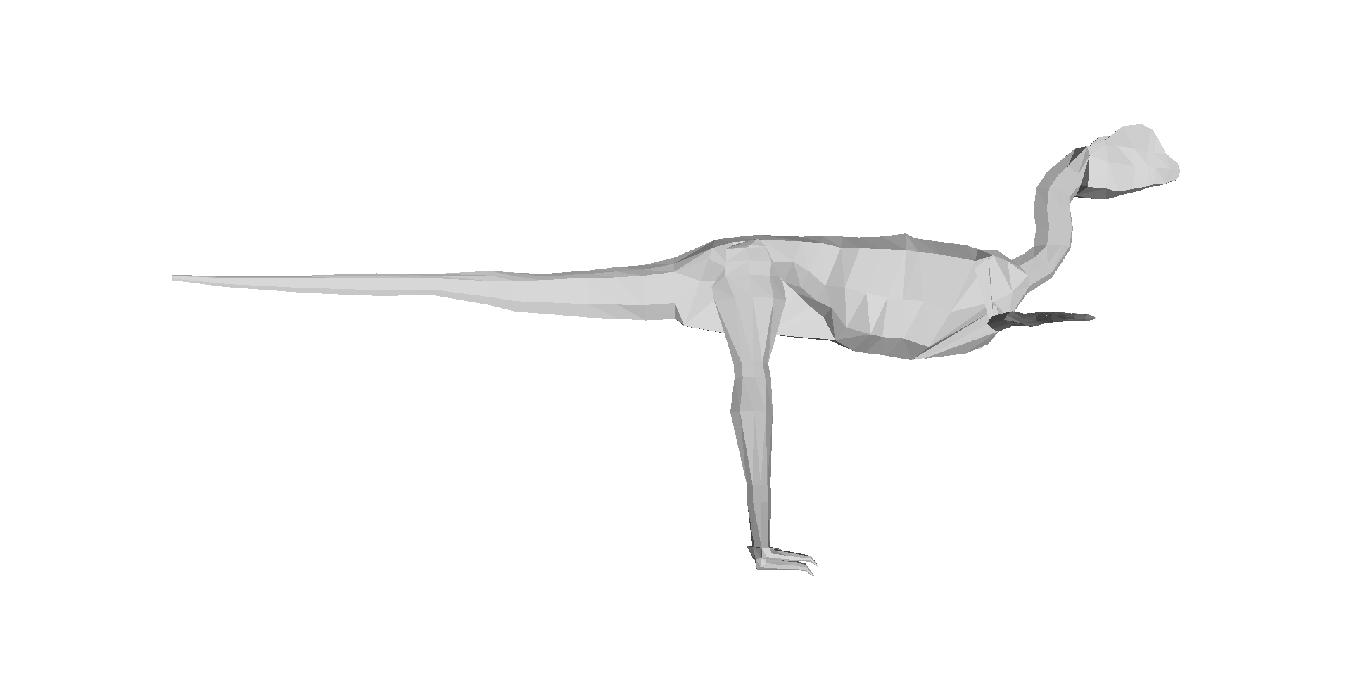
Me and a Mystery Scientist (then an undergrad; now a very successful palaeontologist!), measuring up a successful Cretaceous hypercarnivore at the UCMP; from my PhD days at Berkeley, ~2000 or so.

In all this research, I was inspired by not only my thesis committee and others at Berkeley, but also to a HUGE degree by Steve Gatesy, a very influential mentor and role model at Brown University. I owe a lot to him, and in a sense this paper is an homage to his trailblazing research; particularly his 1990 Paleobiology paper.

In 2001, I got the NSF bioinformatics postdoc I badly wanted, to go to the Neuromuscular Biomechanics lab at Stanford and learn the very latest 3D computational methods in biomechanics from Prof. Scott Delp’s team. This was a pivotal moment in my career; I became partly-an-engineer from that experience, and published some papers that I still look back fondly upon. Those papers, and many since (focused on validating and testing the accuracy/reliability of computer models of dinosaurs), set the stage for the present paper, which is one of the ones I’ve dreamed to do since the 1990s. So you may understand my excitement here…

Stanford's Neuromuscular Biomechanics Lab, just before I left in 2003.

Stanford’s Neuromuscular Biomechanics Lab, just before I left in 2003.

But the new paper is a team effort, and was driven by a very talented and fun then-PhD-student, now postdoc, Dr Vivian Allen. Viv’s PhD (2005-2009ish) was essentially intended to do all the things in biomechanics/evolution that I had run out of time/expertise to do in my PhD and postdoc, in regards to the evolution of dinosaur (especially theropod) locomotor biomechanics. And as I’d hoped, Viv put his own unique spin on the project, proving himself far better than me at writing software code and working with 3D graphics and biomechanical models. He’s now everything that I had hoped I’d become by the end of my postdoc, but didn’t really achieve, and more than that, too. So huge credit goes to Viv for this paper; it would never have happened without him.

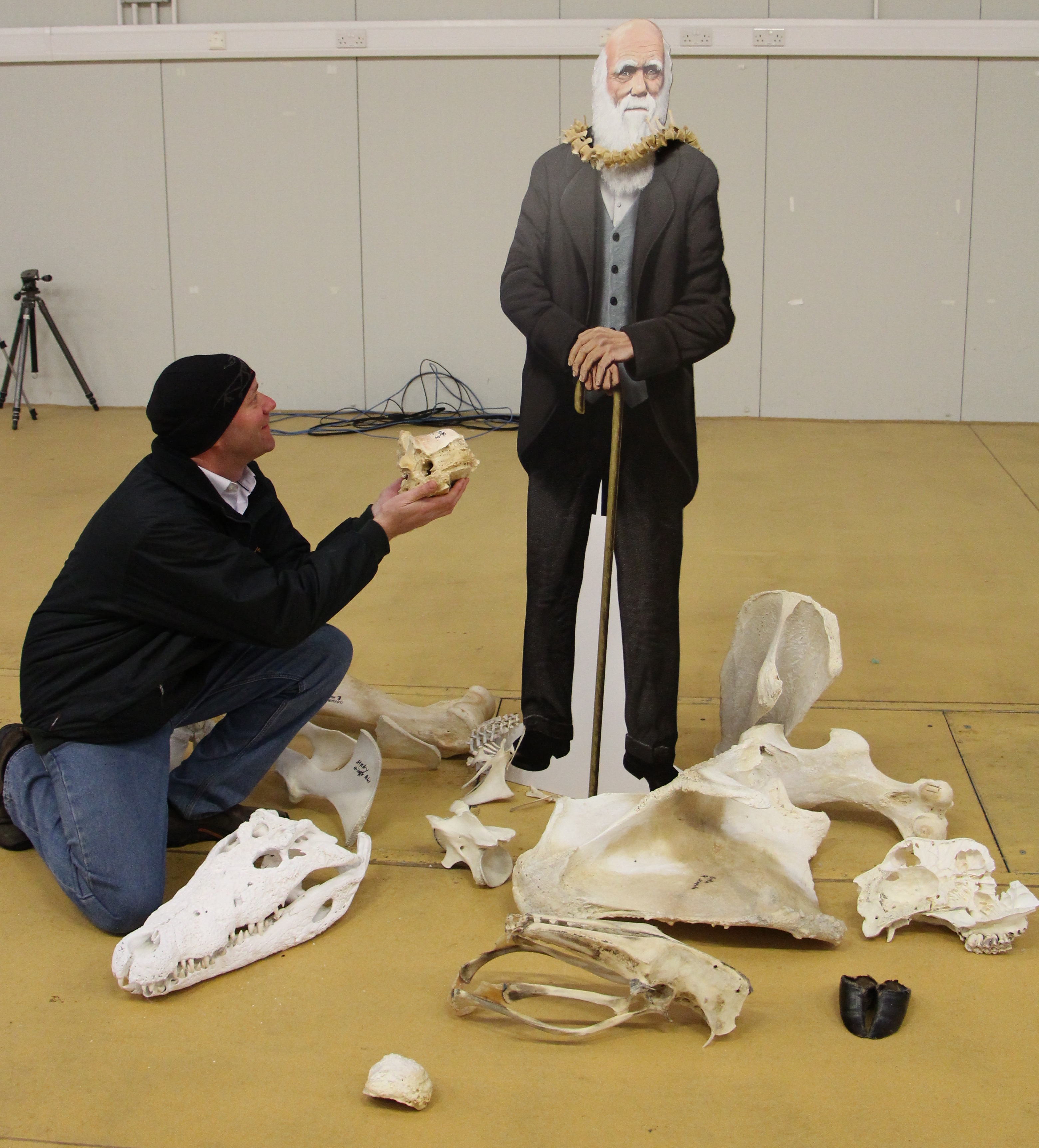
We also got Karl Bates, another proven biomechanics/modelling expert, to contribute diverse ideas and data. Furthermore, Zhiheng Li (now at UT-Austin doing a PhD with Dr Julia Clarke) brought some awesome fossil birds (Pengornis and Yixianornis) from the IVPP in Beijing in order to microCT scan them in London. Zhiheng thus earned coauthorship on the paper — and I give big thanks to the Royal Society for funding this as an International Joint Project, with Dr Zhonghe Zhou at the IVPP.

That’s the team and the background, and I’ve already given you the punchlines for the paper; these are the primitive and the derived states of the paper. The rest of this post is about what happened behind the scenes. No huge drama or anything, but hard, cautious work and perseverance.

Me shortly after I moved to the RVC; video still frame from a dinosaur exhibit I was featured in. Embarassingly goofy pic, but I like the blurb at the bottom. It's all about the evolutionary polarity, baby!

Me shortly after I moved to the RVC; video still frame from a dinosaur exhibit (c. 2004) I was featured in. Embarassingly goofy pic, but I like the blurb at the bottom. It’s all about the evolutionary polarity, baby!

The paper of course got started during Viv’s PhD thesis; it was one of his chapters. However, back then it was just a focus on how the centre of mass changed, and the results for those simple patterns weren’t very different from those we present in the paper. We did spot, as our Nature supplementary information notes,  a strange trend in early theropods (like Dilophosaurus; to a lesser degree Coelophysis too) related to some unusual traits (e.g. a long torso) and suggested that there was a forward shift of centre of mass in these animals, but that wasn’t strongly upheld as we began to write the Nature paper.

On the urging of the PhD exam committee (and later the paper reviewers, too), Viv looked at the contributions of segment (i.e. head, neck, trunk, limbs, tail) mass and centre of mass to the overall whole body centre of mass. And I’m glad he did, since that uncovered the trend we did not expect to find: that the forelimb masses were far more important for moving the centre of mass forwards than the mass (or centre of mass) of the tail was– in other words, the statistical correlation of forelimb mass and centre of mass was strong, whereas changes of tail size didn’t correlate with the centre of mass nearly as much. We scrutinized those results quite carefully, even finding a very annoying bug in the 3D graphics files that required a major re-analysis during peer review (delaying the paper by ~6 months).

The paper was submitted to Nature first, passing a presubmission inquiry to check if the editor felt it fit the journal well enough. We had 3 anonymous peer reviewers; 1 gave extensive, detailed comments in the 3 rounds of review and was very fair and constructive, 1 gave helpful comments on writing style and other aspects of presentation as well as elements of the science, and 1 wasn’t that impressed by the paper’s novelty but wanted lots more species added, to investigate changes within different lineages of maniraptorans (e.g. therizinosaurs, oviraptorosaurs). That third reviewer only reviewed the paper for the first round (AFAIK), so I guess we won them over or else the editor overruled their concerns. We argued that 17 taxa were probably good enough to get the general evolutionary trends that we were after, and that number was ~16 more species than any prior studies had really done.

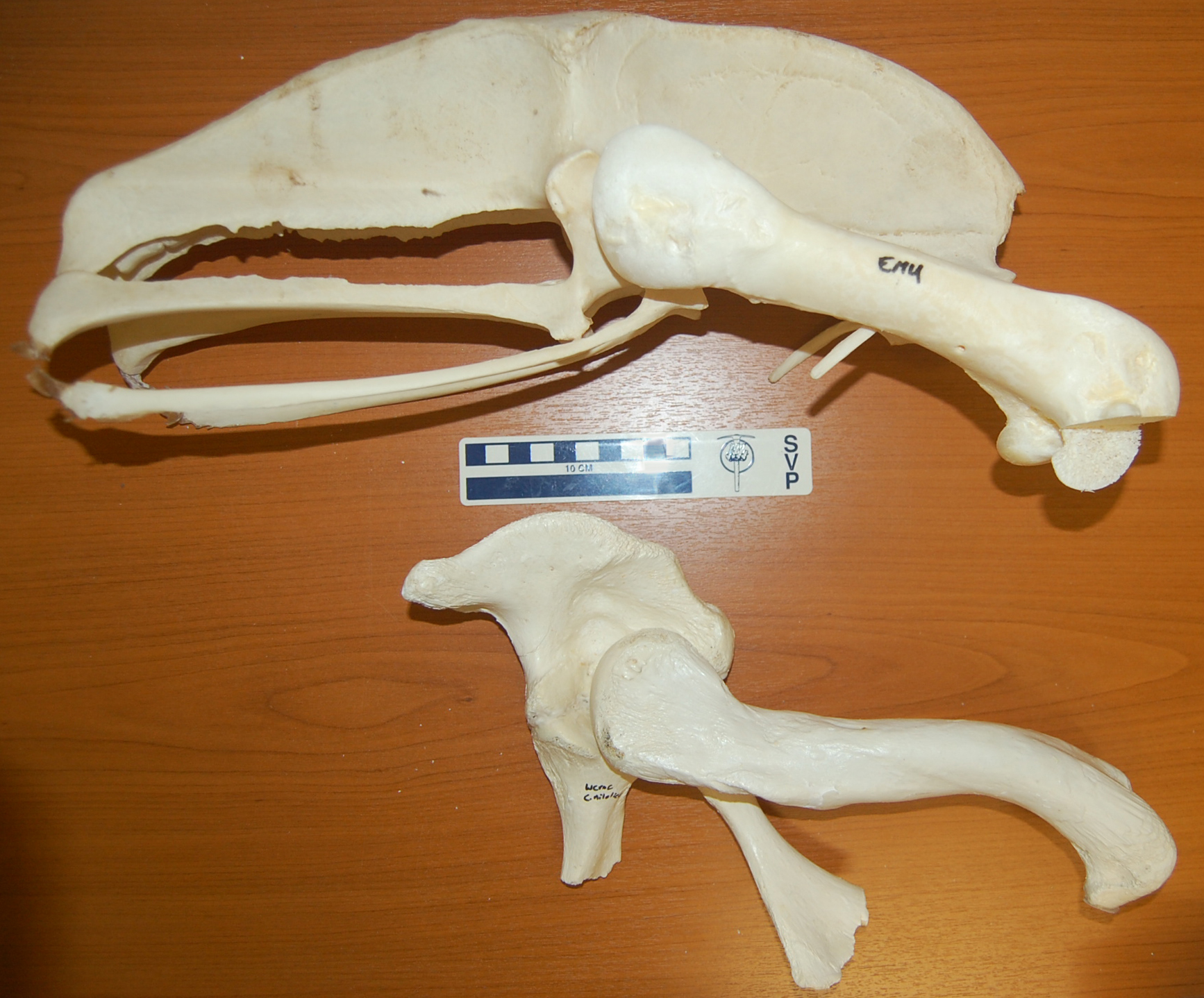
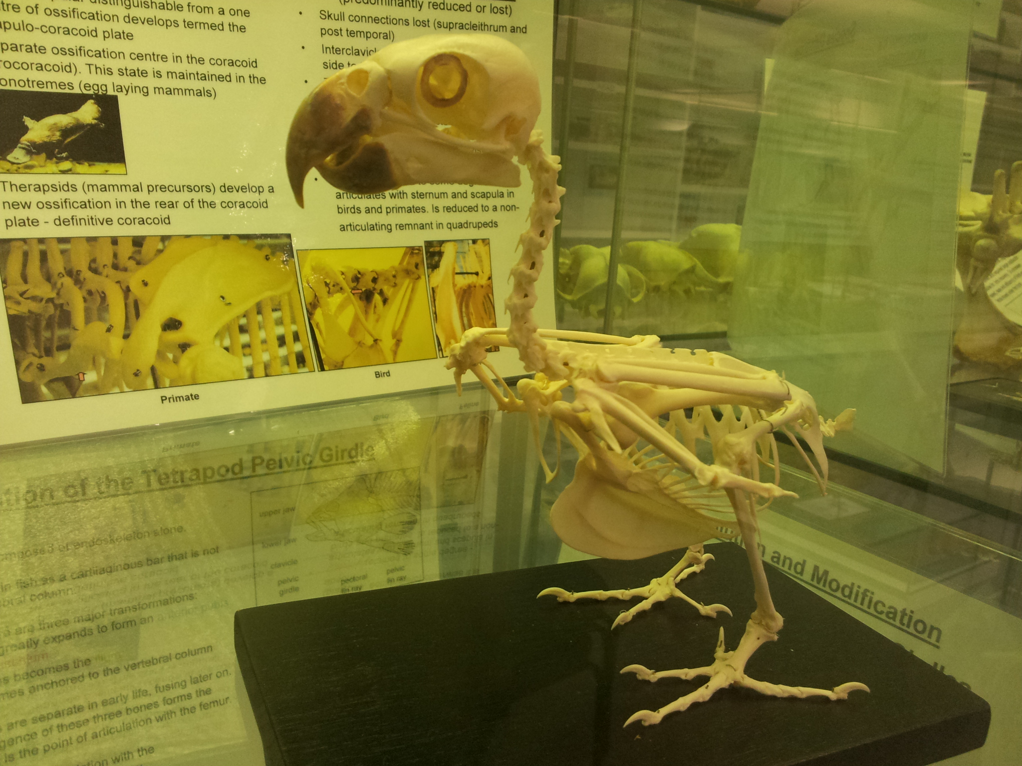
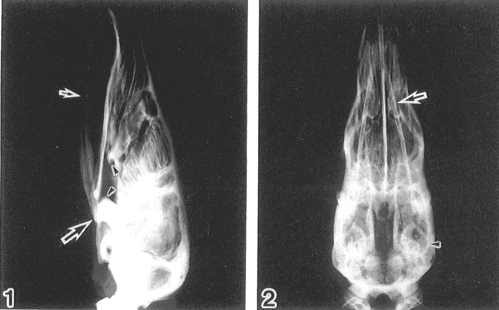
Above: CT scan reconstruction of the early extinct bird Yixianornis in slab conformation, and then Below: 3D skeletal reconstruction by Julia Molnar, missing just the final head (I find this very funny; Daffy Duck-esque) which we scaled to the fossil’s dimensions from the better data in our Archaeopteryx images. Yixianornis reconstruction There is also the concern, which the reviewers didn’t focus on but I could see other colleagues worrying about, that some of the specimens we used were either composites, sculpted, or otherwise not based on 100% complete, perfectly intact specimens. The latter are hard to come by for a diversity of extinct animals, especially in the maniraptoran/early bird group. We discussed some of these problems in our 3D Tyrannosaurus paper. The early dinosauromorph Marasuchus that we used was a cast/sculpted NHMUK specimen based on original material, as was our Coelophysis, Microraptor and Archaeopteryx; and our Carnegie ??Caenagnathus??Anzu (now published) specimen was based more on measurements from 1 specimen than from direct scans, and there were a few other issues with our other specimens, all detailed in our paper’s Supplementary Information.

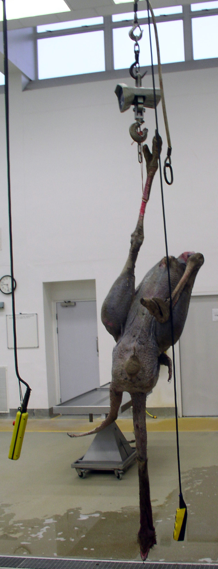
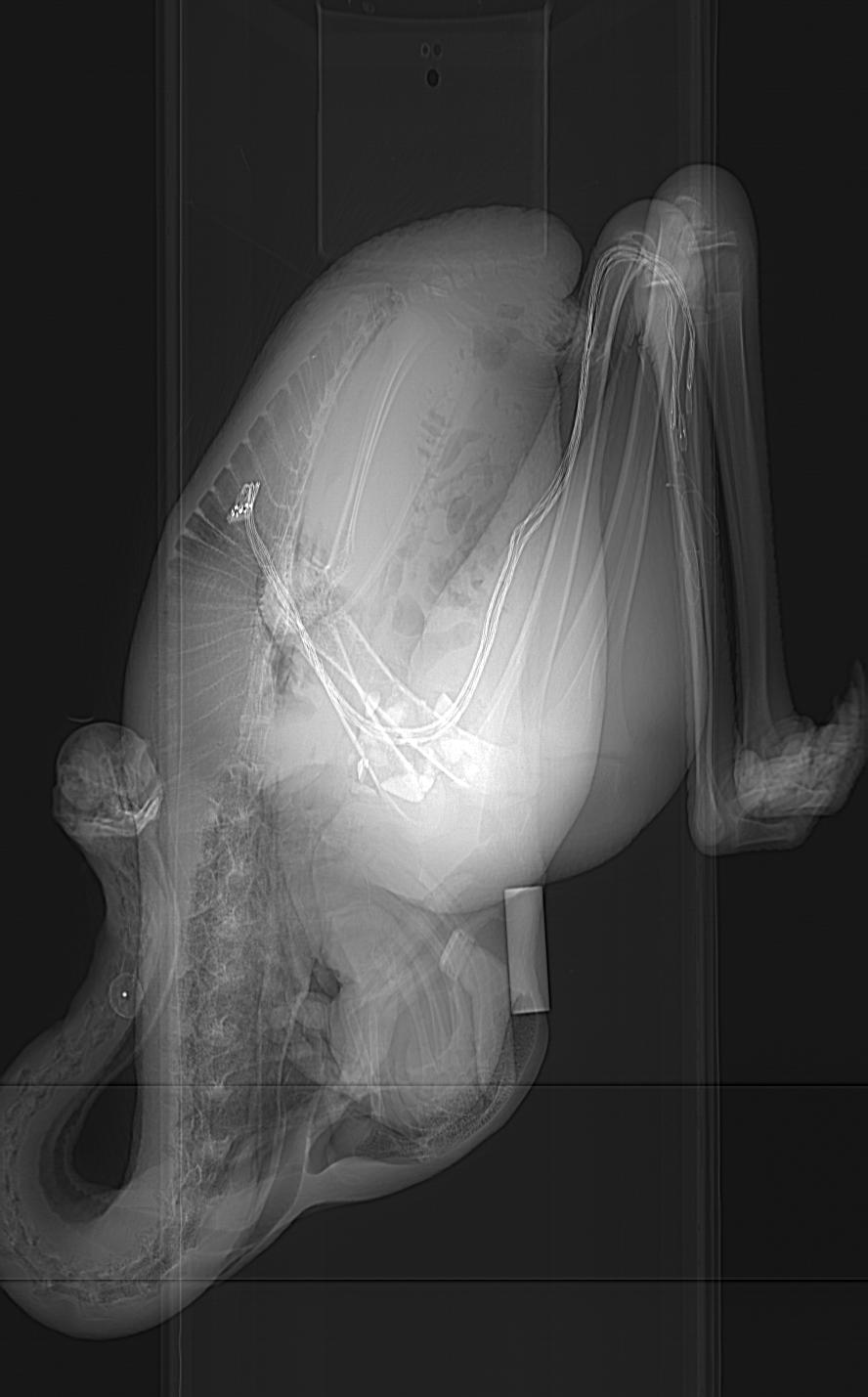
But our intuition, based on a lot of time spent with these models and the analysis of their data, is that these anatomical imperfections matter far, far less than the statistical methods that we employed– because we add a lot of flesh (like real animals have!) outside of the skeleton in our method, the precise morphology of the skeleton doesn’t matter much. It’s not like you need the kind of quality of anatomical detail that you need to do systematic analyses or osteological descriptive papers. The broad dimensions can matter, but those tend to be covered by the (overly, we suspect) broad error bars that our study had (see graph above). Hence while anyone could quibble ad infinitum about the accuracy of our skeletal data, I doubt it’s that bad– and it’s still a huge leap beyond previous studies, which did not present quantitative data, did not do comparative studies of multiple species, or did not construct models based on actual 3D skeletons as opposed to artists’ 2D shrinkwrapped reconstructions (the “Greg Paul method”). We also did directly measure the bodies of two extant archosaurs in our paper: a freshwater crocodile and a junglefowl (CT scan of the latter is reconstructed below in 3D).

One thing we still need to do, in future studies, is to look more carefully inside of the bird clade (Aves/Avialae) to see what’s going on there, especially as one moves closer to the crown group (modern birds). We represented modern birds with simply 1 bird: the “wild-type chicken” Red junglefowl, which isn’t drastically different in body shape from other basal modern birds such as a tinamou. Our paper was not about how diversity of body shape and centre of mass evolved within modern birds. But inspecting trends within Palaeognathae would be super interesting, because a lot of locomotor, size and body shape changes evolved therein; ostriches are probably a very, very poor proxy for the size and shape of the most recent common ancestor of all extant birds, for example, even though they seem to be fairly basal within that whole lineage. And, naturally, our study opens up opportunities for anyone to add feathers to our models and investigate aerodynamics, or to apply our methods to other dinosaur/vertebrate/metazoan groups. If the funding gods are kind to us, later this year we will be looking more closely, in particular, at the base of Archosauria and what was happening to locomotor mechanics in Triassic archosaurs…

Clickum to embiggum:

Australian freshwater crocodile, Crocodylus johnstoni; we CT scanned it in 3 pieces.

Australian freshwater crocodile, Crocodylus johnstoni; we CT scanned it in 3 pieces while visiting the Witmer lab in Ohio.

A Red junglefowl cockerel, spotted in Lampang, Thailand during one of my elephant gait research excursions there. Svelte, muscular and fast as hell.

A Red junglefowl cockerel, spotted in Lampang, Thailand during one of my elephant gait research excursions there. Svelte, muscular and fast as hell. This photo is here to remind me to TAKE BLOODY PICTURES OF MY ACTUAL RESEARCH SPECIMENS SO I CAN SHOW THEM!

I’d bore you with the statistical intricacies of the paper, but that’s not very fun and it’s not the style of this blog, which is not called “What’s in John’s Software Code?”. Viv really worked his butt off to get the stats right, and we did many rounds of revisions and checking together, in addition to consultations with statistics experts. So I feel we did a good job. See the paper if that kind of thing floats your boat. Someone could find a flaw or alternative method, and if that changed our major conclusions that would be a bummer– but that’s science. We took the plunge and put all of our data online, as noted above, so anyone can do that, and that optimizes the reproducibility of science.

What I hope people do, in particular, is to use the 3D graphics of our paper’s 17 specimen-based archosaur bodies for other things– new and original research, video games, animations, whatever. It has been very satisfying to finally, from fairly early in the paper-writing process onwards, present all of the complex data in an analysis like this so someone else can use it. My past modelling papers have not done this, but I aim to backtrack and bring them up to snuff like this. We couldn’t publish open access in Nature, but we achieved reasonably open data at least, and to me that’s as important. I am really excited at a personal level, and intrigued from a professional standpoint, to see how our data and tools get used. We’ll be posting refinements of our (Matlab software-based) tools, which we’re still finding ways to enhance, as we proceed with future research.

Velociraptor-model-min Dilophosaurus-model-min00

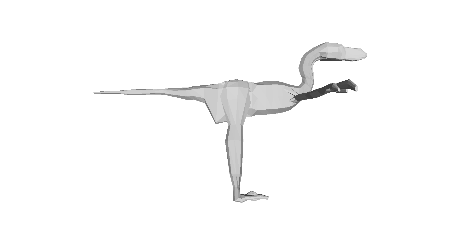
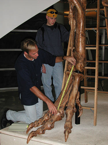
Above: Two of the 17 archosaur 3D models (the skinny “mininal” models; shrinkwrapped for your protection) that you can download and examine and do stuff with! Dilophosaurus on the left; Velociraptor on the right. Maybe you can use these to make a Jurassic Park 4 film that is better, or at least more scientifically accurate, than Hollywood’s version! 😉 Just download free software like Meshlab, drop the OBJ files in and go!

Now, to bring the story full circle, the paper is out at last! A 4 year journey from Viv’s PhD thesis to the journal, and for me a ~14 year journey from my mind’s eye to realization. Phew! The real fun begins now, as we see how the paper is received! I hope you like it, and if you work in this area I hope you like the big dataset that comes with it, too. Perhaps more than any other paper I’ve written, because of the long voyage this paper has taken, it has a special place in my heart. I’m proud of it and the work our team did together to produce it. Now it is also yours. And all 3200ish words of this lengthy blog post are, as well!

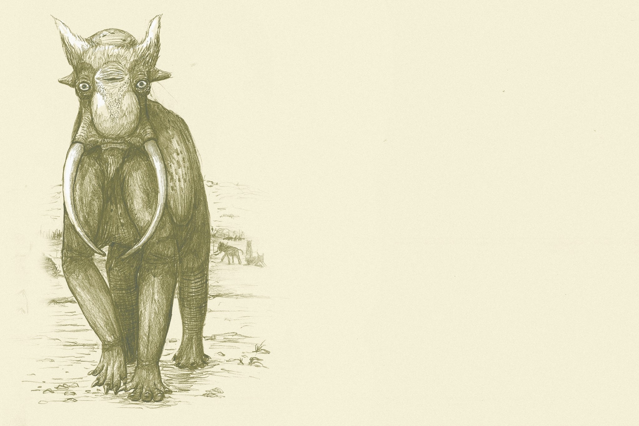
Last but not least, enjoy the wonderful digital painting that Luis Rey did for this paper (another of my team’s many failed attempts to get on the cover of a journal!); he has now blogged about it, too!

Dinosaur posture and body shape evolving up the evolutionary tree, with example taxa depicted.

Dinosaur posture and body shape evolving up the evolutionary tree, with example taxa depicted. By Luis Rey.


News stories about this paper will be added below as they come out, featuring our favourites:

1) NERC’s Planet Earth, by Harriet Jarlett: “Dinosaur body shape changed the way birds stand

2) Ed Yong on Phenomena: “Crouching  bird, hidden dinosaur

3) Charles Choi on Live Science: “Crouching bird, hidden evolutionary purpose?

4) Brian Handwerk on Nat Geo Daily News: “Birds’ “Crouching” Gait Born in Dinosaur Ancestors

5) Jennifer Viegas on Discovery News: “Heavier dino arms led evolution to birds

6) Puneet Kollipara on Science News: “Birds may have had to crouch before they could fly

And some superb videos- we’re really happy with these:

1) Nature’s “Crouching Turkey, Hidden Dragon

2) Reuters TV’s “3D study shows forelimb enlargement key to evolution of dinosaurs into birds

Synopsis: Decent coverage, but negligible coverage in the general press; just science-specialist media, more or less. I think the story was judged to be too complex/esoteric for the general public. You’d think dinosaurs, evolution, computers plus physics would be an “easy sell” but it was not, and I don’t think we made any big errors “selling” it. Interesting– I continue to learn more about how unpredictable the media can be.

Regardless, the paper has had a great response from scientist colleagues/science afficionados, which was the target audience anyway. I’m very pleased with it, too– it’s one of my team’s best papers in my ~18 year career.

Read Full Post »

frontcover

The Unfeathered Bird book by Katrina van Grouw proclaims immediately in its Introduction that it “is not an anatomy of birds.”  True– it is far more than that, and it would be a shame if it had just been a dry, technical avian osteology reference book. It is a unique blend of art and science- particularly avian anatomy, evolution, taxonomy, natural history and more. The Unfeathered Bird is written for a general audience; birders/twitchers or just natural history buffs would be ideal targets of its unfettered passion for all things avian. A 12-year-old who is very keen on animals could enjoy it, and it may ignite the flames of ornithological excitement in many young or older readers. I am glad it was not called “The Naked Bird” as that would have caused some serious misconceptions (badum-tish!). The book is dripping with illustrations (at least one every two pages, often more). Almost all of the illustrations (except some paintings in the style of the cover) are in the same brownish sketch style that, like much of the book, evokes a bygone era of dark wooden cabinets and shadowed halls packed with skeletons, with nary an interactive graphics display, animatronic dinosaur or hyperdetailed cladogram in sight. It feels like an homage to the Victorian naturalists’ joy for anatomical detail conveyed through painstakingly detailed woodcuts. And while many still think of feathers as “the defining feature of birds,” enough about feathers already. Seriously. This is a book is about what lies beneath, and how all that non-fluffy stuff is important for birds’ lives, too.

(image-intense post; all can be clicked to embiggritate)

Katrina with peacock feather headdress? (back cover pic and rear view of same skeleton)

Katrina with peacock feather headdress? (back cover pic and rear view of same skeleton)

Katrina with front cover framed pic and the peacock skeleton that went with it.

Katrina with front cover framed pic and the peacock skeleton that went with it.

The Introduction continues to explain that the book is truly about how the external anatomy of birds is linked to the bony anatomy, which might remind astute readers of modern approaches like the extant phylogenetic bracket. The rest of the book uses both skeletal and unfeathered, quasi-myological illustrations to get this point across vividly. The explanatory text is written at a basic enough level for the average reader and is just the right length, with interesting anecdotes and natural history facts that even the expert reader will find interesting or even inspirational (e.g. possibly a goldmine for research ideas). First there is a 26 page “Basic” section with an introduction to avian osteology, with bountiful sketches to illustrate key organs and text explaining how it all fits together in the fully accoutered bird. The decision to use classical Linnean taxonomy (defunct or re-arranged taxa from the Systema Naturae like Accipitres, Picae, Anseres, Grallae, Gallinae and Passeres; which are the six “Specific” chapters in the second section of the book) was a good one- it enhances the classical feel of the tome and gives the author a great opportunity to discuss convergent evolution and how that misled past ornithologists.

But for me, the book is most pleasurable for the visualizations and the passion for all things birdy that weaves through them and the accompanying text. The removal of feathers, or even all soft tissues, from bird bodies (posed in naturalistic behaviours) that van Grouw renders in her illustrations shows birds in a new light, emphasizing the strangeness and diversity that lie beneath. The author begins the book with a touching Acknowledgments section in which her husband Hein van Grouw, curator of birds at the Natural History Museum’s Tring collection, features very prominently, making it clear that the book was a team operation and comes from the heart after a 25-year journey. This gives the book a special warmth that is preserved throughout the remainder- although the illustrations are of flayed bodies or boiled / beetle-macerated skeletons, the tone is nothing less than an earnest love for birds of all kinds, and a zest for portraying those feelings to the reader in sketches and prose. It is a joyous celebration, not a somber litany, of the wonder of birds that can be gleaned from dead bodies. There is so much powerful, awesome imagery stuffed into those pages that it is hard to summarize. I’ll let five of my favourite images from the book (more are in her gallery and her book’s Facebook page; but even these are just the tip of the icebird) help get this across (used with permission of the author):

Naked kiwi in action.

Naked kiwi in action.

The unscaled bird: guineafowl feet.

The unscaled bird: guineafowl feet.

Deplumed sparrowhawk with dove trophy, exalting in its triumph.

Deplumed sparrowhawk with dove trophy, exalting in its triumph.

Budgerigar has made a friend? Or came to grips with its own mortality?

Budgerigar has made a friend? Or came to grips with its own mortality?

Trumpet Manucode WTF anatomy! Spiraling tracheal coil made me gasp in awe when I saw this image in the book.

Trumpet Manucode’s WTF anatomy! Spiraling tracheal coil made me gasp in awe when I saw this image in the book.

Now I’ll depart from this post just being a book review. I went to the Tring collection to do some research, and arranged my trip so I’d also get to see the debut of a Tring special exhibit featuring The Unfeathered Bird, and also to meet Katrina as well as Hein van Grouw. The placement of the exhibit at Tring is apropos, because Katrina was a curator at the museum until a few years ago and Hein still is. But the inspiration for the work and the specimens used (with a few exceptions, including from other museums) are Katrina’s. She (with Hein’s help) procured bodies of birds to dissect, macerate and sketch for the book over its 25 year fledging period, noting in the Acknowledgments that “no birds were harmed” to do this– do read those acknowledgments, as there are some amusing tales there of how she obtained some specimens.

I was fortunate to be able to take some photos of the exhibit while they set it up, and grabbed some candid images of Katrina and colleagues during that process. The following images show off the exhibit, which is all in one clean, bright, simply adorned room in the Tring that lets Katrina’s framed sketches be the focus. Here are some examples:

Poster advert for the book in the Tring collections.

Poster advert for the book in the Tring collections.

Tring exhibit setup, with Katrina, husband Hein, and helper finishing it up.

Tring exhibit setup, with Katrina, husband Hein, and helper finishing it up.

Tring exhibit now ready.

Tring exhibit now ready.

Tring exhibit case.

Tring exhibit case.

Framed sketches at Tring exhibit.

Framed sketches at Tring exhibit.

Framed sketches at Tring exhibit.

More framed sketches at Tring exhibit.

The exhibit is fun for people who are already Unfeathered Bird fans, and a good way of drawing in new ones. The book is a precious thing that any fan of birds, especially scientists, really needs to have a hard copy of. While it claims not to be an anatomy text, its illustrations provide ample opportunities to use it for that purpose. But really the point of owning all 287-plus pages is to bask in the warmth of true, pure appreciation for classic ornithology, which I found infectious. It is a book by and for bird lovers, but also for those that find the interface of art and science to be fascinating.

I confess I used to hate birds. I found them annoying and boring; all that flitting and twitting and pretentious feathers. “Get over yourselves, already, and calm down too!” was my reaction to them. When I started grad school, I had an open disdain for birds, even moreso than for mammals (OK, except cats). I was a “herp” fan through and through, for most of my life (childhood spent catching anoles in Florida, or stalking frogs in Ohio; during visits to my grandparents). What won me over was studying birds (and eventually mammals, too) as a young scientist, and learning how incredible they are– not just as endpoints in the story of theropod dinosaur evolution, as my thesis focused on, but as amazing animals with spectacular form-function relationships.  The Unfeathered Bird is saturated with that amazement, so we’re birds of an unfeather.

Framed sketch of dodo head at Tring exhibit.

Framed sketch of dodo head at Tring exhibit.

Entirely unfeathered Indian peafowl in matching views.

Entirely unfeathered Indian peafowl in matching views.

Painted Stork and Toco Toucan sketches.

Painted Stork and Great Hornbill sketches.

Red junglefowl, wild ancestor of domestic chickens (and the book ends with several such breeds illustrated),

Red junglefowl, wild ancestor of domestic chickens (and the book ends with several such breeds illustrated).

Katrina told me that she is already deep into writing the next book, whose subject I won’t spoil for you here but maybe we will be lucky enough to have her appear in the Comments and plug it? 🙂 (Her website does say “It was Hein’s stroke of genius to include domestic birds and they’ve provided the inspiration for my next project.” so the cat is out of the bag and amongst the pigeons!) It is great to hear that the book has done quite well sales-wise and critically, such as ~#67 on the Amazon sales list at one point– I hope this paves the way for more such books not only from Katrina, but from others engaged in lateral thinking (and still others) on the boundaries of science-art.

Read Full Post »

I’ve been either super busy or on holiday and low on creative energy, so although I have five or so blog posts frozen in my mind, I haven’t progressed them far enough yet. To whet your appetite, they include a review of the bird book AND exhibit “The Unfeathered Bird”, a summary of our recent PeerJ paper on croc lungs (Schachner, Hutchinson and Farmer; see here and also here), a rant on optimality in biomechanics, and a summary of a new and (to us) very exciting dinosaur paper that is very-soon-to-come, and something else that I can’t remember right now but it probably is totally awesome.

But here’s an interlude to keep you stocked on freezer-related imagery. We did an annual inventory and massive cleanup (and clean-out!) of all our freezers, throwing out some 300ish chickens and other odds and ends, and finally loading all cadaveric material I have into a single database, which I’ll share here shortly! It was a long time coming, and took ~6 people about 4 exhausting hours; last year’s attempt was just a holding maneuver by comparison. Here is how it went.

Freezersaurus gut contents being sorted.

Freezersaurus gut contents being sorted. A cold drizzle was falling. It was not pleasant work.

Large specimens, especially horse legs, being moved into the walkin freezer.

Large specimens, especially horse legs and the remnants of an ostrich hind end or two, being moved into the walk-in freezer.

Research Fellow Jeff Rankin wrangles some horse legs into their freezer.

Research Fellow Jeff Rankin wrangles some horse legs into their freezer. I like this photo for his knowing smile as he stands amidst horse limbs spread akimbo.

Postdoc Heather Paxton helps sort out elephant foot tendons and "predigits" in their freezer.

Postdoc Heather Paxton helps sort out elephant foot tendons and “predigits” in their freezer. Nice view of our long line of chest freezers in action, too.

And, as an extra reward if you made it to the end, here’s what I was doing for the past week (check out my Twitter feed for more)– seeing amazing art and architecture and food and stuff in Rome, which is just dripping with wicked anatomical portrayals (e.g. in this image; click to embiggen and oggle the classical physiques).

Rome's Trevi Fountain

Rome’s Trevi Fountain

Read Full Post »

If you want to see a new/reinvigorated, exciting direction that palaeoart is headed, check out the All Yesterdays book by Conway, Kosemen and Naish. This review is fully cognizant that I’m late to the party of hailing this book as part of a palaeoart renaissance. I confess I haven’t read any of the many reviews of this book; I just know it is highly regarded and popular, from excitement on social media sites I frequent. So if my review covers ground others have too, so be it; it’s purely my own thoughts but I expect that mine fall in line with many others’. I’m reviewing the book on this blog because I love the interface between science and art (which is very important in anatomy), and because anatomy, and how one infers it when it is unknown, is the fundamental theme of the book.

You can buy All Yesterdays for around £18 (ASIDE: oddly, used copies (“May not include CD, access code, or DJ”– ???) are around £42 on the same site; perhaps those are artist-signed??? I have no idea!). It is a good deal at that price. While you’re at it, get “Dinosaur Art: The World’s Best Paleoart” by White et al. (including Conway) for a similar price. My review will return to some comparisons between these two books, released just a few months apart.

All-Yesterdays-coverworlds-greatest-paleoart

All Yesterdays is about not only how we reconstruct dinosaurs and other prehistoric animals, but also about thinking outside-the-box in the ways we reconstruct them and thereby bucking some recent clichés and tropes. Some of those outside-the-box ideas might seem ludicrous, and some probably are. But one of the main points of All Yesterdays is that there is plenty about extinct animals, and even living animals, that we don’t know, even though the field of paleoart has matured into greater scientific rigor than in the days of Knight, Zallinger and others (1920s-1950s). There is a focus on uncertainties about integument (e.g. feathering, spines, colour/patterning, body contours) and behaviour (e.g. avoiding stereotypes like perpetually aggressive predators and frightened prey animals– amen to that!). And the capstone of the book, which in some ways I loved the most, is turning the issue on its head and pretending that we only had skeletons of extant animals, then proceeding to reconstruct those animals (elephants, whales, horses and swans stand out prominently in this section; some of these are shown below). I wish more scientists in my general area would practice this; e.g. validation of a methodology used to reconstruct extinct animals in science.

The ‘speculative zoology’ of All Yesterdays deserves favourable comparison to one of my favourite science-art books, 1981’s After Man by Dougal Dixon. I fell so in love with that book as a 10-year-old that I wouldn’t let my parents return it to the library and I made them pay the hefty lost-book-fee (yes, I was a little bastard!). I still have it, too. (Sorry, Sequoya Branch Public Library of Madison, WI!) Likewise, the whimsy of the Rhinogrades is evoked by this work, and of course Tetrapod Zoology blog readers will be no strangers to it, either.

The book begins with a clear, succinct (7 page) summary of the history and science of reconstructing animals, with a focus on paleoart’s approach rather than science’s. I would have found it interesting (but space constraints presumably precluded) to feature more of the interface/parallels with scientists at the same time, such as the careful reconstructions of musculature in A.S. Romer’s masterful work in the 1920s (e.g. below), or later efforts by palaeontologists like Alick Walker and Walter Coombs. Many of these luminaries sought not to reconstruct animals for artistic purposes, but for almost purely scientific ones: to understand what skeletal anatomy meant in terms of broader biology (e.g. comparative anatomy) and phylogeny (e.g. origin of birds or archosaur evolution). The quality of their own artistic representations as well as scientific interpretations varied a lot. Indeed, sometimes the choice of model organisms (crocodile for Romer; lizard for Walker; birds in the post-1960’s) reveals much about the author’s preconceptions about phylogeny, marshalled towards a favoured hypothesis (e.g. a crocodile origin of birds for Walker; or an avian origin amongst dinosaurs for Bakker, Paul and others), rather than a circumspect assessment of all relevant evidence.

Romer1923-fig6

Figure 6 from Romer, 1923; very crocodylian T. rex right hindlimb muscles.

But eventually the “model organism” approach to reconstructing extinct animals gave way to the extant phylogenetic bracket; very popular today; which itself is an adaptation of the outgroup method for polarity assessment in phylogenetic systematics (cladistics). I am sure many modern paleoartists explicitly consider the “EPB” in their reconstructions, although this leaves many ambiguities (e.g. integument of crocodiles and birds being totally different!) that they must overcome, whereas scientists might just give up. This interface of art and science is part of what make palaeontology so enjoyable.

The EPB mindset has been a big step forward for evolutionary morphology and palaeontology, but still some of the greatest questions (e.g. what were the actual sizes, colour patterns, or behaviours of extinct animals? How did novelties arise and which novelties did dinosaurs have that extant relatives lack?) are left ambiguous by the EPB. This is because either the EPB itself is ambiguous (crocodiles or other taxa do one thing; birds do something altogether different), or because features leave no osteological correlates (e.g. muscle/tendon/ligament scars) on fossils that can be compared with the EPB.  This quandary leads to the fun side of this book– filling in the huge gaps left by both basic anatomical interpretation and the restrictions imposed by the EPB, and then playing with the frontiers of anatomical, behavioural and ecological reconstruction, using informed speculation.

The extant phylogenetic bracket for archosaurs.

The extant phylogenetic bracket for archosaurs.

In addition to the startling, bizarre “All Todays” reconstructions at the end of the book, the highlights for me were the camouflaged Majungasaurus and plesiosaur, the “feathered mountain” (below) of a therizinosaur (can anyone illustrate a plausible therizinosaur and make it normal and boring? I wager not!) and the neck-swinging elasmosaurs engaged in “honest signalling” of their fitness. Many of the illustrations riff on notions popular in the modern palaeo-zeitgeist (and subject of many conversations at conferences, or even publications), such as evidence for the spiny integument of some ornithischians, fat ornithopods, Microraptor of somewhat-known-colouration, and so on. But plenty of other images riff on a “well why not?” theme, challenging the viewer to consider that extinct animals could have many surprises left in store for us with future discoveries, or else plausible features that we’ll never know of but might seem laughable or unfashionable to illustrate now. Each image has text explaining the logic behind it- this is not just a montage of pictures. This is a thinking person’s book- you should buy it for rumination, to challenge your preconceptions, not to have a flashy coffee table book. It’s not eye candy — it’s more like brain jerky.

John Conway's mountain-of-feathers therizinosaurs: eerily beautiful.

John Conway‘s mountain-of-feathers therizinosaurs: eerily beautiful.

I think this is a bold, fun (re)new(ed) direction for palaeoart. There’s always a place for rigorous, conservatively evidence-based, by-comparison-almost-uncreative scientific illustration of extinct organisms. The World’s Best Paleoart presents loads of this, often in vividly colourful, photo-realistic, lavish, glossy detail, whereas the approach in All Yesterdays tends toward a more soft, matte, informal style including sketches or abstractions, toning down the serious and intense (even cluttered?) approach that can characterize modern palaeoart, including The World’s Best Paleoart.  Sometimes those reconstructing life of the past (scientists included!) may emphasize that detailed realism too much and lose some of the joyful playfulness that palaeoart can revel in, at its best, most inspirational or thought-provoking. The former style might be considered the more “safe” or technical practice; the latter more risky or unconstrained.

Memo Kosemen's "All Todays" swans, with tadpolefish, might haunt your nightmares.

C.M. Kosemen‘s “All Todays” swans, with tadpolefish, might haunt your nightmares.

I’m not casting negative judgement on either style; both are absolutely wonderful — and valuable. I love both books! I’m glad we’re in a new age where the fun is waltzing back into palaeoart, that’s all. All Yesterdays doesn’t just waltz, either. It pounces into your field of view, wiggles its rainbow-coloured, mandrill-esque ankylosaurid bottom at you with a cheeky grin, and proceeds to make you smirk, be bemused, and even gasp at its adventurousness in rapid succession as you turn its pages. At 100 pages it doesn’t overstay its welcome either– that kaleidoscopic thyreophoran rump cartwheels off into the sunset at an opportune moment.

You won't forget Memo Kosemen's "All Todays" elephant.

You won’t forget C.M. Kosemen‘s “All Todays” elephant.

If All Yesterdays makes someone uncomfortable with its swashbucklerish daring, they’re probably taking palaeontology way too seriously– and maybe missing not only some good fun, but also some potential truths. Dogma is a terrible thing, and All Yesterdays slaughters it with delightful relish. Bring on the next installment! If you have  All Yesterdays too, what’s your favourite part? Or if you don’t have it, I’d be happy to answer queries in the Comments.

Read Full Post »

Chilly 1st Birthday to You, WIJF Blog!

It has indeed been a year of blogging now! And it has been a very fun year at that. Here is my look back at past events on this blog.

Stomach-Churning Rating: 8/10- some heavy-hitters in here, but to regular Freezerinos they will mostly be familiar.

giraffe-leg-CT

This blog’s first image and subject: giraffe legs and modelling.

NUMB WITH NUMBERS:

First, the usual consideration of statistics: wow! I never expected the blog to be this successful! I’d sort of hoped as much, but for such a niche blog it was far from guaranteed in my mind. However, the initial response was overwhelming: 4210 hits in its first month, many of them on the first day!

Since then, although the usual number of blog views are around 100-200/day, there are now 76 blog followers,  and a total of ~111,000 views! According to ImpactStory and Topsy, the blog has had 48 tweets (7 of them “Influential”), 111 Facebook likes, 105 Facebook comments, and 53 Facebook shares. Nice!

The biggest day was April 27, 2012: 10,564 views– ZOWIE! That was fun. More about that below. I’m amused that my very first post only has 85 views even to this date, but it didn’t really contain much.

Visitors tended to come from browser searches (23,243 hits!), in particular hunting for images of the feet/limbs of elephants, rhinos, giraffes and other megafauna (looks like my intended purpose worked– vets and other anatomists want this rare information!). Oddly, from a few of my tweets that got listed on my blog, “deepstaria enigmatica” (remember that craze?) became one of the most common terms (214 to date!) that brings people here via the intertubez. Giraffe anatomy and patella are also major sources of search strikes. Interesting!

But don’t dismiss the power of Facebook (4,399 oggles on WIJF total) versus the somewhat surprisingly smaller impact of Twitter (2,036 pings). I say surprising because I push the blog much harder on Twitter than anywhere else, but Facebook pages like Perez’s Veterinary Anatomy (>33,000 members/likes!) have done far more than my mere ~1,300 Twitter followers can. Other blogs like the Chinleana palaeo blog (1,008 palaeo-hits here) and the ubiquitous Pharyngula (791 athe-hits) have helped a lot, too– thanks to all those bloggers and science writers who have linked to my humble little blog!!!

Who are YOU? You mostly come from the USA and UK, of course, but Japan is 3rd on my visitors ranking, followed by Canada, Germany, Australia and the Netherlands. Russia: we want more of you, too. Monaco, Nauru, Tuvalu and Liechtenstein- you as well, please! North Korea, keep trying.

So this is all great; really great. I’m rather amazed.

Definitely the blog has succeeded in what I aimed: to present the fun, awesome, curious side of anatomy in all its raw glory, using the freezer as a common theme (although I’ve felt free to deviate from purely freezer-based science when it pleases me). And it has crystallized for me just how important and powerful a single picture of anatomy can be.

That is what this post emphasizes- the pictures of the year from this blog. Enjoy the walk down morphology lane.

MOST POPULAR POSTS AND PERIODS:

Certainly the post; indeed the single photograph; that stands out for this blog is that of the elephant with its guts spilling out, from my Inside Nature’s Giants post on 13 April (so it took 2 weeks to gather momentum before the views spilled out like so many bowels). In a single day I had thousands of visitors from Boing Boing, Metafilter, Reddit, Gizmodo, io9, pinterest (which still sends me a lot of hits daily), and more! So here once again is that beastly image:

Stunning emergence of The Guts

Stunning emergence of The Guts

The post even got re-discovered by Reddit (the dreaded repost), leading to another surge. 24,330 views of the blog post so far!

A distant second to the elephant guts in terms of broad popularity was the “how thick is a rhino’s skin?” image; another Reddit favourite; with 4,719 views of the post from World Rhino Day 2012:

Skinning a White rhino forelimb

But also the “Animal: Inside Out” review did very well here (2,338 views to date), which was quite gratifying because I did a lot of detailed but enjoyable research for that one. It continues to bring people here, long after the NHM exhibit closed (it is now at Chicago’s excellent Museum of Science & Industry), which is quite cool.

Thanks to the poll results from last week, I’ll be doing more exhibition reviews like this– see below. My favourite image from that post is this: the bull (but don’t forget the camel, either):

Great exhibit. No bullshit.

Great exhibit. No bullshit.

Once we’re past those top 3 pages, things settle down to numerous posts with ~1000 or less views to date– highlights include the big rhinos and giant rhinos post: Rhino humeri

And the post on WCROC the big Nile crocodile got a fair amount of attention, as well as my posts on our Ichthyostega research and vertebral evolution discoveries, naked dinosaurian ostriches, chicken meat, giraffe anatomy (many pages, but this one is relatively most popular), and then the series I did on the RVC’s Anatomy Museum (first post here).

Here are a few thumbnails of the greatest hits from those posts and some others– which do you remember and why?

DSC_0203 Mystery Dissection 3  DSC_0963a  Whole 2 Gratuitious Melanosuchus (black caiman) shot. chicken-viscera-myopathy Gratuitious rhinoceros leg.  Kitty Hedz it is defunct rhino_front  hippo_L_knee Wolpertingers Jenny Hanniver- "face" windfall-croc (4) The nuchal ligament, which runs along the spine and helps hold up that long neck.  The left cheek's teeth-- and check out the spines on the inside of the cheek! Keratinous growths to aid in chewing, food movement, digestion etc. These extend into the stomach, too! Amazed me first time I saw them, in an okapi (giraffe cousin). my-brain2 If this post bummed you out, just focus on these contented cats. An offering to The Master

…and we’ll never speak of the freezer-penis again…

Of course, there were the puzzles and mysteries, too. When I think of those, the image I think of most is this one; one of the first. Remember what it is? DSCN0880

I’ll be defrosting some new ways to puzzle you this year.

Personally, my post about my brain means a lot to me (and any zombies out there) of course, but also I’m rather keen on my entry on elephant biomechanical models (cheeseburger units!), and the posts about elephant foot pathologies and the rhino crisis also carried a strong, semi-personal urgency.

I also featured a lot of movies here- if you want to peruse them, they’re always on my Youtube account here— >22,000 views so far; not bad. One of my favourites is this one, of a pumpkin being smashed in slow motion:

Furthermore, in terms of effort writing and researching, my very detailed post on chimeras and Jenny Hannivers and such is very memorable for me, and more recently the Freezermas series was a huge undertaking– which gave me needed breaks but also soaked up a lot of time during some intensive grant-writing!

I predict that the pangolin post, in particular, will proceed to provoke a promiscuous proportion of people to pass by this blog.

But the WIJF blog has always been about including you too, my loyal Freezerinos– what about you? Please thaw out your memories of past posts and comment below on what sticks out as your favourites and why. I’d love to hear about it!

Eggs: full of bountiful promise and symbolism for the future.

Eggs: full of bountiful promise and symbolism for the future.

THE FROZEN TUNDRA OF THE FUTURE:

A final duty for this post, heralded by my poll earlier, is for me to peer into my frosty crystal ball and report on the future of this blog:

As promised, it will continue for a year or more; as long as I feel I have something new to say and someone to tell it to.

The poll convinced me, as I’d hoped, to venture into more reviews of museum/other exhibits that I visit locally or abroad. Now and then I’ll also tackle a new or classic paper, good or bad, that tickles my anatomical fancy, and give my perspective on it. The mysteries and puzzles will continue; I was checking in that poll to see what the enjoyment level was, and it is clearly still reasonably high. I’ll continue presenting my own research here, especially when it’s quite anatomical (stay tuned for something new and VERY exciting in a few weeks!). As I’d hoped, hardly anyone found the self-promotional aspect of this blog (presenting my own research) to need downplaying, but I think over the coming year you’ll see more diversity of what is presented in terms of current research by anyone. I welcome suggestions of cool anatomical science to cover. I will try to cover mostly postcranial anatomy, since other blogs/Facebook pages already do such a good job with cranial morphology, and postcranial is much more my expertise.

But generally I will just keep on keepin’ on with what I’ve been doing!

Examples of what’s yet to come: some close encounters with my collection of specimens– the cast of characters that populate my freezers. What exactly is there, and what are the odd things I haven’t yet even mentioned here? I’ll also just grab some specimens and thaw and dissect them for the purpose of blogging it (live-tweeting too?), and going through some of the anatomical talking points for each. And much more! You may even see Cryogenics, Yetis, or Snowball Earth come up in features touching on the theme of freezerness, general science and critical thinking.

But– IMPRESSIVE IMAGERY, again, is what WIJF is truly about. It’s what I’m about, too- I became an anatomist partly because the visually arresting nature of anatomy grabbed me and won’t let go.

Here are some NEW images to ponder. One is… unpleasant; one is more abstractly technical; but both are about the bewitching power of anatomy. The coming year will run the gamut between these extremes:

PigsHeads
PURPLE EMU WHOLE 1 _Se1_Im002

Thanks, everyone here, for helping to make blogging fun for me and for others, and for enduring my self-indulgence – especially in this post – but I hope you enjoyed a ramble through this past year in my freezers.

claimtoken-5140fe20ed3db

Read Full Post »

astro-dino

Keith Feinstein of Eureka Exhibits sent me this sketch he did, and when I expressed my reaction of hey-dude-this-is-awesome, he welcomed me to put it up on the blog. Well why the hell not? Awesome belongs here. Dinosaurs (especially space-tyrannosaurs) belong here. Astronauts go into space and space is cold– like an ultra-freezer? So there you are. Additional context: we’ve worked together on Eureka’s “Be the Dinosaur” simulation/exhibit and are now doing “Be the Astronaut.” I suppose this sketch foreshadows the next step: “Be The Astronaut Riding the Dinosaur in the Land of Badassedness”.

Stay tuned for a big post tomorrow…

Read Full Post »

We are coming up on the 1 year anniversary of this blog. I’ll discuss that anniversary and do a retrospective when the time comes; I have plans…

But first: Frosty feedback time! I want to involve current blog readers in guiding where this blog goes in the future. I renewed the URL for another year, so there will at least be that much more, and probably more than 1 year, since I have plenty of ideas and energy left and am enjoying this.

Let’s let the poll do the talking – and you, too! I hope for some discussion- please, your constructive criticism and suggestions! What do you honestly want more/less of, or totally new, within reason? Maybe I haven’t thought of everything I could do. In fact, I’m sure I haven’t.

3 choices at most per user (you don’t have to use all 3), please. And I think the poll will allow new entries (“Other”) to be added (EDIT: Hmm, doesn’t work the way I thought it did. If you do click “Other”, please add a comment below explaining it. Otherwise I don’t know what “other” means… EDIT EDIT– OK, if you’ve read this far, I will find out what you put in the “Other” box (it shows up in my WordPress admin stuff) but it isn’t made publically visible- see here if curious).

If there’s something you feel strongly about, such that 1 vote just doesn’t cut the mustard, speak out in the comments below. (Sorry, no, the bad jokes and terrible puns are here to stay 🙂 )

Go for it! I’ll let the poll run for a week or so. Blog lurkers, please de-lurk! I want to hear from you, too.

Thanks,

John of the Freezers

Read Full Post »

…a daily picture of anatomy! And today the pictures are a mysteryyyyyyy! ♫

Welcome back againagain, and again (gasp, pant)– and again (exhausted howl)… and… aaaaaagaiiiiin… to Freezermas

This is the end. I’ve worked hard all week to bring you all-new content for Freezermas, and on the Seventh Day I get drunk rest— and make you do the work! Off into the hoary wilderness you go, seeking answers to eternal trivial mysteries.

Seven mystery photos of museum specimens today, each from a different museum (or other institution whose role it is to display critters, in 2/7 cases) and animal! I’ve visited all these facilities and taken these photos myself. Which specimens can you identify, and (ultra difficult) can you identify the institution it’s from?

Stomach-Churning Rating: 2/10. Super tame.

You had some impromptu practice on day 2. Very well, then. This session counts for points. If you want a recap of points, see last Mystery Dissection.

But because the pictures are small and numerous (refer to them by number 1-7, please), the points/correct answer are simplified: 2 pts for correct answer, and maybe 1 bonus pt for something clever but incorrect, 0 pts here just for shooting the breeze (“Superhuman effort isn’t worth a damn unless it achieves results.”–Ernest Shackleton), plus 1 pt extra credit if you correctly ID the museum/institution. Being first does not matter here. Just being correct. With 7 mysteries, you can freeze up a lot of points here! But…

Difficulty: Cropping. Lots of cropping. And therefore quite pixellated if you zoom in much; don’t even bother clicking to embigitate. However, there may or may not be themes between some pictures, or critical clues. They are identifiable.

Off you venture, brave Freezerinos! Wear multiple layers.

1) Freezermas7-1 2) Freezermas7-2

3) Freezermas7-3

4) Freezermas7-4 

5)Freezermas7-5 6)Freezermas7-6

7)Freezermas7-7

But wait– there is a mystery eighth specimen, which even I am not completely sure what it is! No points for figuring it out, but mucho respect!

Freezermas-MysteryXtra

And…

Happy Freezermas! One last time– sing it: “On the seventh day of Freezermas, this blo-og gave to me: one tibiotarsustwo silly Darwinsthree muscle layersfour gory heartsfive doggie models, six mangled pangl’ins a-aaaaaaand seven specimens that are mysteries!” ♪

I hope you enjoyed Freezermas. Let’s hope we’re all thawed out in time for the next one.


CLUES/ANSWERS: Click these thumbnails to embigrinate them if you need help–

snapperpareiasaurs frogfish  Sclerocephalus  Suedinosauroidaardvarks

Read Full Post »

…a daily picture of anatomy! And today it is six picture-facts; doo-raa-dee! ♫

Welcome back againagain, and again (gasp, pant)— and again (exhausted howl) to Freezermas

And Happy World Pangolin Day!

Stomach-Churning Rating: 4/10; pretty tame images of anatomy today– but 9/10 if you consider how vile a practice it is to eat pangolins.

Much like rhinoceroses are, pangolins (“scaly anteaters”) are threatened with extinction across Africa and Asia largely because tradition holds that they have magical skin. It comes down to that. It’s simply pathetic.

Pangolin in Borneo

Sunda pangolin, Manis javanica; from Wikipedia. It’s not a pig in a convenient artichoke-like wrapper. It’s a precious, rare creature.

To make matters worse, pangolins are smaller than rhinos and covered in the tough armour that makes them so desirable, and hence they are more portable and easy to hide. They also are thought to taste delicious — or just have the social cachet that it is a sign of affluence to be able to afford to eat them — to some people, especially from some southeast Asian cultures. Habitat loss/growing populations/deforestation/climate change aren’t helping, either.

Around 60,000 pangolins were illegally smuggled or otherwise slaughtered for human uses in 2012 worldwide; contrasting with 668 rhinos in South Africa that year (perhaps 2,000 worldwide?); so the scale of the problem is immense. Smaller-bodied pangolins will be more numerous in the wild than large, wide-roaming rhinos, but the drain on those numbers is obviously not sustainable. Sometimes pangolins are smuggled alive, a cruel practice that delivers them fresh but in a poor welfare state at the point of sale, compounding the urgency to turn the tide of exploitation.

Please take the time today to lend your hand to a good conservation group. Learn about the crisis facing pangolins (e.g. this recent article; and this video) and speak out about it. Of course, don’t eat pangolins, either.

Let’s not let humanity fail in its moral imperative of stewardship.

Pangolin body and skeleton

My photo of a pangolin body and skeleton, from the University of Cambridge Museum of Zoology’s exhibits.

In celebration of World Pangolin Day, for today’s Freezermas we have six impressive facts about pangolin anatomy. Much like rhinos, these are animals we don’t know as well as we should. I’ve never had one in my freezers, and would feel a bit weird if I did, since I find them so adorable, but they do have fascinating anatomy, natural history and evolutionary heritage. All the more reason to preserve them as they should be: alive and with the freedom they deserve.


Pangolin Fact 1: Pangolins have highly modified skulls with myrmecophagous adaptations-– these are specializations for eating arthropods (especially ants/termites): toothless, tubular snout, reduced mandibles, and more– shown below.

Pangolin skull x-ray

X-ray of Malayan pangolin (Manis javanica) skull in side(1) and top(2) views, modified from Endo et al., 1998. The small arrow denotes the V-shaped, splint-like mandible, and the large arrow is directed at the jaw joint (zygomatic process on the temporal bone). The zygomatic arch, crossing from the jaw joint toward the front of the upper jaw (maxilla), is incomplete, so there is no bony bridge across the cheek as in many mammals. The large masseter and temporalis muscles run across this region, forming a more flexible, muscular cheek involved in feeding. Some nice labelled skull photos are here.

EDIT: Aaagh! Of course I should have checked Digimorph, which has a kickass CT scan/movie of the skull. Play with that; hours of anatomy-tainment!


Pangolin Fact 2: Pangolins have long tongues whose attachment extends way across the breastbone.

Pangolin tongue dissection

Tongue anatomy of a Malayan pangolin, from Prapong et al., 2009. This shows the chest region in ventral view (head is to the right side), with the main body of the sternum removed. A indicates muscles forming a sac around the tongue base; B is where the tongue finally inserts on the sternum/xiphoid processes; C is the ribcage; D is the xiphisternal joint (middle of the sternum parts).

Your tongue, even Gene Simmons‘s, just extends a little ways down your chin. It is, however,  a common misconception that a pangolin’s tongue is longer than the animal. It can’t be longer than the distance between its sternal origin and the tip of the snout, so it might extend up to 40cm out of the mouth when fully extended in a large pangolin. Around 1988, there was the scientific misconception that the tongue extended way back to the pelvis (hips) or stomach. This is not true according to the latest literature I’ve read (e.g. in caption above), but is widespread in pangolin information on the internet. If someone has secondary confirmation of this either way, I’d love to see some concrete evidence.

EDIT: This image of a dissected pangolin fetus indicates a quite long tongue, maybe even long enough to attach near the pelvis, although that site agrees that there is no pelvic attachment. The latter site also depicts a fascinating cartilaginous sheath for the tongue. The misinformation about pangolin tongues does make me wonder: perhaps there is a lot of diversity in tongue attachments/lengths among the 8 pangolin species? Who knows.


Pangolin Fact 3: Pangolins have toughened, keratinized stomach linings.

Pangolin stomach histology

Click to embigitate. Histology of the stomach lining in Manis tricuspis (modified from Ofusori et al., 2008), showing layers of keratinized stratified squamous epithelium (thick stomach lining). These layers seems to act as a protective coating against the rasping, chitinous exoskeletons of the ants and termites that are consumed, helping to reduce the risk of ulcers while reportedly eating up to 200,000 ants/per night. There is also an increased preponderance of elastic and collagenous fibers in layers of the stomach, helping it to expand to enclose many ants from one feeding.


Pangolin Fact(ish) 4: Pangolins are not closely related to other ant-eating living mammals, but to carnivores.

Eurotamandua fossil

Eurotamandua; a possible fossil relative of pangolins from the early Eocene (Messel, Germany); image from Wikipedia.

Together, the eight living pangolin species are remnants of the group Pholidota, which has a respectable fossil record– particularly considering that they lack teeth, which are often such a diagnostic feature for mammalian fossils. Controversy persisted for many years about whether they were related to anteaters, sloths and armadillos (Xenarthra) within the group Edentata, along with possibly aardvarks (Tubulidentata) and other digging and/or myrmecophagous animals. There has also been controversy about some fossil mammals and their relationships, including Eurotamandua (above) and the Palaeonodonta— the latter seems to be approaching a consensus, though, as an extinct sister group to Pholidota.

Nonetheless, the main features that were once thought to unite ant-eating mammals as close relatives now seem to be a prime example of convergent evolution. Xenarthans are definitely closely related to each other, but aardvarks are afrotherians (closer related to hyraxes and elephants), and pangolins seem not to be closely related to either of those groups.

More conclusively, with the addition of genetic data, it has emerged that Pholidota is most closely related to Carnivora (mongooses, dogs, cats, bears, pinnipeds, etc.) among living mammals. A good example of this conclusion is the very recent paper by O’Leary et al. in Science. Furthermore, this image shows a nice example of such a phylogenetic result. This relationship with Carnivora raises fascinating questions about the tempo and mode of the evolution of all their digging/ant-eating specializations- when, where and how did they become so much like other ant-eating mammals?


Pangolin Fact 5: Pangolins have many digging (fossorial) and climbing (scansorial) adaptations, especially in their forelimbs.

Pangolin hindfeet Feet of anteating mammals

Click to embignify. Above (modified from Gaudin et al., 2006; by Julia Morgan-Scott): drawings of hind feet of (left) the Eocene fossil pangolin Cryptomanis and (right) Manis; Below: line drawings of front feet (from Humphrey, 1869) showing the convergent evolution of digging/climbing hands in (left to right) pangolins, an anteater (2-toed; Myrmecophaga), Ai (3-toed sloth; Bradypus) and Unau (2-toed sloth; Choloepus).

A striking feature of pangolin claw bones (unguals); evident above; is their characteristic fissured anatomy (split ends), which even the fossils have. This probably is how they develop strong, keratinous digging claws that remain anchored to those bony claw cores. If you look really closely, you may be able to see the fused scaphoid and lunar (scapholunar) bones of the wrist in the manus of Manis. Cryptomanis (above left) had more climbing specializations than living pangolins; is this how pangolins first evolved, and then later added more ant-eating features? This makes sense in terms of their phylogeny (above), as they are related to primitively climbing carnivores.

Other possibly digging/climbing-related features characteristic of pangolins include the loss of a coracoid process on the pectoral girdle, and curious enrolled zygapophyses (joints) on the lumbar (lower back) vertebrae — the functional significance of the latter feature is almost unstudied, but is reminscent of the complex xenarthrous vertebrae that gave Xenarthra their name (see above and this past post). A nice photo of a pangolin ribcage/vertebrae is here. There is an exceptional page on pangolins and their once-thought-to-be-close relatives among Xenarthra here, with lots of anatomical detail.

A feature that first got me scientifically curious about pangolins in my research is the presence of “predigits“- prepollex and prehallux- in their hands and feet (“prh” in upper left two figures). Many mammals have these, and some have expanded them into larger structures like the “sixth toes of elephants” (hence my interest), but precious little is known about their evolution or function in many other groups.


Pangolin Fact 6: Pangolin skin armour, like rhinoceros horns, is just modified skin (hair/epidermis) keratin; shaped into imbricating scales.

Pangolin scales closeup Coat of pangolin scales

That apocryphally “magic skin”. Images from Wikipedia: closeup above, and below it a suit of armour made from those scales–  coated in gold and given to King George III in 1820.

These scales, the double-edge sword of pangolins (both protecting them in nature and making them desirable in part of the human world for silly reasons), form as pangolins grow. In the fetus they are still soft, making fetuses more of a delicacy in some Asian cultures. Much like the stomach lining described above, the skin is formed from keratinized, stratified squamous epithelium– much more densely formed than in our skin, but more like in our fingernails. Asian pangolins, unlike African species, may have some more normal hairs beneath the scales, too.

There is no convincing evidence that the scales are any more healthy to eat, in any form, than your own fingernails, dead skin, or hair. Given the ready availability of the latter to any humans, we’re all wearing, and growing, our own goldmine…

I’ve barely dug into the fascinating biology of pangolins. I haven’t talked about their bipedal locomotion, much as it fascinates me, because we know next to nothing about that. I’m not aware of good scientific studies on their prehensile tail, either. A great page on pangolin biology, with a focus on reproduction and anatomy, is here. A lovely illustration and discussion of the convergent evolution between anteaters and pangolins is here. Awesome photos and facts are here. More about pangolins’ plight here, and very thoroughly here.

If you have favourite links to more material, or want to provide more information or especially questions, don’t hold back and experience painful pang(olin)s of remorse– chime in in the comments below!

Happy World Pangolin Day! Visit these great pages, please! Here, here and hereAnd…

Happy Freezermas! Sing it: “On the sixth day of Freezermas, this blo-og gave to me: one tibiotarsus, two silly Darwins, three muscle layers, four gory heartsfive doggie models a-and six facts of pangolin anatomy!” ♪

Read Full Post »

…a daily picture of anatomy! And today it is five pictures; zza-zza-zee! ♫

Welcome back againagain, (gasp, pant) and again to Freezermas

I’m letting the dogs out today. Science gone barking mad! Hopefully my puns will not screw the pooch.

Stomach-Churning Rating: 4/10; a dog cadaver’s leg (not messy), then just tame digital images of anatomy.

I am working with Rich Ellis, a former MSc student at Univ. Colorado (see his cool new paper here!), for a fun new collaboration this year. He was awarded a prestigious Whitaker Foundation scholarship to do this research, which focuses on how different animals stand up from a squatting position, with the legs about as bent as they can be.

We want to know how animals do this standing up movement, because it is in some ways a very demanding activity. Very flexed/bent limb joints mean that the muscles (and some tendons) are stretched about as far as they ever will be. So this places them at disadvantageous lengths (and leverage, or mechanical advantage) for producing force. We know almost nothing about how any animal, even humans, does this-– how close to their limits of length are their muscles? Which muscles are closest? Does this change in animals with different numbers of legs, postures, anatomy, size, etc? Such fundamental questions are totally unaddressed. It’s an exciting area to blaze a new trail in, as Rich is doing. So far, we’ve worked with quail, humans, and now greyhounds; in the past I did some simple studies with horses and elephants, too. Jeff Rankin from my team and other collaborators have also worked on six species of birds, of varying sizes, to see how their squat-stand mechanics change.  Thus we’ve covered a wide diversity of animals, and now we’re learning from that diversity. “Diversity enables discovery,” one of my former PhD mentors Prof. Bob Full always says. Too true.

Greyhounds are interesting because they are medium-sized, long-legged, quadrupedal, quite erect in posture, and very specialized for fast running. Fast runners tend to have big muscles with fairly short fibres. Short fibres are bad for moving the joints through very large ranges of motion. So how does a greyhound stand up? Obviously they can do it, but they might have some interesting strategies for doing so- the demands for large joint motion may require a compromise with the demands for fast running. Or maybe the two demands actually can both be optimized without conflict. We don’t know. But we’re going to find out, and then we’ll see how greyhounds compare with other animals.

To find out, we first have to measure some dogs standing up. We’ve done that for about 8 greyhounds. Here is an example of a cooperative pooch:

Those harmless experiments, if you follow me on Twitter, were live-tweeted under the hashtag #StandSpotStand… I dropped the ball there and didn’t continue the tweeting long after data collection, but we got the point across– it’s fun science addressing useful questions. Anyway, the experiments went well, thanks to cooperative pooches like the one above, and Rich has analyzed most of the data.

Now the next step involves the cadaver of a dog. We could anaesthetize our subjects and do this next procedure to obtain subject-specific anatomy. But it really wouldn’t be ethically justified (and if I were an owner I wouldn’t allow it either!) and so we don’t. A greyhound is a greyhound as far as we’re concerned; they’ll be more like each other than either is like a quail or a human. Individual variation is a whole other subject, and there are published data on this that we can compare with.

We get a dead dog’s leg — we don’t kill them; we get cadavers and re-use them:

Greyhound hindlimb for CT

We study the hindlimb because birds and humans don’t use their forelimbs much to stand up normally, so this makes comparisons simpler. We’re collecting forelimb data, though, as we work with quadrupeds, for a rainy day.

We then CT scan the leg, getting a stack of slices like this– see what you can identify here:

It’s not so clear in these images, but I was impressed to see that the muscles showed up very clearly with this leg. That was doggone cool! Perhaps some combination of formalin preservation, fresh condition, and freezing made the CT images clearer than I am used to. Anyway, this turned out to be a treat for our research, as follows.

We then use commercial software (we like Mimics; others use Amira or other packages) to “segment” (make digital representations in 3D) the CT scan data into 3D anatomy, partitioning the greyscale CT images into coloured individual objects– two views of one part of the thigh are shown below.

What can you identify as different colours here? There are lots of clues in the images (click to embiggen):

Hindlimb segmentation of greyhound

And here is what the whole thigh looks like when you switch to the 3D imaging view:

Quite fetching image, eh?!

The next steps after we finish the limb segmentation are to apply the experimental data we observed for greyhounds of comparable size by importing the model and those data into biomechanics software (SIMM/OpenSim). We’ve done about 40 models like this for various species. I detailed this procedure for an elephant here.

Then, at long last, science will know how a greyhound stands up! Wahoo! Waise the woof! Stay tuned as we hound you with more progress on this research-as-it-happens. Rich just finished the above thigh model this week, and the rest of the leg will be done soon.

Many thanks to Rich Ellis for providing images used here. And thank you for persevering my puns; they will now be cur tailed.

Happy Freezermas! Sing it: “On the fifth day of Freezermas, this blo-og gave to me: one tibiotarsus, two silly Darwins, three muscle layers, four gory hearts, a-and five stages modelling a doggie!” ♪

Read Full Post »

« Newer Posts - Older Posts »