Feeds:
Posts
Comments

Archive for June, 2016

Sorry about the title. It’s the best I could do. In case you missed it on our Anatomy to You blog, we unleashed a hefty database of CT (and some MRI) scans of our frozen crocodile cadavers last week, for free public usage. In total, it’s about 34 individuals from 5 species, in 53 databases constituting around 26,000 individual DICOM file format slices of data. This page has a table of what the data/specimens are. I am writing this post to share some more images and ensure that word gets out. We’re thrilled to be able to finally release this first dataset. We have plans to let loose a LOT more such data in the future, for various organisms that we study.

Stomach-Churning Rating: 2/10- be glad that these data don’t come with an olfactory component, especially the five rotten, maggot-ridden Morelet’s croc specimens, which are among the stinkiest things I’ve dealt with.

Crocodiles are no strangers to this blog, of course, as these past links testify. Indeed, most of the crocodile images I’ve blogged with come from specimens that are in this scan dataset. We even released a “celebrity crocodile, “WCROC” or FNC7 in our dataset, which is the 3.7m long Nile croc from “Inside Nature’s Giants”. It broke our CT scanner back in ~2009 but we got the data, except for the torso, and we also got some MRI scans from it, so we’re chuffed.

Above: The only spectacled caiman (Caiman crocodilus); and indeed the only alligatorid; in our dataset. To watch for: stomach contents/gastroliths, and all the damn osteoderms that I did/didn’t segment in this quickly processed file. This specimen had its limbs dissected for one of our studies, so only the right limbs are visible.

There are some more specimens to come- e.g. five baby Nile crocs‘ datasets (“GNC1-5”) are hiding somewhere in our drives and we just need to dig them up. You might also know that we published some scan data for crocodile vertebral columns (including fossils) in our recent paper with Julia Molnar et al. (and related biomechanical data discussed here), and we published all of our anatomical measurements for a huge set of crocodylian species in our papers by Vivian Allen et al. And then I had an enjoyable collaboration with Colleen Farmer and Emma Schachner on the lung anatomy of various crocodylian species, using these same specimens and related scan datasets.


Above: rotating Crocodylus moreletii (specimen FMC5 from our database) in a happy colour.

Sharing these kind of huge datasets isn’t so easy. Not only do few websites host them cheaply, and with reasonable file size limits, and limited headaches for what info you have to provide, and with some confidence that the websites/databases will still exist in 5-20 years, but also we were hesitant to release the dataset until we felt that it was nicely curated. Researchers can now visit my lab and study the skeletons (or in some cases, the still-frozen specimens) matched up with the scan data, and known body masses or other metadata. We’re not a museum with dedicated curatorial staff, so that was not trivial to reliably organize, and I still worry that somewhere in the dataset we mis-identified a specimen or something. But we’ve done our best, and I’m happy with that for now.

Above: rotating Osteolaemus tetraspis (specimen FDC2 from our database), which was obviously dissected a bit postmortem before we could scan it, but still shows some cool features like the extensive bony armour and the cute little doglike (to me, anyway) skull. I worked with these animals (live) a bit >10 years ago and came to love them. Compared to some other crocodiles we worked with, they had a pleasant demeanour. Like this guy:

Osteolaemus (resting) set up with motion capture markers for a yet-to-be-published study that we did in 2005 (ugh!). It wasn't harmed by this.

Osteolaemus (resting) set up with motion capture markers for a yet-to-be-published gait study that we did in 2005 (ugh!). It wasn’t harmed by this.

Anyway, as a person who likes to maintain quality in the science we do, I also was hesitant to “just” release the DICOM file data rather than beautiful segmented 3D skeletal (or other tissue) geometry that is ready for 3D printing or animation or other uses, or interactive online tools like Sketchfab. Other labs (e.g. Witmerlab) do these kind of things better than we do and they inspire us to raise our game in the future, but I am sure that we will be forgiven for releasing big datasets without gorgeous visuals and more practical, processed files — this time. 🙂  We agree with many other scientists that sharing data is part of modern, responsible science– and it can be fun, too! Oddly enough, in this case we hadn’t used the CT/MRI data much for our own studies; most of the scans were never fully digitized. We just scan everything we get and figured it was time to share these scans.

Enjoy. If you do something cool with the data that we’ve made accessible, please let us know so we can spread the joy!

And if you’re a researcher headed to ICVM next week, I hope to see you there!

Read Full Post »

Who needs “Ice Road Truckers” when you have the “John’s Freezer” team on the road with fossils, amphibians, felids and 3D phenotype fun? No one, that’s who. We’re rocking the Cheltenham Science Festival for our first time (as a group), and pulling out all the stops by presenting two events! Here’s the skinny on them, with updates as the week proceeds.

Stomach-Churning Rating: 2/10 for now (just bones), but it could change once the cheetah dissection is under way… 8/10 bloody cheetah bits but only at the end (updated)

Right now, Lauren Sumner-Rooney (of “Anatomy To You” and other fame) is on-site with a rotating team of others from our lab, in the “Free Activity Tents” area of the Imperial Gardens/Square, inside a marquee where we’ll be showing off our NERC-funded tetrapod research all week. This “First Steps” event features not only our past and present work with Jenny Clack, Stephanie Pierce, Julia Molnar and others on Ichthyostega & its “fishapod” mates, but also our “scampering salamanders” research in Spain, Germany and England. I’ve blogged a lot about all that, and won’t repeat it here, but you can see a 3D-printed Ichthyostega skeleton, view the skeleton in a virtual reality 3D environment, see related specimens and engage in kid-friendly activities, and talk to our team about this and other related research.

Ichthyostega 3D printed backbone is born!

Ichthyostega 3D printed backbone is born!

The central themes of that event are how bone structure relates to function and how we can use such information, along with experimental measurements and computer models of real salamanders, to reconstruct how extinct animals might have behaved as well as how swimming animals became walking ones. How did fins transform into limbs and what did that mean for how vertebrates made the evolutionary transition onto land? If you know my team’s work, that encapsulates our general approach to many other problems in evolutionary biomechanics (e.g. how did avian bipedalism evolve?). Added benefits are that you too can explore this theme in a hands-on way, and you can talk with us about it in person. That continues all week (i.e. until Saturday evening); I’ll be around from Thursday afternoon onwards, too. Kids of all ages are welcome!

Ichthyostega 3D print taking shape!

Ichthyostega 3D print taking shape!

Then, on Saturday for our second free event we join forces with Ben Garrod (master of primate evolution, the secrets of bones, and “Attenborough and the Giant Dinosaur”) and RVC’s forensic pathologist Alexander Stoll as well as Sophie Regnault (“sesamoid street” PhD student w/me). As the “Large Animal Dissection” title hints, it’s not the right kind of gig to bring small kids to. There will be blood and stuff— we’ll be dissecting a cheetah together from 10am-4pm. This will involve walking through all the major organ systems, giving evolutionary anecdotes, and plenty more, with an aim to understand how the magnificent adaptations of cheetahs evolved—but also to investigate what problem(s) this animal faced that led to its sad demise. By the day’s end, there will just be a skeleton left. Get a front row seat early for this event, which serendipitously ties into “Team Cat”’s Leverhulme Trust-funded research project (we wanted a big animal and it just happened to be a cheetah; I had hoped for a giant croc or a shark or something but can’t complain!).

Ichthyostega 3D print is ready!

Ichthyostega 3D print is ready!

If you miss these events, please do cry bitter tears of regret. But don’t despair, there will be another “big cat dissection” in the London area in ~November (watch here for details), and plenty more fossil tetrapod stuff to come, plus a LOT more dinosaurs on the horizon!

Guess the bones! (photo by Zoe Self)

Guess the bones! (photo by Zoe Self)

And please come back to this blog post for more pics and stories as the week carries on… For hashtag afficionados, you can follow the fun on Twitter etc. at #firststepsCSF16. What a world we live in!

Update 1: While you’re here, check out our Youtube playlists of tetrapod-related videos:

Lobe-finned fishes

Ichthyostega‘s awesome anatomy

Tetrapod evolution: Tiktaalik to salamanders!

Update 2: Photos of our main stand (about tetrapod evolution)

csf2016-display

Our poster/banner display looks nice.

20160609_143548

Our tent brings in some punters.

20160609_151508

Our bones excite people young and old, sighted and blind.

20160610_090009

Fun with stickers and lab t-shirts.

And…

Update 3: Cheetah meat & greet
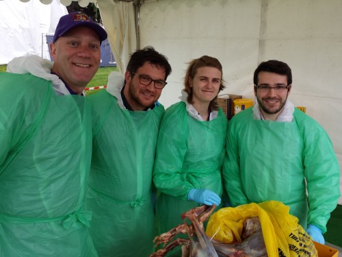
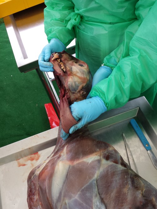
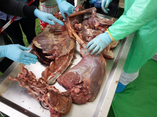
Ben, Alex, Sophie and I tackled the cheetah dissection today and it went GREAT! Much better than I’d optimistically expected. Rain didn’t scare the crowds off and neither did the gore, which there was some of (gelatinous spinal cords, lumpy tumors and at least one flying tiny bit of cheetah flesh that landed on a good-natured audience member!). Photos will tell the tale:

20160611_091345

Peek-a-boo!

20160611_093828

Sophie and Alex help us get set up in our tent.

20160611_094554

Our initial rough schedule- although we ended up improvising more after lunch.

20160611_095500

Dissectors assemble!

20160611_105126

The beast revealed. It was skinned by the museum that loaned it to us.

20160611_110748

Alex showing his talent: removing the viscera in one piece from end to end, starting with the tongue.

20160611_153148

Impressive finding of a surgical fixture (plate and wires) on the tibia, which had been used to hold the shattered bone back together long enough for it to heal. Added to the kidney disease and liver-spleen-lung cancer, this cheetah was in the sorriest shape of any cadaver I’ve seen yet.

20160611_153213

Cheetah coming to pieces: (from bottom) lumbar/pelvic region, hindlimb, thorax, forelimb and other bits.

20160611_155452

Dr Adam Rutherford, an eye expert, did a nice dissection of the cheetah’s eye, here showing the tapetum lucidum (reflective membrane), which shows up as light blue colour. Its small size befits the not-very-nocturnal habits of cheetahs.

20160611_155459

The lens of the cheetah’s eye. Now cloudy because of dehydration and crystalization, but still fascinating to see.

Want to see more images and the enthusiastic responses from the audience (we got some great feedback)? Check out Twitter’s #cheltscifest feed, or more simply my Storify condensation of the cheetah-related tweets here.

Read Full Post »

I hadn’t been feeling very well for several weeks and then last night it happened. This post is a description of what it’s like to be an epileptic, written simply to document my experience. My goal here is to do that, almost in a dispassionate scientific way, and if it helps others going through similar experiences — feedback I often receive from such posts — that’s wonderful. My post is not a call for sympathy or help, although those are understandable and kind responses, and it’s not a complaint either. It just is, because what I am is what it is.

I’ve realized that my blog has become about not just documenting how amazing, freakish and immensely fallible that anatomy can be in other species, but also about my own experiences with my anatomy (and physiology) failing, as per these two prior posts about my shoulder and brain (more links therein). Sharing these experiences gives me strength and clarity, even if some of that emerges from partly detaching myself from the emotional nature of the experience and trying to look at it from outside of myself. I can be a private person, so feeling like I can discuss something uncomfortable and vulnerable makes me feel like I am growing, much as I innately resist that.

Stomach-Churning Rating: 1/10; no fun events described, but no images either.

I’ve had enough experience now as an epileptic that I look back on my seizures with disappointment (“Oh damn, not again.”) but also familiarity (“OK that happened; I know how things will go now.”). They are terrifying at the time, especially for my family, and my disorientation when emerging from unconsciousness with strangers around and with a gap in my memory is nightmarish.

I was watching a documentary about the Jutland battle in WWI while my daughter was put to bed. Then… I woke up, maybe 20 minutes later, unsure what was going on. There were two “first responders” (emergency non-paramedics) present, one of whom I eventually recognized from my prior emergency experiences in recent months, trying to talk to me with my wife. I was impressed to later hear that they’d come within 5 minutes of being called; not bad for life in a small English town. I came to realize that my right shoulder hurt again (from violent spasms), reducing it to almost a one-degree-of-freedom joint (mostly able to move fore and aft; almost zero pronation/supination without intense pain), reminscent of Ichthyostega‘s. I was surrounded by tissues wet with blood from my lip, where I’d again bitten myself during my fit. I could sense my racing heartbeat and fluctuating temperature, other hallmarks of my pre- and post-seizure symptoms. My vision was blurry, with my eyes usually becoming dilated during a seizure.

But the predominant feeling that takes an uncomfortably long time to pass is the “post-ictal state“, a mind clouded by confusion, slowly becoming aware that my neurons are misfiring but are beginning to sort themselves out. I sometimes irrationally want to just go back to sleep and not talk, and need some rational insistence from carers that we can’t do that right now. It is this vacillation between consciousness and unconsciousness, in a grey area in between, that I find most disturbing, as I cannot completely trust my own mind, disbelieving what is happening (“Is this real?”), and sometimes I lapse back into seizure(s) again. This is a powerful example of the frightfulness of uncertainty. As a scientist, so reliant on my mind, it is horrifying to feel like it is out of control. It also conjures up memories of observing my mother’s mind declining with Alzheimer’s syndrome, and those are vastly painful.

As I became able to put words together semi-coherently, and as the medics poked and prodded me to do tests on my symptoms (I had a cannula in my left arm’s blood vessel by now), discussion turned to whether to take me to the hospital once the paramedics arrived with the ambulance. In the past, there was no question of the need for a trip to Accident & Emergency (A&E in the UK; same as the ER in the USA).

Yet now, with almost 2 years of experiences behind me, I (and my carers) have come to know my better seizures from my worst ones. And given that A&E normally involves >4 hours of lying around in a noisy room, constantly disturbed by checkups or screaming patients, it is far from restful and rest is what I tend to need most. After an hour of vigilance, my symptoms faded and I became more able to answer queries, even to talk over options. We agreed that I could stay home, try to rest, and go to A&E if I had another seizure.

I am glad to say that I got a full night of rest and I feel a lot better today. That I am able to think clearly enough to write this post gives me reassurance. After past seizures, I’d often be unable to do much except take naps and gawk slack-jawed at the TV screen, with my vision still blurred (one eye even seemed to change shape post-seizure once, and I began seeing things in the corner of my field of view that are not truly there). So the bright side of this post is, maybe my medications are working better now, and maybe we will get this epilepsy under control, but I keep saying that every 2-3 months and then being proven wrong by another seizure, so a lot of uncertainty looms.

Nonetheless, seizures involve a “refractory period” that makes further seizures less likely for some time period, so odds are good that I can feel more secure while I recover from this event, which usually takes two weeks or so to get my brain feeling closer to “normal”. Even so, my mind remains clouded by these post-ictal feelings, weighting me down with fatigue that is the most chronic challenge I struggle against now as an epileptic. It leaves me unable to do as much as I once could, with a backlog of work growing behind me like never before. This is the “diminishing” that I lamented before; it is not just old age.

That’s what one experience was like for me, and I’m glad that it was far from the worst “neural storms” I’ve suffered. I hope that readers find it interesting. Now that my battle is over for now, I’ll take some time to find out how that Jutland battle turned out.

Read Full Post »