Feeds:
Posts
Comments

Posts Tagged ‘in all seriousness’

Hey, a short post here to say go check this new blog out! I love it. The first main post-introductory post is a dissection of a snow leopard, documenting a real vet case attempting to figure out why it died. The “Veterinary Forensics blog” is going cool places, and it is a kindred spirit to this blog. You might, as I do sometimes when walking into a veterinary pathology/postmortem facility, see surprising and rare stuff– like in this photo of urban foxes:

troop of foxes


Read Full Post »

…a daily picture of anatomy! And today it is six picture-facts; doo-raa-dee! ♫

Welcome back againagain, and again (gasp, pant)— and again (exhausted howl) to Freezermas

And Happy World Pangolin Day!

Stomach-Churning Rating: 4/10; pretty tame images of anatomy today– but 9/10 if you consider how vile a practice it is to eat pangolins.

Much like rhinoceroses are, pangolins (“scaly anteaters”) are threatened with extinction across Africa and Asia largely because tradition holds that they have magical skin. It comes down to that. It’s simply pathetic.

Pangolin in Borneo

Sunda pangolin, Manis javanica; from Wikipedia. It’s not a pig in a convenient artichoke-like wrapper. It’s a precious, rare creature.

To make matters worse, pangolins are smaller than rhinos and covered in the tough armour that makes them so desirable, and hence they are more portable and easy to hide. They also are thought to taste delicious — or just have the social cachet that it is a sign of affluence to be able to afford to eat them — to some people, especially from some southeast Asian cultures. Habitat loss/growing populations/deforestation/climate change aren’t helping, either.

Around 60,000 pangolins were illegally smuggled or otherwise slaughtered for human uses in 2012 worldwide; contrasting with 668 rhinos in South Africa that year (perhaps 2,000 worldwide?); so the scale of the problem is immense. Smaller-bodied pangolins will be more numerous in the wild than large, wide-roaming rhinos, but the drain on those numbers is obviously not sustainable. Sometimes pangolins are smuggled alive, a cruel practice that delivers them fresh but in a poor welfare state at the point of sale, compounding the urgency to turn the tide of exploitation.

Please take the time today to lend your hand to a good conservation group. Learn about the crisis facing pangolins (e.g. this recent article; and this video) and speak out about it. Of course, don’t eat pangolins, either.

Let’s not let humanity fail in its moral imperative of stewardship.

Pangolin body and skeleton

My photo of a pangolin body and skeleton, from the University of Cambridge Museum of Zoology’s exhibits.

In celebration of World Pangolin Day, for today’s Freezermas we have six impressive facts about pangolin anatomy. Much like rhinos, these are animals we don’t know as well as we should. I’ve never had one in my freezers, and would feel a bit weird if I did, since I find them so adorable, but they do have fascinating anatomy, natural history and evolutionary heritage. All the more reason to preserve them as they should be: alive and with the freedom they deserve.


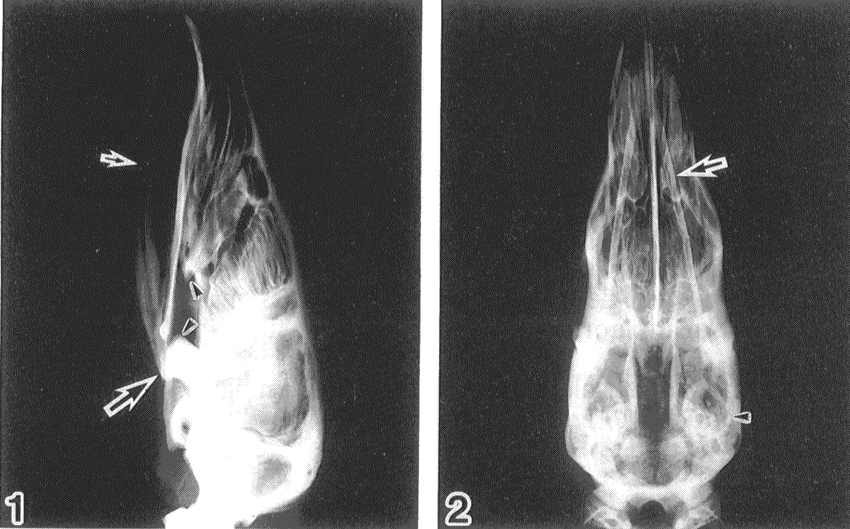
Pangolin Fact 1: Pangolins have highly modified skulls with myrmecophagous adaptations-– these are specializations for eating arthropods (especially ants/termites): toothless, tubular snout, reduced mandibles, and more– shown below.

Pangolin skull x-ray

X-ray of Malayan pangolin (Manis javanica) skull in side(1) and top(2) views, modified from Endo et al., 1998. The small arrow denotes the V-shaped, splint-like mandible, and the large arrow is directed at the jaw joint (zygomatic process on the temporal bone). The zygomatic arch, crossing from the jaw joint toward the front of the upper jaw (maxilla), is incomplete, so there is no bony bridge across the cheek as in many mammals. The large masseter and temporalis muscles run across this region, forming a more flexible, muscular cheek involved in feeding. Some nice labelled skull photos are here.

EDIT: Aaagh! Of course I should have checked Digimorph, which has a kickass CT scan/movie of the skull. Play with that; hours of anatomy-tainment!


Pangolin Fact 2: Pangolins have long tongues whose attachment extends way across the breastbone.

Pangolin tongue dissection

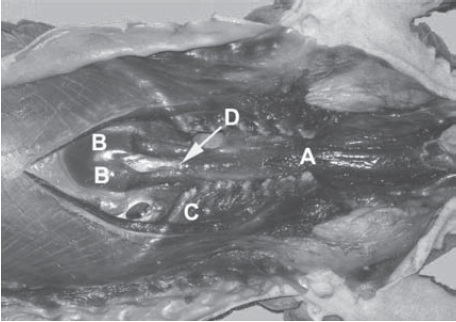
Tongue anatomy of a Malayan pangolin, from Prapong et al., 2009. This shows the chest region in ventral view (head is to the right side), with the main body of the sternum removed. A indicates muscles forming a sac around the tongue base; B is where the tongue finally inserts on the sternum/xiphoid processes; C is the ribcage; D is the xiphisternal joint (middle of the sternum parts).

Your tongue, even Gene Simmons‘s, just extends a little ways down your chin. It is, however,  a common misconception that a pangolin’s tongue is longer than the animal. It can’t be longer than the distance between its sternal origin and the tip of the snout, so it might extend up to 40cm out of the mouth when fully extended in a large pangolin. Around 1988, there was the scientific misconception that the tongue extended way back to the pelvis (hips) or stomach. This is not true according to the latest literature I’ve read (e.g. in caption above), but is widespread in pangolin information on the internet. If someone has secondary confirmation of this either way, I’d love to see some concrete evidence.

EDIT: This image of a dissected pangolin fetus indicates a quite long tongue, maybe even long enough to attach near the pelvis, although that site agrees that there is no pelvic attachment. The latter site also depicts a fascinating cartilaginous sheath for the tongue. The misinformation about pangolin tongues does make me wonder: perhaps there is a lot of diversity in tongue attachments/lengths among the 8 pangolin species? Who knows.


Pangolin Fact 3: Pangolins have toughened, keratinized stomach linings.

Pangolin stomach histology

Click to embigitate. Histology of the stomach lining in Manis tricuspis (modified from Ofusori et al., 2008), showing layers of keratinized stratified squamous epithelium (thick stomach lining). These layers seems to act as a protective coating against the rasping, chitinous exoskeletons of the ants and termites that are consumed, helping to reduce the risk of ulcers while reportedly eating up to 200,000 ants/per night. There is also an increased preponderance of elastic and collagenous fibers in layers of the stomach, helping it to expand to enclose many ants from one feeding.


Pangolin Fact(ish) 4: Pangolins are not closely related to other ant-eating living mammals, but to carnivores.

Eurotamandua fossil

Eurotamandua; a possible fossil relative of pangolins from the early Eocene (Messel, Germany); image from Wikipedia.

Together, the eight living pangolin species are remnants of the group Pholidota, which has a respectable fossil record– particularly considering that they lack teeth, which are often such a diagnostic feature for mammalian fossils. Controversy persisted for many years about whether they were related to anteaters, sloths and armadillos (Xenarthra) within the group Edentata, along with possibly aardvarks (Tubulidentata) and other digging and/or myrmecophagous animals. There has also been controversy about some fossil mammals and their relationships, including Eurotamandua (above) and the Palaeonodonta— the latter seems to be approaching a consensus, though, as an extinct sister group to Pholidota.

Nonetheless, the main features that were once thought to unite ant-eating mammals as close relatives now seem to be a prime example of convergent evolution. Xenarthans are definitely closely related to each other, but aardvarks are afrotherians (closer related to hyraxes and elephants), and pangolins seem not to be closely related to either of those groups.

More conclusively, with the addition of genetic data, it has emerged that Pholidota is most closely related to Carnivora (mongooses, dogs, cats, bears, pinnipeds, etc.) among living mammals. A good example of this conclusion is the very recent paper by O’Leary et al. in Science. Furthermore, this image shows a nice example of such a phylogenetic result. This relationship with Carnivora raises fascinating questions about the tempo and mode of the evolution of all their digging/ant-eating specializations- when, where and how did they become so much like other ant-eating mammals?


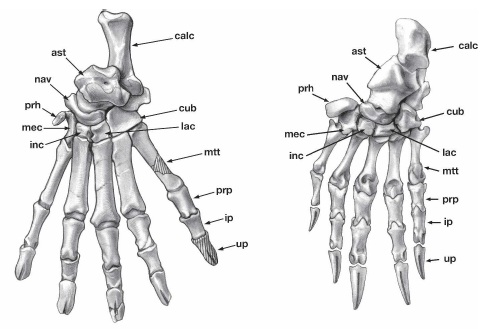
Pangolin Fact 5: Pangolins have many digging (fossorial) and climbing (scansorial) adaptations, especially in their forelimbs.

Pangolin hindfeet Feet of anteating mammals

Click to embignify. Above (modified from Gaudin et al., 2006; by Julia Morgan-Scott): drawings of hind feet of (left) the Eocene fossil pangolin Cryptomanis and (right) Manis; Below: line drawings of front feet (from Humphrey, 1869) showing the convergent evolution of digging/climbing hands in (left to right) pangolins, an anteater (2-toed; Myrmecophaga), Ai (3-toed sloth; Bradypus) and Unau (2-toed sloth; Choloepus).

A striking feature of pangolin claw bones (unguals); evident above; is their characteristic fissured anatomy (split ends), which even the fossils have. This probably is how they develop strong, keratinous digging claws that remain anchored to those bony claw cores. If you look really closely, you may be able to see the fused scaphoid and lunar (scapholunar) bones of the wrist in the manus of Manis. Cryptomanis (above left) had more climbing specializations than living pangolins; is this how pangolins first evolved, and then later added more ant-eating features? This makes sense in terms of their phylogeny (above), as they are related to primitively climbing carnivores.

Other possibly digging/climbing-related features characteristic of pangolins include the loss of a coracoid process on the pectoral girdle, and curious enrolled zygapophyses (joints) on the lumbar (lower back) vertebrae — the functional significance of the latter feature is almost unstudied, but is reminscent of the complex xenarthrous vertebrae that gave Xenarthra their name (see above and this past post). A nice photo of a pangolin ribcage/vertebrae is here. There is an exceptional page on pangolins and their once-thought-to-be-close relatives among Xenarthra here, with lots of anatomical detail.

A feature that first got me scientifically curious about pangolins in my research is the presence of “predigits“- prepollex and prehallux- in their hands and feet (“prh” in upper left two figures). Many mammals have these, and some have expanded them into larger structures like the “sixth toes of elephants” (hence my interest), but precious little is known about their evolution or function in many other groups.


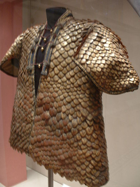
Pangolin Fact 6: Pangolin skin armour, like rhinoceros horns, is just modified skin (hair/epidermis) keratin; shaped into imbricating scales.

Pangolin scales closeup Coat of pangolin scales

That apocryphally “magic skin”. Images from Wikipedia: closeup above, and below it a suit of armour made from those scales–  coated in gold and given to King George III in 1820.

These scales, the double-edge sword of pangolins (both protecting them in nature and making them desirable in part of the human world for silly reasons), form as pangolins grow. In the fetus they are still soft, making fetuses more of a delicacy in some Asian cultures. Much like the stomach lining described above, the skin is formed from keratinized, stratified squamous epithelium– much more densely formed than in our skin, but more like in our fingernails. Asian pangolins, unlike African species, may have some more normal hairs beneath the scales, too.

There is no convincing evidence that the scales are any more healthy to eat, in any form, than your own fingernails, dead skin, or hair. Given the ready availability of the latter to any humans, we’re all wearing, and growing, our own goldmine…

I’ve barely dug into the fascinating biology of pangolins. I haven’t talked about their bipedal locomotion, much as it fascinates me, because we know next to nothing about that. I’m not aware of good scientific studies on their prehensile tail, either. A great page on pangolin biology, with a focus on reproduction and anatomy, is here. A lovely illustration and discussion of the convergent evolution between anteaters and pangolins is here. Awesome photos and facts are here. More about pangolins’ plight here, and very thoroughly here.

If you have favourite links to more material, or want to provide more information or especially questions, don’t hold back and experience painful pang(olin)s of remorse– chime in in the comments below!

Happy World Pangolin Day! Visit these great pages, please! Here, here and hereAnd…

Happy Freezermas! Sing it: “On the sixth day of Freezermas, this blo-og gave to me: one tibiotarsus, two silly Darwins, three muscle layers, four gory heartsfive doggie models a-and six facts of pangolin anatomy!” ♪

Read Full Post »

Good day, everyone. Maybe by the end of this post if you don’t agree that it is a good day, you will at least see why I think it is.

Ten years ago today, something Really Bad happened to my brain. I don’t need to go into details, but it is very fair to say that I almost died. And that was the second close call in my adult life; there was another, years earlier, with a different vital organ system. So I celebrate December 16th each year as “Not Dead Yet Day“. As this is this blog’s first NDYD, I figure you can all join in the celebration, for any reason you might have to celebrate life. It can be hard to love some aspects of life sometimes, especially in pretty depressing times like the 21st century can be (so far). This can especially feel true in light of recent events in Connecticut, or ongoing nightmares in Syria and many other lands, with vanishing innocents, vanishing wildlife and vanishing habitats, the inexorable heat death of the universe… shit I’d better stop now or I’ll lose it!

This day helps to remind me to stay focused, as much as I can, on what matters in my life, and what I can control in my life to make things better for the little bubble of the world that I exist in. Some things are far beyond even our hope, let alone our means, to control. And sometimes we get broadsided by Really Bad Shit. But in between any of that powerlessness or inauspicious shit, there can be joy from many sources– for me (like many others), it comes from family and friends, science and the natural world’s wonders, delicious food and amazing travel, and much more. It comes from experiencing reality with all its facets.

Here is my brain. You can’t see much. Feel free to make jokes about that, I’ve set myself up nicely with that last sentence!

my-brain1

These are MRI scan images from a routine checkup I had about 3 years ago. I suppose you can consider it a game of “Mystery MRI slices”, but one in which I give you the answer (my brain). You can see lots of cool anatomy here; if you know your anatomy feel free to mention what’s visible (or not) in the Comments, and make jokes– I will probably enjoy any of them. I like self-deprecatory humour. And happily, I checked out fine in that scan, and continue to be fine… relatively. I’m not the same person I was >10 years ago— in 2002 I got married (but missed my bachelor party because I was hospitalized for another problem), got an important paper (“Tyrannosaurus was not a fast runner”) published in Nature that changed my career (and arguably got me my job today), had this Really Bad thing happen, and plenty more. It was an eventful year.

my-brain2

At the time the Really Bad thing happened, I was feeling poorly but working very hard on final revisions/re-analysis of elephant gait data for a paper that ended up being published in Nature in 2003; so things ended up looking even better for my career. But I made a decision that day that, in a fortunate way, ended up having a greater impact than any mere publication. Rather than sit in my house with our cats and feel poorly, I made the choice to drive in to work and process more elephant video data. Just as I was parking my car on the Berkeley campus (illegally; I was feeling very poorly by that point) to go in to do the work… I woke up in an ambulance.

I was lucky. I was somewhere public where I was spotted having trouble, not alone in my house for >8 hours until my newlywed-wife came home to discover me. So I got help, and medical science saved my ass — and my brain, and thus other regions of my anatomy and my mortal existence. If I’d adopted the other choice, and stayed home alone, our cats probably would have witnessed something terrible and been unable to help, awesome as kitties can be.

I’ve never felt the same after that day. I’m certainly a case of “scarred but smarter.” I can say smarter mainly because my brain survived the trauma OK and I learned from the experience. I can say scarred because I still feel repercussions of all sorts from that Really Bad day. Although I’ve always had a dark sense of humour, strongly connected with my eccentric passions in science (e.g. this blog! Go figure.), I think it’s fair to say that my humour darkened. I’m not as bubbling with joy as I used to be. I used to almost always grin and exclaim “Excellent!” when someone asked me “how’s it going?”. I can still burble with frabjous joy, but not quite as often.

That day brought me closer in touch with the darker side of life, and the brighter side too. I think I’d been overlooking both. Closer in touch with reality, and with the serendipity and calamity that accompany it. There have been other, terrible events in my life since then, too, that have brought new existentialist focus to my mind, but that’s a part of most people’s middle age period (e.g. losing many loved ones).  I’ve had a great career so far, too, thanks in part to good things that happened 10 years ago, and to good things that have happened since thanks to hard work and some good fortune. But that doesn’t mean life has been a nonstop joyride, or even easy.

So today I take some special time to think about what life is about, what is real and must be faced wide awake vs. what is self-deceitful slumber, and why life is still worth loving– which I do love, with all my brain. And every day I think about the big changes that 2002 wrought on my life, and how so many other seemingly important things that happen in my life don’t matter one fucking bit– hence I try to just have fun, be a good human and not worry so much.

Have the best day you can have, everyone. I’m off to have some fun family time, but wanted to share my brain’s thoughts with you today. Maybe you have a similar story to share, too, or maybe my brain’s thoughts inspire some in your own brain. It’s wonderful how that glistening anatomy can do such things, and it’s wonderful how resilient that anatomy is, much as we need to be… because we are one and the same, our brains and our selves that dwell inside them, and the love of life that they can conjure.

If this post bummed you out, just focus on these contented cats.

If this post bummed you out, just focus on these contented cats.

Read Full Post »

In case you haven’t heard, Saturday, September 22nd, 2012 (today, at this writing) is World Rhino Day! The main websites include here and here.  Ivan Kwan has also posted a fantastic blog entry “Rhinos are not prehistoric survivors” for WRD2012- check it out! And if you haven’t seen the WitmerLab’s AWESOME Visible Interactive Rhino site, you really really need to (in fact, quit reading this and go there first; it is soooooo good!).

I’ve written about the global rhino crisis before, and about rhino foot pathologies. The title of today’s post may be “cute”, or at least goofy, but the real situation is as grim as the images I’ll share. I won’t repeat the explanation, but all five living species of rhinoceroses are in serious trouble. There’s a good chance that most or all of them will go extinct quite soon– see the previous links for more information on this. Javan and Sumatran rhinos are dangling the most precariously over the precipice of extinction. My goal in this post is to share the beautiful, complex and exotic anatomy of rhinoceros anatomy and movement, and the joy of contributing new scientific information about poorly understood species.

Stomach-Churning Rating: 7/10— dissections, and there are a couple of pics where the specimens are not so fresh, and there’s big skin, and a huge heart.

Baby white rhinoceros. Will frozen specimens like this be all we have of rhinos someday?

The purpose of today’s rhino post is to share a bit more; especially images; of the work my team has done on rhinoceros gait and limb anatomy; all of it unpublished but hopefully coming soon. We’ve steadily been collecting data since ~2005. Because my previous post went through some of this, I’ll keep it brief and image-focused.

First, a video of one of our amusing encounters with a white rhinoceros, at Woburn Safari Park. In this study, we wanted to measure, for the first time really, the gaits (footfall patterns) that a white rhinoceros uses at different speeds, and how often it uses those different gaits. We attached a GPS unit on a horse surcingle around the rhino’s torso, which measured the animal’s speed once a second. We then observed 5 individuals (1 at a time over various days), following them in my station wagon (estate car) across the safari park. We filmed them with a conventional camcorder to document their gaits, and concentrated on the two periods of the day that they’d normally be active: when released from their overnight barn, and when coming in for the night back to that barn. They got rather excited and frisky some of those times. The GPS belt then kept recording speeds for the rest of the day; unsurprisingly, the rhinos generally did not do much. I have to thank Nick Whiting, rhino handler, for his help making this research happen. I’ve been meaning  for too long to finish the final paper… soon, I hope! Enjoy this tense scene of a rhino investigating my car (driven by me and with an undergraduate student filming) then having a nice canter/gallop across the field (accompanied by my jubilant narration).

Like our foot pressure research, we aim that this work provides baseline data useful to caretakers of rhinos; for example, to test if a particular animal is lame. This follows what we’ve successfully done with elephant gaits and feet, translating basic research into more clinical application. But my major scientific interest is in understanding more about what makes any rhinoceros, even a 2-tonne White rhino, so much more athletic than any elephant (even a baby or 2-tonne small adult Asian elephant). As the video shows, they can use a variety of gaits including cantering and galloping, and trotting at slower running speeds. No elephant ever does that, and no one knows precisely why. The leg bones are more robust, but the muscles aren’t that dramatically larger in rhinos.

An Indian rhinoceros forelimb- note the characteristic knobbly hide, unlike the smoother, more elephant-like hide of a White rhinoceros.

Similarly, the anatomical work we do with rhinos is intended to not only be useful science for comparative biologists like me, showing how rhino limbs work and how they differ from those of other animals, but also to aid clinicians in comparing normal vs. pathological anatomy. For conveying that anatomical work, I’m lucky to have been granted permission to use a professional photographer’s pictures of some of my freezers’ rhino specimens– big thanks to James King-Holmes and the Science Photo Library. The watermarked images below belong to them. I ask that you do not use them elsewhere, honouring their license to me for personal usage on this website (and I will only use them here). I’m in all the images, which makes me feel weird putting them up here, but it’s about the rhinos (and freezers), not me. First: the infamous “rhino foot freezer”, featuring some of its denizens:

Second, a re-introduction to multifarious contents of Freezersaurus, but this time featuring rhino feet (here, a skinned white rhino foot that we had already studied):

…and inside we go (and I begin to get frosty and numb-fingered from holding a foot; my smile soon fades):

Taking a rest with the skinned white rhinoceros foot:

And now warming up at the “digital freezer”, our CT scanner, and preparing to scan another rhinoceros foot, which segues nicely out of this image sequence:

Now over to some 3D anatomy– segmented reconstructions of rhinoceros fore (top) and hind (bottom) feet, from CT scans; if you’ve frequented this blog you know the drill. Here, the longest bones are the metacarpals/metatarsals and the upper bones are the carpals/tarsals, then the bones near the botttom are the phalanges, which connect to the hooves (visible in the bottom image):

I’ll wrap up with a series of images of basic limb muscle anatomy from dissections we’ve done of baby and adult Indian and White rhinoceroses. First, here’s what a rhino looks like underneath the skin:

But ahh that skin, that fabled “pachyderm” skin! A rhino’s greatest defense is also a real chore to get through in a dissection.  Here, we enlist the help of a crane and hook, hurrying to get down to the muscles of this forelimb before rotting takes over too much (as with other big animals, this is a tough race against time even in chilly England!):

Here is a closer look at that amazing armoured skin; sometimes 10cm or so thick:

Back to the forelimb muscles– stocky and well-defined for this athletic animal:

(late addition) Here are the massive shoulder muscles, such as the serratus and latissimus dorsi (this is a left limb in side view; head is toward the left):

And now a close look at the forearm muscles:

And then over to the hindlimb, here from an adult Indian rhino, whose thigh bone (femur) shows the characteristic giant “third trochanter” (toward the bottom centre of the image), which is an expanded bony attachment for the giant “gluteobiceps” muscle complex that retracts the femur for the power stroke in locomotion. Also, this specimen showed fascinating anatomy that I’d never seen before: the third trochanter has a thin bar of bone that extends up (toward the bottom left in the image) to fuse with the greater trochanter, opposite the head of the femur (upper left corner):

Damn my photography skills, cutting off the edge of that image and instead giving a view of my boots! Anyway, another interesting feature of that femur: the medial (inner) condyle of the femur (knee joint surface) has a pink stripe of worn cartilage. This is indicative of at least a moderate stage of arthritis, shown here (look for the pinkness amidst the shiny, healthy white cartilage on the upper right side). It is an exemplar of serious welfare problems that some captive, and probably some wild as well, rhinos face:

(late addition) Back up the limb, this baby White rhino shows the massive thigh muscles, especially that “gluteobiceps” that attaches to the third trochanter, noted above, and also showing the hamstrings:

Moving down the limb, we encounter the glorious three-toed perissodactyl foot of rhinos, and the robust hooves/nails, which are reasonably healthy in this animal– unlike others I’ve seen:

And the sole of that foot, showing a fairly healthy pad, below. Toward the rear (away from the nails), it culminates in a modest-sized fat pad, or digital cushion, akin to that in elephants but far less well developed and lacking the false “sixth toe” (predigit) (see also CT scan movie of the hindfoot above):

Here’s a view inside that marvelous foot, showing the HUGE digital flexor tendons. These help support the toes against gravity and, in theory, can act to curl them up– although in a rhino’s foot, as in an elephant’s, the toes are more like a single functional hoof, with reduced independence compared to a carnivore or primate:

And that ends our tour of rhinoceros limb anatomy and function. Help spread the word of how precious and threated rhinos are; educate yourself and others! And if you overhear someone talking about using rhino horn for medicine, try to politely educate them on the utter fallacy of this tradition. It is this cruel, greedy, ignorant practice that needs to die; not rhinos. I don’t enjoy receiving dead rhinos, on a personal level, even though the science excites me. I’d rather have many more alive and living good, healthy lives. And my team is trying to do what we can to help others on the “front lines” of rhino conservation make that happen.

For example, Will Fowlds, vet and co-owner of Amakhala Game Reserve, South Africa, recently sent us some images of a white rhino that had been caught in a poacher’s foot snare some years ago. The poor rhino still was having problems healing– we inspected x-ray images and external photos and helped to make an initial diagnosis of osteomyelitis, a nasty infectious, inflammatory foot bone/joint disease. We are following this case to hope that the rhino recovers and contribute help where we can, but the tough job belongs to the keepers/vets on the ground, not to mention the rhinos…

Furthermore, we’ve done foot pressure research covered here, and here is an example of the data we’ve collected (image credit: Dr Olga Panagiotopoulou), showing high pressures on the toes and low pressures on the foot pads:

Big thanks to people on my team that have helped with this and related research: Dr Olga Panagiotopoulou (and Dr Todd Pataky at Shinshu University, Japan), Dr Renate Weller in the VCS Dept at the RVC, Liz Ferrer at Berkeley, and former undergraduate student researchers Sophie Regnault, Richard Harvey, Hinnah Rehman, Richard Sheehan, Kate Jones, Bryony Armson and Suzannah Williams.

A White rhino’s heart, with more images below, all courtesy of William Perez’s Veterinary Anatomy Facebook pages. A mass of around 10kg (22 lbs weight) is not unusual! (Compare with even larger elephant heart)

White rhino closeup: coronary arteries

White rhino: branches of left coronary artery

White rhino heart: right atrium

Read Full Post »

Here is a little vignette for you; a taster of the BBSRC-funded chicken biomechanics project my team has underway in collaboration with Jonathan Codd’s team at Uni Manchester. I did not know about the following situation until a couple of years ago during my former PhD student (now postdoc) Heather Paxton‘s research on chicken mechanics.

Regard this chicken, slit open along the midline to show the viscera. The huge pectoralis muscles (breast meat) have been pushed aside; the right side’s are clumsily outlined (I blame caffeine?) in blue.

Then consider the heart, within the jagged, shamefully and ineptly drawn green circle. I’ll come back to that.

So this broiler chicken took 6 weeks to reach this size, of about 3 kilograms (6.6 lbs). Fifty years ago, before artificial selection was imposed on a MASSIVE scale (many billions of chickens per year worldwide, bred in a complex pyramid scheme of crossed strains), that same chicken strain would have taken 15 weeks to reach a normal slaughter mass of roughly 2 kg (4.4 lbs). The major selection, of course, has been for edible meat, especially that lovely breast muscle’s white meat.

If we look at red junglefowl, to a large degree the “wild type” ancestors of domestic chickens that are native to southeast Asia, the leg muscles take up about 7.7% body mass per leg vs. about 6.3% in the broiler. Just a small decrease, but probably an important one, and something our research focuses a lot on (walking ability, lameness, activity levels etc). But that’s a subject for a future post. In stark contrast, the breast muscles (back to the blue ellipse above)  have gone from 7% to up to 11.6% body mass per wing; a huge change!

Now let’s return to another large muscle, the one within the green circle above; the heart. Not only must the heart, which has become relatively larger by perhaps 25%, pump blood to a body that has enlarged by >50%, but it also must perfuse the giant pectoral muscles, which have enlarged by >65%.

Herein lies the problem… You probably can predict what happens.

Several syndromes may develop, but the one I want to cover here is called deep pectoral myopathy (AKA “Oregon disease” or better yet “green muscle disease”, a very appropriate term as you’ll see below). Basically, the giant pectoralis muscles receive inadequate blood flow from the smallish heart, because the muscles are so big and under so much pressure, creating resistance to flow, and so the muscles begin dying from within. A picture tells the story:

While surely uncomfortable for the birds and hence a welfare problem, it is usually not found until the animals are slaughtered, and then of course the meat is destroyed rather than delivered for human consumption. Because of the welfare problems and loss of meat (i.e. financial loss), the poultry industry is trying to remedy this problem. W’e’re working on aspects of this as well, as part of our study of how the locomotor and ventilatory systems of chickens develop and have evolved.

I am blogging this as a great example of how anatomy can go haywire and become imbalanced when evolutionary selection pressures are intense and highly specific (e.g. almost single-minded human selection for large breast muscle). It is also a conundrum that human society faces: while chicken meat seems more efficient and more ecologically sound than some other meats, and there is growing demand for meat as the human population grows, how do we balance welfare concerns with food security, economics and other factors? And how do we judge when artificial selection has gone too far? I do not present an answer because the answer is not easy, and because my team is still learning about how to answer it.

Read Full Post »

Here now is the promised blog post, which uses the rhino foot mystery pic as a springboard to address a phenomenon that is a bit better known, partly because it is an even worse situation and involving (arguably) even more charismatic critters: elephants.

A rotating movie of a CT scan reconstruction is a good way to kick this off:

This shows the right hind foot of an Asian elephant that had mild pathology; mostly a roughening of some of the bone surfaces that is called osteitis (proliferative bone growth possibly due to infection or other irritation) and perhaps a mild case of degenerative joint disease such as osteoarthritis. But this is nothing compared to the severe cases we’ve observed in other elephant feet, and indeed may not have anything to do with why this elephant died (I’m not sure; I was given very little medical history for this one).

If you want more elephant anatomy lessons, see the videos from the posting on six-toed elephants. I will proceed assuming some basic familiarity with bones of the feet in animals, although you may be just fine even without that.

About 50% of elephants in captivity die from foot disorders of one kind or another. Elephant keepers spend a huge amount of time and energy taking the best care of elephant feet that they can, but a variety of factors including anatomy, biomechanics, exercise, obesity, ground surface, hygiene, “hoof” care including trimming, nutrition, and much more are part of the very complex causal nexus underlying these disorders. Wild elephants get similar problems, too, but less frequently (e.g. in drought periods, I’m told); there are few solid data on this, however.

Onwards, then! I shall present a cavalcade of horrific examples of the kinds of elephant foot pathology that we have observed in specimens that have come through my freezers at the RVC.

Let’s start with what one of our vets might see on examination of a live elephant at a zoo:

This is an x-ray image of the third (on the left) and fourth (on the right) toes of an elephant’s front foot. The RVC (Dr. Renate Weller and myself) have developed protocols to take such x-rays on live elephants.  The anatomy shown here is pretty normal and non-pathological. So with that in mind, check this out; toes four (on the left) and five (on the right), different animal:

Ouch! Digit 4 (“ring finger”) has a proliferation of bone that is characteristic of an animal with osteomyelitis: a flowering of bone in response to infection and painful swelling, probably caused by an abscess on the toe’s sole/nail. This animal was put down because of its unresolvable misery from this disorder. Oddly, we see toe 4 as well as 3 and 5 as the most commonly pathological; toes 1 and 2 seldom are. We’ll be discussing this in a new paper coming out soon; I’ll get back to that another day.

Assuming such conditions don’t resolve, the next place the foot may end up is in The Freezer at the RVC, and then into our CT scanner before we do postmortem dissections and a report on the pathologies so the zoo knows what went wrong. Here’s an example of what we cut off the end of the fourth toe of such an animal:

Just looks like a glob of tissue, right? The joint between two segments of the toe is visible as a pinkish white structure on the right side, with some bleeding on the cartilage where it wore down to the bone surface. But it gets worse. Here is how that same toe bone looked when we cleaned it up (boiling and bleaching away soft tissues):

Here, that same roughened joint surface is visible at the top of the specimen. Two toe bones have become fused together (the bottom one is not visible), encased in a cocoon of lacy, spiny bone. Again, ouch. The next specimen had a different kind of “ouch”- its fifth toe basically shattered:

That toe is almost unrecognizable, having disintegrated rather than proliferated its bony scaffolding. Other specimens may be in less extreme states of pathology but still likely to have been in pain:

The label here says it all; third toe with a cyst where an infection entered the bone.

This one, the end of a third metatarsal, shows degenerative joint disease with a loss of articular cartilage, and holes where abrasion has worn down into the bone and caused bleeding. In contrast, and to give you a breather from the horrors, here is a healthy, younger elephant’s similar joint surface:

Nice white, fresh, shiny cartilage! Ahhh…

But then we dive back into Grand Guignol-level aberrations:

Here we’re looking at the back side of a right hind foot of an elephant, at the level of the ankle joint. The joint capsule surrounding the ankle joint has been cut open in my dissection to expose the terribly pathological, but still somewhat white and shiny, cartilages (middle of the image) which have been abraded (in some regions) but also extended by new bone formation (in other regions) to creep around the back of the ankle. Here, the bone growth was fulfulling a role to limit joint mobility and thereby restrict painful joint motions- the joint was fusing into an ankylosis (no, not an ankylosaur,  but same Greek root). Here is a closer look, removing the tibia and fibula that were at the top of the screen in the above image, and looking down onto the ankle joint surface:

You should be able to more clearly see how the cartilage and underlying bone are not forming a smooth edge, as they should on the talus (ankle bone), but rather an irregular, jagged contour (area to the right of the label). This animal would have been visibly lame, to say the least; elephant ankles can’t move much even in normal animals but this one was even less mobile. We’ve had some specimens where the ankle was so fused it was totally immobile and took a saw to separate the two sides of the joint. Oddly, I haven’t seen an ankylosis like that in the wrist, which in normal elephants is as flexible a joint as the ankle is inflexible.

Pathologies like these sadly aren’t uncommon in elephant feet but zoo/park keepers are doing their best to turn the trend around. Zoo conditions generally were a lot worse 50 years ago. The pictures below document this, from museum specimens we’ve studied (among many others) at the University Museum of Zoology at Cambridge and the Natural History Museum in London. See what pathologies you can spot! Some are from wild-shot animals, reinforcing that foot pathologies are not just a zoo thing. (click to embiggen)


Zoo/park conditions are improving now— in the UK for example, elephants are being moved into more safari park-like environs and given more varied surfaces to walk on or even dig (e.g. sand at Chester Zoo). But because elephants live long lives, and foot pathologies sometimes cannot be reversible (or even detectable, sometimes), any pathologies existing now may well still be evident, or even worsen despite the best care, for decades to come. The lag time for fixing the global problem of elephant foot pathologies is not a short one. I won’t get into the controversy over whether elephants should be in zoos/parks or not, but at least for the short to medium term they are, and we need to make the best of that. The images in this post help show why, and perhaps point a way toward how.

Read Full Post »

On a serious note, in case you aren’t aware of it, there is a huge crisis afoot with the world’s rhino populations, because of demonstrably false claims about the health benefits of rhino horn (e.g. curing cancer) leading to a surge in its value worldwide, and thereby a massive upswing in poaching- as well as theft of museum specimens(!?). It may just be a matter of time before zoos’ and safari parks’ rhinos outside of Africa/Asia get hit, too. If sustained, this poaching could wipe out multiple rhino species in a matter of years; it is that severe and seems to already be worse this year than it was last year- and that was a Very Bad Year for rhinos.

However, a lot of people are uniting against the cruel greed and gross ignorance that has fueled the decline of rhinos, and public support seems to be growing. You can contribute, too; one example is this cause, or this one, to name but two. I’ve tried to make this my #1 cause, both on personal/ethical and scientific grounds (e.g. our work on rhino foot mechanics, health and care, to be detailed here later), and have been educating myself about it.

Anatomist extraordinaire Professor Larry Witmer of Ohio University has been contributing scientifically to helping rhinos, in an unexpected (but very sensible) way. This is a perfect example of how important basic science is; if we didn’t know rhino horn/nose anatomy we’d be less able to treat problems when they arise, and such problems can come from unexpected directions. His team’s contributions in the understanding of rhino anatomy are helping in one horrible case (see video below), in which three rhinos had their horns slashed off (along with part of the top of their skulls!) and two have survived so far. The vets in South Africa are trying to treat these two mutilated animals, and Larry’s group has been providing anatomical advice along with superb pictures on their Facebook page, which I want to publicize here because it is such GREAT freezer-based anatomical science and stunning imagery, and a seriously urgent cause. Please take a look. And while you’re at it, check out the Kariega Game Reserve’s Facebook page with more info on the plight of poor Themba and Thandi.

Edit: also check out this great story on our research, by Ann and Steve Toon, rhino conservationists/photographers/journalists.

Read Full Post »

« Newer Posts