I’ve described our “Walking the Cat Back” Leverhulme Trust-funded project with Dr. Anjali Goswami and colleagues before, but today we really got stuck into it. We’re dissecting a 46kg male Snow Leopard (Panthera uncia) as the first “data point” (actually several hundred data points, but anyway, first individual) in our study of how limb and back muscles change with size in felids. No April Fools’ pranks here; real science-as-it-happens.
Stomach-Churning Rating: 7/10 for skinned leopard and globs of fat. Much worse in person, hence the downgrading from what could be a higher score. Don’t click the photos to emkitten them if you don’t want to see the details.
This leopard is the same one that Veterinary Forensics blogged about. It died in a UK cat conservation/recovery centre. Today is simply a short post, but it is the first in what will surely be a continued series of posts on felid postcranial anatomy and musculoskeletal biomechanics by our felid research team, with bits of natural history and evolution thrown in when we can manage. As befits one of my curt “Anatomy Vignette” posts, pictures will tell the story.

Skinned and mostly de-fatted snow leopard, with fat piled up on the lower left hand corner near the hind feet. Here we are identifying and then removing and measuring the individual muscles. Project postdoc Andrew Cuff is hard at work on the forelimb while I’m mucking around with the hindlimb. The fat here is about 3kg subcutaneous fat, so around 6.5% of body mass. And as the cat has been around for a while, that fat has gone a bit rancid and that is not nice. Not nice at all, no… Usually smells do not bother me, but this took some adjustment. Fortunately, the muscles are still OK, and work is coming along well.
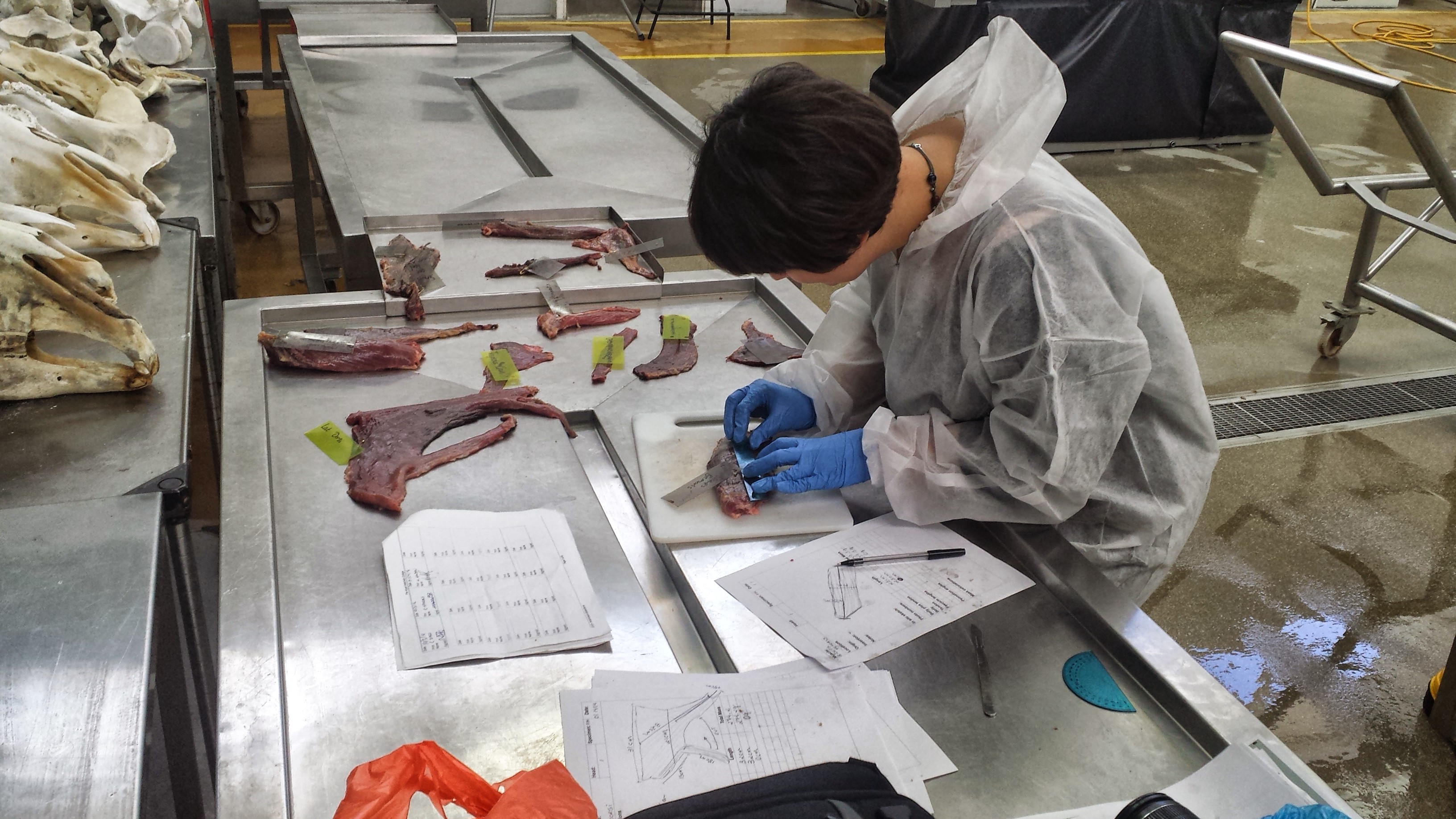
UCL PhD student Marcela Randau, carving up our cat’s limb muscles. As usual in comparative biomechanics, we measure the “architecture”- parameters of the muscle that relate in a somewhat straightforward fashion to function. This muscular architecture includes things like muscle mass, the lengths of the fibers (fascicles) that make up the muscles, and the angle of the fascicles to the muscle’s line of action. These parameters correlate reasonably well with the force and power that the muscle can develop, and its working range of length change. Other posts here have discussed this more, but by measuring the architecture of many muscles in many felids of different sizes, we can determine how felids large and small adapt their anatomy to support their bodies and move their limbs. This will help to solve some lingering mysteries about the odd ways that cats move and how their movement changes with body size.
This research is being driven forward mainly by Andrew and Marcela, shown above, so I wanted to introduce them and our odoriferous fat cat. Upcoming dissections: 1-2 more snow leopards, tiger, various lions, ocelot, black-footed cat, leopard, and a bunch of moggies, and whatever else comes our way. All were EU zoo/park mortalities (there are a LOT of big cats out there!).
EDIT: Had to add a photo of the CLAWS! Whoa dude.


After viewing the full size picture, I can appreciate the problem. That lot certainly looks a bit ripe. If you’ll pardon my ignorance, is 3kg a lot of subcutaneous fat for a cat this size?
Yes that’s quite a bit; a fat cat indeed!
For some reason I assumed the thigh muscles would be larger; they seem relatively compact in the photo. The front end seems more heavily muscled, is this where most of the power comes from when the animal is moving quickly, or am I way off?
Well spotted. We’d taken some hindlimb muscles off (the hamstrings, mainly) so it looks more svelte than it really was. The hindlimb muscles do tend to be a bit larger in felids, by overall mass; about 20% vs 15% body mass per pair, respectively. And indeed, as in most mammals, the hindlimbs are the main power source for locomotion (maximal power [Force * velocity] and muscle mass are closely related).
Thanks for the claw shot. One of my favorite things from Inside Nature’s Giants was the big cat dissection where someone pulled the tendons that retract the claws. It was aso cool to see the mechanism in a large format example that happens on a smaller scale in my own fat cat’s paws (20 pounder). Would love to see the cheetah musculature in comparison to the others you are planning.
The ING scene, if memory serves, was with Penny Hudson from our lab, who published the key work on cheetah musculature (and dissected many other cats):
P.E. Hudson, S.A. Corr, R.C. Payne-Davis, S.N. Clancy, E. Lane, A.M. Wilson (2011). Functional anatomy of the cheetah (Acinonyx jubatus) hindlimb. Journal of Anatomy. 218(4): p. 363-74.
P.E. Hudson, S.A. Corr, R.C. Payne-Davis, S.N. Clancy, E. Lane, A.M. Wilson (2011). Functional anatomy of the cheetah (Acinonyx jubatus) forelimb. Journal of Anatomy. 218(4): p. 375-85.