To kick off the New Year just right, our tetrapod team has a new paper in Nature, following up on last year’s Ichthyostega not-so-good-at-walking study (also see here). Yet this paper has a more anatomically descriptive — and also an “evo-devo” — twist to it. For brevity, I’ll let our press release tell the story, since I think it does a good job of it (like I always preach scientists should do, we worked with our PR company to write this together, so we’re happy with how the press release came out). In a nutshell, our study used some very fancy synchotron radiation techniques to image the 3D anatomy of the backbone in early land vertebrates. Our findings surprised even us, and ended up turning around palaeontology/comparative anatomy’s view of how the backbone evolved, giving us a new glimpse into our inner tetrapod.
Stick around for the videos at the end, which are the first four supplementary movies from the paper and are rather pretty (there are two more, for imaging/segmenting afficionados, but they are not as pretty or interesting for most of this blog’s readership). The final figure (Figure 1 from our paper) gives some extra visual context.
The paper is:
Pierce, S.E., Ahlberg, P.E., Hutchinson, J.R., Molnar, J.L., Sanchez, S., Tafforeau, P., Clack, J.A. 2013. Vertebral architecture in the earliest stem tetrapods. Nature, published online [here].
I should note that I’m just 3rd author, so I deserve only modest credit. But I helped. Even though no freezers were involved, or harmed, in the process.
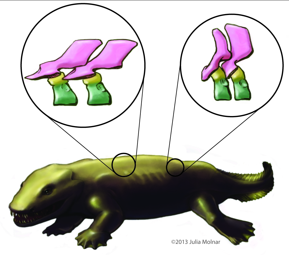
Above image: Julia Molnar‘s illustration of Ichthyostega showing anatomical changes of its spine from front to back, with neural arch/spine in pink, twin pleurocentra in yellow, and intercentrum in green. These four parts, three kinds of bones, made up the backbone of the first land vertebrates. These parts evolved in different ways in later animals, but formed one main bone in all living lineages of vertebrates.
RVC PRESS RELEASE:
Scientists reassemble the backbone of life using a particle accelerator
Research published today (Sunday 13 January 2013) in the journal Nature documents, for the first time, the intricate three-dimensional structure of the backbone in the earliest four-legged animals (tetrapods).
The international team of scientists, led by Dr Stephanie E. Pierce from The Royal Veterinary College and Professor Jennifer A. Clack from the University of Cambridge, bombarded 360 million year old early tetrapod fossils with high energy synchrotron radiation. The resulting high resolution X-ray images allowed the researchers to reconstruct the backbones of the extinct animals in exceptional detail.
The backbone, also known as the spine or vertebral column, is a bony structure found in all tetrapods, along with other vertebrates such as fish. It is formed from many elements or vertebrae all connected in a row – from head to tail. Unlike the backbone of living tetrapods (e.g. humans), in which each vertebra is composed of only one bone, early tetrapods had vertebrae made up of multiple parts.
Lead author Dr Pierce says: “For more than 100 years, early tetrapods were thought to have vertebrae composed of three sets of bones – one bone in front, one on top, and a pair behind. But, by peering inside the fossils using synchrotron X-rays we have discovered that this traditional view literally got it back-to-front.”
For the analysis, the European Synchrotron Radiation Facility (ESRF) in France, where the three fossil fragments were scanned with X-rays, used a new protocol to reveal tiny details of the fossil bones buried deep inside the rock matrix.
Using this new technology, the team of scientists discovered that what was thought to be the first bone – known as the intercentrum – is actually the last in the series. And, although this might seem like a trivial oversight, this re-arrangement in vertebral structure has over-arching ramifications for the functional evolution of the tetrapod backbone. (see here for a now out-of-date image from Wikipedia)
Dr. Pierce explains: “By understanding how each of the bones fit together we can begin to explore the mobility of the spine and test how it may have transferred forces between the limbs during the early stages of land movement”.
But, the findings didn’t end there. One of the animals – known as Ichthyostega – was also found to have an assortment of hitherto unknown skeletal features including a string of bones extending down the middle of its chest.
Professor Clack says: “These chest bones turned out to be the earliest evolutionary attempt to produce a bony sternum. Such a structure would have strengthened the ribcage of Ichthyostega, permitting it to support its body weight on its chest while moving about on land.”
This unexpected discovery supports recent work done by the same authors that showed Ichthyostega probably moved by dragging itself across flat ground using synchronous ‘crutching’ motions of its front legs – much like that of a mudskipper or seal.
Dr Pierce adds: “The results of this study force us to re-write the textbook on backbone evolution in the earliest limbed animals.”
The next step, the researchers say, is to understand how the backbone aided locomotion in these early tetrapods using sophisticated biomechanical analysis.
The study was funded by the Natural Environment Research Council.
Additional support was provided by the European Research Council and the ESRF, of which the Science and Technology Facilities Council (STFC) is the UK shareholder.
MOVIES:
These are rotating images of the anatomy, colour-coded, of the four species of early tetrapod that we examined for this study. Each shows the same basic pattern of having a “reverse rhachitomous” (pleurocentra in the front, intercentrum in the back; trying to think of a mullet joke…) anatomy. This is opposite the pattern that essentially all studies since famed evolutionary biologist/palaeontologist Edward Drinker Cope coined the term “rhachitomous” in 1878 have portrayed these and related animals as having. And this realization forces a re-examination of how the backbone structures first evolved in tetrapods and which parts (intercentra? pleurocentra? And where?) formed the spines of later animals.
For once, as authors we all felt that this finding really deserved the painfully hackneyed “rewrite the textbooks” label. It changes a lot of what we thought we knew about this classic evolutionary transition of anatomy. Check a vertebrate palaeontology/comparative anatomy textbook and you’ll likely find rhachitomous vertebrae and/or changes of pleurocentra vs. intercentra told in a way that we now are pretty sure is wrong.
You can also see the “sternebrae” (sternal elements; parts of the sternum that evolved independently in later land animals) in the first movie. This, to my knowledge, is by far the oldest such evidence. I know of ossified sternal plates in Early Permian mesosaurs like Stereosternum, but nothing earlier although perhaps in some synapsid I don’t know, or a basal diapsid of some kind? Chime in in the comments if you know of something I missed. Regardless, the sternebrae in Ichthyostega have nothing to do directly with those convergently evolved in lissamphibians, lepidosaurs, synapsids and archosaurs, although there may be some parallel developmental mechanisms involved and at least similar dermal tissues recruited into ossification patterns. Even so, these sternebrae are further evidence of how that taxon, at least, was beginning to make forays onto land, as they’d have helped it to support its belly on land and breathe.
The segmented PPC-SRµCT of Ichthyostega stensioi MGUH VP 6115 spinning in yaw and roll.
The segmented PPC-SRµCT of Ichthyostega eigili MGUH VP 29017a spinning in yaw and roll.
The segmented PPC-SRµCT of Acanthostega gunnari MGUH f.n. 1227 spinning in yaw.
The segmented µCT of Pederpes finneyae GLAHMS 100815 spinning in yaw.
FIGURE:
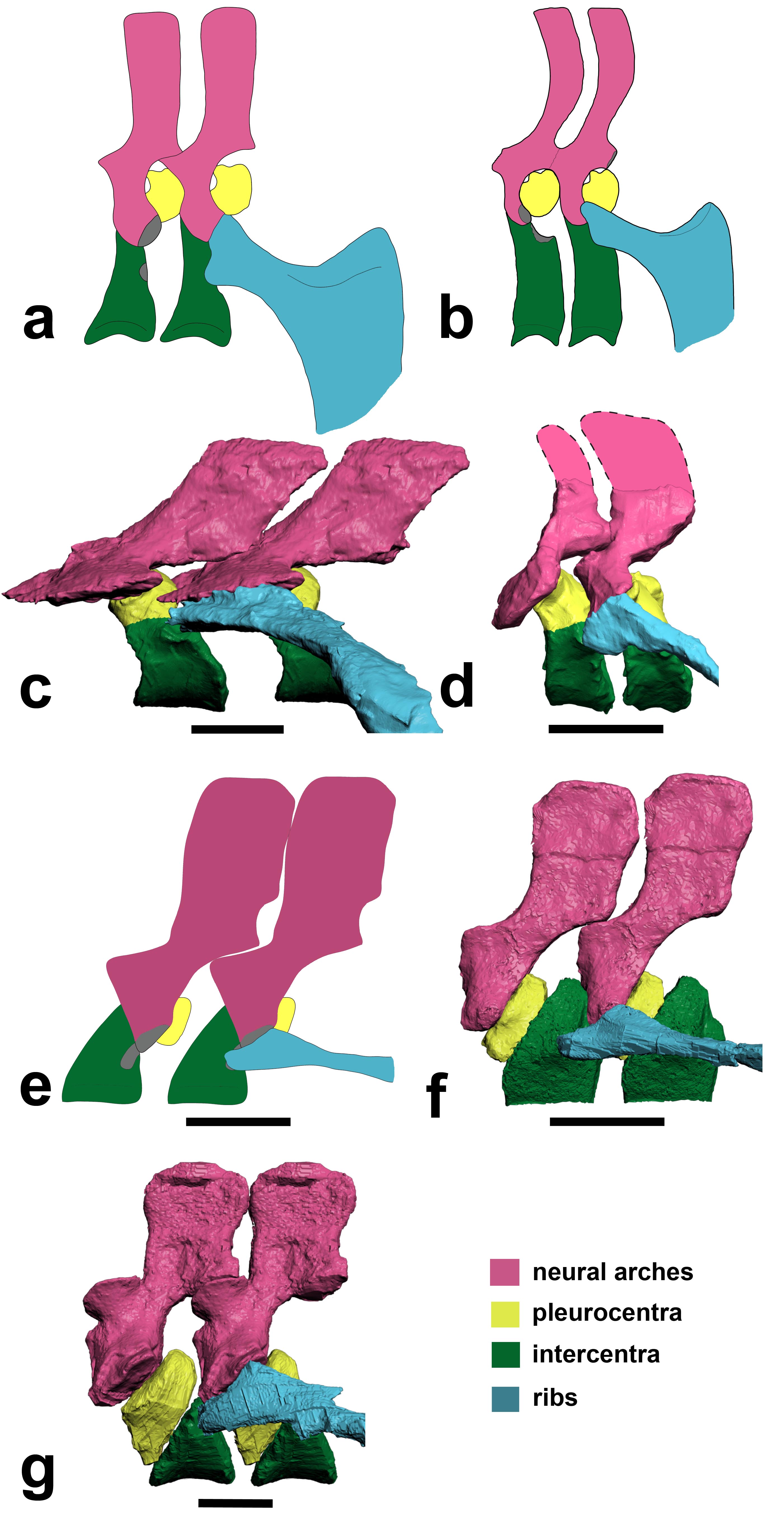
Above: (a,b) How we used to think the vertebrae were composed in early tetrapods like Ichthyostega. (c) How we found that Ichthyostega‘s posterior thoracic vertebrae actually tend to look. (d) Ichthyostega‘s anterior lumbar vertebral morphology. (e) Acanthostega according to Coates’s important description. (f) Our revision of the anatomy of Acanthostega (anterior dorsal). (g) Our new interpretation of Pederpes‘s morphology, from a posterior dorsal. Focus on the yellow vs. green elements. In a,b and e they are in different positions (reversed) compared with our new versions in c,d,f,g.
To put the above figure and movies into broader context, check this Wikipedia image. We think the red/pink bones (pleurocentra) are in the wrong place relative to the blue ones (intercentrum); the ones currently there in this image actually belong to the vertebral unit behind that one, so the pleurocentra should be moved to the front (left end) of each unit. But also look down toward the bottom of the figure. Some of those vertebrae may need to have their blue/pink bits re-examined and interpreted, too. Is it turtles intercentra all the way down?
There you have it! Welcome to your new, revised, irradiated, reverse-rhachitomous inner tetrapod’s vertebrae. Propagation phase-contrast X-ray synchrotron microtomography FTW!!!!
Science media articles arising from this study–
I like to keep track of media stories covering our research, using this blog, so here are some of the stories about this paper. It’s funny… this was one of the most broadly important papers I’ve ever been on, but the coverage was relatively scant. It was too technical. We knew that would be a problem, and really had a hard time putting into words why the study was so surprising even to us! Most writers wanted the “how did the animals move?” angle, which was not what the study was about. I still feel that this angle was not even needed; the study (and again I take minimal credit for it) is exciting without it. To comparative anatomy and evo-devo specialists, anyway. Well, that’s science for you; sometimes it is just too hard to explain its value to the outside world, even when you feel its importance in your very spine… And the press coverage was not terrible by any means; no sour grapes from me. Regardless, we’re glad it has been well received by specialist researcher colleagues we’ve spoken to, and that matters a lot.
NERC’s Planet Earth (nice story from our funder)- “Scientists had fossil backbone backwards”
BBC online (the only story aside from NERC’s that did more than read the press release) “Tetrapod anatomy: Backbone back-to-front in early animals”
Discovery News online– “First Land Animals Shuffled Like Seals” (good, but is sort of mixing up our this study, our 2012 one and Ahlberg et al’s 2005 seal-analogue study; latter two were more about movement. As often happens, a lot of other media stories basically copied this one’s headline/angle.)
Discover 80beats– “Paleontologists Use 3-D Models to Rewrite Evolution” (also in “top stories”)
Popsci– “Particle Accelerator Reveals That First Land Animals Walked Like Seals”
Daily FMail (nice pics)- “Astonishing 3D images reveal the first four-legged land animals in amazing detail – and overturn a century of research” (wins longest headline award)
Red Orbit– “Study Reveals First Ever Images Of Early Tetrapod Backbone And How It Helped In Land Evolution”
Examiner.com– “X-ray study rewrites tetrapod backbone evolution (Photos)”
Everything Dinosaur– “Ichthyostega Gets a Re-Think”
Business Standard– “Scientists recreate earliest quadraped’s backbone” (Proofread, editors! Quadruped.)
Geekosystem– “Early Land-Dwelling Animals Moved About Like Seals, Probably Didn’t Balance Balls on Their Noses” (scores some pts for humour)
…and the PR-copying, non-spellchecking fail of the week award goes to… Physorg! “Scientists reassemble the backbone of life with a particle acceleratorynchrotron [sic] X-rays”
Warming up the acceleratorynchrotron for our next study… 🙂


Wow! This is so, so cool.
Thanks Stella! We’re keen on it, too. 🙂
Revolutionary stuff, John! Massive implications for how we view the early development of the weight-bearing skeleton. Nice that you rotated the images around two separate axes so that we got to see all of it.
I just wanted to say that the red blob in the first vid is the creature’s heart and I can prob find other soft tissues via Digital Graphic Segregation if I adjust the contrast on my monitor and stare at the screen hard enough. 😉
Thanks man! We had a great team, otherwise we never would have noticed this stuff and realized its importance.
I think you’re on to something with the heart! Roll with it. 😉
[…] I’ve done my best to do this discovery justice, you should go check out the original press release for images and more […]
[…] E. Pierce, Per E. Ahlberg, John R. Hutchinson, Julia L. Monar, Sophie Sanchez, Paul Tafforeau, and Jennifer A. […]
[…] researchers to look at body parts in new ways. For instance, using such techniques, we were able torewrite the evolution of the backbone of early land animals called […]
[…] to look at body parts in new ways. For instance, using such techniques, we were able to rewrite the evolution of the backbone of early land animals called […]
[…] to look at body parts in new ways. For instance, using such techniques, we were able to rewrite the evolution of the backbone of early land animals called […]
[…] Variação de vértebras desde peixes ancestrais até grupos terrestres. Nós, humanos, e os dinossa… […]