This post was just published yesterday in a shorter, edited form in The Conversation UK, with the addition of some of my latest thoughts and the application of the editor’s keen scalpel. Check that out, but check this out too if you really like the topic and want the raw original version! I’ve changed some images, just for fun. The text here is about 2/3 longer.
Recently, the anatomy of animals comes up a lot, at least implicitly, in science news stories or internet blogs. Anatomy, if you look for it, is everywhere in organismal and evolutionary biology. The study of anatomy has undergone a renaissance lately, in a dynamic phase energized by new technologies that enable new discoveries and spark renewed interest. It is the zombie science, risen from what some had assumed was its eternal grave!
Stomach-Churning Rating: 4/10; there’s a dead elephant but no gore.
My own team has re-discovered how elephants have a false “sixth toe” that has been a mystery since it was first mentioned in 1710, and we’ve illuminated how that odd bit of bone evolved in the elephant lineage. This “sixth toe” is a modified sesamoid kind of bone; a small, tendon-anchoring lever. Typical mammals just have a little nubbin of sesamoid bone around their ankles and wrists that is easily overlooked by anatomists, but evolution sometimes co-opts as raw material to turn into false fingers or toes. In several groups of mammals, these sesamoids lost their role as a tendon’s lever and gained a new function, more like that of a finger, by becoming drastically enlarged and elongated during evolution. Giant pandas use similar structures to grasp bamboo, and moles use them to dig. We’ve shown that elephants evolved these giant toe-like structures as they became larger and more terrestrial, starting to stand up on tip-toe, supported by “high-heels” made of fat. Those fatty heels benefit from a stiff, toe-like structure that helps control and support them, while the fatty pads spread out elephants’ ponderous weight.

Crocodile lung anatomy and air flow, by Emma Schachner.
I’ve also helped colleagues at the University of Utah (Drs. Emma Schachner and Colleen Farmer) reveal, to much astonishment, that crocodiles have remarkably “bird-like” lungs in which air flows in a one-way loop rather than tidally back and forth as in mammalian lungs. They originally discovered this by questioning what the real anatomy of crocodile lungs was like- was it just a simple sac-like structure, perhaps more like the fractal pattern in mammalian lungs, and how did it work? This question bears directly on how birds evolved their remarkable system of lungs and air sacs that in many ways move air around more effectively than mammalian lungs do. Crocodile lungs indicate that “avian” hallmarks of lung form and function, including one-way air flow, were already present in the distant ancestors of dinosaurs; these traits were thus inherited by birds and crocodiles. Those same colleagues have gone on to show that this feature also exists in monitor lizards, raising the question (almost unthinkable 10-20 years ago) of whether those bird-like lungs are actually a very ancient and common feature for land animals.
Speaking of monitor lizards, anatomy has revealed how they (and some other lizards) all have venom glands that make their bites even nastier, and these organs probably were inherited by snakes. For decades, scientists had thought that some monitor lizards, especially the huge Komodo dragons, drooled bacteria-laden saliva that killed their victims with septic shock. Detailed anatomical and molecular investigations showed instead that modified salivary glands produced highly effective venom, and in many species of lizards, not just the big Komodos. So the victims of numerous toothy lizard species die not only from vicious wounds, but also from worsened bleeding and other circulatory problems promoted by the venomous saliva. And furthermore, this would mean that venom did not evolve separately in the two known venomous lizards (Gila monster and beaded lizard) and snakes, but was inherited from their common ancestor and became more enhanced in those more venomous species—an inference that general lizard anatomy supports, but which came as a big surprise when revealed by Bryan Fry and colleagues in 2005.
There’s so much more. Anatomy has recently uncovered how lunge-feeding whales have a special sense organ in their chin that helps them detect how expansive their gape is, aiding them to engulf vast amounts of food. Scientists have discovered tiny gears in the legs of leafhoppers that help them make astounding and precise leaps. Who knew that crocodilians have tiny sense organs in the outer skin of their jaws (and other parts of their bodies) that help them detect vibrations in the water, probably aiding in communication and feeding? Science knows, thanks to anatomy.
Just two decades or so ago, when I was starting my PhD studies at the University of California in Berkeley, there was talk about the death of anatomy as a research subject; both among scientists and the general public. What happened? Why did anatomy “die” and what has resuscitated it?

TH Huxley, anatomist extraordinaire, caricatured in a lecture about “bones and stones, and such-like things” (source)
Anatomy’s Legacy
In the 16th through 19th centuries, the field of gross anatomy as applied to humans or other organisms was one of the premier sciences. Doctor-anatomist Jean Francois Fernel, who invented the word “physiology”, wrote in 1542 that (translation) “Anatomy is to physiology as geography is to history; it describes the theatre of events.” This theatric analogy justified the study of anatomy for many early scientists, some of whom also sought to understand it to bring them closer to understanding the nature of God. Anatomy gained impetus, even catapulting scientists like Thomas Henry Huxley (“Darwin’s bulldog”) into celebrity status, from the realisation that organisms had a common evolutionary history and thus their anatomy did too. Thus comparative anatomy became a central focus of evolutionary biology.
But then something happened to anatomical research that can be hard to put a finger on. Gradually, anatomy became a field that was scoffed at as outmoded, irrelevant, or just “solved”; nothing important being left to discover. As a graduate student in the 1990s, I remember encountering this attitude. This apparent eclipse of anatomy accelerated with the ascent of genetics, with anatomy reaching its nadir in the 1950s-1970s as techniques to study molecular and cellular biology (especially DNA) flourished.
One could argue that molecular and cellular biology are anatomy to some degree, especially for single-celled organisms and viruses. Yet today anatomy at the whole organ, organism or lineage level revels in a renaissance that deserves inspection and reflection on its own terms.
Anatomy’s Rise
Surely, we now know the anatomy of humans and some other species quite well, but even with these species scientists continue to learn new things and rediscover old aspects of anatomy that laid forgotten in classic studies. For example, last year Belgian scientists re-discovered the anterolateral ligament of the human knee, overlooked since 1879. They described it, and its importance for how our knees function, in novel detail, and a lot of media attention was drawn to this realisation that there are some things we still don’t understand about our own bodies.
A huge part of this resurgence of anatomical science is technology, especially imaging techniques- we are no longer simply limited to the dissecting knife and light microscope as tools, but armed with digital technology such as 3-D computer graphics, computed tomography (series of x-rays) and other imaging modalities. Do you have a spare particle accelerator? Well then you can do amazing synchrotron imaging studies of micro-anatomy, even in fairly large specimens. Last year, my co-worker Stephanie Pierce and colleagues (including myself) used this synchrotron approach to substantially rewrite our understanding of how the backbone evolved in early land animals (tetrapods). We found that the four individual bones that made up the vertebrae of Devonian tetrapods (such as the iconic Ichthyostega) had been misunderstood by the previous 100+ years of anatomical research. Parts that were thought to lie at the front of the vertebra actually lay at the rear, and vice versa. We also discovered that, hidden inside the ribcage of one gorgeous specimen of Ichthyostega, there was the first evidence of a sternum, or breastbone; a structure that would have been important for supporting the chest of the first land vertebrates when they ventured out of water.
Recently, anatomists have become very excited by the realization that a standard tissue staining solution, “Lugol’s” or potassium iodide iodine, can be used to reveal soft tissue details in CT scans. Prior to this recognition, CT scans were mainly used in anatomical research to study bone morphology, because the density contrast within calcified tissues and between them and soft tissues gives clearer images. To study soft tissue anatomy, you typically needed an MRI scanner, which is less commonly accessible, often slower and more expensive, and sometimes lower resolution than a CT scanner. But now we can turn our CT scanners into soft tissue scanners by soaking our specimens in this contrast solution, allowing highly detailed studies of muscles and bones, completely intact and in 3D. Colleagues at Bristol just published a gorgeous study of the head of a common buzzard, sharing 3D pdf files of the gross anatomy of this raptorial bird and promoting a new way to study and illustrate anatomy via digital dissections- you can view their beautiful results here. Or below (by Stephan Lautenschlager et al.)!

These examples show how anatomy has been transformed as a field because we now can peer inside the bodies of organisms in unprecedented detail, sharing and preserve those data in high-resolution digital formats. We can do this without the concern that a unique new species from Brazilian rainforests or exciting fossil discovery from the Cambrian period would be destroyed if we probed certain questions about its anatomy that are not visible from the outside– a perspective in which science had often remained trapped for centuries. These tools became rapidly more diverse and accessible from the 1990s onward, so as a young scientist I got to see some of the “before” and “after” influences on anatomical research—these have been very exciting times!
When I started my PhD in 1995, it was an amazing luxury to first get a digital camera to use to take photographs for research, and then a small laser scanner for making 3D digital models of fossils, with intermittent access to a CT scanner in 2001 and now full-time access to one since 2003. These stepwise improvements in technology have totally transformed the way I study anatomy. In the 1990s, you dissected a specimen and it was reduced to little scraps; at best you might have some decent two-dimensional photographs of the dissection and some beetle-cleaned bones as a museum specimen. Now, we CT or MRI scan specimens as routine practice, preserving many mega- or gigabytes of data on its internal and external, three-dimensional anatomy in lush detail, before scalpel ever touches skin. Computational power, too, has grown to the point where incredibly detailed 3D digital models produced from imaging real specimens can be manipulated with ease, so science can better address what anatomy means for animal physiology, behaviour, biomechanics and evolution. We’re at the point now where anatomical research seems no longer impeded by technology– the kinds of questions we can ask are more limited by access to good anatomical data (such as rare specimens) than by the ways we acquire and use those data.
My experience mirrors my colleagues’. Larry Witmer at Ohio University in the USA, past president of the International Society for Vertebrate Morphologists, has gone from dissecting bird heads in the 1990s to becoming a master of digital head anatomy, having collected 3D digital scans of hundreds of specimens, fossil and otherwise. His team has used these data to great success, for example revealing how dinosaurs’ fleshy nostrils were located in the front of their snouts (not high up on the skull, as some anatomists had speculated based on external bony anatomy alone). They have also contributed new, gorgeous data on the 3D anatomy of living animals such as opossums, ostriches, iguanas and us, freely available on their “Visible Interactive Animal” anatomy website. Witmer comments on the changes of anatomical techniques and practice: “For extinct animals like dinosaurs, these approaches are finally putting the exploration of the evolution of function and behavior on a sound scientific footing.”
I write an anatomy-based blog called “What’s in John’s Freezer?” (haha, so meta!), in which I recount the studies of animal form and function that my research team and others conduct, often using valuable specimens stored in our lab’s many freezers. I started this blog almost two years ago because I noticed a keen interest, or even hunger for, stories about anatomy amongst the general public; and yet few blogs explicitly were about anatomy for its own sake. This interest became very clear to me when I was a consultant for the BAFTA award-winning documentary series “Inside Nature’s Giants” in 2009, and I was noticing more documentaries and other programmes presenting anatomy in explicit detail that would have been considered too risky 10 years earlier. So not only is anatomy a vigorous, rigorous science today, but people want to hear about it. Just in recent weeks, the UK has had “Dissected” as two 1-hour documentaries and “Secrets of Bones” as back-to-back six 30-minute episodes, all very explicitly about anatomy, and on PRIME TIME television! And PBS in the USA has had “Your Inner Fish,” chock full of anatomy. I. Love. This.

Before the scalpel: the elephant from Inside Nature’s Giants
There are many ways to hear about anatomy on the internet these days, reinforcing the notion that it enjoys strong public engagement. Anatomical illustrators play a vital role now much as they did in the dawn of anatomical sciences– conveying anatomy clearly requires good artistic sensibilities, so it is foolish to undervalue these skills. The internet age has made disseminating such imagery routine and high-resolution, but we can all be better about giving due credit (and payment) to artists who create the images that make our work so much more accessible. Social media groups on the internet have sprung up to celebrate new discoveries- watch the Facebook or Twitter feeds of “I F@*%$ing Love Science” or “The Featured Creature,” to name but two popular venues, and you’ll see a lot of fascinating comparative animal anatomy there, even if the word “anatomy” isn’t necessarily used. I’d be remiss not to cite Emily Graslie’s popular, unflinchingly fun social media-based explorations of gooey animal anatomy in “The Brain Scoop”. I’d like to celebrate that these three highly successful disseminators of (at least partly) anatomical outreach are all run by women—anatomical science can (and should!) defy the hackneyed stereotype that only boys like messy stuff like dissections. There are many more such examples. Anatomy is for everyone! It is easy to relate to, because we all live in fleshy anatomical bodies that rouse our curiosity from an early age, and everywhere in nature there are surprising parallels with — as well as bizarre differences from — our anatomical body-plans.
Anatomy’s Relevance
What good is anatomical knowledge? A great example comes from gecko toes, but I could pick many others. Millions of fine filaments, modified toe scales called setae, use micro-molecular forces called van der Waals interactions to help geckos cling to seemingly un-clingable surfaces like smooth glass. Gecko setae have been studied in such detail that we can now create their anatomy in sufficient detail to make revolutionary super-adhesives, such as the product “Geckskin”, 16 square inches of which can currently suspend 700 pounds aloft. This is perhaps the most famous example from recent applications of anatomy, but Robert Full’s Poly-Pedal laboratory at Berkeley, among many other research groups excelling at bio-inspired innovation in robotics and other fields of engineering and design, regularly spins off new ideas from the principle that “diversity enables discovery”, as applied to the sundry forms and functions found in organisms. By studying the humble cockroach, they have created new ways of building legged robots that can scour earthquake wreckage for survivors or explore faraway planets. By asking “how does a lizard use its big tail during leaping?” they have discovered principles that they then use to construct robots that can jump over or between obstacles. Much of this research relates to how anatomical traits determine the behaviours that a whole, living, dynamic organism is capable of performing.
Whereas when I was a graduate student, anatomists and molecular biologists butted heads more often than was healthy for either of them, competing for importance (and funding!), today the scene is changing. With the rise of “evo devo”, evolutionary developmental biology, and the ubiquity of genomic data as well as epigenetic perspectives, scientists want to explain “the phenotype”—what the genome helps to produce via seemingly endless developmental and genetic mechanisms. Phenotypes often are simply anatomy, and so anatomists now have new relevance, often collaborating with those skilled in molecular techniques or other methods such as computational biology. One example of a hot topic in this field is, “how do turtles build their shells and how did that shell evolve?” To resolve this still controversial issue, we need to know what a shell is made of, what features in fossils could have been precursors to a modern shell, how turtles are related to other living and extinct animals, how a living turtle makes its shell, and how the molecular signals involved are composed and used in animals that have or lack shells. The first three questions require a lot of anatomical data, and the others involve their fair share, too.
Questions like these draw scientists from disparate disciplines closer together, and thanks to that proximity we’re inching closer to an answer to this longstanding question in evolutionary biology and anatomy, illustrated above in the video. As a consequence, the lines between anatomists and molecular/cellular biologists increasingly are becoming blurred, and that synthesis of people, techniques and perspectives seems to be a healthy (and inevitable?) trend for science. But there’s still a long way to go in finding a happy marriage between anatomists and the molecular/cellular biologists whose work eclipsed theirs in past decades. Old controversies like “should we use molecules or morphology to figure out how animals are related to each other?” are slowly dying out, as the answer becomes evident to be “Yes. Both.” (especially when fossils can be included!) Such dwindling controversies contribute to the healing of disciplinary rifts and the unruffling of parochial feathers.
Yet many anatomists would point to lingering obstacles that give them concern for their future; funding is but one of them (few would argue that gross anatomical research is as well off in provision of funding as genetics is, for example). There are clear mismatches between the hefty importance, vitality, popularity and rigour of anatomical science and its perception or its role in academia.
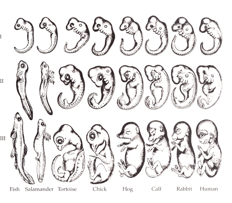
Romane 1892, covering Haeckel’s classic, early evo-devo work (probably partly faked, but still hugely influential) (source)
Anatomy’s Future
One worry the trend that anatomy as a scientific discipline is clearly flourishing in research while it dwindles in teaching. Fewer and fewer universities seem to be teaching the basics of comparative anatomy that were a mainstay of biology programmes a century ago. Yet anatomy is everywhere now in biology, and in the public eye. It inspires us with its beauty and wonder—when you marvel at the glory of beholding a newly discovered species, you are captivated by its phenotypic pulchritude. Anatomy is still the theatre in which function and physiology are enacted, and the physical encapsulation of the phenotype that evolution moulds through interactions with the environment. But there is cause for concern that biology students are not learning much about that theatre, or that medical schools increasingly seem to eschew hands-on anatomical dissection in favour of digital learning. Would you want a doctor to treat you if they mainly knew human anatomy from a CGI version on an LCD screen in medical school, and hence were less aware of all the complexity and variation that a real body can house?
Anatomy has an identity problem, too, stemming from decades of (Western?) cultural attitudes (e.g. the “dead science” meme) and from its own success—by being so integral to so many aspects of biology, anatomy seems to have integrated itself toward academic oblivion, feeding the perception of its own obsolescence. I myself struggled with what label to apply to myself as an early career researcher- I was afraid that calling myself an “anatomist” would render me quaint or unambitious in the eyes of faculty job interview panels, and I know that many of my peers felt the same. I resolved that inner crisis years ago and came to love identifying myself at least partly as an anatomist. I settled on the label “evolutionary biomechanist” as the best term for my speciality. In order to reconstruct evolution or how animals work (biomechanics), we first often need to describe key aspects of anatomy, and we still discover new, awesome things about anatomy in the process. I still openly cheer on anatomy as a discipline because its importance is so fundamental to what I do, and I am far from alone in that attitude. Other colleagues that do anatomical research use other labels for themselves like “biomechanist”, “physiologist,” or “palaeontologist”, because those words better capture the wide range of research and teaching that they do, but I bet also because some of them likely still fear the perceived stigma of the word “anatomy” among judgemental scientists, or even the public. At the same time, many of us get hired at medical, veterinary or biology schools/departments because we can teach anatomy-based courses, so there is still hope.
Few would now agree with Honoré de Balzac’s 19th century opinion that “No man should marry until he has studied anatomy and dissected at least one woman”, but we should hearken back to what classical scientists knew well: it is to the benefit of science, humanity and the world to treasure the anatomy that is all around us. We inherit that treasure through teaching; to abscond this duty is to abandon this trove. With millions of species around today and countless more in the past, there should always be a wealth of anatomy for everyone to learn from, teach about, and rejoice.

X-ray technology has revolutionized anatomical studies; what’s next? Ponder that as this ostrich wing x-ray waves goodbye.
Like this post? You might also find my Slideshare talk on the popularity of anatomy interesting- see my old post here for info!


Excellent piece, and it is great to see your additional thoughts. Two comments:
1) I wonder if the resurgence in anatomy is due in part to a renewed methodological rigor–not that old studies lacked rigor, but that today we have new techniques for testing hypotheses in both functional and evolutionary context. One example would be phylogenetic comparative methods–where it used to be enough to show casual correlations as evidence of function, an extra phylogenetic (and often quantitative) dimension has proven invaluable. Basic descriptions are still our bread-and-butter, but it is rare indeed to see even the most detail-laden description ignore phylogeny or statistical methods. I suspect that anatomy (justifiably) has regained some credibility as a result.
2) One rather unfortunate attitude that has hindered anatomical education is a view that teaching anatomy is a fall-back from a “real” job doing “real” science. A colleague pointed this out in a quote from Neil Shubin, where Shubin says, “It’s not unusual to have paleontologists teach anatomy because we know anatomy pretty well. It’s a good backup for a paleontologist to have, in case you can’t get a regular appointment. There aren’t a lot of jobs.” (source) I of course don’t think Shubin meant anything horrible by this (and he has done amazing things at all levels for public education on evolutionary anatomy), but it does speak to a common attitude across academia. As a graduate student, I was often congratulated for my decision to study anatomy, because it would provide job security down the line. This is of course true (ironically, I ended up in a museum job), but along the way I discovered that teaching anatomy was deeply rewarding, relevant to my research, and a dynamic field in its own right. Not at all a “fall-back.” There are probably a couple of blog posts to be written on this topic alone…
One addendum to my comments above – I was incredibly fortunate to have landed in a grad program where anatomy education was just as valued as research (Stony Brook U). From the department chair down, the anatomy faculty sets a fantastic example for how high-quality teaching is just as important and fulfilling as research. The “anatomy as fallback” attitude was not something I really experienced at Stony Brook!
Yes this varies a lot; you can encounter the full spectrum of attitudes depending on what uni you go to, what dept and who your supervisor is at the time or who you hang out with most. I still encounter the teaching vs. research tension in my environment, which is a false dichotomy… although my present working role is heavily focused on research (including undergraduate projects, which are teaching too), it has changed before and will change again, and I view my outreach efforts as teaching at least as much as research activities; to separate the two (e.g. for beancounting) sometimes is not so helpful, because in a real person they often are blended.
Quick response (almost bedtime)- 1) I think that is also true, yes; rigor in non-3D-techy ways has given anatomy some boost, but the big boost from 1990s onward (phylogenetic methods starting to come in earlier than that) still seems to be imaging technology.
2) Oh I totally agree! Zero shame in anatomy jobs- in this market, any job you can get is a good one, and we should be counselling students to optimize their chances of getting what they want/can get. I was pushed, very firmly, at Berkeley to get human anatomy teaching experience, and I am lucky I got it; it helped a LOT.
This was on a thread yesterday for baby names…
Kent
4 Fans
My 7 year old Son, Huxley Spenser Bown. Named after Darwin’s bulldog.
9 Apr 10:36 AM
In reply to Kent_Bown
Eric S. (alex98)
285 Fans· Dulce bellum inexpertis
You named your kid after a dog ????
9 Apr 4:28 PM
kind of says it all…..
Let’s keep that between us and not let old TH Huxley know; he’d go barking mad.
wanna laugh?
http://www.noahTheMovie.com
be “kind”
Great post. Nuff said.
[…] of evidence. The history & ongoing importance of anatomy. Excellent case made by John […]
Reposted on the Huff Post http://www.huffingtonpost.com/dr-john-r-hutchinson/science-of-anatomy_b_5154450.html and io9 http://io9.com/the-science-of-anatomy-is-undergoing-a-revival-1562557653/! Cool!
[…] Well, my first thought is that it’s beautiful. I don’t tend to think of it as gross. I’ve rhapsodized about this before. Animals are wonderful inside and out, and I regularly pause during a dissection to marvel at how […]