Giraffe limb from previous post, now shown via movie of DICOM (CT image data) files. Axial slices every 2.5mm, from toes to knee/stifle. Darker areas are lower density (black is air); white is very dense– bone, artefact, metal, Santorum, etc.
Posted in Frozen Mammals, tagged CT, giraffe on March 5, 2012| 1 Comment »
Giraffe limb from previous post, now shown via movie of DICOM (CT image data) files. Axial slices every 2.5mm, from toes to knee/stifle. Darker areas are lower density (black is air); white is very dense– bone, artefact, metal, Santorum, etc.
Posted in Frozen Mammals, tagged CT, giraffe on March 3, 2012| 8 Comments »
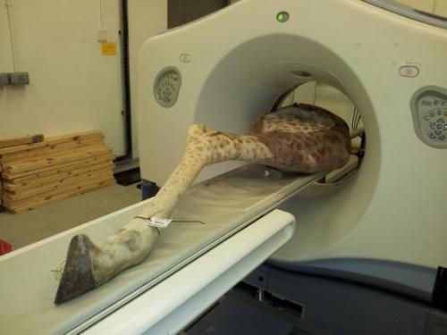
I’m preparing to do anatomically-realistic computer modelling of giraffe locomotor mechanics with some colleagues. To do that, we of course need the 3D anatomy of bones, muscles and tendons, for which CT can be pretty useful. Here, we put our first frozen leg through the motions. It was a 3 person job to lift the sucker, but the CT bed managed to move it through the scanner with minimal hiccups. Inside the ring that the upper end of the leg is lined up with are 8 x-ray detectors, so 8 CT slices can be imaged at once, speeding the procedure.
This specimen died in a UK zoo recently, apparently from trauma (falling?), which we’re trying to help them figure out in the course of our scans and future dissections. We often provide a pretty detailed postmortem service in return for being given cadavers, since we are a vet school with a lot of expertise in pathology and anatomy. Also, we have been describing the kinds of pathologies we observe along the way, because terribly little is known about some diseases/injuries in non-domestic animals, so there is plenty we can contribute to the scientific literature as a result. We’re also interested in documenting how pathologies in wild vs. captive animals differ (if at all).

